 Phytotoxic potential of leaf extracts from species occurring in two phytophysiognomies of the Atlantic Forest: a chemical and biological evaluation
Abstract
Some plants synthesize phytotoxic compounds that can inhibit the development of neighboring species. Phenolic compounds from some botanical families have been the focus of studies because of their effect on surrounding plants. This work aims to analyze the phytochemical profile of leaf methanolic extracts through the identification of major phenolic compounds, evaluation of their phytotoxicity, and their intraspecific variation. Leaves from Calophyllum brasiliense, Psidium guineense, and Miconia cinnamomifolia from the Ombrophilous Forest and Restinga phytophysiognomies were sampled. The phytotoxic activity was also tested against Solanum lycopersicum. The extract of M. cinnamomifolia presented the greatest chemical diversity and intraspecific variation. The M. cinnamomifolia forest extract presented potent inhibition of seed germination and root growth of the target species. Both C. brasiliense extracts affected the target species in higher concentrations. In the P. guineense Restinga extract, which had greater activity than the species' forest extract, 8-hydroxyluteolin-8-sulfate was identified. This is the first report of flavonoid sulphate for the Myrtaceae family, with reported biological activities and indicated for studies in bioprospection. Our study highlights the importance of conservation of Atlantic Forest species, due to the diversity of phenolic compounds and their phytotoxic effects, in response to distinct environmental conditions.
Keywords:
chemical traits; Dense Ombrophilous Forest; intraspecific variation; phenolic compounds; Restinga
Introduction
Knowledge of the level of interaction among plant species belonging to a natural system is limited because the full potential of the special metabolites they produce in response to different environmental filters is unknown (Marsh et al., 2019). The degree of interaction among species can be informed indirectly by floristic and phytosociological surveys since the plant communities studied are a result of interactions among species, e.g., competition and chemical signalization (Imatomi et al., 2013a; Gillani et al., 2024).
Plants synthesize a wide variety of secondary or special metabolites, whose diversity varies according to plant family and species (Kessler & Kalske, 2018). Such compounds play an essential role in the interaction of plants with their environment by triggering mechanisms that promote survival and adaptation (Verma & Shukla, 2015). Among the special metabolites, phenols and flavonoids stand out for their notable structural diversity (Lattanzio, 2013).
Phenolic compounds are widely distributed in bryophytes and mainly in vascular plants (Cheynier et al., 2013). They act as signals in plant-microorganism interactions and in plant-plant relationships (Mousavi et al., 2021), as well as in defense mechanisms against pathogens and herbivores. They are essential functional traits that attract pollinators and fruit and seed dispersers and contribute to responses to environmental filters (Cheynier et al., 2013; Verma & Shukla, 2015). They are also considered a cause of ecological and economic problems, such as interfering with the regeneration of natural forests and annual crops due to soil sickness (Li et al., 2010). Phytotoxic phenols tend to cause negative effects on other plants, such as delaying germination, hindering root and shoot growth (Cheynier et al., 2013). Such compounds include flavonoids, hydroxycinnamic and benzoic acids, phenylpropanoids, coumarins, and tannins (Barral & Paradelo, 2011; Cheng & Cheng, 2015; Mousavi et al., 2021).
The expression of special metabolites by individual plants of the same species in different locations may be affected by abiotic factors (Arnold et al., 2019; Yang et al., 2018), which may reflect differences in the production of phytotoxic compounds (Pilatti et al., 2018). Abiotic factors such as light, temperature, and water influence, directly or indirectly, plant growth and metabolism, and the chemical diversity in plants is a result of their plasticity (Arnold et al., 2019; Yang et al., 2018).
Complex biomes, with multiple phytophysiognomies, present marked changes in plant chemical profiles due to differences in tree canopy and water availability (Cerqueira et al., 2021). One example is the Brazilian Atlantic Forest, which comprises diverse phytophysiognomies that vary largely in vegetation structure, microclimate, and edaphic conditions (Pireda et al., 2019). Coastal sandbanks, known as Restingas (RE), are characterized by high temperatures and irradiance, as well as lower availability of water and nutrients (Oliveira et al., 2023). In contrast, the Dense Ombrophilous Forest (OF) is an Atlantic Forest phytophysiognomy characterized by higher water availability and low light irradiation due to the forest canopy (Pireda et al., 2019; Borges et al., 2022; Xavier et al., 2023).
Thus, we hypothesize that individuals of the same species grown in different Atlantic Forest phytophysiognomies will present intraspecific variation at the chemical level, which may result in distinct phytotoxic activities of phenolic compounds from both studied areas. To test this hypothesis, species found in both OF and RE areas belonging to families with known occurrence of phenolic compounds were studied: Melastomataceae (Vasconcelos et al., 2006; Sobrinho et al., 2017; Bomfim et al., 2021), Myrtaceae (Santos et al., 2018; Macedo et al., 2021; Imatomi et al., 2013a; Imatomi et al., 2013b), and Calophyllaceae (García-Zebadúa et al., 2014; Liu et al., 2022; Zailan et al., 2022). In addition, the studied families are characterized by their richness and abundance, as well as their high endemism in Brazilian forest formations (Lucas & Bünger, 2015; Cabral et al., 2021; Wagner et al., 2022; Goldenberg et al., 2025).
As for the focus species, Psidium guineense Sw. (Myrtaceae) may present phenolic acids (caffeic, vanillic, and gallic acids), catechins, flavones, and isoflavonoids (Senanayake et al., 2018). Calophyllum brasiliense Cambess. (Calophyllaceae) is reported as a producer of phenolic acids, flavonoids, xanthones, chromanones, and coumarins (García-Zebadúa et al., 2014; Santos et al., 2025). The chemical composition of Miconia cinnamomifolia (DC.) Naudin (Melastomataceae) is unknown until this date, although flavonoids may be found in other Miconia species (Bomfim et al., 2021).
Therefore, this study aimed to evaluate intraspecific variation between two distinct Atlantic Forest phytophysiognomies, with basis on the chemical composition of methanolic fractions of crude leaf extracts of individuals of Calophyllum brasiliense Cambess. (Calophyllaceae), Psidium guineense Sw. (Myrtaceae) and Miconia cinnamomifolia (DC.) Naudin (Melastomataceae). It also aimed to assess the phytotoxic potential of the extracts.
Materials and Methods
Plant Material
Leaves of Psidium guineense, Miconia cinnamomifolia, and Calophyllum brasiliense were sampled in April 2022, at approximately 9 a.m., from five individuals from each of the following two Atlantic Forest phytophysiognomies in the state of Rio de Janeiro, Brazil: Dense Ombrophilous Forest (OF), at Reserva Biológica de Poço das Antas (Rebio Poço das Antas), in the municipality of Silva Jardim (22º30' S - 22º33' S; 42º15' W - 42º19' W); and Restinga (RE) at Reserva Particular do Patrimônio Nacional Fazenda Caruara (RPPN Faz. Caruara), in the municipality of São João da Barra (21º75' S; 41º03' W). A voucher specimen of each species was deposited at the HUERJ Herbarium of the Universidade do Estado do Rio de Janeiro (Table 1).
During plant sampling, microclimatic data were collected using a digital thermohygrometer (TH01) and a radiometer (LI-250). The absolute mean values of the measured parameters revealed marked contrasts between the vegetation physiognomies (Table 1).
Leaf Extracts
Leaves of C. brasiliense, P. guineense, and M. cinnamomifolia from each phytophysiognomy were pooled and dried in a forced air oven for three days at 40 °C and ground using a Marconi® MA330/1 mill. The powder obtained (12 g) was submitted to extraction with 60 mL of ethanol in an Ultronique® Q5.9/40 Eco-Sonics ultrasonic bath for 30 min at 60 °C and filtered using a Buchner funnel, to yield one crude extract per species.
Crude dry extracts of P. guineense and M. cinnamomifolia were dissolved in a minimum quantity of ethanol and subsequently added to microcrystalline cellulose (4.0 g). The solvent was eliminated by a Fisatom ® 802 rotary evaporator, using approximately 8 rpm rotation at 45 °C. Subsequently, the cellulose coated with the extract was deposited on a layer of cellulose (200 mg moistened with 3 mL of distilled water) that had been previously prepared in a Büchner funnel with a porous plate. The column was successively eluted in increasing order of polarity using 200 mL of hexane, chloroform, ethyl acetate, methanol, and water solvents. Individual fractions were collected, concentrated in a rotary evaporator, and lyophilized.
The Calophyllum brasiliense crude extract was initially subjected to the same chromatography procedure as the other plants; however, the extract obstructed the Büchner funnel with cellulose. The extract was subsequently subjected to a liquid-liquid fractionation in the separation funnel using the solvent system hexane/methanol/water (1/0.5/0.5 v/v). In this procedure, 2 g of the dry extract was solubilized in 50 mL of the low phase, and the solution was washed four times with 50 mL of the upper phase (UP). The UP was collected and reserved, and the lower phase (LP) was concentrated in a rotary evaporator and lyophilized. Following this, the LP fraction was subjected to a second liquid-liquid fractionation using the solvent system hexane/ethyl acetate/methanol/water (1/2/0.5/1 v/v). Next, the procedure outlined above was executed, and the new lower phase was collected and concentrated for drying. This second fraction was employed in the analysis to clarify the chemical compounds.
Phytochemical Analysis
High-performance liquid chromatography (HPLC) analysis was performed using solutions of the methanolic fractions of M. cinnamomifolia and P. guineense, and the LP precipitate of C. brasiliense, prepared at a concentration of 5 mg. mL-1 in a mixture of 5 % acetonitrile in ultrapure water in 2.0 ml Eppendorf microtubes. HPLC analysis was performed using a Hitachi LaChrom Elite® HPLC System chromatograph coupled to a diode array detector (DAD). A Luna Omega C-18 column (5 cm x 2 mm i.d.), 1.6 μm particle, and 100 A, equipped with a compatible 2.1 mm pre-column, was used. The column temperature was 40 °C, and the flow rate was 0.3 mL.min-1 during 65 minutes of analysis. The mobile phase consisted of two solvents: (A) deionized water with 0.1 % formic acid; and (B) acetonitrile (HPLC grade) with 0.1 % formic acid (Tédia® and Sigma-Aldrich®). The solutions were injected into the HPLC system (5 μL, in triplicate). The chromatogram data showed absorption signal I at 254 nm, with detection of ellagic tannins at 280 nm, and signal I for UV absorption at 340 nm, referring to the B ring of flavonoids at 343 nm.
UPLC-DAD-MS analysis
UPLC-UV-ESI-MS/MS analysis was performed on the same chromatographic apparatus described in the previous section, coupled to a high-resolution mass spectrometer equipped with an electrospray ion source (ESI) and quadrupole time of flight mass analyzer (Q-TOF) (Bruker Daltonics, Bremen, Germany). Column effluent was introduced into the mass spectrometer at 0.25 mL.min-1.
Nebulizer gas was high-purity nitrogen (N2) produced online by a Peak Scientific NM32LA nitrogen generator. Analysis parameters were set using negative ionization mode with spectra acquired over a mass range from m/z 50 to 1400. The optimum ESI-MS parameters were capillary voltage, +3.5 kV; drying gas temperature, 210°C; drying gas flow, 10.0 L/min; nebulizing gas pressure, 72.5 psi; collision RF, 200 Vpp; transfer time 120 µs; and pre-pulse storage, 3 µs. Moreover, automatic MS/MS experiments were performed, adjusting the collision-energy values as follows: m/z 500, 30 eV; above m/z 500, 50 eV; and using nitrogen as collision gas. The MS data were processed through Data Analysis 4.0 software (Bruker Daltonics, Bremen, Germany). External instrument calibration was performed using a Cole Palmer syringe pump (Vernon Hills, IL, USA) directly connected to the interface, eluting a sodium formate (NaCHO2) solution cluster containing 5 mM sodium hydroxide (NaOH) and 0.2 % formic acid in water/isopropanol 1/1 (v/v).
The LC/MS system was controlled by HyStar 3.2 software (Bruker Daltonics, Bremen, Germany).
Phytotoxicity Bioassays
The methanolic fractions of the crude extracts from C. brasiliense, M. cinnamomifolia, and P. guineense (16 mg) were dissolved in 0.1 % dimethyl sulfoxide (DMSO, 5 µL per mL) and diluted in distilled water until reaching the concentrations of 2000, 1000, 500, 250, and 125 ppm. DMSO is widely used in phytotoxicity bioassays as it improves the dissolution of extract compounds (Lorenzo et al., 2016). DMSO at 0.1 % does not affect the growth of target species, as reported by Castellano (2001). This bioassay adopted distilled water containing DMSO, as above, as a negative control. The positive control was the herbicide glyphosate (Roundup®), dissolved in DMSO and diluted as the plant extracts above (Macías et al., 2000; Castellano, 2001).
The bioassays were performed in Petri dishes (Ø = 6 cm), each equipped with two layers of 80 g filter paper, with four replicates per treatment. Twenty seeds of Solanum lycopersicum L. cv. Santa Cruz Kada (Isla Sementes, Porto Alegre, Brazil), pre-disinfected with 1 % sodium hypochlorite, were placed directly on the filter paper of each dish along with 1 mL of the respective treatment. All dishes were labeled, sealed with Parafilm®, and placed in a germination chamber (Solab SL-224/210, Piracicaba, Brazil) set at 27 °C, under a 12/12 h photoperiod, for a period of seven days (Macías et al., 2000; Silva et al., 2017; Feitoza et al., 2020).
During the bioassay, the number of germinated seeds was counted twice a day using a 12 h-interval between counts, until the end of the 7 days. The seeds were considered germinated with 2 mm of radicle protrusion. The percentage of Germination (PG %) was calculated as (1):
where Xi is the number of germinated seeds and N is the total number of seeds, with the values expressed as percentages (Anjum & Bajwa, 2005).
To evaluate seed germination delay, the Germination Speed Index (GSI) was calculated as (2):
where G is the number of germinated seeds at time t (in hours) during observation i (Anjum & Bajwa, 2005).
At day 7 of the bioassay, root length was measured (n=20, r=4) using a digital pachymeter, from the root-shoot transition zone to the root tip. Non-germinated seeds were counted as zeros in root length measures.
Statistical Analysis
Data were statistically analyzed using R 4.4.1v (R Core Team, 2024). Sample normality was evaluated by the Shapiro-Wilk test (p = 0.05). Non-normal data were compared using the Kruskal-Wallis test and Dunn’s post-hoc test (p = 0.05). Bar graphs were produced in Excel® by calculating the percentage of the control, which is given by (3):
where 𝑋 𝑇 is the mean of measurements of each treatment, and 𝑋 𝐶 is the mean of measurements of the negative control. Results are expressed as percentages. Negative values denote inhibition, whereas positive values indicate stimulation (Feitoza et al., 2018). Inhibition values were further submitted to Unweighted Pair Group Method with Arithmetic Mean (UPGMA) analysis using Euclidean distance to evaluate similarities among extract effects.
Results
Phytochemical Analysis
The phenolic compounds profile of this species was obtained by UPLC-DAD-ESI-Q-TOF-MS analysis of the methanolic fractions of Psidium guineense and Miconia cinnamomifolia, and the Calophyllum brasiliense second low phase. Chromatograms with molecular absorption detection at wavelengths of 254 nm and 340 nm are shown in Figures S1-S6. The compounds are summarized in Table 2, which includes the compound name, molecular formula, MS/MS fragments, experimental mode m/z in negative mode [M-H]-, and retention time. The fragmentation profiles of all compounds were compared to those in the literature and databases, including Mass Bank and GNPS.
List of compounds identified in the UPLC chromatograms at 254 nm and 340 nm of extracts of Calophyllum brasiliense Cambess., Psidium guineense Sw., and Miconia cinnamomifolia (DC.) Naudin. originating from two phytophysiognomies of the Atlantic Forest: Dense Ombrophilous Forest (OF) and Restinga (RE).
After analysis of the C. brasiliense leaf extract, three compounds were identified in the two phytophysiognomies: astilbin 3-O rhamnoside (5), quercetin hexoside (6), and quercetin rhamnoside (7). Two compounds were identified only in OF: gallic acid and dihydroxybenzoic acid hexoside (Table 2; Figure S1). The extract of C. brasiliense from RE indicated variation with the presence of protocatechuic acid (Table 2; Figure S2).
Some phenolic compounds of P. guineense leaf extracts were found common to both phytophysiognomies, namely quinic acid (1), galloyl hexoside (2), myricetin-3-glucoside (3), and myricetin 3-arabinoside (4) (Table 2; Figure S3 and S4). One compound was found only in RE, 8-hydroxyluteolin-8-sulfate (6) (Table 2; Figure S4).
Miconia cinnamomifolia exhibited the most diverse phenolic composition in both phytophysionomies. Gallic acid, quercetin rutinoside, and quercetin pentoside I were among the phenolic compounds identified in both phytophysiognomies. The following compounds were found only in OF: peduncalagin, casuarictin, tellimagrandin II, quercetin hexoside I, quercetin pentoside II, quercetin pentoside I, and quercetin rhamnoside (Figure S5). The following compounds were identified only in RE: vescalaginin (2), castalagin (4), methoxygalic acid (5), hydrolyzed tannin (6), isoorientin (8), ellagic acid (9), vitexin (11), rutin (12), and isovitexin (13) (Figure S6).
Phytotoxicity Analysis
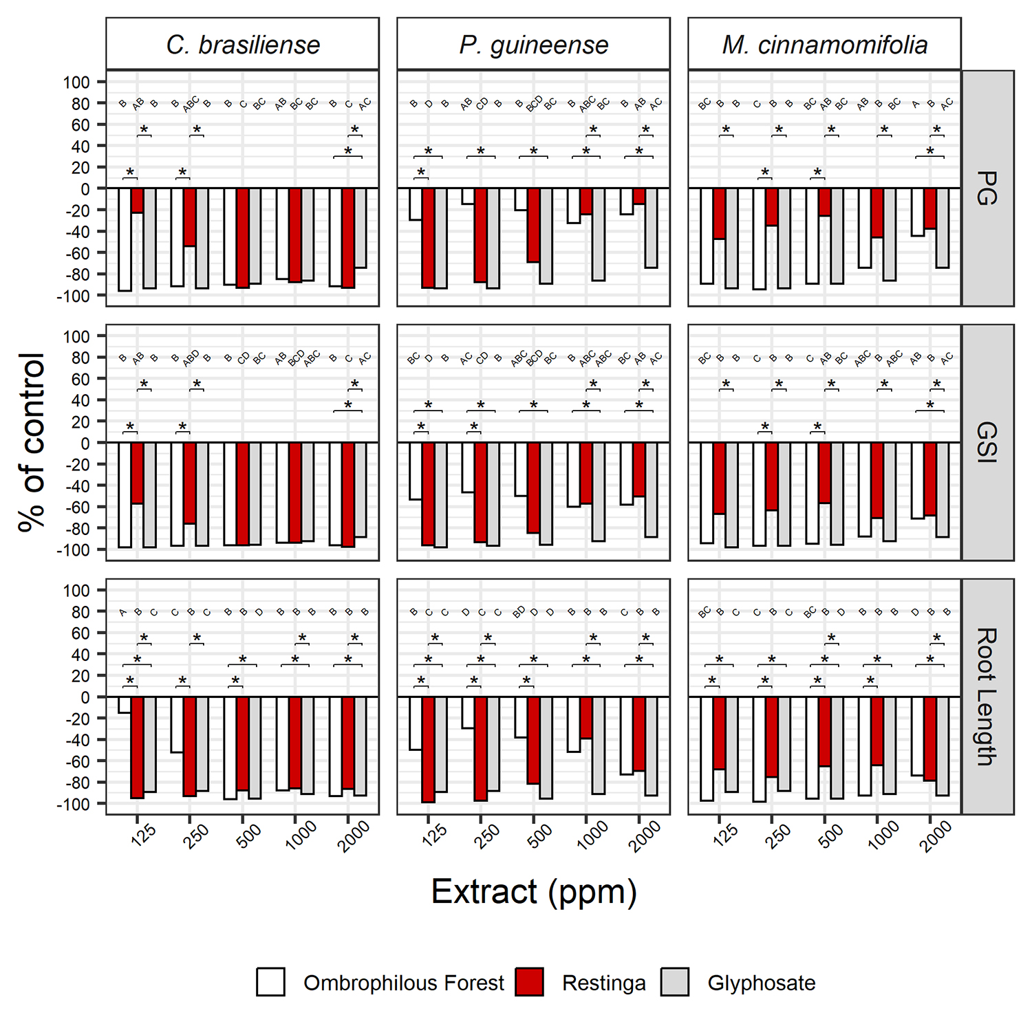
The leaf extracts from P. guineense, M. cinnamomifolia, and C. brasiliense showed significant phytotoxic activity (p < 0.05) against Solanum lycopersicum (Fig. 1; Table S1). Calophyllum brasiliense, from both studied phytophysiognomies, demonstrated high phytotoxicity in particular, as evidenced by high inhibition (i.e., highly negative percentages of negative control) for percentage of germination (PG), Germination Speed Index (GSI), and root length for concentrations 500, 1000, and 2000 ppm. The OF and RE leaf extracts differed in effect at lower doses (125 and 250 ppm); the OF extract presented greater inhibition for PG and GSI than the RE extract, whereas the inverse was observed for root length. The OF extract and the herbicide glyphosate (positive control) had similar inhibition effects on PG and GSI at the lower concentrations (125 and 250 ppm). However, both OF and RE extracts at 2000 ppm presented greater inhibition of PG and GSI than glyphosate. In addition, the RE extract caused a greater decrease in root length than glyphosate at 125 and 250 ppm, and the OF extract caused greater inhibition than its positive control at 500 and 2000 ppm (Fig. 1).
Effect of the methanolic partitions of Miconia cinnamomifolia and Psidium guineense and lower fraction of Calophyllum brasiliense, at concentrations of 2000, 1000, 500, 250, 125 ppm, on the Germination Percentage (PG), Germination Speed Index (GSI), and Root Length of Solanum lycopersicum. Bars are displayed as percentages of the negative control (distilled water). Negative percentage values indicate inhibition. Distinct uppercase letters indicate significant differences among concentrations after Kruskal-Wallis test, followed by the post-hoc Dunn test (p < 0.05), where the negative control (distilled water) represents “A”. (*) indicates significant differences among Ombrophilous Forest and Restinga extracts and the herbicide Glyphosate, after Kruskal-Wallis test, followed by the post-hoc Dunn test (p < 0.05).
In general, the effects of the leaf extract of P. guineense from OF differed significantly from the extract from RE and from glyphosate (Fig. 1; Table S1). The OF extracts exhibited the lowest phytotoxic effects on PG, GSI, and root length of 125 to 500 ppm, whereas no significant differences (p > 0.05) were found between OF and RE extracts above 1000 ppm. Moreover, the RE leaf extract significantly decreased PG and GSI of 125 to 500 ppm, whereas root length was reduced in all concentrations in comparison to the negative control. The RE extract was less phytotoxic than glyphosate in most cases; however, this extract exhibited greater inhibition of root length than glyphosate at 125 and 250 ppm.
The OF leaf extract of M. cinnamomifolia significantly reduced PG and GSI of S. lycopersicum from 125 to 500 ppm, compared to the negative control (Fig. 1; Table S1). The RE extract generally exhibited the lowest inhibition values (i.e., less negative percentages of control), in comparison to the OF extract and glyphosate. When comparing the two extracts, the effect of OF on PG and GSI was greater than that of the RE extract, at 250 and 500 ppm, and the same pattern was observed for root length at 125-1000 ppm. Moreover, the OF extract presented similar percentages of control to glyphosate for PG and GSI, and its inhibitory effect on root length was greater than that observed for the herbicide at 125, 250, and 1000 ppm.
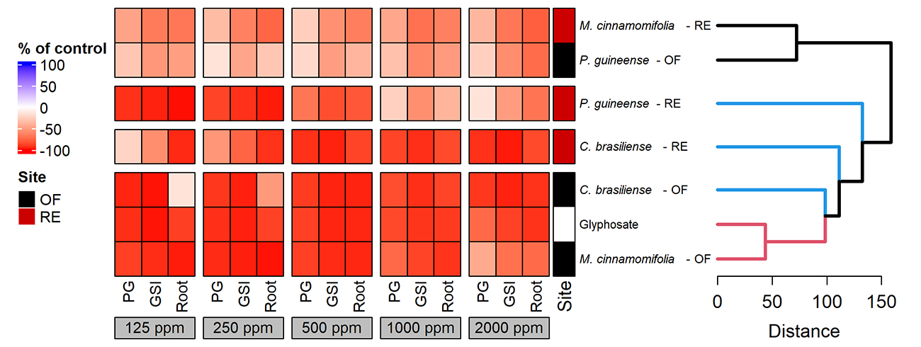
The hierarchical UPGMA cluster analysis compared the similarities among the extracts of C. brasiliense, P. guineense, and M. cinnamomifolia, from both OF and RE, and the herbicide glyphosate, based on their effects on germination and initial development of tomato (Fig. 2). The analysis indicated the formation of two groups. Group A gathers the M. cinnamomifolia RE and the P. guineense OF extracts, based on lower inhibition percentages than the other treatments. Group B is the largest group and contains the remaining treatments. The P. guineense RE extract is the most distant treatment in Group B, due to its mild inhibitory effect at 1000 and 2000 ppm, whereas the lowest concentrations (125 and 250 ppm) were highly inhibitory. The C. brasiliense RE extract is separated from the other remaining treatments in Group B, since its 125 and 250 ppm concentrations caused lower inhibition in PG and GSI. Still inside Group B, the M. cinnamomifolia OF extract, the C. brasiliense OF extract, and the glyphosate herbicide were clustered together due to their high inhibition values at most concentrations. Since the C. brasiliense OF extract caused less inhibition of root length at 125 and 250 ppm (below -50 % of control), the M. cinnamomifolia OF extract and glyphosate were the most similar, as their phytotoxicity was high at all concentrations.
Hierarchical cluster analysis of the effects of the methanolic partitions of Miconia cinnamomifolia and Psidium guineense and lower fraction of Calophyllum brasiliense, at concentrations of 2000, 1000, 500, 250, 125 ppm, on the Germination Percentage (PG), Germination Speed Index (GSI), and Root Length (Root) of Solanum lycopersicum. The dendrogram was built using a Euclidean distance coefficient and the UPGMA (unweighted pair group method using arithmetic averages) method. Values used were percentage differences from the negative control (distilled water) and are displayed as a heatmap varying from 100 % inhibition (-100, red) to 100 % stimulation (100, blue).
Discussion
The fractions of the analyzed extracts exhibited chemical profiles rich in phenolic compounds. All three species showcased hydrolysable tannins and flavonoids, but with some differences in flavonoid types. Flavonoids, phenolic compounds, and their degradation products play a significant role in phytotoxicity (Fiorentino et al., 2008; Li et al., 2010; Mousavi et al., 2021; Weston & Mathesius, 2013). This is further evidenced by the inhibitory effect of their extracts on S. lycopersicum seed germination and root growth, regardless of their phytophysiognomy of origin. Solanum lycopersicum (tomato) is considered a suitable target species in phytotoxicity bioassays, due to its uniform germination and rapid growth (Macías et al., 2000). Phytotoxicity studies suggest that phenolic compounds and flavonoids present inhibitory or stimulatory effects depending on the target species. Macías et al. (1997) found that methylflavonoids from Helianthus annuus L. inhibited tomato seed germination and growth, whereas they caused growth stimulation in L. sativa and Lepidium sativum L.
In this study, the C. brasiliense leaf extracts from both phytophysiognomies significantly inhibited S. lycopersicum germination and root growth. Above 500 ppm, those extracts caused similar effects as the glyphosate herbicide. Such direct interferences are indicative of the phytotoxic potential of C. brasiliense compounds to be explored in the development of bioherbicides. It is worth noting that gallic acid and dihydroxybenzoic acid hexoside were identified in the OF extract of C. brasiliense. The high inhibition in seed germination after the C. brasiliense OF extract can be attributed in part to the presence of these compounds. Pinho et al. (2017) investigated the phytotoxicity of 11 phenolic acids, including gallic acid, protocatechuic acid, and 4-hydroxybenzoic acid, and observed that the phytotoxicity of these acids increased with their hydrophobicity. On the other hand, protocatechuic acid was identified in the RE extract of the same species. Wang et al. (2013) observed that protocatechuic acid is a biodegradation product from (-)-epicatechin, and this process increased the inhibitory effect on Lactuca sativa L. seed germination and radicle growth. Among metabolites found in both OF and RE leaf extracts from this species, we detected quercetin glycosides and astibin 3-O-rhamnoside. Previous studies indicate that O-glycosyl ligands in flavonoids seem to be less phytotoxic than their aglycone counterparts (Martino et al., 2012; Feitoza et al., 2018).
Myrtaceae species have been the focus of studies on phytotoxicity (Imatomi et al., 2013a;b). Extracts from Myrcia tomentosa Glaz. leaves, which were reported to produce 3-O-glycosylflavonols such as juglanin and avicularin, inhibited root length of S. lycopersicum seedlings (Imatomi et al., 2013b). The literature highlights the presence of flavonoids in Psidium, including different types, such as isoflavones (Lapčík et al., 2005), dihydrochalcone derivatives, flavonols (kaemperol, quercetin, and myricetin derivatives), dihydroflavonoids, and catechin (Beltrame et al., 2021). The phytochemical analysis carried out on P. guineense leaves in the present study revealed the presence of flavonols, mainly myricetin, in samples from both phytophysiognomies. The bioassay performed here showed more pronounced phytotoxic action for the RE extract, which exhibited greater inhibition of root length than glyphosate at 125 and 250 ppm. Myricetin is considered phytotoxic (Einhellig et al., 2004) and exhibits the ability to oxidize Fe2+ ions, impacting mitochondria and chloroplasts (Ximenez et al., 2022). This disrupts homeostatic balance, inflicts damage to the photosynthetic apparatus, and triggers oxidative stress, culminating in seedling growth inhibition (Ximenez et al., 2022).
Flavonoid sulphates are a large group of lesser-known compounds that may be found in marine and land plants, including various monocots and eudicots (Barron et al., 1988). The 8-hydroxyluteolin-8-sulfate found in this study is the first report of flavonoid sulphate in Myrtaceae. Moreover, this metabolite was found only in P. guineense individuals in the Restinga environment. Among the functions associated with flavonoid sulphates in plants, other studies cite the relevance of such compounds in salt-tolerant plants, such as mangrove species, and in response to high reactive oxygen species (ROS) (Manurung et al., 2021; Mohammed et al., 2025). Supikova et al. (2022) suggest that, as seen in glycosylated flavonoids, sulfation of flavonoids is associated with compound storage in the vacuole, presenting less toxicity or biological activity.
Regarding luteolin, Beninger & Hall (2005) previously isolated and identified, from Chrysanthemum morifolium L. v. Ramat leaves, the luteolin 7-O-β-glucuronide. These authors demonstrated that the leaf extract of this species, at concentrations of 0.2 and 2.0 mM, significantly reduced the frond number and chlorophyll content of Lemna gibba L. The conclusion about the allelopathic activity of luteolin was likely attributed to the ortho-3′,4′-dihydroxy arrangement of the B-flavonoid ring in the compound. In our study, the 8-hydroxyluteolin-8-sulfate presents the aforementioned arrangement at the B-ring, as well as the sulfate group at the C-8 position. Perhaps this compound may exhibit phytotoxic effects as an isolated compound or synergistically with other compounds found. The results found in this previous study serve as a guide for future studies with the flavone identified in the RE extract of P. guineense.
Phytochemical studies of the genus Miconia have identified 148 metabolites, mostly flavonoids and phenolic acids, as well as terpenoids and steroids (da Silva et al., 2022). Despite the extensive metabolite profile, allelopathic and phytotoxic studies are scarce for the genus (Gatti et al., 2004; Isaza et al., 2007; Santos et al., 2015). The present study found M. cinnamomifolia to present the greatest chemical diversity and intraspecific variation in foliar extracts from both environments, including gallic and ellagic acid, tannins, flavones, and flavonols.
The OF extract of M. cinnamomifolia is the most promising treatment among all extracts, due to its diversity of phenolic compounds, its high phytotoxicity in all concentrations tested, and the high similarity of phytotoxic effect with the glyphosate herbicide. This inhibitory effect may be associated with the presence of flavonoids, mainly quercetin. The accumulation of quercetin in the meristematic zone of the root apex interferes with the transport of auxin to adjacent cells, causing changes in the hormonal gradient and, therefore, limiting cell proliferation and radicle growth (Cheynier et al., 2013; Agati et al., 2021; Singh et al., 2021). This extract also contained pedunculagin and tellimagrandin II. These compounds demand close attention since they have been previously reported for their phytotoxic potential, with impacts on the development of target species through interactions with proteins or metals (Barry et al., 2001; Grundhöfer et al., 2001).
The present bioassay with the RE extract of M. cinnamomifolia showed inhibitory effects on the root length of S. lycopersicum at all concentrations. Individuals of M. cinnamomifolia from RE exhibited a diverse array of phenolic compounds, with a predominance of hydrolyzed tannins, identified as vescalaginin and castalagin. Studies on the phytotoxic activity of these compounds are scarce. Our novel findings about M. cinnamomifolia phytochemical composition and phytotoxic potential highlight the importance of further elucidation of plant chemical diversity, plant chemical traits, and their potential biological activities.
The present bioassay analysis revealed significant inhibitory effects on germination potential, germination speed, and root growth after treatment with OF leaf extracts of C. brasiliense and M. cinnamomifolia. A similar negative impact on seedling development was observed following treatment with the RE leaf extract of P. guineense. Compounds such as quercetin (Fernández-Aparicio et al., 2021), gallic acid (Patterson, 1981; Sodaeizadeh et al., 2009), dihydroxybenzoic acid hexoside (Hussain et al., 2020; Vieites‐Álvarez et al., 2023), luteolin and its derivatives (Beninger & Hall, 2005; Hussain et al., 2020), which were found in all three of the studied species, have been cited in the literature as phytotoxic compounds on target plants. It is important to note that plant chemicals may interact with each other, either antagonizing their effect or presenting a synergic effect (Silva et al., 2013; Feitoza et al., 2018). In other words, mixtures of plant chemicals may present an increased or decreased phytotoxicity in comparison to their isolated effect. These unprecedented results may guide future experiments with isolated and/or combined compounds found in the studied species, are important guidelines for new ecological studies, and show the chemical potential produced by these species that allows their establishment in two different phytophysiognomies.
The results regarding abiotic factors in the two phytophysiognomies reiterate the same pattern of variation observed for the Restinga, characterized by high irradiance, and for the Ombrophilous Forest, where air humidity is higher (Pireda et al., 2019; Oliveira et al., 2023; Xavier et al., 2023). Changes in abiotic factors, such as light and water availability, may be reflected in distinct chemical profiles (Arnold et al., 2019; Yang et al., 2018). Biosynthesis of plant phenolics occurs from the shikimate-phenylpropanoid pathway, and this biosynthetic route is affected by environmental stresses, including high light irradiation (Lattanzio, 2013). Phenylalanine ammonia-lyase (PAL) is a crucial enzyme of the phenylpropanoid pathway, and its activity is increased in response to biotic and abiotic factors, enhancing protection against UV radiation and ROS (Barros & Dixon, 2020).
In response to our hypothesis, only M. cinnamomifolia exhibited chemical intraspecific variation between the Atlantic Forest phytophysiognomies. The M. cinnamomifolia OF extract presented higher phytotoxicity, similar to the herbicide glyphosate. Moreover, C. brasiliense and P. guineense had little variation in the chemical profiles. While C. brasiliense extracts had similar phytotoxic effects at high extract concentrations, the P. guineense RE extract was more inhibitory in lower concentrations. We reported for the first time the occurrence of 8-hydroxyluteolin-8-sulfate in P. guineense RE extract. This uncommon flavonoid group, which is restricted to some plant families, is characterized by its biological activities, highlighting its potential in the development of new pharmaceuticals.
The most bioactive extracts in this study can be manipulated to produce bioherbicides through the isolation of promising compounds and their structural modification. Phenolic compounds, as the structures identified in this study, are generally associated with the response to UV radiation and radical scavenging ability, but our findings indicate a significant role of phenolic compounds in the mediation of plant-plant interaction processes in natural plant communities. As the comprehension of chemical diversity in a plant community can be as important as knowing the taxonomic diversity, knowledge of the chemical profile can subsidize the selection of plant species for ecosystem restoration, preventing harmful effects to species diversity and associated components. Studies must be addressed to enhance the knowledge about the functionality of chemical traits and their biological activities.
Supplementary Material
The following online material is available for this article:
Table S1:
Figure S1:
Figure S2:
Figure S3:
Figure S4:
Figure S5:
Figure S6:
Acknowledgements
We thank Herick Viana and Adilson Martins for collecting the species in the study areas. We thank Dr. Jose Fernando Andrade Baumgratz for taxonomic classification. We thank B. F. Ribeiro and Dr. Marcelo da Silva Mathias for technical work in the laboratory of LBCT/CBB/UENF and LCQ/CCT/UENF. We thank the Multiuser Center (CME-LBCT) for providing infrastructure and the Advanced Microscopy Unit (AMU-CENABIO) of the Federal University of Rio de Janeiro. This study is a part of the Ph.D. dissertation of R.P.A. at UERJ.
References
-
Abdulla R, Mansur S, Lai H et al. 2017. Qualitative análises of polyphenols in Macroporous resin pomegranate husks extract by HPLC-QTOF-MS. Phytochemical Analysis 28:5. 465-473. doi: 10.1002/pra.2695.
» https://doi.org/10.1002/pra.2695 -
Agati G, Guidi L, Landi M, Tattini M. 2021. Anthocyanins in photoprotection: Knowing the actors in play to solve this complex ecophysiological issue. The New Phytologist 232: 2228. doi: 10.1111/nph.17648
» https://doi.org/10.1111/nph.17648 -
Anjum T, Bajwa R. 2005. Importance of germination indices in interpretation of allelochemical effects. International Journal of Agriculture & Biology 7: 417-419. doi: 1560-8530/2005/07-3-417-419
» https://doi.org/1560-8530/2005/07-3-417-419 -
Arnold PA, Kruuk LEB, Nicotra AB. 2019. How to analyse plant phenotypic plasticity in response to a changing climate. New Phytologist 222: 1235-1241. doi: 10.1111/nph.15656
» https://doi.org/10.1111/nph.15656 - Barral MT, Paradelo R. 2011. A review on the use of phytotoxicity as a compost quality indicator. Dynamic Soil, Dynamic Plant 5: 36-44.
-
Barron D, Varin L, Ibrahim RK, Harborne JB, Williams CA. 1988. Sulphated flavonoids - an update. Phytochemistry 27: 2375-2395. doi: 10.1016/0031-9422(88)87003-1
» https://doi.org/10.1016/0031-9422(88)87003-1 -
Barros J, Dixon RA. 2020. Plant Phenylalanine/Tyrosine Ammonia-lyases. Trends in Plant Science 20: 66-79. doi: 10.1016/j.tplants.2019.09.011
» https://doi.org/10.1016/j.tplants.2019.09.011 -
Barry KM, Davies NW, Mohammed CL. 2001. Identification of hydrolysable tannins in the reaction zone of Eucalyptus nitens wood by high performance liquid chromatography - electrospray ionisation mass spectrometry. Phytochemical Analysis 12: 120-127. doi: 10.1002/pca.548
» https://doi.org/10.1002/pca.548 -
Beltrame BM, Klein‐Junior LC, Schwanz M, Henriques AT. 2021. Psidium L. genus: A review on its chemical characterization, preclinical and clinical studies. Phytotherapy Research 35: 4795-4803. doi: 10.1002/ptr.7112
» https://doi.org/10.1002/ptr.7112 -
Beninger CW, Hall JC. 2005. Allelopathic activity of luteolin 7-O-β-glucuronide isolated from Chrysanthemum morifolium L. Biochemical Systematics and Ecology 33: 103-111. doi: 10.1016/j.bse.2004.06.016
» https://doi.org/10.1016/j.bse.2004.06.016 -
Bomfim EMS, Coelho AAOP, Silva MC, Marques EJ, Vale VLC. 2021. Phytochemical composition and biological activities of extracts from ten species of the family Melastomataceae Juss. Brazilian Journal of Biology 82: e242112. doi: 10.1590/1519-6984.242112
» https://doi.org/10.1590/1519-6984.242112 -
Borges NL, Pireda S, Oliveira DS et al. 2022. The functional variability of the morphoanatomical and physiological traits of native species leaves in a flooded tropical forest. Trees 36: 1837-1851. doi: 10.1007/s00468-022-02332-x
» https://doi.org/10.1007/s00468-022-02332-x -
Breiter T, Laue C, Kressel G. et al. 2011. Bioavailabity and antioxidant potencial of different rooibos formulation. Food Chemistry 128:2. 338-347. doi: 10.1016/j.foodchem.2011.03.029
» https://doi.org/10.1016/j.foodchem.2011.03.029 -
Cabral FN, Trad RJ, Amorim BS, Fantin C, Albuquerque PM, Cardozo NMD. 2021. Flora da Reserva Ducke, Amazonas, Brasil: Calophyllaceae. Rodriguésia 72: e01942018. doi: 10.1590/2175-7860202172002
» https://doi.org/10.1590/2175-7860202172002 - Castellano D. 2001. Optimización de bioensayos alelopáticos. Aplicación en la búsqueda de herbicidas naturales. PhD Thesis, Universidad de Cádiz, Spain.
-
Cerqueira AF, Rocha-Santos L, Benchimol M, Mielke MS. 2021. Habitat loss and canopy openness mediate leaf trait plasticity of an endangered palm in the Brazilian Atlantic Forest. Oecologia 196: 619-631. doi: 10.1007/s00442-021-04879-x
» https://doi.org/10.1007/s00442-021-04879-x -
Cheng F, Cheng Z. 2015. Research progress on the use of plant allelopathy in agriculture and the physiological and ecological mechanisms of allelopathy. Frontiers in Plant Science 6: 1020. doi: 10.3389/fpls.2015.01020
» https://doi.org/10.3389/fpls.2015.01020 -
Cheynier V, Comte G, Davies KM, Lattanzio V, Martens S. 2013. Plant phenolics: Recent advances on their biosynthesis, genetics, and ecophysiology. Plant Physiology and Biochemistry 72: 1-20. doi: 10.1016/j.plaphy.2013.05.009
» https://doi.org/10.1016/j.plaphy.2013.05.009 -
da Silva VB, Almeida-Bezerra JW, Costa AR et al. 2022. The genus Miconia Ruiz & Pav. (Melastomataceae): Ethnomedicinal uses, pharmacology, and phytochemistry. Molecules 27: 4132. doi: 10.3390/molecules27134132
» https://doi.org/10.3390/molecules27134132 -
da Silva Mathias M, Oliveira RR. 2018. Differentiation of the phenolic chemical profiles of Cecropia pachystachya and Cecropia hololeuca. Phytochemical Analysis 30: 73-82. doi: 10.1002/pca.2791
» https://doi.org/10.1002/pca.2791 -
Deng X, Gao G, Zheng S, Li F. 2008. Qualitative and quantitative analysis of flavonoids in the leaves of Isatis indigatica Fort. by ultra-performance liquid chromatography with PDA and electrospray ionization tandem mass spectrometry detection. Journal of Pharmaceutical and Biomedical Analysis 48: 562-567. doi: 10.1016/j.jpba.2008.05.020
» https://doi.org/10.1016/j.jpba.2008.05.020 - Einhellig FA, Galindo JC, Molinillo JM, Cutler HG. 2004. Mode of allelochemical action of phenolic compounds. In: Macías FA, Galindo JCG, Molinillo JMG (eds.). Allelopathy: Chemistry and mode of action of allelochemicals. Boca Ratón, CRC Press. p. 217-38.
-
Feitoza RBB, Lima HRP, Oliveira EAG et al. 2018. Structural and ultrastructural variations in roots of Calopogonium mucunoides Desv. treated with phenolic compounds from Urochloa humidicola (Rendle) Morrone & Zuloaga and phenolic commercial standards. South African Journal of Botany 116: 142-149. doi: 10.1016/j.sajb.2018.03.005
» https://doi.org/10.1016/j.sajb.2018.03.005 -
Feitoza RBB, Varela RM, Torres A et al. 2020. Evaluation of the phytotoxicity of Urochloa humidicola roots by bioassays and microscopic analysis. Characterization of new compounds. Journal of Agricultural and Food Chemistry 68: 4851-4864. doi: 10.1021/acs.jafc.0c00307
» https://doi.org/10.1021/acs.jafc.0c00307 -
Fernández-Aparicio M, Masi M, Cimmino A, Vilariño S, Evidente A. 2021. Allelopathic effect of quercetin, a flavonoid from Fagopyrum esculentum roots in the radicle growth of Phelipanche ramosa: Quercetin natural and semisynthetic analogues were used for a structure-activity relationship investigation. Plants 10: 543. doi: 10.3390/plants10030543
» https://doi.org/10.3390/plants10030543 -
Fiorentino A, D'Abrosca B, Pacifico S et al. 2008. Potential allelopatic effects of stilbenoids and flavonoids from leaves of Carex distachya Desf. Biochemical Systematics and Ecology 36: 691-698. doi: 10.1016/j.bse.2008.07.002
» https://doi.org/10.1016/j.bse.2008.07.002 - García-Zebadúa JC, Reyes-Chilpa R, Huerta-Reyes M et al. 2014. El árbol tropical Calophyllum brasiliense: una revisión botánica, química y farmacológica. Vitae 21: 126-145.
-
Gatti AB, Perez SCJGA, Lima MIS. 2004. Atividade alelopática de extratos aquosos de Aristolochia esperanzae O. Kuntze na germinação e no crescimento de Lactuca sativa e Raphanus sativus L. Acta Botanica Brasilica 18: 459-472. doi: 10.1590/S0102-33062004000300006
» https://doi.org/10.1590/S0102-33062004000300006 -
Gillani SW, Ahmad M, Ajmal Ali M et al. 2024. Phyto-ecological studies and distribution patterns of subfamily Polygonoideae in relation to edaphic factors across diverse ecological zones. Heliyon 10: e36571. doi: 10.1016/j.heliyon.2024.e36571
» https://doi.org/10.1016/j.heliyon.2024.e36571 -
Goldenberg R, Baumgratz JFA, Michelangeli FA et al. 2025. Melastomataceae. https://floradobrasil.jbrj.gov.br/FB161 21 May 2025.
» https://floradobrasil.jbrj.gov.br/FB161 -
Grundhöfer P, Niemetz R, Schilling G, Gross GG. 2001. Biosynthesis and subcellular distribution of hydrolyzable tannins. Phytochemistry 57: 915-927. doi: 10.1016/s0031-9422(01)00099-1
» https://doi.org/10.1016/s0031-9422(01)00099-1 -
Hussain MI, El-Sheikh MA, Reigosa MJ. 2020. Allelopathic potential of aqueous extract from Acacia melanoxylon R. Br. on Lactuca sativa. Plants 9: 1228. doi: 10.3390/plants9091228
» https://doi.org/10.3390/plants9091228 -
Imatomi M, Novaes P, Gualtieri SCJ. 2013a. Interspecific variation in the allelopathic potential of the family Myrtaceae. Acta Botanica Brasilica 27: 54-61. doi: 10.1590/S0102-33062013000100008
» https://doi.org/10.1590/S0102-33062013000100008 -
Imatomi M, Novaes P, Matos AP et al. 2013b. Phytotoxic effect of bioactive compounds isolated from Myrcia tomentosa (Myrtaceae) leaves. Biochemical Systematics and Ecology 46: 29-35. doi: 10.1016/j.bse.2012.09.005
» https://doi.org/10.1016/j.bse.2012.09.005 - Isaza JH, Qi FJJG, Usma JLG, Restrepo JC. 2007. Actividad alelopática de algunas especies de los géneros Miconia, Tibouchina, Henriettella, Tococa, Aciotis y Bellucia (Melastomataceae). Scientia Et Technica 13: 409-413.
-
Kessler A, Kalske A. 2018. Plant secondary metabolite diversity and species interactions. Annual Review of Ecology, Evolution, and Systematics 49: 115-138. doi: 10.1146/annurev-ecolsys-110617-062406
» https://doi.org/10.1146/annurev-ecolsys-110617-062406 -
Lapčík O, Klejdus B, Kokoška L et al. 2005. Identification of isoflavones in Acca sellowiana and two Psidium species (Myrtaceae). Biochemical Systematics and Ecology 33: 983-992. doi: 10.1016/j.bse.2005.03.007
» https://doi.org/10.1016/j.bse.2005.03.007 -
Lattanzio V. 2013. Phenolic Compounds: Introduction. In: Ramawat K, Mérillon JM (eds.). Natural Products. Berlin, Heidelberg, Springer. p. 1543-1580. doi: 10.1007/978-3-642-22144-6_57
» https://doi.org/10.1007/978-3-642-22144-6_57 -
Li H, Liu Y, Zhang Z, Liao H, Nie L, Yao S. 2005. Separation and purification of chlorogenic acid by molecularly imprinted polymer monolithic stationary phase. Journal of Chromatography A 1098: 66-74. doi: 10.1016/j.chroma.2005.08.046
» https://doi.org/10.1016/j.chroma.2005.08.046 -
Li ZH, Wang Q, Ruan X, Pan CD, Jiang DA. 2010. Phenolics and plant allelopathy. Molecules 15: 8933-8952. doi: 10.3390/molecules15128933
» https://doi.org/10.3390/molecules15128933 -
Liu X, Shen J, Zhu K. 2022. Antibacterial activities of plant-derived xanthones. RSC Medicinal Chemistry 13: 107-116. doi: 10.1039/d1md00351h
» https://doi.org/10.1039/d1md00351h -
Lorenzo P, Reboredo-Durán J, Múñoz L, González L, Freitas H, Rodríguez-Echeverría S. 2016. Inconsistency in the detection of phytotoxic effects: A test with Acacia dealbata extracts using two different methods. Phytochemistry Letters 15: 190-198. doi: 10.1016/j.phytol.2015.11.001
» https://doi.org/10.1016/j.phytol.2015.11.001 -
Lucas EJ, Bünger MO. 2015. Myrtaceae in the Atlantic Forest: Their role as a ‘model’ group. Biodiversity and Conservation 24: 2165-2180. doi: 10.1007/s10531-015-0992-7
» https://doi.org/10.1007/s10531-015-0992-7 -
Macedo JGF, Rangel JML, Santos MO, Camilo CJ, Costa JGM, Souza MMA. 2021. Therapeutic indications, chemical composition and biological activity of native Brazilian species from Psidium genus (Myrtaceae): A review. Journal of Ethnopharmacology 278: 114248. doi: 10.1016/j.jep.2021.114248
» https://doi.org/10.1016/j.jep.2021.114248 -
Macías FA, Molinillo JMG, Torres A, Varela RM, Castellano D. 1997. Bioactive flavonoids from Helianthus annuus cultivars. Phytochemistry 88: 683-687. doi: 10.1016/S0031-9422(97)00011-3
» https://doi.org/10.1016/S0031-9422(97)00011-3 -
Macías FA, Castellano D, Molinillo JMG. 2000. Search for a standard phytotoxic bioassay for allelochemicals. Selection of standard target species. Journal of Agricultural and Food Chemistry 48: 2512-2521. doi: 10.1021/jf9903051
» https://doi.org/10.1021/jf9903051 -
Manurung J, Kappen J, Schnitzler J et al. 2021. Analysis of Unusual Sulfated Constituents and Anti-infective Properties of Two Indonesian Mangroves, Lumnitzera littorea and Lumnitzera racemosa (Combretaceae). Separations 8: 82. doi: 10.3390/separations8060082
» https://doi.org/10.3390/separations8060082 -
Manyelo TG, Sebola NA, Hassan ZM, Mabelebele M. 2020. Characterization of the phenolic compounds in different plant parts of Amaranthus cruentus grown under cultivated conditions. Molecules 25: 4273. doi: 10.3390/molecules25184273
» https://doi.org/10.3390/molecules25184273 -
Marsh KJ, Saraf I, Hocart CH, Youngentob K, Singh IP, Foley WJ. 2019. Occurrence and distribution of unsubstituted B-ring flavanones in Eucalyptus foliage. Phytochemistry 160: 31-39. doi: 10.1016/j.phytochem.2019.01.005
» https://doi.org/10.1016/j.phytochem.2019.01.005 -
Martins CC, Kahmann A, Anzanello MJ, Rodrigues RC, Rodrigues E, Mercali GD. 2023. Acid hydrolysis conditions do affect the non-extractable phenolic compounds composition from grape peel and seed. Food Research International 174: 113636. doi: 10.1016/j.foodres.2023.113636
» https://doi.org/10.1016/j.foodres.2023.113636 -
Martino L, Mencherini T, Mancini E, Aquino RP, De Almeida LFR, De Feo V. 2012. In vitro phytotoxicity and antioxidant activity of selected flavonoids. International Journal of Molecular Sciences 13: 5406-5419. doi: 10.3390/ijms13055406
» https://doi.org/10.3390/ijms13055406 -
Mohammed HA, Ragab EA, Dutta A et al. 2025. Sulfated flavonoids: An updated and comprehensive review of their chemistry and bioactivities. Phytochemistry Reviews. doi: 10.1007/s11101-025-10112-x
» https://doi.org/10.1007/s11101-025-10112-x -
Mousavi SS, Karami A, Haghighi TM, Alizadeh S, Maggi F. 2021. Phytotoxic potential and phenolic profile of extracts from Scrophularia striata. Plants 10: 135. doi: 10.3390/plants10010135
» https://doi.org/10.3390/plants10010135 -
Olatunde OO, Tan SLD, Shiekh KA, Benjakul S, Nirmal NP. 2021. Ethanolic guava leaf extracts with different chlorophyll removal processes: Anti-melanosis, antibacterial properties and the impact on qualities of Pacific white shrimp during refrigerated storage. Food Chemistry 341: 128251. doi: 10.1016/j.foodchem.2020.128251
» https://doi.org/10.1016/j.foodchem.2020.128251 -
Oliveira DS, Simioni PF, Araújo I et al. 2023. Effects of microclimatic variation on plant leaf traits at the community level along a tropical forest gradient. Trees 37: 1499-1513. doi: 10.1007/s00468-023-02445-x
» https://doi.org/10.1007/s00468-023-02445-x -
Pannakal ST, Eilstein J, Hubert J et al. 2023. Rapid Chemical Profiling of Filipendula ulmaria Using CPC Fractionation, 2-D Mapping of 13C NMR Data, and High-Resolution LC-MS. Molecules 28: 6349. doi: 10.3390/molecules28176349
» https://doi.org/10.3390/molecules28176349 -
Patterson DT. 1981. Effects of allelopathic chemicals on growth and physiological responses of soybean (Glycine max). Weed Science 29: 53-59. doi: 10.1017/S0043174500025820
» https://doi.org/10.1017/S0043174500025820 -
Pavlović AV, Papetti A, Zagorac DČD et al. 2016. Phenolics composition of leaf extracts of raspberry and blackberry cultivars grown in Serbia. Industrial Crops and Products 87: 304-314. doi: 10.1016/j.indcrop.2016.04.052
» https://doi.org/10.1016/j.indcrop.2016.04.052 -
Pereira LD, Barbosa JMG, Silva AJR, Ferri PH, Santos SC. 2017. Polyphenol and ellagitannin constituents of jabuticaba (Myrciaria cauliflora) and chemical variability at different stages of fruit development. Journal of Agricultural and Food Chemistry 65: 1209-1219. doi: 10.1021/acs.jafc.6b02929
» https://doi.org/10.1021/acs.jafc.6b02929 -
Pilatti DM, Fortes AMT, Jorge TCM, Boiago NP. 2018. Comparison of the phytochemical profiles of five native plant species in two different forest formations. Brazilian Journal of Biology 79: 233-242. doi: 10.1590/1519-6984.179526
» https://doi.org/10.1590/1519-6984.179526 -
Pinho IA, Lopes DV, Martins RC, Quina MJ. 2017. Phytotoxicity assessment of olive mill solid wastes and the influence of phenolic compounds. Chemosphere 185: 258-267. doi: 10.1016/j.chemosphere.2017.07.002
» https://doi.org/10.1016/j.chemosphere.2017.07.002 -
Pireda S, Oliveira DS, Borges NL et al. 2019. Acclimatization capacity of leaf traits of species co-occurring in restinga and seasonal semideciduous forest ecosystems. Environmental and Experimental Botany 164: 190-202. doi: 10.1016/j.envexpbot.2019.05.012
» https://doi.org/10.1016/j.envexpbot.2019.05.012 -
R Core Team. 2024. R: A language and environment for statistical computing. R Foundation for Statistical Computing. https://www.R-project.org/ Accessed on 28 Aug. 2025.
» https://www.R-project.org/ -
Dos Santos C, Galaverna RS, Angolini CF et al. 2018. Antioxidative, antiproliferative and antimicrobial activities of phenolic compounds from three Myrcia species. Molecules 23: 986. doi: 10.3390/molecules23050986
» https://doi.org/10.3390/molecules23050986 -
Santos LE, Trindade MGP, Santos VS, Vieira IMC, Kuster RM, Batitucci MCP. 2025. Phytochemical and chemometric investigation of morphotypes of Calophyllum brasiliense Cambess. (Calophyllaceae) in the mountainous region of Espírito Santo, Brazil. Biochemical Systematics and Ecology 120: 104982. doi: 10.1016/j.bse.2025.104982
» https://doi.org/10.1016/j.bse.2025.104982 -
Santos MAF, Silva MAP, Santos ACB et al. 2015. Allelopathy of Miconia spp. (Melastomataceae) in Lactuca sativa L. (Asteraceae). Journal of Agricultural Science 7: 151. doi: 10.5539/jas.v7n12p151
» https://doi.org/10.5539/jas.v7n12p151 -
Santos PF, Gomes LN, Mazzei JL et al. 2018. Polyphenol and triterpenoid constituents of Eugenia florida DC. (Myrtaceae) leaves and their antioxidant and cytotoxic potential. Química Nova 41: 1140-1149. doi: 10.21577/0100-4042.20170284
» https://doi.org/10.21577/0100-4042.20170284 -
Senanayake CM, Hapugaswatta H, Jayathilaka N, Seneviratne KN. 2018. Phenolic extracts of the leaves of Psidium guineense Sw. improve the shelf life of sunflower oil and baked cake and antioxidant status of Wistar rats. Journal of Food Biochemistry 42: e12632. doi: 10.1111/jfbc.12632
» https://doi.org/10.1111/jfbc.12632 -
Sheng F, Hu B, Jin Q, Wang J, Wu C, Luo Z. 2021. The analysis of phenolic compounds in walnut husk and pellicle by UPLC-Q-Orbitrap HRMS and HPLC. Molecules 26: 3013. doi: 10.3390/molecules26103013
» https://doi.org/10.3390/molecules26103013 -
Silva EAS, Lôbo LT, Silva GA et al. 2013. Flavonoids from leaves of Derris urucu: Assessment of potential effects on seed germination and development of weeds. Anais da Academia Brasileira de Ciências 85: 881-889. doi: 10.1590/S0001-37652013000300004
» https://doi.org/10.1590/S0001-37652013000300004 -
Silva BP, Nepomuceno MP, Varela RM et al. 2017. Phytotoxicity study on Bidens sulphurea Sch. Bip. as a preliminary approach for weed control. Journal of Agricultural and Food Chemistry 65: 5161-5172. doi: 10.1021/acs.jafc.7b01922
» https://doi.org/10.1021/acs.jafc.7b01922 -
Singh P, Arif Y, Bajguz A, Hayat S. 2021. The role of quercetin in plants. Plant Physiology and Biochemistry 166: 10-19. doi: 10.1016/j.plaphy.2021.05.023
» https://doi.org/10.1016/j.plaphy.2021.05.023 -
Soares JC, Rosalen PL, Lazarini JG et al. 2019. Comprehensive characterization of bioactive phenols from new Brazilian superfruits by LC-ESI-QTOF-MS, and their ROS and RNS scavenging effects and anti-inflammatory activity. Food Chemistry 281: 178-188. doi: 10.1016/j.foodchem.2018.12.106
» https://doi.org/10.1016/j.foodchem.2018.12.106 -
Sobrinho AP, Minho AS, Ferreira LL, Martins GR, Boylan F, Fernandes PD. 2017. Characterization of anti-inflammatory effect and possible mechanism of action of Tibouchina granulosa. Journal of Pharmacy and Pharmacology 69: 706-713. doi: 10.1111/jphp.12712
» https://doi.org/10.1111/jphp.12712 -
Sodaeizadeh H, Rafieiolhossaini M, Havlík J, Van Damme P. 2009. Allelopathic activity of different plant parts of Peganum harmala L. and identification of their growth inhibitors substances. Plant Growth Regulation 59: 227-236. doi: 10.1007/s10725-009-9408-6
» https://doi.org/10.1007/s10725-009-9408-6 -
Supikova K, Kosinova A, Vavrusa M et al. 2022. Sulphated phenolic acids in plants. Planta 255: 124. doi: 10.1007/s00425-022-03902-6
» https://doi.org/10.1007/s00425-022-03902-6 -
Vasconcelos MAL, Royo VA, Ferreira DS et al. 2006. In vivo analgesic and anti-inflammatory activities of ursolic acid and oleanoic acid from Miconia albicans (Melastomataceae). Zeitschrift für Naturforschung C 61: 477-482. doi: 10.1515/znc-2006-7-803
» https://doi.org/10.1515/znc-2006-7-803 -
Verma N, Shukla S. 2015. Impact of various factors responsible for fluctuation in plant secondary metabolites. Journal of Applied Research on Medicinal and Aromatic Plants 2: 105-113. doi: 10.1016/j.jarmap.2015.09.002
» https://doi.org/10.1016/j.jarmap.2015.09.002 -
Vieites‐Álvarez Y, Otero P, Prieto MA et al. 2023. Testing the role of allelochemicals in different wheat cultivars to sustainably manage weeds. Pest Management Science 79: 2625-2638. doi: 10.1002/ps.7444
» https://doi.org/10.1002/ps.7444 -
Wagner MA, Bogoni JA, Fiaschi P. 2022. Myrtaceae richness and distribution across the Atlantic Forest Domain are constrained by geoclimatic variables. Plant Ecology 223: 1079-1092. doi: 10.1007/s11258-022-01258-1
» https://doi.org/10.1007/s11258-022-01258-1 -
Wang C-M, Li T-C, Jhan Y-L, Weng J-H, Chou C-H. 2013. The Impact of Microbial Biotransformation of Catechin in Enhancing the Allelopathic Effects of Rhododendron formosanum. PLoS One 8: e85162. doi: 10.1371/journal.pone.0085162
» https://doi.org/10.1371/journal.pone.0085162 -
Weston LA, Mathesius U. 2013. Flavonoids: Their structure, biosynthesis and role in the rhizosphere, including allelopathy. Journal of Chemical Ecology 39: 283-297. doi: 10.1007/s10886-013-0248-5
» https://doi.org/10.1007/s10886-013-0248-5 -
Xavier V, Pireda S, Oliveira DS, Vitória AP, Da Cunha M. 2023. Leaf and wood functional traits explain the strategies developed by Byrsonima sericea (Malpighiaceae) to survive in Atlantic Forest ecosystems under water and light variations. Flora 308: 152386. doi: 10.1016/j.flora.2023.152386
» https://doi.org/10.1016/j.flora.2023.152386 -
Ximenez GR, Bianchin M, Carmona JMP, Oliveira SM, Ferrarese-Filho O, Pastorini LH. 2022. Reduction of Weed Growth under the Influence of Extracts and Metabolites Isolated from Miconia spp. Molecules 27: 5356. doi: 10.3390/molecules27175356
» https://doi.org/10.3390/molecules27175356 -
Yang L, Wen KS, Ruan X, Zhao YX, Wei F, Wang Q. 2018. Response of plant secondary metabolites to environmental factors. Molecules 23: 762. doi: 10.3390/molecules23040762
» https://doi.org/10.3390/molecules23040762 -
Yeo J, Shahidi F. 2020. Identification and quantification of soluble and insoluble-bound phenolics in lentil hulls using HPLC-ESI-MS/MS and their antioxidant potential. Food Chemistry 315: 126202. doi: 10.1016/j.foodchem.2020.126202
» https://doi.org/10.1016/j.foodchem.2020.126202 -
Zailan AAD, Karunakaran T, Abu Bakar MH, Mian VJY. 2022. The Malaysian genus Calophyllum (Calophyllaceae): A review on its phytochemistry and pharmacological activities. Natural Product Research 36: 4569-4579. doi: 10.1080/14786419.2021.1982936
» https://doi.org/10.1080/14786419.2021.1982936 -
Zengin G, Cvetanović A, Gašić U et al. 2019. Chemical composition and bio-functional perspectives of Erica arborea L. extracts obtained by different extraction techniques: Innovative insights. Industrial Crops and Products 142: 111843. doi: 10.1016/j.indcrop.2019.111843
» https://doi.org/10.1016/j.indcrop.2019.111843
-
Data Availability
Data and R scripts related to this article will be available upon request to the corresponding author.
-
Funding Information
This study was financed in part by the Coordenação de Aperfeiçoamento de Pessoal de Nível Superior - Brasil (CAPES) - Finance Code 001; Conselho Nacional de Desenvolvimento Científico e Tecnológico -CNPq(308267/2021-4); and Fundação de Amparo à Pesquisa do Rio de Janeiro -FAPERJ(CNE - E26/203.995/2024; Temáticos - E-26/211.339/2021).
Data and R scripts related to this article will be available upon request to the corresponding author.