 ADVANCING THE ONTOGENY OF HOP FLOWERS (HUMULUS LUPULUS L., CANNABACEAE) WITH NEW INSIGHTS INTO PERIANTH AND GYNOECIUM STRUCTURE
Abstract
Humulus lupulus L., commonly known as Hop, is an economically important species in the Cannabaceae family, characterized by diclinous, monochlamydeous, and pentamerous flowers. This study updates the descriptions of the developmental stages of staminate and pistillate flowers in H. lupulus, with a focus on the processes leading to its unique floral features, such as apetaly and a gynoecium containing a single ovule and two long stigmatic branches. Flower buds and flowers of various sizes were processed for surface and histological examination. In addition, the vascularization of the calyx and gynoecium was studied using X-ray micro-computed tomography and clearing techniques. The vascularization and ontogeny confirm the sepal nature of the perianth and reveal dimorphism between the carpels of the flower. This dimorphism supports the pseudomonomerous nature of the gynoecium, where one carpel nourishes the ovule while the other contributes only to the ovary wall. Each carpel produces a stigmatic branch, a feature also observed in other species of Cannabaceae. A notable finding is the presence of an intragynoecial compitum. This study enhances our understanding of the floral structure in H. lupulus through the use of new protocols for floral organ dissection and analysis, including a modified protocol for x-ray micro-computed tomography.
Keywords:
anatomy; apetaly; floral morphology; floral ontogeny; Rosales; Urticalean rosids
Introduction
Humulus lupulus L., commonly known as hop, has been cultivated by human communities for thousands of years (Kubitzki, 1993) and is currently valued primarily as a flavoring agent in beer production (Almaguer et al., 2014). Along with Cannabis sativa L. and approximately 100 other species, it belongs to Cannabaceae, an economically important family of angiosperms. This family includes herbs, shrubs, trees, and vines with cosmopolitan distribution (The Plant List, 2013).
The genus Humulus comprises three species: H. lupulus, H. yunnanensis Hu, and H. japonicus Siebold & Zucc. (Zanoli & Zavatti, 2008). Humulus lupulus and H. japonicus, cultivated as an ornamental plant, are the most economically significant species, particularly in the pharmaceutical and beverage industries (Zanoli & Zavatti, 2008; Almaguer et al., 2014; Karabín et al., 2016). Hops are vines whose stems coil clockwise around the support, with simple 3-5 lobed, long-petiolate opposite leaves, with serrated margins. The species is dioecious, with racemose inflorescences. Individuals bearing pistillate flowers have inflorescences protected by large bracts (Zanoli & Zavatti, 2008). The flowers are monoclamydeous and pentamerous, with a bicarpellate, syncarpous gynoecium, as well as one bract and two prophylls in the pistillate flowers (Shephard et al., 2000).
Advancing studies on the formation of the Hop flower is important from various perspectives. From an economic standpoint, various bioactive compounds are produced in the inflorescence, which may vary in quantity and concentration throughout the developmental stages (Kavalier et al., 2011). From a morphological perspective, this study can contribute to understanding the floral architecture patterns of members of the Cannabaceae family (see Leme et al., 2020) and even of other families belonging to the Rosales order (APG IV, 2016). From an evolutionary perspective, floral development data of Cannabaceae species such as Cannabis sativa, Celtis iguanaea (Jacq.) Sarg., Trema micrantha (L.) Blume (Leme et al., 2020), and Humulus lupulus (Shephard et al., 2000) can provide useful morphological data to formulate phylogenetic hypotheses for the family (De Pinna, 1991). Therefore, more details about the flower development of these species are highly welcome.
The flower of most representatives of Cannabaceae has only one perianth whorl that corresponds to the calyx (Shephard et al., 2000; Leme et al., 2020). The presence of two floral morph types has two possible developmental paths. One involves the abortion of one of the whorls that houses the gametophytes (androecium/gynoecium), as seen in Celtis iguanaea and Trema micrantha, which leads to the late differentiation of pistillate and staminate flowers (Leme et al., 2020). The second ontogenetic pathway for dicliny is observed in Cannabis sativa (Leme et al., 2020) and Humulus lupulus (Shephard et al., 2000), with the absence of stamens or carpels from the beginning of floral development, characterizing an early differentiation of floral morphotypes and the absence of vestigial organs.
The vegetative and floral development of H. lupulus has been previously described using light and scanning electron microscopy techniques, permitting an early distinction between developing pistillate and staminate flowers and thus making it possible to determine whether an individual produces pistillate or staminate flowers by the analysis of inflorescence meristems. Abortion of stamens and carpels was reported (Shephard et al., 2000). However, the perianth of pistillate flowers has not been unanimously characterized, with authors describing it as sepalar or as an undifferentiated perianth (Bechtel, 1921; Shephard et al., 2000). Also, the gynoecium, whether monomerous or pseudomonomerous (= presence of two or more carpels, with a unilocular and uniovulate ovary), and the two stigmatic branches origin (two to six described for members of the order), which are important morphological conditions for the evolutionary success of this group, were not specifically addressed in previous papers about Hop morphology and deserve detailed investigation. Such conditions have been studied by Leme et al. (2018; 2020), Pedersoli et al. (2020; 2022), Pedersoli & Teixeira (2020), and Leite et al. (2018; 2020) in other species of Cannabaceae and of the Rosales order.
This study aimed to update the understanding of the development of staminate and pistillate flowers of Humulus lupulus, with a particular focus on the perianth and gynoecium. We aimed to elucidate the ontogenetic pathways that lead to the distinctive floral characteristics observed in this species. To achieve this, four main questions were addressed: (a) Are the pistillate and staminate flowers truly apetalous? (b) Is the gynoecium pseudomonomerous, as observed in other species within Cannabaceae and Rosales (see Leite et al., 2020)? (c) Do the stigmatic branches result from the fusion of two carpels or the division of the stigma of a single carpel, as occurs in Ulmaceae (Leme et al., 2018) and Moraceae (Leite et al., 2018; 2020)? (d) Do the ontogenetic characteristics of Hop flowers support the current taxonomic placement of the genus Humulus within Cannabaceae, particularly its close relationship to Cannabis sativa?
Material and methods
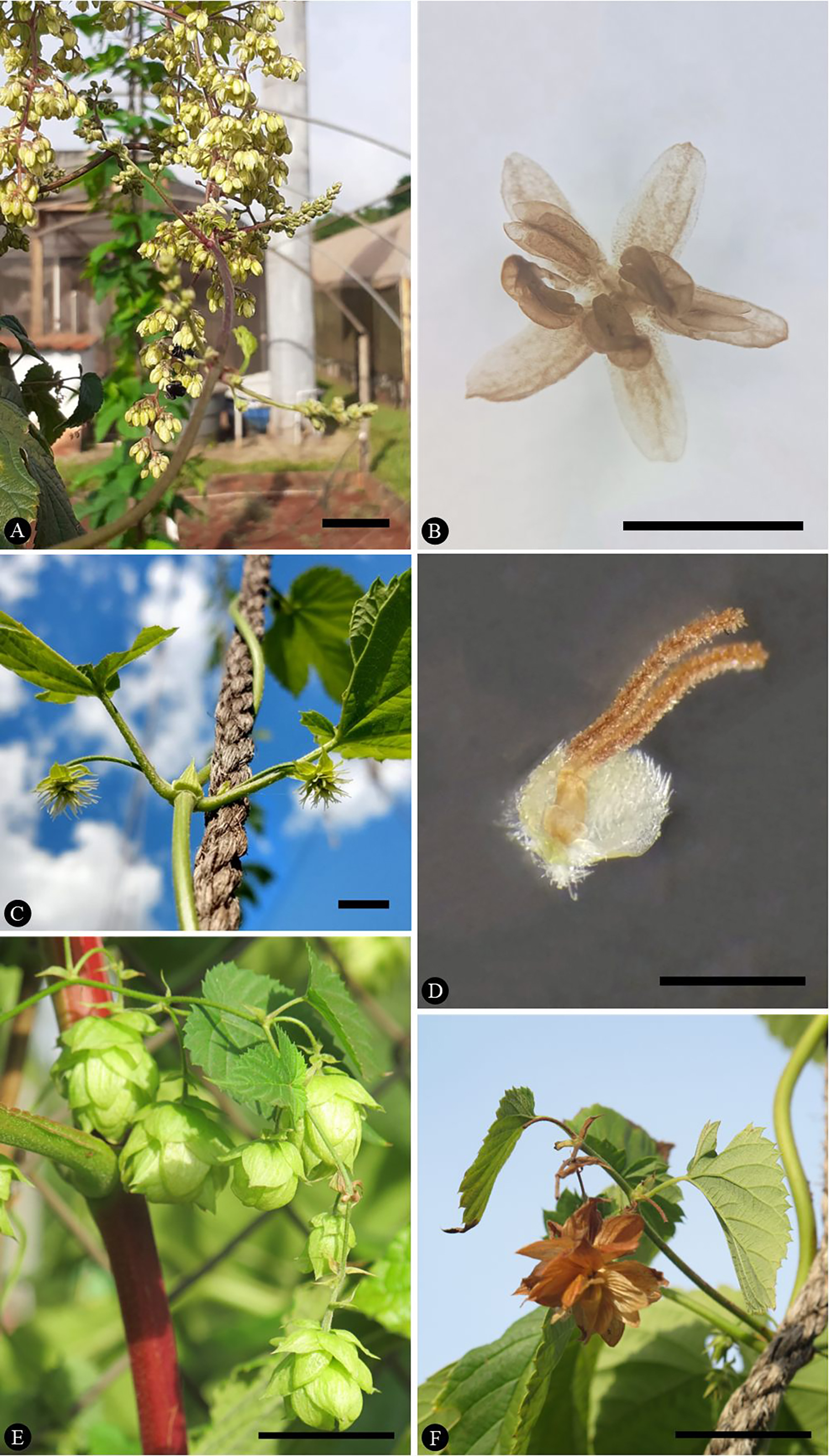
The collected material consisted of inflorescences with staminate flowers collected from individuals cultivated at UNESP, Jaboticabal, Brazil (Figure 1 A , B) and inflorescences with pistillate flowers collected from individuals cultivated in the Medicinal Garden of FCFRP/USP, Ribeirão Preto, Brazil (Figure 1 C -F). Vouchers are deposited in the LabBot-FCFRP/USP spirit collection with numbers 226 (Jaboticabal population) and 227 (Ribeirão Preto population), and in SPFR herbarium (FFCLRP/USP) with accession number 17861.
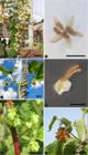
Photographs of Humulus lupulus plants. A. Branch of a staminate plant showing staminate flowers B. A staminate flower in closeup. C. Branch of a pistillate plant showing pistillate flowers. D. A pistillate flower in closeup. E. Branch of a pistillate plant showing an inflorescence with bracteates, pistillate flowers in the process of dehiscence. F. Branch of a pistillate plant showing an old inflorescence with dehiscing bracts. Scale bars: A, B = 2 cm; C = 1 cm, D = 0.1 cm; E, F = 3 cm.
The samples were fixed in buffered formalin (Lillie, 1954 apud Clark, 1981) or Karnovsky’s solution (Karnovsky, 1965), dissected with the aid of a Leica MZ 75 stereomicroscope (Heerbrugg, Switzerland), and processed for surface (scanning electron microscopy - SEM), anatomical (light microscopy - LM) and vascularization (microscopic computed tomography - micro-CT) analyses.
For SEM observations, samples were dissected under a stereomicroscope, dehydrated in an ethanol series, critical point dried with a Bal Tec CPD 030 instrument (Schalksmühle, Märkischer Kreis, North Rhine-Westphalia, Germany), mounted on metal supports on carbon adhesive tape and sputtered with gold with a Bal Tec SCD 050 sputter coater (Balzers, Liechtenstein). Electron micrographs were obtained with a Jeol JSM 6610LV (Jeol Ltd., Tokyo, Japan) scanning electron microscope under 25 kV.
For LM observation, samples were dehydrated in an ethanol series, embedded in historesin (Gerrits & Horobin, 1991), and transversely and longitudinally cut into 5-μm thick sections with a rotary microtome (Leica RM 2245 - Nussloch, Germany). Sections were stained with 0.05% Toluidine Blue in phosphate buffer, pH 6.8 (O’Brien et al., 1964), mounted on water (Gerlach, 1969), and observed under a Leica DM 5000B (Wetzlar, Hesse, Germany) light microscope. Photomicrographs were taken with a Leica DFC 295 digital camera attached to a microscope (Leica DM 5000B - Wetzlar, Hesse, Germany).
For Micro-CT analysis, the gynoecia of anthetic pistillate flowers were treated with a solution of 1% phosphotungstic acid in 70% ethanol for 1 week (Staedler et al., 2013), dehydrated in an ethanol series with 1% phosphotungstic acid as a contrasting agent, critical point dried (Bal Tec CPD 030), mounted on a wooden skewer or microtubules with two-component Loctite Durepoxi adhesive paste and Leica historesin (see supplementary material 1), and scanned. The scans were obtained with a Phoenix v|tome|x S240 imaging system (General Electric) with a 180kV Microfocus X-ray source using the following settings: voltage, 60 kV; source current, 200 μA exposure time, 0.3 ms; pictures per sample, 1000; camera binning, 1; and optical magnification 108.8×, with a voxel size of 1.8, 1,99 and 2,7 μm. The datos x|2 software (General Electric) was used to perform the 3D reconstruction from the scan data. The AMIRA software was used for visualization of the scan data.
For diaphanization/clearance, the gynoecium and sepals of pistillate and staminate flowers were cleared and stained with safranin to analyze the vascular bundles. Previously fixed materials were dissected and rehydrated in an ethanol series. The material was left in 50% alcohol for two hours, in 30% alcohol for two hours, in 10% alcohol for 2 hours, and then left in water overnight. Next, the protocol by Monteiro et al. (1979) was adapted, using different concentrations of NaOH and sodium hypochlorite solutions, without dehydrating the material again for staining with safranin. The material was inserted into a 50% NaOH solution, left in an oven at 50ºC for 2 hours, washed three times with distilled water, placed in a 50% sodium hypochlorite solution, and left at 50°C until clarification. Next, the material was washed thrice with distilled water and stained with 1% safranin. Photomicrographs were taken with a Leica DFC 295 digital camera.
Results
Flower morphology
The staminate flowers (Figs. 1A, B) consist of two whorls: the calyx and the androecium, with the corolla and gynoecium absent. The calyx comprises five free, slightly yellow sepals, approximately 2.8 mm in length, while the androecium consists of five free yellow stamens measuring about 2.4 mm in length. Each flower is accompanied by a small bract that partially protects the flower. The anthers are elongated, with longitudinal dehiscence, and inserted at the base into a short filament (Fig. 1 B ).
The pistillate flowers (Figs. 1C-F) are sessile and occur in the axils of bracts, which occur in pairs in the axils of bracts on the inflorescence. The flowers consist of two whorls: the calyx and the gynoecium. The corolla and stamens are completely absent. The calyx consists of three fully united sepals, forming a campanulate structure that surrounds the lower portion of the ovary. Although not completely covered by the sepals, the ovary is protected by a large, pale green bracteole. The gynoecium comprises a unilocular, uniovulate, slightly green ovary measuring approximately 0.4 mm in length, and two long, papillose stigmatic branches, about 2 mm in length, which are slightly green and turn brown with senescence.
Development of pistillate flower
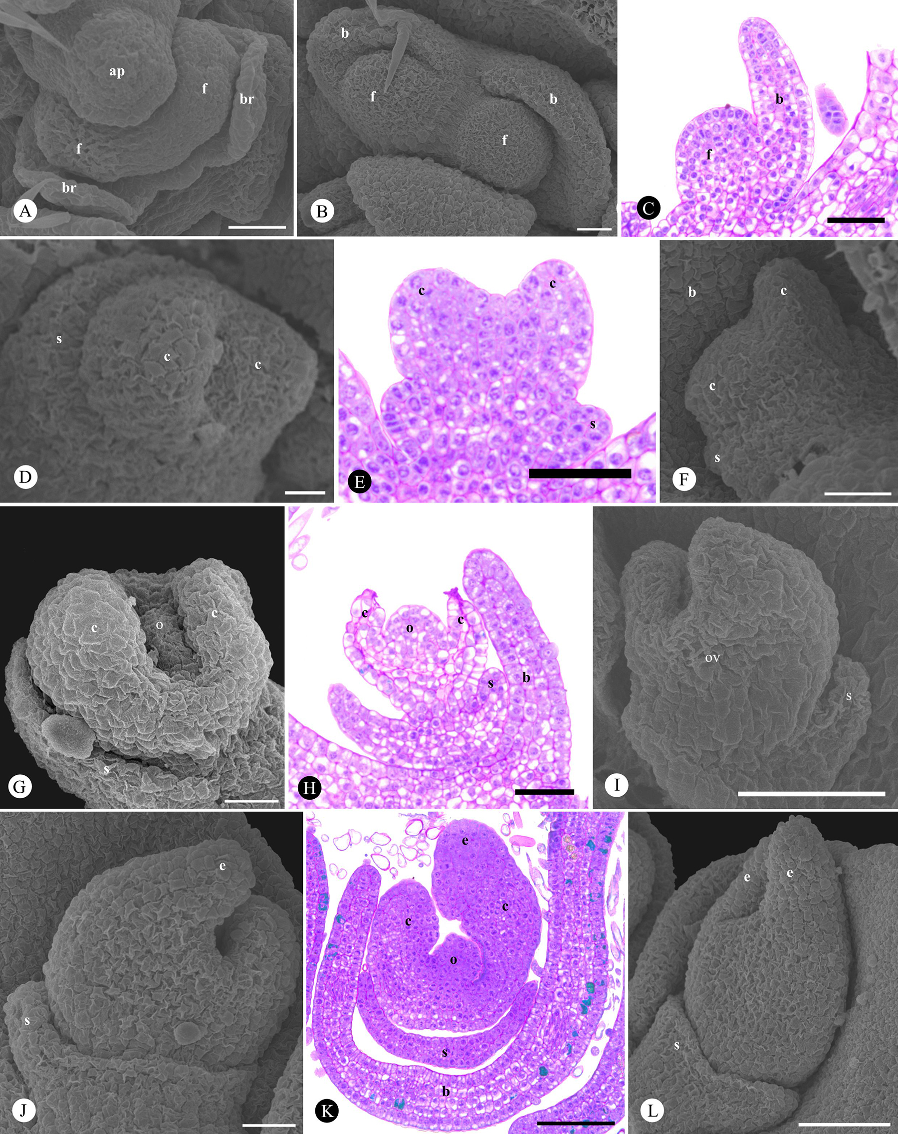
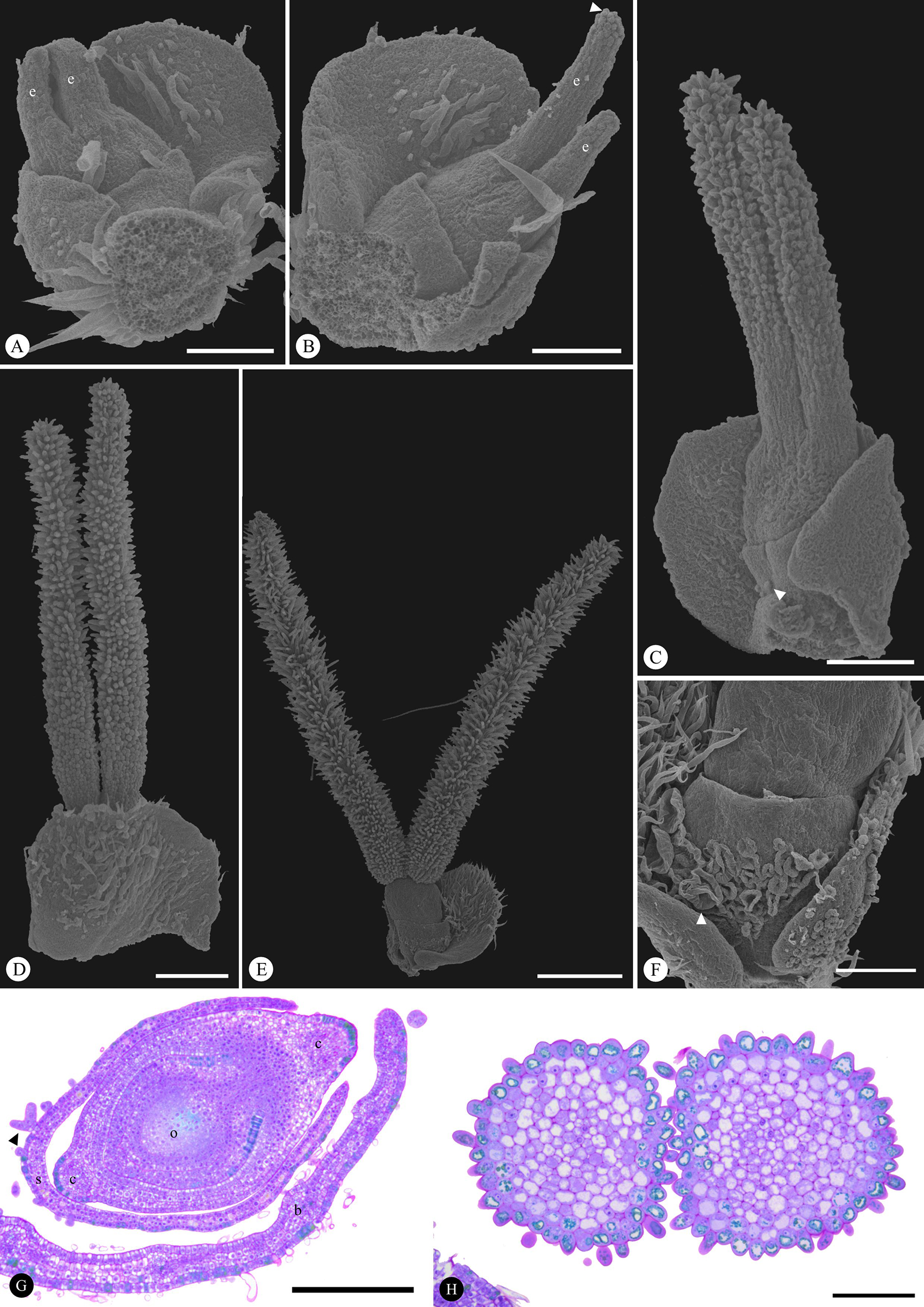
The apex of each inflorescence is protected by four bracts (Fig. 2 A ). In the axil of each bract there is an oval inflorescence primordium from which two floral primordia and two bracteoles originate, each primordium located in the axil of one of the bracteoles. Initially, the floral primordium has a rounded shape (Figs. 2B, C). Organ initiation begins with the differentiation of the base and center of the floral primordium. The peripheral region of the meristem expands, forming a unified ring-like primordium of the three sepals, while two carpel primordia appear in the center (Figs. 2D, E). The bracteole elongates and covers the floral organs at the time of carpel emergence, and the sepal differentiates at the periphery of the floral primordium (Fig. 2 F ). The carpels elongate unevenly (Figs. 2F-H), resulting in two distinct shapes (Fig. 2 I ). They extend around the space occupied by the developing ovule, and one carpel begins differentiating a stigmatic branch before the other, making their asymmetry evident once again (Figs. 2J, K). The elongation of the sepals begins simultaneously (Figs. 2I, J). At this stage, both apexes of the carpels differentiate into stigmas, which lengthen during development (Fig. 2 L ). The sepals are arranged in a short cup, surrounding the base of the ovary, while the stigmas elongate and surpass the bracteole in height (Fig. 3 A ). As sepals lengthen, they begin to develop trichomes and the stigmatic surface differentiates into papillae (Figs. 3B-D). At anthesis, the stigmas separate and become completely covered with papillae (Figs. 3E, H), while the gamosepalous calyx fully encloses the ovary (Fig. 3 F ). The developed ovary contains a single ovule, which is lateralized concerning the direction of stigma elongation (Fig. 3 G ).
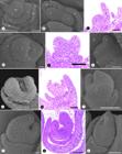
Development of pistillate flowers of Humulus lupulus. Initial and intermediate stages. A. View of the apical bud (Ap) of the inflorescence and its bracts (B) as well as floral buds (f) (SEM). B. Apical view of a pair of bracteoles (b) and a pair of floral apices (f) (SEM). C. Longitudinal section of a bracteole (b) and a floral apex (f), stained with toluidine blue (LM) D. View of a sepal primordium (s) at the periphery and two carpel primordia (c) at the center (SEM). E. Longitudinal section of carpel primordia (c) and sepal primordium (s), stained with toluidine blue (LM). F. View of the asymmetric elongation of the carpels (c) completely covered by the bracteole (b) at the bottom of the image (SEM). G. View of early ovule initiation (o) and carpel elongation (c) (SEM). H. Longitudinal section of a developing pistillate flower covered by the bracteole (b), showing carpels (c), ovule (o), and sepals (s), stained with toluidine blue (LM). I. Final stage of ovary formation (ov) and sepal elongation (s) (SEM). J. Beginning of differentiation of the first stigma (e) (SEM). K. Longitudinal section of a flower with the stigmatic branch (e) starting to differentiate, stained with toluidine blue, showing the ovule (o), sepal (p), carpels (c), and bracteole (b) (LM). L. Differentiation of the two stigmatic branches (e). Note the ring perianth (SEM). Symbols: ap: inflorescence apex - b: bracteole - br: bract - c: carpel - e: stigma - f: floral primordium, o: ovule, ov: ovary - s: sepal. Scale bars: A = 50 µm; B = 20 µm; C = 50 µm; D = 10 µm; E = 50 µm; F = 20 µm; G = 20 µm; H = 50 µm; I = 50 µm; J = 20 µm; K = 100 µm; L = 50 µm.
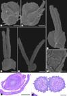
Development of pistillate flowers of Humulus lupulus. Final stages and specializations. A-B. View of the flower in the style elongation phase (e) (SEM). B. beginning of differentiation of papillae on one of the stigmas (arrow). C-E. Stages of differentiation of the two papillary stigmatic branches (SEM). Note in C the lupulin-secreting trichomes (arrow) begin to develop on the sepals (SEM) and trichomes completely covered by papillae in E (SEM). F. Fully developed glandular trichomes on the sepal (SEM). G. Cross section of the ovary, stained with toluidine blue, showing the ovule (o), sepal (p), carpels (c) and bracteole (b) (LM). H. Cross section of the base of the stigmatic branches of an adult flower, showing papillae on the surface, stained with toluidine blue (LM). Symbols: ap: inflorescence apex - c: carpel - e: stigma - o: ovule s: sepal. Scale bars: A-C = 100 µm; D = 200 µm; E = 500 µm; F-G = 200 µm; H = 100 µm.
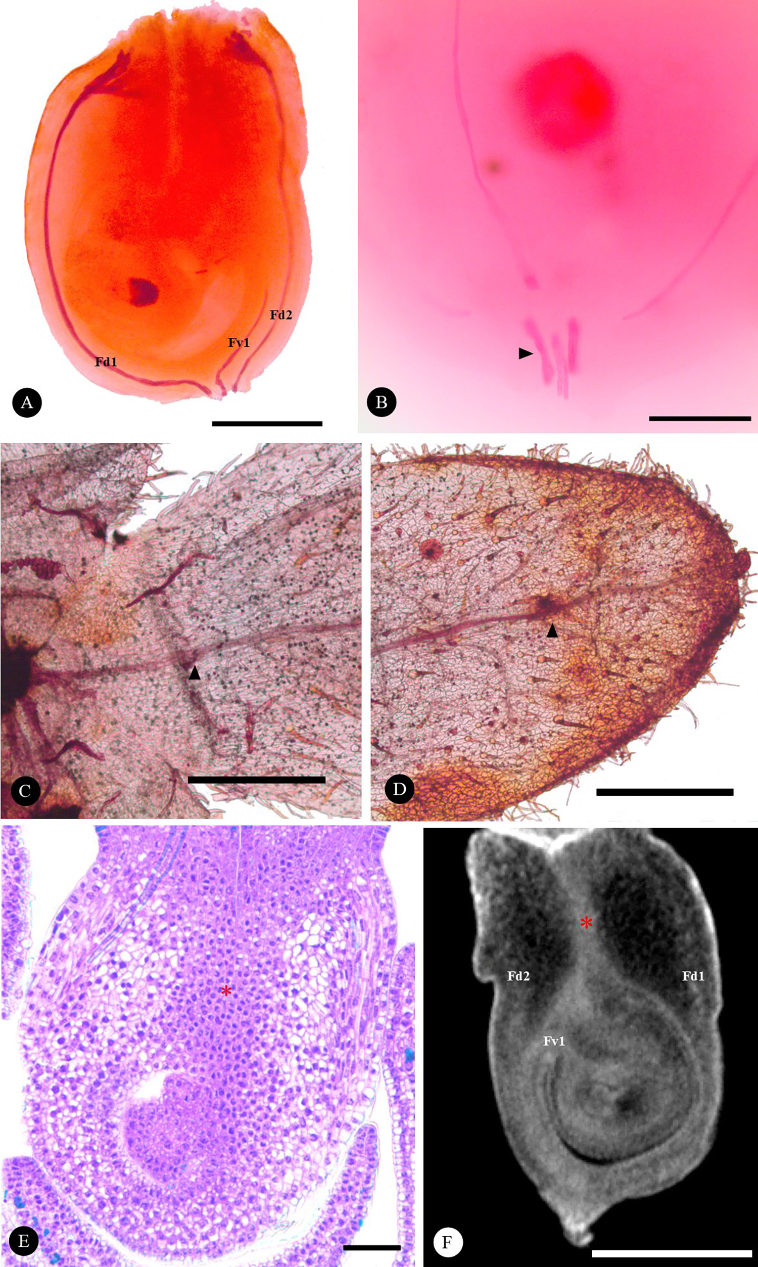
Each carpel is vascularized by a dorsal bundle. One of the carpels has a ventral vascular bundle that runs parallel to the dorsal bundle at the base of the carpel and is connected to the ovule (Figs. 4A, B, E). The sepals of the staminate flower are vascularized by one vascular bundle, while those of the pistillate flower are vascularized by three bundles, parallel at its base (Figs. 4C, D).
A well-developed intragynoecial secretory compitum crosses the center of the ovary, formed by secretory cells, characterized by prominent nuclei and dense cytoplasm (Fig. 4 A ). It can also be observed as a darker region of the tissue in three-dimensional reconstructions (Fig. 4 B ).
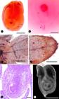
Sepal and ovary anatomy of Humulus lupulus. A. Lateral view of the ovary cleared and stained with Safranin (LM). B. Lateral view of the sepals from a pistillate flower, cleared and stained with Safranin, evidencing vascular bundles (arrow) (LM). C-D. Lateral view of a sepal from a staminate flower, evidencing vascular bundle (arrow) E. Longitudinal ovary section stained with toluidine blue showing the compitum region (*) (LM). F. Longitudinal section of the ovary showing vascular bundles (tomography). G. Tridimensional ovary reconstruction revealing contrast retention at the compitum region (*) (CT scanning Microtomography). Symbols: Fd1: dorsal bundle 1 - Fd2: dorsal bundle 2 - Fv1: ventral bundle 1 * = compitum region. Scale bars: A=200 µm; B=100 µm; C=200 µm; D=100 µm; E = 50 µm; F=230 µm.
Development of staminate flower
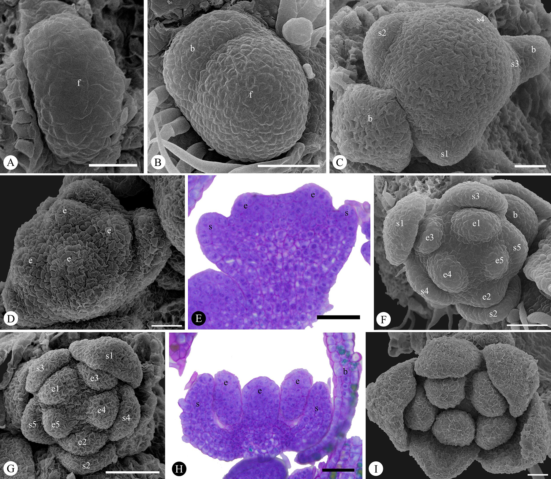
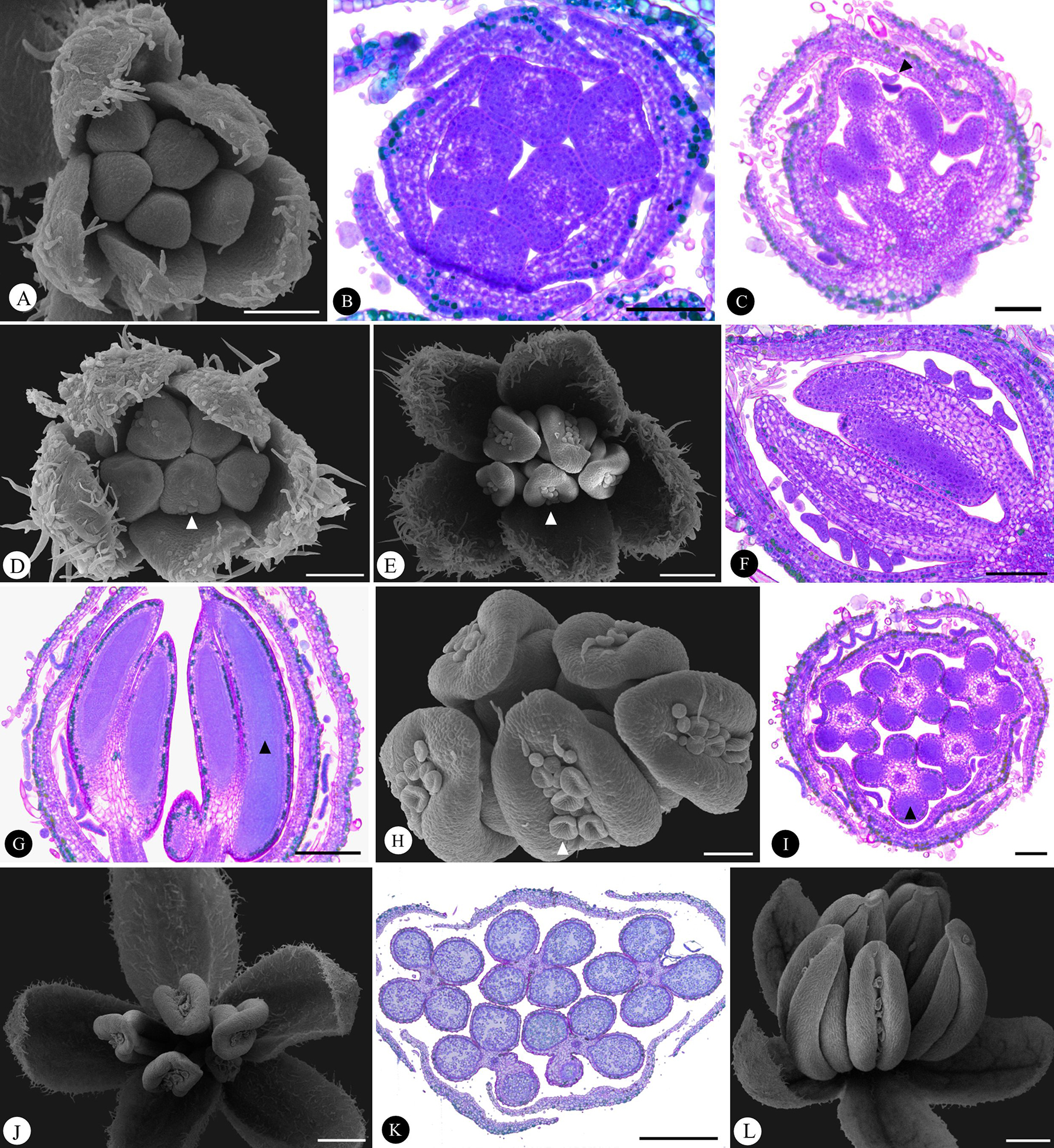
Each staminate inflorescence primordium (Fig. 5 A ) is in the axil of a bract and produces one floral primordium and two bract primordia. Initially, the floral primordium has a rounded shape (Fig. 5 B ). Organogenesis begins with the differentiation of five sepals at the periphery of the floral primordium in a helical pattern (Fig. 5 C ). After sepal initiation, five stamen primordia appear in the central region of the floral primordium (Figs. 5D-F), also in a helical pattern. The sepals and stamens elongate (Figs. 5G, H) and the sepals expand rapidly, covering the developing stamens (Fig. 5 I ). At this time, glandular trichomes appear on the surface of the sepals (Figs. 6A-E) and then on the surface of the anther connective (Figs. 6D-I). All the organs elongate, and the cells of the sporogenic tissue in the anthers become visible (Figs. 6G, I), which will originate the pollen grains (Fig. 6 K ). At this stage, the flowers have sepals and anthers covered by glandular trichomes (Fig. 6 J , L).
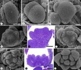
Development of staminate flowers of Humulus lupulus - initial and intermediate stages. A. Apical view of a floral primordium (f) (SEM). B. Apical view of a floral primordium (f) and a bract primordium (b) (SEM). C. Apical view of the spiral initiation of the sepals (s) at the periphery of the floral primordium and the pair of bracts (b) in the axils of the floral primordium (SEM). D. Emergence of stamens (e) in the floral primordium (SEM). E. Longitudinal section showing the beginning of the development of sepals (s) and stamens (e), stained with toluidine blue (LM). F-G. Apical view of calyx and developing androecium (SEM). H. Longitudinal section showing the elongation of the primordia of sepals (s) and stamens (e), stained with toluidine blue (LM). I.: Apical view showing elongation of stamens and sepals (SEM). Symbols: b: bract - e: stamen - f: floral primordium - s: sepal. Scale bars: A = 20 µm; B = 50 µm; C, D = 20 µm; E-H = 50 µm; I = 20 µm.
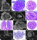
Development of the staminate flower of Humulus lupulus - final stages and cell specializations. A. Apical view of a flower bud with sepals covered by trichomes (SEM). B. Cross section of a flower bud showing sepals with trichomes, stained with toluidine blue (LM). C. Longitudinal section of a flower bud showing the beginning of the development of glandular trichomes in the anther connective, stained with toluidine blue (arrow) (LM). D-E. Stages of elongation of stigmas and sepals, as well as development of glandular trichomes (arrow) in anthers, and development of tector trichomes in sepals (SEM). F. Longitudinal section of a flower bud showing a more elongated stamen and a greater amount of glandular trichomes, stained with toluidine blue (LM). G. Longitudinal section of fully expanded stamens with developing pollen mother cells (arrow), stained with toluidine blue (LM). H. Apical view of fully expanded stamens containing glandular trichomes (arrow) (SEM). I. Cross section of a staminate bud with fully expanded stamens. Presence of glandular trichomes on sepals and stamens; developing pollen mother cells (arrow) stained with toluidine blue (LM). J. Apical view of a staminate flower in its final stage (SEM). K. Cross-section of a staminate flower in its final stage, with differentiated pollen, stained with toluidine blue (LM). L. Lateral view of a flower in the final stage of development (SEM). Scale bars: A-D = 100 µm; E = 200 µm; F = 100 µm; G-I = 200 µm; J-L = 500 µm.
Discussion
This study advances the floral ontogenic investigation of Humulus lupulus beyond that of Bechtel (1921) and Shephard et al. (2000) by using new protocols and technology to address our research questions and by individually dissecting and analyzing flower organs. We provide information about the nature of the perianth and gynoecium and about the previously undocumented occurrence of an intragynoecial compitum. This information provides a better understanding of the construction of the two floral morphotypes of Hops, the spatial constraints acting in their development, and the phylogenetic relationships among species within the family, which will be addressed in the following sections of this discussion.
The pistillate and staminate flowers of Hop has a sepalar perianth
According to studies on floral structure, Hops (Humulus lupulus) possess only two whorls: the gynoecium/androecium and whorl of perianth, which was considered sepalar and vestigial by Bechtel (1921) and Shephard et al. (2000), because of the restrict length and vascular supply of the sepals. In this study, we characterized the perianth of pistillate flowers as a calyx composed of three sepals completely united, forming a ring since the inception and vascularized by three bundles that run through the entire organ. Thus, our data diverge from previous studies on the morphology and vascularization of Cannabaceae (Bechtel, 1921; Leme et al., 2020; Shephard et al., 2000) and identify three sepals that develop together, united by a congenital process, characterizing Humulus lupulus autapomorphy within the family.
In the pistillate flower of H. lupulus, the sepals are neither robust nor broad, nor do they completely cover the gynoecium, probably because the bracteoles accompanying each flower serve as protection for the ovary, as in Cannabis sativa (Leme et al., 2020). However, the fruit develops surrounded by a persistent calyx in pollinated flowers (Shephard et al., 2000).
In addition to morphological studies, molecular tools could be useful for determining perianth identity. The ABCDE model proposes that the combination of MADS-box transcription factors determines the development of each floral whorl. This model states that the genes designated as E are expressed in all whorls, and the A-class genes expressed only in conjunction with E demarcate the development of sepals, while co-expression of AB generates petals, co-expression of BC generates stamens, expression of C only with E generates carpels, and D-class genes are responsible for ovule development (Theissen & Saedler, 2013; Gutiérrez et al., 2022).
Our morphological data demonstrate that perianth differentiation does occur, evidenced by the presence of vascularization and specializations (glandular trichomes) in the calyx. Therefore, a more comprehensive study of homeotic genes could assess the expression of AP1-FULL genes specifically in the sepals of pistillate flowers to accurately determine perianth identity.
The gynoecium of Hop is pseudomonomerous and has a compitum
The gynoecium of Humulus lupulus is bicarpellate, originating from the initiation of two dimorphic carpel primordia with unequal elongation and lateralization of the single ovule. Dimorphism is also characterized by the arrangement of vascular bundles, as evidenced by light microscopy images of cleared and Safranin-stained material, and by computed tomography images, where it is possible to observe that only one carpel has a vascular bundle connected to the ovule.
Our data indicate that one carpel contributes to the formation of the ovary wall and one stigma, while the other also contributes to ovule formation, in addition to the ovary wall and one stigma branch formation. These carpels are united at their base, a condition that characterizes the pseudomonomery of the gynoecium, where one carpel develops an ovule with an embryo sac, while the other carpel is considered "sterile" (Sokoloff et al., 2017).
The presence of a carpel that does not contribute to ovule formation may suggest that the stigma formed by that carpel is not functional, since the stigma is not directly united to the ovule. However, the presence of a well-defined intra-gynoecial compitum, also found in Moraceae species (Leite et al., 2020), is strong evidence of the functionality of both stigmas. The compitum is a common feature in syncarpous gynoecia and provides an even distribution of pollen tubes from different carpels, increasing their competition and improving the quality and quantity of offspring (Armbruster et al., 2002). In the case of the pseudomonomerous gynoecium, the compitum is a key factor that ensures the functionality of the morphological dimorphism between the carpels, preventing any pollen captured by one carpel from being eliminated and creating a pathway for pollen tubes from the non-ovulated carpel to reach the ovule (Endress, 2011; Sokoloff et al., 2017).
A pseudomonomerous gynoecium has been documented in Rosales, mainly in the Urticoid clade. Cannabaceae present pseudomonomery documented by floral ontogeny analysis in the genera Cannabis, Celtis, and Trema. In these cases, there is a congenital initiation of the carpels, followed by their separation during development. Celtis and Trema also show the presence of vestigial organs during flower development, which is not observed in Cannabis (Leme et al., 2020). Floral ontogeny in Humulus lupulus has been studied in less detail, showing, for example, the absence of vestigial organs during development (Shephard et al., 2000). Also, although previous authors reported morphological evidence for pseudomonomery in Hops, specific documentation of it has not been stated clearly before the present study (see Bechtel, 1921; Kubitzki, 1993; Shephard et al., 2000).
Moraceae has two congenitally fused carpels, which vary in their contribution to ovary formation. They have a short style and a long stigma among species, consistently exhibiting dimorphism between the two carpels, and a uniovulate and unilocular ovary, which characterizes the gynoecium as pseudomonomerous (Leite et al., 2018; 2020). Pseudomonomery in Ulmaceae has been well-documented and shows variations in vascularization and carpel contributions (Fukuoka, 1982; Okamoto et al., 1992; Leme et al., 2018). Urticaceae exhibits cases of both pseudomonomery and monomery, which can result from the abortion of one carpel or the non-division of the carpel primordium (Pedersoli et al., 2022).
Therefore, the elucidation of Hop’s controversial morphological data brought by this paper significantly improves the quality of comparative analyses in Cannabaceae and the urticoid clade.
Spatial contingencies in the formation of staminate and pistillate flowers
Celtis and Trema, which are the sister group of Humulus and Cannabis in Cannabaceae (Yang et al., 2013; Zhang et al., 2018), have flowers with a fixed merosity of five sepals (Leme et al., 2020), while Humulus lupulus and Cannabis sativa exhibit different merosity between pistillate (three sepals) and staminate (five sepals) flowers. Considering that their common ancestors have a fixed merosity of five sepals, it is likely that sepals have been lost in pistillate flowers. In Hops, the processes leading to this reduction of the perianth may be regulated by mechanical forces acting during development (Bull-Hereñu et al., 2022).
In staminate flowers, the physical forces exerted by the bracts may regulate the initiation of sepals. Sepal initiation occurs first in the areas with lower pressure exerted by the bracts of the inflorescence, between them, while the last sepal emerges in the region of the floral primordium that is in contact with the developing bract, which represents the location with the highest pressure of the floral primordium (Bull-Hereñu et al., 2022).
This type of influence on the perianth of pistillate flowers is also present but in different ways. The mechanical force exerted by the extensive protective bracts and the axis of the inflorescence on the floral primordia may explain their initial oval shape (Ronse De Craene, 2018) and possibly regulate the division of the floral primordium that precedes the initiation of bracteoles and floral organs. The perianth of the pistillate flower is influenced by the mechanical force generated by the initiation and elongation of the carpels, which start almost at the same time as the bracteole and exert great pressure by belonging to the largest and most conspicuous whorl of the flower (Rudall, 2010; Ronse De Craene, 2018; Bull-Hereñu et al., 2022). Furthermore, the perianth is constrained by the influence of the bracteole, whose presence reduces the space available for the floral primordium (Ronse De Craene, 2018; Bull-Hereñu et al., 2022). Therefore, evidence indicates that the loss and reduction of sepals are regulated by the physical forces acting during development.
In addition to the differences in the perianth, there is also a difference in the number of organs in the whorls responsible for reproduction. While the androecium has five stamens, the gynoecium has only two carpels, and only one of them houses a gametophyte. This difference results in a relationship where there is much more production of male gametophytes than female gametophytes, which is a common characteristic among wind-pollinated plants (Culley et al., 2002; Friedman & Barret, 2009), demonstrating the relationship between pollination and floral morphology in the case of anemophily (Ronse De Craene, 2018; Leme et al., 2020).
Phylogenetic proximity of Humulus lupulus and Cannabis sativa
The phylogenetic proximity of Humulus lupulus and Cannabis sativa is supported by molecular data and by the morphological characters derived from comparative analysis of floral development (Yang et al., 2013).
This study provides consistent data with the comparative analysis of floral ontogeny conducted by Leme et al. (2020), reaffirming the similarity of floral ontogeny between Humulus lupulus and Cannabis sativa in the absence of vestigial reproductive whorls and the presence of a reduced calyx. The vascularization of the calyx in Humulus lupulus follows the pattern of the family, with sepals vascularized by a single bundle, and the union between the sepals indicates its proximity to Cannabis sativa (Leme et al., 2020), except for the joint development of the sepals. The gamosepalous calyx is a shared characteristic between Humulus and Cannabis, while the joint development of the sepals, for this analysis, can be considered as an autapomorphy of Humulus lupulus.
In addition to the notable morphological similarities in the reproductive structures of Humulus and Cannabis (present study; Leme et al., 2020), molecular analyses support the proximity of the genera Humulus and Cannabis (Yang et al., 2013; Zhang et al., 2018). It was also found that the system of sex chromosomes is homologous and shared by Humulus lupulus and Cannabis sativa, with parts of the chromosomes that have ceased recombination in a common ancestor of these two species (Prentout et al., 2021).
Our study revealed that ovule initiation occurs while the carpels are still elongating and before the ovary is fully formed, a condition not previously reported in Cannabaceae (Bechtel, 1921; Shephard et al., 2000; Leme et al., 2020) and therefore considered a unique feature of Hops. This condition appears to follow the general trend of early ovule initiation in uniovulate ovaries (Endress, 2015). Additionally, the presence of a pseudomonomerous gynoecium in Humulus lupulus further reaffirms the previously raised proposal that this condition represents a synapomorphy of the Urticoid clade, as it is also found in other species of Cannabaceae (Leme et al., 2018; 2020), Moraceae (Leite et al., 2018; 2020), Ulmaceae (Leme et al., 2018), and Urticaceae (Pedersoli et al., 2022).
Acknowledgements
We are grateful to Rodrigo Ferreira Silva (FFCLRP-USP), Maria Dolores Seabra Ferreira, José Augusto Maulin (FMRP-USP), and Edimárcio da Silva Campos (FCFRP-USP) for their technical assistance; to Elettra Greene (in memoriam) for revising the English text; and to two anonymous reviewers for their valuable comments. This study was supported by the Sao Paulo Research Foundation (Fapesp - grant numbers 2018/03691-8, 2021/14731-3 and 2022/12533-2), National Council for Scientific and Technological Development (CNPq - grant numbers 304029/2023-8), and Coordination for the Improvement of Higher Education Personnel (CAPES - finance code 001).
References
- Almaguer C, Schönberger C, Gastl M, Arendt EK, Becker T. 2014. Humulus lupulus - a story that begs to be told: A review. Journal of the Institute of Brewing 120: 289-314.
-
Armbruster WS, Debevec EM, Willson MF. 2002. Evolution of syncarpy in angiosperms: Theoretical and phylogenetic analyses of the effects of carpel fusion on offspring quantity and quality. Journal of Evolutionary Biology 15: 657-672. doi: 10.1046/j.1420-9101.2002.00414.x.
» https://doi.org/10.1046/j.1420-9101.2002.00414.x. -
APG IV, 2016. An update of the Angiosperm Phylogeny Group classification for the orders and families of flowering plants. Botanical Journal of the Linnean Society 181: 1-20. doi: 10.1111/boj.12385.
» https://doi.org/10.1111/boj.12385. -
Bechtel AR. 1921. The floral anatomy of the Urticales. American Journal of Botany 8: 386-410. doi: 10.2307/2435455.
» https://doi.org/10.2307/2435455 -
Bull-Hereñu K, Dos Santos P, Toni JFG et al 2022. Mechanical forces in floral development. Plants 11: 661. doi: 10.3390/plants11050661.
» https://doi.org/10.3390/plants11050661 - Clark, G. 1981. Staining procedures. 4th. edn. Baltimore, Williams & Wilkins.
-
Culley TM, Weller SG, Sakai AK. 2002. The evolution of wind pollination in angiosperms. Trends in Ecology & Evolution 17: 361-369. doi: 10.1016/S0169-5347(02)02540-5.
» https://doi.org/10.1016/S0169-5347(02)02540-5. -
De Pinna MC. 1991. Concepts and tests of homology in the cladistic paradigm. Cladistics 7: 367-394. doi: 10.1111/j.1096-0031.1991.tb00045.x.
» https://doi.org/10.1111/j.1096-0031.1991.tb00045.x. -
Endress PK. 2011. Evolutionary diversification of the flowers in angiosperms. American Journal of Botany 98: 370-396. doi: 10.3732/ajb.1000299.
» https://doi.org/10.3732/ajb.1000299 -
Endress PK. 2015. Patterns of angiospermy development before carpel sealing across living angiosperms: Diversity, and morphological and systematic aspects. Botanical Journal of the Linnean Society 178: 556-591. doi: 10.1111/boj.12294.
» https://doi.org/10.1111/boj.12294 -
Friedman J, Barret SC. 2009. Wind of change: New insights on the ecology and evolution of pollination and mating in wind-pollinated plants. Annals of Botany 103: 1515-1527. doi: 10.1093/aob/mcp035.
» https://doi.org/10.1093/aob/mcp035. -
Fukuoka N. 1982. On pseudomonomerous pistil of the Ulmaceae. Acta Phytotaxonomica et Geobotanica 56: 84-91. doi: 10.18942/bunruichiri.KJ00001079140.
» https://doi.org/10.18942/bunruichiri.KJ00001079140. - Gerlach G. 1969. Botanische Mikrotechnik. Stuttgard, Georg Thieme Verlag.
- Gerrits PO, Horobin RW. 1991. The application of glycol methacrylate in histotechnology; some fundamental principles. Groningen, Rijksuniversiteit Groningen/Faculteit der Geneeskunde.
-
Gutiérrez RM, Ribeiro THC, Oliveira RR, Benedito VA, Chalfun- Junior A. 2022. Genome-wide analyses of MADS-box genes in Humulus lupulus L. reveal potential participation in plant development, floral architecture, and lupulin gland metabolism. Plants 11: 1237. doi: 10.3390/plants11091237.
» https://doi.org/10.3390/plants11091237. -
Karabín M, Hudcová T, Jelínek L, Dostálek P. 2016. Biologically active compounds from hops and prospects for their use. Comprehensive Reviews in Food Science and Food Safety 15: 542-567. doi: 10.1111/1541-4337.12201.
» https://doi.org/10.1111/1541-4337.12201. - Karnovsky MJ. 1965. A formaldehyde-glutaraldehyde fixative of high osmolarity for use in electron microscopy. Journal of Cell Biology 27: 1A-149A.
-
Kavalier AR, Litt A, Ma C et al 2011. Phytochemical and morphological characterization of hop (Humulus lupulus L.) cones over five developmental stages using high performance liquid chromatography coupled to time-of-flight mass spectrometry, ultrahigh performance liquid chromatography photodiode array detection, and light microscopy techniques. Journal of Agricultural and Food Chemistry 59: 4783-4793. doi: 10.1021/jf1049084.
» https://doi.org/10.1021/jf1049084. - Kubitzki K. Cannabaceae. 1993. In: Kubitzki K, Rohwer JG, Bittrich V. Flowering Plants· Dicotyledons. The Families and Genera of Vascular Plants. Berlin/Heidelberg, Springer. vol. II, p. 204-206.
-
Leite VG, Mansano VF, Teixeira SP. 2018. Floral development of Moraceae species with emphasis on the perianth and androecium. Flora 240: 116-132. doi: 10.1016/j.flora.2018.01.009.
» https://doi.org/10.1016/j.flora.2018.01.009. -
Leite VG, Basso-Alves JP, Gualberto ARS, Teixeira SP. 2020. A comparative ontogenetic approach to understanding the pseudomonomerous gynoecium in Moraceae. International Journal of Plant Sciences 181: 241-255. doi: 10.1016/S0169-5347(02)02540-5.
» https://doi.org/10.1016/S0169-5347(02)02540-5. -
Leme FM, Staedler YM, Schönenberger J, Teixeira SP. 2018. Ontogeny and vascularization elucidate the atypical floral structure of Ampelocera glabra, a tropical species of Ulmaceae. International Journal of Plant Sciences 179: 461-476. doi: 10.1086/697899.
» https://doi.org/10.1086/697899. -
Leme FM, Schönenberger J, Staedler YM, Teixeira SP. 2020. Comparative floral development reveals novel aspects of structure and diversity of flowers in Cannabaceae. Botanical Journal of the Linnean Society 193: 64-83. doi: 10.1093/botlinnean/boaa004.
» https://doi.org/10.1093/botlinnean/boaa004. - Lillie RD. 1954. Histopathology technic and practical histochemistry. New York, McGraw-Hill Book Co.
- Monteiro WR, Giulietti AM, Mazzoni SC, Castro MM. 1979. Hairs on reproductive organs of some Eriocaulaceae and their taxonomic significance. Boletim de Botânica da Universidade de São Paulo 7: 43-59.
-
O'Brien TP, Feder N, Mcccull YME. 1964. Polychromatic staining of plant cell walls by Toluidine Blue O. Protoplasma 59: 368-373. doi: 10.1007/BF01248568.
» https://doi.org/10.1007/BF01248568. -
Okamoto M, Kosuge K, Fukuoka N. 1992. Pistil development and parietal placentation in the pseudomonomerous ovary of Zelkova serrata (Ulmaceae). American Journal of Botany 79: 921-927. doi: 10.1002/j.1537-2197.1992.tb13675.x.
» https://doi.org/10.1002/j.1537-2197.1992.tb13675.x. -
Pedersoli GD, Teixeira SP. 2020. Flower morphogenesis and gametophyte fertility attest to the rare condition of gynomonoecy in Parietaria debilis (Urticaceae). Botany 98: 703-715. doi: 10.1139/cjb-2020-0082.
» https://doi.org/10.1139/cjb-2020-0082. -
Pedersoli GD, Gaglioti AL, Teixeira SP. 2020. Floral development elucidates the formation of a tubular calyx and connate stamens in Cecropieae species (Urticaceae). Plant Systematics and Evolution 306: 72. doi: 10.1007/s00606-020-01697-1.
» https://doi.org/10.1007/s00606-020-01697-1. -
Pedersoli GD, Staedler YM, Schönenberger J, Teixeira SP. 2022. A comparative approach reveals diversity of floral developmental processes in Urticaceae. Botanical Journal of the Linnean Society 200: 465-490. doi: 10.1093/botlinnean/boac028.
» https://doi.org/10.1093/botlinnean/boac028. -
Prentout D, Stajner N, Cerenak A et al (2021). Plant genera Cannabis and Humulus share the same pair of well‐differentiated sex chromosomes. New Phytologist 231: 1599-1611. doi: 10.1111/nph.17456.
» https://doi.org/10.1111/nph.17456. -
Ronse De Craene L. 2018. Understanding the role of floral development in the evolution of angiosperm flowers: clarifications from a historical and physico-dynamic perspective. Journal of Plant Research 131: 367-393. doi: 10.1007/s10265-018-1021-1.
» https://doi.org/10.1007/s10265-018-1021-1. -
Rudall PJ. 2010. All in a spin: Centrifugal organ formation and floral patterning. Current Opinion in Plant Biology 13: 108-114. doi: 10.1016/j.pbi.2009.09.019.
» https://doi.org/10.1016/j.pbi.2009.09.019. -
Shephard HL, Parker JS, Darby P, Ainsworth CC. 2000. Sexual development and sex chromosomes in hop. New Phytologist 148: 397-411. doi: 10.1046/j.1469-8137.2000.00771.x.
» https://doi.org/10.1046/j.1469-8137.2000.00771.x. -
Sokoloff DD, Nuraliev MS, Oskolski AA, Remizowa MV. 2017. Gynoecium evolution in angiosperms: Monomery, pseudomonomery, and mixomery. Moscow University Biological Sciences Bulletin 72: 97-108. doi: 10.3103/s0096392517030105.
» https://doi.org/10.3103/s0096392517030105. -
Staedler YM, Masson D, Schönenberger J. 2013. Plant tissues in 3D via X-ray tomography: Simple contrasting methods allow high resolution imaging. PLoS One 8: e75295. doi: 10.1371/journal.pone.0075295.
» https://doi.org/10.1371/journal.pone.0075295. -
Theissen G, Saedler H. 2013. Floral quartets. Nature 409: 469-471. doi: 10.1038/35054172.
» https://doi.org/10.1038/35054172. -
The Plant List. 2013. Version 1.1. http://www.theplantlist.org/ 15 Apr. 2024.
» http://www.theplantlist.org/ -
Yang MQ, van Velzen R, Bakker FT, Sattarian A, Li DZ, Yi TS. 2013. Molecular phylogenetics and character evolution of Cannabaceae. Taxon 62: 473-485. doi: 10.12705/623.9.
» https://doi.org/10.12705/623.9. -
Zanoli P, Zavatti M. 2008. Pharmacognostic and pharmacological profile of Humulus lupulus L. Journal of ethnopharmacology 116: 383-396. doi: 10.1016/j.jep.2008.01.011.
» https://doi.org/10.1016/j.jep.2008.01.011. -
Zhang H, Jin J, Moore MJ, Yi T, Li D. 2018. Plastome characteristics of Cannabaceae. Plant diversity 40: 127-137. doi: 10.1016/j.pld.2018.04.003.
» https://doi.org/10.1016/j.pld.2018.04.003.
The datasets related to this article will be available upon request to the author.