 Brazilian Guideline for Exercise Test in the Adult Population – 2024
Class of Recommendation
Class I: Conclusive evidence, or, failing that, general consensus that the procedure is safe and useful/effective.
Class II: Conflicting evidence and/or divergence of opinion about the procedure’s safety and usefulness/effectiveness.
Class IIa: Evidence/opinion is in favor of the procedure. Most approve of it.
Class IIb: Safety and usefulness/effectiveness less well established, with divergence of opinions.
Class III: Evidence and/or consensus that the procedure is not useful/effective and, in some cases, is harmful.
Level of Evidence
Level A: Data obtained from multiple large randomized studies, concordant and/or robust meta-analysis of randomized clinical studies.
Level B: Data derived from less robust meta-analyses, from a single randomized study or from non-randomized (observational) studies.
Level C: Data obtained from expert consensus.
Table of Contents
Part 1 – Indications, Legal Aspects, and Training in Exercise Test 08
1. Introduction 08
2. Indications and Contraindications for ET, CPET, and Cardiac Stress Imaging 08
2.1. General Indications for ET 08
2.2. Specific Clinical Indications for ET 09
2.2.1. Indications for ET in Coronary Artery Disease 09
2.2.2. Indications for ET in Asymptomatic Patients 09
2.2.3. Indications for ET in Athletes 09
2.2.4. Indications for ET in Hypertension 09
2.2.5. Indications for ET in Valvular Heart Disease 09
2.2.6. Indications for ET in Heart Failure and Cardiomyopathies 11
2.2.7. Indications for ET in the Context of Arrhythmias and Conduction Disorders 11
2.2.8. Indications for ET in Other Clinical Conditions 11
2.3. Relative and Absolute Contraindications 11
2.3.1. Relative Contraindications to ET/CPET 12
2.3.2. Absolute Contraindications to ET/CPET 12
2.4. Indications for CPET 12
2.4.1. General Indications for CPET 12
2.4.2. Specific Clinical Indications for CPET 12
2.5. Indications for Cardiac Stress Imaging 12
2.5.1. Myocardial Perfusion Imaging 12
2.5.2. Indications for Stress Echocardiography 13
3. Legal Aspects and Essential Conditions for Performing ET, CPET, and Cardiac Stress Imaging 15
3.1. Legal Aspects Involved in the Practice of ET and CPET 15
3.2. Essential Conditions for Performing ET and CPET 15
3.3. Informed Consent Forms for ET/CPET 18
3.4. Informed Consent Forms for Cardiac Stress Imaging 18
4. Aspects Related to Training in Exercise Test as an Area of Focused Practice 18
Part 2 – The Exercise Test 21
1. ET methodology 21
1.1. Core Conditions to Perform ET 22
1.1.1. Team 22
1.1.2. Physical Infrastructure 23
1.1.3. Equipment 23
1.1.4. Emergency Supplies 23
1.1.5. Emergency Drugs 23
1.1.6. Orientations for Patients when Scheduling ET 23
1.2. Basic ET Procedure 24
1.2.1. Pre-test Phase 24
1.2.2. Initial Assessment 24
1.2.3. Targeted and Specific Physical Examination 24
1.2.4. Electrocardiographic Monitoring and Recording System 24
1.2.4.1. Three-lead Systems 25
1.2.4.2. Twelve-lead Systems 25
1.2.4.3. Placements Including 13 or More Leads 27
1.2.4.4. Skin Preparation for ECG Monitoring 27
1.2.4.5. ECG Recording 27
1.2.5. Hemodynamic Monitoring 27
1.2.5.1. Heart Rate Monitoring 27
1.2.5.2. Blood Pressure Monitoring 27
1.2.6. Monitoring for Signs and Symptoms 28
1.2.7. Prevention of ET Complications 28
1.3. Ergometers 28
1.3.1. Cycle Ergometer 28
1.3.2. Treadmill 28
1.3.3. Upper-Body Ergometer 28
1.3.4. Other Ergometers 28
1.4. Choice of Protocol 29
1.4.1. Cycle Ergometer Protocols 29
1.4.2. Treadmill Protocols 29
1.4.2.1. Step Protocols 29
1.4.2.1.1. Bruce Protocol 29
1.4.2.1.2. Modified Bruce Protocol 29
1.4.2.1.3. Ellestad Protocol 29
1.4.2.1.4. Naughton Protocol 30
1.4.2.2. Ramp Protocol 30
1.4.3. Upper-Body Ergometer Protocol 30
1.4.4. Test Cessation/Termination 30
2. Test Accuracy, Pretest Probability, and Pretest Scores 31
2.1. Pretest Probability of CAD 31
2.2. Sensitivity, Specificity, and Predictive Value 31
2.3. Pretest CVD Scores and Risk Factors 32
3. Clinical and Hemodynamic Responses to Exercise in the Adult Population 32
3.1. Clinical Responses 32
3.1.1. Exercise Tolerance 32
3.1.2. Cardiorespiratory Fitness/Functional Classification 33
3.1.3. Symptoms 34
3.1.4. Inspection and Auscultation 34
3.2. Hemodynamic Responses 35
3.2.1. Heart Rate 35
3.2.1.1. Resting Heart Rate 35
3.2.1.2. Chronotropic Response 36
3.2.2. Blood Pressure Response 36
3.2.3. Double Product 39
4. ECG Responses 41
4.1. P Wave 41
4.1.1. Normal Responses 42
4.1.2. Abnormal Responses 42
4.2. PR Interval/Segment 43
4.2.1. Normal Responses 43
4.2.2. Abnormal Responses 43
4.3. Q Wave 43
4.3.1. Normal Responses 43
4.3.2. Abnormal Responses 43
4.4. R Wave 43
4.4.1. Normal Responses 43
4.4.2. Abnormal Responses 43
4.5. S Wave 43
4.5.1. Normal Responses 44
4.5.2. Abnormal Responses 44
4.6. QRS Duration 44
4.6.1. Normal Responses 44
4.6.2. Abnormal Responses 44
4.7. High-frequency Assessment of QRS Fragmentation 44
4.7.1. Normal Responses 44
4.7.2. Abnormal Responses 44
4.8. T Wave 45
4.8.1. Normal Responses 45
4.8.2. Abnormal Responses 45
4.9. U Wave 45
4.9.1. Normal Responses 45
4.9.2. Abnormal Responses 45
4.10. Early Repolarization 46
4.11. ST-segment Elevation 46
4.12. J Point and Upsloping Depression 47
4.13. Slow Upsloping, Horizontal, and Downsloping ST-segment Depression 47
4.13.1. ST Hump Sign 48
4.14. Normalization of ST-segment Changes 49
4.15. ST/HR Slope, ST/HR Index, ST/HR Loop, and ST/HR Hysteresis 49
4.15.1. ST/HR Slope 49
4.15.2. ST/HR Index 49
4.15.3. ST/HR Loop 49
4.15.4. ST/HR Hysteresis 50
4.16. QT Interval, QTc, QT Hysteresis, and QT Dispersion 50
4.16.1. Abnormal Responses 50
4.17. Disorders of Atrioventricular Conduction, Intraventricular Conduction, and Impulse Formation 51
4.17.1. Atrioventricular Conduction Disorders 51
4.17.1.1. First-degree Atrioventricular (AV) Block 51
4.17.1.2. Type I Second-degree AV Block (Mobitz I) 52
4.17.1.3. Type II Second-degree AV Block (Mobitz II) 52
4.17.1.4. Type 2:1 AV Block/Advanced or High-grade AV Block/Third-degree AV Block or Complete AV Block 52
4.17.2. Intraventricular Conduction Disorders 52
4.17.2.1. Left Bundle Branch Block 52
4.17.2.1.1. Pre-existing Left Bundle Branch Block 53
4.17.2.1.2. Exercise-induced Left Bundle Branch Block 53
4.17.2.2. Fascicular Blocks 53
4.17.2.3. Right Bundle Branch Block 54
4.17.2.3.1. Pre-existing Right Bundle Branch Block 54
4.17.2.3.2. Exercise-induced Right Bundle Branch Block 54
4.17.3. Disorders of Impulse Formation 54
4.17.3.1. Ventricular Arrhythmias 55
4.17.3.2. Supraventricular Arrhythmias 56
4.17.3.3. Atrial Fibrillation/Atrial Flutter 57
4.17.3.4. Bradyarrhythmias/Chronic Chronotropic Incompetence 58
4.17.3.5. Inappropriate Sinus Tachycardia 58
4.18. Indirect Metabolic Assessment 58
4.18.1. VO2/METs 58
4.18.2. Functional Aerobic Impairment (FAI) 59
4.18.3. Myocardial Aerobic Impairment (MAI) 59
4.19. Post-test Risk Scores and ET Prognostic Variables 60
4.19.1. Duke Score 60
4.19.2. Athens Score/QRS Score 60
4.19.3. Raxwal and Morise Score 61
5. Test Cessation Criteria 61
6. ET Reporting 61
6.1. General Information 61
6.2. Observed, Measured, and Recorded Data 62
6.3. Descriptive Report 63
6.4. Conclusion 63
6.5. ECG Recording 64
7. Other Diagnostic Tests Performed Simultaneously with or in Addition to ET 64
7.1. Ankle-brachial Index 64
7.1.1. Method of ABI Measurement 64
7.1.1.1. Resting ABI 64
7.1.1.2. Post-exercise ABI 65
7.1.2. Patient Preparation and Measurement Technique 65
7.2. Noninvasive Oximetry 66
7.2.1. Equipment 66
7.2.2. Procedures for Noninvasive Oximetry 67
7.2.3. Interpretation of Findings 67
7.3. Biomarkers and Other Laboratory Tests 67
7.3.1. Blood Lactate 68
7.3.2. Arterial Blood Gas Analysis 68
8. Particular Aspects of ET Performance and Interpretation in Specific Clinical Conditions 70
8.1. Dextrocardia/Situs Inversus 70
8.2. Chagas Disease/Chagas Cardiomyopathy 71
8.3. Peripheral Artery Disease 72
8.4. Parkinson’s Disease 73
8.5. Valvular Heart Disease 74
8.5.1. Aortic Stenosis 74
8.5.2. Aortic Regurgitation 75
8.5.3. Mitral Stenosis 75
8.5.4. Mitral Regurgitation 76
8.5.5. Mitral Valve Prolapse 76
8.6. ET after Myocardial Revascularization 77
8.6.1. ET after Percutaneous Coronary Intervention 77
8.6.2. ET after Coronary Artery Bypass Grafting 78
Part 3 – Cardiopulmonary Exercise Test 78
1. Introduction 78
2. Exercise Physiology Applied to CPET 78
3. Pulmonary Ventilation, Expired Gas Analysis, and Derived Variables 79
3.1. Pulmonary Ventilation 79
3.1.1. Baseline Spirometry 79
3.1.2. Ergospirometry 80
3.1.3. Ventilatory Reserve 80
3.2. Oxygen Consumption 81
3.3. Carbon Dioxide Production 81
3.4. Ventilatory Thresholds 81
3.4.1. First Ventilatory Threshold 81
3.4.2. Second Ventilatory Threshold 81
3.5. Respiratory Quotient 82
3.6. Ventilatory Equivalents of Oxygen and Carbon Dioxide 82
3.7. End-tidal Oxygen and Carbon Dioxide Pressures 83
3.8. Oxygen Pulse 84
3.9. Delta VO2to Delta Work Rate (ΔVO2/ΔWR) Ratio 84
3.10. Cardiorespiratory Optimal Point 84
3.11. Oxygen Uptake Efficiency Slope (OUES) 84
3.12. Exercise Oscillatory Ventilation 85
3.13. Oxygen Uptake Recovery Delay 85
3.14. Circulatory Power and Ventilatory Power 85
3.15. Reference Ranges for CPET Variables 85
4. Equipment and Methods 86
4.1. Ergometers 86
4.2. Airflow or Volume Transducers 86
4.3. Gas Analyzers 88
4.4. Measurements of Gas Exchange 88
4.5. Calibration, Quality Control, and Sanitization 88
4.6. Protocols 88
4.7. Data Analysis Software 88
4.8. Guidelines for Patients when Scheduling CPET 88
5. CPET in Selected Specific Situations 89
5.1. Heart Failure 89
5.2. Coronary Artery Disease 89
5.3. Hypertrophic Cardiomyopathy 90
5.4. Valvular Heart Disease 90
5.5. Lung Diseases 90
5.5.1. Chronic Obstructive Pulmonary Disease 90
5.5.2. Pulmonary Vascular Disease 91
5.6. Differential Diagnosis of Dyspnea 91
5.7. Athletes and Exercise Enthusiasts 92
5.8. Cardiorespiratory Rehabilitation 92
6. Interpretation and Preparation of the CPET Report 92
Part 4 – Exercise Test Combined with Cardiac Imaging Methods 93
1. Cardiovascular Stress Combined with Cardiac Imaging Methods 93
1.1. Myocardial Perfusion Imaging 93
1.1.1. Physical Stress Methods (Exercise Test) 93
1.1.1.1. Contraindications to Physical Stress in MPI 93
1.1.1.2. Guidelines for Patients When Scheduing MPI with Physical Stress 93
1.1.1.3. Methodological Aspects of Physical Stress in MPI 93
1.1.1.4. Interpretation of ET Results in MPI 93
1.1.2. Methodological Aspects of Pharmacologic Stress in MPI 94
1.1.2.1. Vasodilators 94
1.1.2.1.1. Dipyridamole 94
1.1.2.1.2. Adenosine 94
1.1.2.2. Drugs that Increase Myocardial Oxygen Consumption 95
1.1.3. Hybrid Stress Methods 96
1.1.4. Novel Drugs 96
1.2. Stress Echocardiography 96
1.2.1. Methodology 96
1.2.1.1. Exercise Stress Methods 96
1.2.1.2. Pharmacologic Stress Methods 97
1.2.1.2.1. Dobutamine 97
1.2.1.2.2. Vasodilators 97
1.2.1.3. Ultrasound Enhancing Agents 98
References 98
Part 1 – Indications, Legal Aspects, and Training in Exercise Test
1. Introduction
Exercise test (ET) is a diagnostic modality, widely used in routine clinical cardiology practice, in which a person is subjected to a planned, individualized degree of physical effort with the purpose of evaluating their clinical, hemodynamic, autonomic, electrocardiographic, indirect metabolic and, occasionally, enzymatic responses to physical exertion.1, 2 When ET also included the assessment of ventilatory parameters and analysis of exhaled gases, it is called cardiopulmonary exercise test (CPET or CPX).3 The term cardiac stress test encompasses both ET and CPET.
Overall, ET and CPET:
– Contribute to the diagnosis and prognosis of cardiovascular diseases, provide guidance to inform the selection of therapeutic interventions, assist in the adoption of preventive measures and support sports practice, are used in forensic medical opinions and in the medical examination of disability claimants, and provide vital information for the follow-up of patients over time.1, 3 - 5
– Provide high reproducibility, well-established cost-benefit and cost-effectiveness, and are available in all regions of Brazil.1, 6
– Are recognized and legally registered as an Area of Focused Practice by the Joint Commission on Medical Specialties.7
– Are of great importance as a cardiovascular stressor for stress perfusion imaging methods in cardiology, especially with a view to the diagnosis and prognosis of ischemic cardiovascular disease.8, 9
This guideline consolidates and updates, in a single document, all information and recommendations present in previous SBC guidelines on TE and CPET, addressing new aspects not considered in previous documents, with particular emphasis on important new information related to testing in the adult population and necessary adaptations to TE/CPET practice in the setting of acute respiratory syndromes.1, 2 This Guideline will be a relevant source of reference for the general cardiologist and, in particular, for physicians training and working in the area of focused practice of exercise test.
2. Indications and Contraindications for ET, CPET, and Cardiac Stress Imaging
2.1. General Indications for ET
ET is widely available in Brazil, at an affordable cost and with recognized utility in clinical practice.10, 11 It is an important diagnostic tool for risk stratification and prognostication in patients with known or suspected heart disease. It allows assessment of the impact of cardiovascular diseases and of the effectiveness of implemented therapies.
General indications and overall objectives for ET include:1, 6, 12 - 18
1) Assessment of exercise-induced symptoms.
2) Determination of functional capacity.
3) Assessment of blood-pressure behavior.
4) Assessment of heart-rate behavior.
5) Detection of myocardial ischemia.
6) Recognition of cardiac arrhythmias and elucidation of their type, density, and complexity.
7) Assessment of the response to exertion in patients with channelopathies.
8) Diagnosis and prognosis of certain cardiovascular diseases.
9) Indication assessment of therapeutic interventions.
10) Assessment of the results of therapeutic interventions.
11) Preoperative assessment.
12) Assessment of cardiorespiratory fitness and physical conditioning.
13) Provide information to support the prescription of physical exercise, including in cardiopulmonary rehabilitation.
14) Provide information to support pre-employment health screenings, periodic check-ups, and disability and worker’s compensation claims.
ET can be performed for the aforementioned purposes in any clinical situations and conditions, as long as its relative and absolute contraindications are followed.
2.2. Specific Clinical Indications for ET
ET has had its effectiveness studied and tested in a range of specific clinical situations. This has allowed determination of the strength of recommendation and level of evidence for these specific indications, which will be presented in detail below.6, 12 - 14, 17, 19
2.2.1. Indications for ET in Coronary Artery Disease
Coronary artery disease remains a leading cause of morbidity and mortality, with an estimated prevalence of angina of 12% to 14% in men and 10% to 12% in women aged 65 to 84. In Brazil, approximately 30% of all deaths are due to cardiovascular causes.20
ET is indicated in the investigation of chest pain of probable cardiac origin due to its relevance, widespread availability, and cost-effectiveness, and has been endorsed as the optimal choice in this setting by the Choosing Wisely Initiative.21
The prevalence of asymptomatic CAD and silent ischemia varies widely depending on the population studied. Asymptomatic diabetics have a relative risk (RR) of 2.0 for CAD, and the prevalence of a positive ET is approximately 23% in these patients.22, 23
The diagnosis of silent myocardial ischemia allows interventions aimed at reducing the risk of future events, including death.24
ET is recommended for risk stratification of patients with stable CAD, prognostication, assessment of the effectiveness of interventions, and investigation of changes in clinical manifestations.25 - 27
Even with a non-ischemic ET, patients with suspected CAD may benefit from ET-enhanced risk stratification, through prognostic variables such as exertional symptoms, functional capacity, blood-pressure and chronotropic response, autonomic function, and musculoskeletal response.28
In patients with CAD, ET is essential for the initial exercise prescription and to inform subsequent adjustments to the cardiovascular rehabilitation program (Table 1).17, 29, 30
2.2.2. Indications for ET in Asymptomatic Patients
ET plays a relevant role in the evaluation of asymptomatic patients, as its variables (HR, blood pressure, ECG, etc.) provide information on prognosis and risk of future abnormalities.57, 58
Cardiorespiratory fitness (functional capacity), as determined by ET, is considered a key marker of health and can be used to define therapeutic and preventive goals. In asymptomatic patients with comorbidities, ET assists in exercise prescription to promote health and well-being. ET is feasible and safe even in patients with advanced age and significant comorbidities.59, 60
It also improves risk stratification of asymptomatic individuals in terms of physical fitness to carry out their duties at work without putting themselves or others at undue risk (Table 2).61
2.2.3. Indications for ET in Athletes
Physical activity (PA) is defined as any bodily movement produced by the musculoskeletal system. Physical exercise, or physical training, is a program of structured, repetitive physical activity, with the aim of recovering, maintaining, or improving one or more components of physical fitness (cardiorespiratory, morphologic, muscular, metabolic, or motor). An athlete is defined an individual of any age, whether amateur or professional, who regularly engages in physical exercise with an emphasis on performance and may participate in competitive sports.13, 34
TE provides important data for cardiologists, sports medicine and preventive medicine specialists regarding the health of elite athletes, Olympic athletes, professional athletes, competitive athletes, members of athletic federations and/or sports clubs, masters athletes, and recreational (pleasure and leisure) athletes. It is used in preparticipation physical evaluation and allows detection of latent pulmonary and cardiovascular diseases (i.e. exercise-induced asthma, hypertension, ischemia, arrhythmias, etc.), monitoring of response to interventions, and prognostic evaluation (Table 3).13, 17, 34, 68
2.2.4. Indications for ET in Hypertension
The systolic blood pressure (SBP) response during ET is considered a risk marker for development of hypertension, death from cardiovascular disease, and stroke.71 - 73 Recent data suggest that the BP response to submaximal-intensity exercise has greater clinical and prognostic significance than the BP achieved during maximal-intensity exercise. Physical performance at the time of ET influences the interpretation of the BP response to exercise. Both hypotension and an exaggerated BP response serve as prognostic markers and indicators of the need to investigate underlying CVD (Table 4).74, 75
Research has found that, in healthy male athletes undergoing ET, the workload-indexed BP response was superior to peak SBP as a predictor of mortality, and was particularly useful for preparticipation screening. A hypertensive response to ET was associated with development of hypertension in young athletes.76
2.2.5. Indications for ET in Valvular Heart Disease
In valvular heart disease, ET should be routinely performed to elucidate questionable symptoms, evaluate indicators that can inform the decision to intervene, clear patients for exercise and inform the exercise prescription (Table 5).92 - 94 ET is useful to unmask so-called “pseudo-asymptomatic” patients and allows serial follow-up of truly asymptomatic patients.94 In symptomatic patients or those with exercise-induced symptoms, intervention, whether surgical or transcatheter, is indicated outright.94
Clearing a patient with valvular heart disease requires assessment of symptoms, functional capacity, and characteristics of the valve lesion and its impact on cardiac function. Asymptomatic individuals with lesions of up to moderate severity can engage in high intensity exercise if the ET reveals good functional capacity and absence of myocardial ischemia, hemodynamic derangements, or arrhythmias.95
2.2.6. Indications for ET in Heart Failure and Cardiomyopathies
In heart failure (HF) and in cardiomyopathies, ET is used to elucidate symptoms, assess exercise tolerance/functional class, support prognostic assessment, and inform adjustment of therapy and prescription of exercise programs (Table 6).29, 116
Exercise intolerance is a typical manifestation of HF, and functional class and HR response to ET are important prognostic variables.117, 118
2.2.7. Indications for ET in the Context of Arrhythmias and Conduction Disorders
Exercise-induced arrhythmias are often caused by cardiovascular conditions that can be evaluated by ET. These may be fully asymptomatic, or may present with symptoms ranging from palpitations to syncope. ET allows investigation of these symptoms, diagnosis and quantification (density) of arrhythmias, and stratification of the risk of sudden cardiac death (SCD). It also plays a relevant role in investigating the causes, impact, and therapeutic options for atrioventricular and intraventricular conduction disorders (Table 7).63, 128 - 132
2.2.8. Indications for ET in Other Clinical Conditions
Table 8 describes other clinical conditions in which ET is recommended, whether for functional assessment, to inform exercise prescription, or for adjustment and optimization of therapy.
2.3. Relative and Absolute Contraindications
ET is generally well-tolerated and safe when properly indicated and performed. However, some specific clinical situations may increase the risk of complications requiring immediate medical intervention (Table 9). The risk of sudden cardiac death is around 1 in every 10,000 exercise tests.6, 184, 185
2.3.1. Relative Contraindications to ET/CPET
These are high-risk clinical scenarios in which preventive and, potentially, therapeutic measures must be adopted before ET/CPET can be performed (Table 10). Such measures include carrying out the ET exclusively in a hospital setting; using modified protocols and a reduced target load; strict observation of emergent symptoms; more frequent BP measurements; and presence of trained personnel and equipment for reprogramming the patient’s pacemaker, ICD or other implantable devices as needed.
2.3.2. Absolute Contraindications to ET/CPET
Chart 1 lists absolute contraindications to the performance of ET/CPET.1, 6, 12 - 17
2.4. Indications for CPET
2.4.1. General Indications for CPET
The general indications for CPET are the same as for ET, but it is particularly indicated when there is a need to assess ventilatory and metabolic variables as well (Chart 2).
2.4.2. Specific Clinical Indications for CPET
Clinical situations for which there is sufficient scientific to determine a class of recommendation for CPET are listed in Table 11.
2.5. Indications for Cardiac Stress Imaging
2.5.1. Myocardial Perfusion Imaging
Myocardial perfusion imaging (MPI) is indicated in different clinical presentations of ischemic heart disease and contributes to defining its severity.9 Other indications include evaluation of potential for revascularization in patients with viable myocardium and as part of preoperative assessment in specific situations (Table 12).26, 31, 128, 251 - 253
Symptomatic patients at intermediate risk for ischemic heart disease benefit most from MPI for diagnostic and prognostic purposes. It should be performed, preferably, under stress, as long as the patient has a functional capacity above 5 METs and the ability to perform the necessary effort on whichever ergometer is available.
Patients with left bundle branch block (LBBB), Wolff-Parkinson-White (WPW) syndrome, or a pacemaker should undergo MPI with pharmacologic stress (dipyridamole or adenosine) instead.9
Among various indications for MPI, it is particularly useful in patients with low functional capacity or conditions that would prevent interpretation of whether an ischemic response to ET/CPET is present. MPI is also superior for CAD risk stratification in patients with a high pretest probability of CAD. MPI should be performed under pharmacologic stress in individuals with intermediate pre-test probability of CAD and an uninterpretable resting ECG or in those incapable or intolerant of physical exertion (Table 13).9
Following the recommendations of the Brazilian Guideline on Nuclear Cardiology, we adopted the following international score: MCI is appropriate for indications with a score of 7 to 9; possibly appropriate for indications with a score of 4 to 6; and rarely appropriate for those indications with a score of 1 to 3.9
Asymptomatic patients with no history of ischemic heart disease and a normal ET/CPET generally do not benefit from MPI. Asymptomatic patients with an abnormal ET may benefit from MPI, especially those at intermediate or high risk (Table 14).9, 254
In patients who are asymptomatic after percutaneous coronary intervention (PCI) and/or CABG, MPI has a favorable cost-benefit ratio in follow-ups longer than 2 and 5 years, respectively. Patients who are symptomatic (angina or anginal equivalents) after PCI and/or CABG are considered to benefit from MPI at any time (Table 15).9, 31, 251, 252, 254
Patients with established CAD and worsening symptoms (or equivalent manifestations) can benefit from MPI at any time, with the main objective of quantifying ischemic burden (extent and intensity of defects) and informing medical management (Class of recommendation-Level of evidence: I-C).252
In patients with acute chest pain with suspected ACS, normal ECG (no ischemic changes or necrosis) or uninterpretable ECG (LBBB, WPW, or pacemaker rhythm), and normal cardiac biomarkers, resting MPI presents has a high negative predictive value (NPV), thus allowing patients to be discharged from the emergency department (Table 16).1, 9, 33, 254 - 258
Assessment of myocardial viability through MPI aids in selection of patients with severe left ventricular dysfunction who are eligible for myocardial revascularization (Table 17).1, 9, 31, 128, 258, 259
Other indications for MPI, such as investigation of heart failure, arrhythmias, syncope, patients with high calcium score (≥400), patients with diabetes, chronic renal failure, or a family history of ischemic heart disease, and preoperative risk assessment before noncardiac surgery and vascular surgery, are covered in the Update of the Brazilian Guideline on Nuclear Cardiology.9, 253
2.5.2. Indications for Stress Echocardiography
Stress echocardiography (EcoE) is a noninvasive imaging method used for diagnosis, risk stratification and prognosis, and assessment of myocardial viability in coronary artery disease (CAD), valvular heart disease, and cardiomyopathies.260
In investigation of ischemia, it provides good accuracy in moderate-to-high-risk patients, with slightly higher specificity compared to other noninvasive imaging methods, such as MPI.260 - 262 However, it should not be considered a substitute for ET; instead, it is indicated in patients with limitations or contraindications to ET.262
The applicable stress testing modalities are exercise (ESE; performed on a treadmill, conventional bicycle/cycle ergometer, or in-bed/supine cycle ergometer) or pharmacologic, either with dobutamine (DSE; atropine can be added to increase sensitivity) or with a vasodilator (adenosine or dipyridamole; rarely performed). Both ESE and DSE have similar diagnostic performance for investigation of ischemia. However, ESE (Class of recommendation-Level of evidence: I-A). allows better interpretation of functional impact, assessment of cardiorespiratory fitness and ventricular dysfunction, and decision-making regarding prognosis and treatment in ischemic heart disease, valvular heart disease, and cardiomyopathies (Table 18).8, 260, 263
Stress echocardiography can be recommended for risk stratification of patients with acute coronary syndrome in chest pain units (Table 19) and as part of the workup for stable CAD (Table 20). The main indications of stress echocardiography in other, nonischemic CVDs are listed in Table 21.
3. Legal Aspects and Essential Conditions for Performing ET, CPET, and Cardiac Stress Imaging
3.1. Legal Aspects Involved in the Practice of ET and CPET
ET and CPET are widely accessible, reproducible, noninvasive methods with a low risk of complications in unselected populations.6, 10, 11, 277 As their performance in Brazil is strictly limited to physicians, they are governed by the Code of Medical Ethics and, therefore, the physician must be aware of their possible ethical and legal implications, duly addressed in the Code of Medical Ethics of the Federal Medical Council ( Conselho Federal de Medicina ), Brazilian Civil Code, Consumer Protection Code, and other applicable laws (Appendix 1).
3.2. Essential Conditions for Performing ET and CPET
Given the specificities of the various stress testing methods and the established legal framework, the following are essential conditions for ET/CPET:
1) Exercise test and cardiopulmonary exercise test are medical procedures under the exclusive responsibility of a qualified physician, who must be physically present at all stages of the procedure. ET and CPET cannot be performed under any circumstance by means of any modality of telemedicine or remote reporting, nor may more than one ET/CPET be conducted simultaneously by the same physician – even in person. This restriction is due to the need to carry out procedures and diagnoses throughout the examination which are the sole and exclusive attribution of physicians, as well as respond to any complications or emergencies.
2) The qualified physician performing the examination must be registered with the Medical Council and must be fit to practice medicine. The Department of Exercise Test, Exercise, Nuclear Cardiology and Cardiovascular Rehabilitation of the Brazilian Society of Cardiology (SBC/DERC) recommends that the physician be board-certified in Cardiology by the Brazilian Medical Association and hold a focused practice designation in exercise test, both duly registered with the Federal Medical Council (FMC).
3) ET and CPET should only be performed upon formal medical request.
4) Written informed consent from the patient or his/her legal guardian (for patients under 18 years of age) is mandatory.
5) If the patient is underage or otherwise legally incapable, it is recommended that their legal guardian or proxy remain in the examination room.
6) The exercise test service or department must have all the recommended equipment available, as well as all of the supplies (including equipment and drugs) necessary for emergency response, as stated in this Guideline.278 - 280
7) The physician responsible for the test must obtain a summary history, perform a targeted physical examination, and perform a pre-test ECG. Current medications, comorbidities, and risk factors must be recorded.
8) Relative and absolute contraindications to ET/CPET must be assessed.
9) When choosing the ET/CPET protocol, the clinical conditions of the patient, the referring physician’s request, the availability of ergometers, and the experience of the performing physician must all be considered.
10) After the test, the patient may only be cleared or discharged once clinically and hemodynamically stable.
11) If serious or life-threatening adverse events arise during the test, the physician responsible for the test will provide the necessary support until the attending physician and/or emergency medical services are able to effectively take over or transfer to the emergency department can be completed. If the event is fatal, the physician responsible for the test is advised to notify the Medical Council and request an opinion from its Ethics Committee.
12) After the test, the patient should be instructed to return to the requesting physician for further management. If the patient or his/her legal guardian or proxy inquires as to the result of the test, the physician performing the test must provide any relevant information.
13) Compensation for the test should include a fair physician’s fee and cover all operating costs.
14) The physician performing the test must follow all recommendations of public health authorities and medical societies regarding any ongoing endemics, epidemics, and pandemics, as well as the applicable rules and regulations of the patient safety system.281
15) ET and/or CPET involves obtaining and processing sensitive patient data, and exercise test services must therefore respect the Brazilian General Data Protection Law and other relevant legislation and FMC ordinances.282 - 284
3.3. Informed Consent Forms for ET/CPET
Informed consent forms (ICFs) for ET/CPET and the consenting process itself must follow the guidelines of the Brazilian Code of Medical Ethics and FMC Recommendation Nº 1/2016.285
3.4. Informed Consent Forms for Cardiac Stress Imaging
ICFs for exercise stress echocardiography, myocardial perfusion imaging, and positron emission tomography must also follow the determinations of the departments and specialty societies and associations involved, in compliance with the guidelines of the Code of Medical Ethics and FMC Recommendation Nº 1/2016.285
4. Aspects Related to Training in Exercise Test as an Area of Focused Practice
Exercise test is recognized as an area of focused practice by the Joint Commission on Medical Specialties (CME) of the FMC, Brazilian Medical Association, and Brazilian National Medical Residency Commission (CNRM).7 Specific training in exercise test as an area of focused practice seeks to provide cardiologists with an opportunity for professional improvement and, consequently, improve the quality of cardiologic diagnostic services and care for patients undergoing ET and CPET.
Such training must follow the legal determinations established by the aforementioned medical organizations and the following SBC/DERC recommendations:
1) Must take place at an educational facility with an active, formally constituted exercise test service, registered with all relevant public authorities, with regular and up-to-date paperwork (including Department of Health clearance). The educational facility may be subject to registration, assessment, and accreditation by SBC/DERC.
2) Routine performance of ET is considered a bare-minimum requirement for the establishment of an educational program providing ET/CPET training. In order to comply with the practical aspects of the program, the facility must also routinely perform CPET and both methods (ET/CPET) in combination with imaging modalities (cardiac stress imaging). The training facility may establish an official agreement with another institution to carry out practical training in CPET and/or cardiac stress imaging.
3) The manner of selection of participants for the training program shall be at the discretion of the facility, and may comprise an interview and/or theoretical test and/or practical test. Facilities are advised to publicize their selection process through public announcements listing the prerequisites, manner of registration, selection criteria, and timetable, and publicize the result of the selection process as well. The facility must ensure a fair, equitable, and transparent process.
4) As a mandatory prerequisite for training in exercise test, candidates must have completed a medical residency in Cardiology or be board-certified in Cardiology and registered as such at the Brazilian Medical Association/FMC.
5) The aim of any training program in exercise test must be for qualified cardiologists to acquire the necessary experience to be responsible for the performance, interpretation, and organization of ET/CPET services. Programs shall be theoretical and practical, with a duration of 12 months (1 year) and a minimum workload of 960 hours (48 weeks of 20 hours of training per week + 30 vacation days).
6) The theoretical portion shall correspond to a minimum of 10% and a maximum of 20% of the total program workload, and shall be dedicated exclusively to theoretical activities: classes, seminars, scientific meetings, conferences, discussion of scholarly articles (journal club), clinical/cardiological grand rounds, and meetings focusing on interpretation of invasive and noninvasive diagnostic tests.
7) The theoretical syllabus must include, at a minimum, all topics and subjects covered in this Guideline. Additional training in basic research techniques, diagnostics, essentials of scientific methodology, basic statistics, ethics, and how to communicate with patients is advised.
8) The practical portion of the program must be under the direct, on-site supervision of a preceptor, who must be board-certified in Cardiology and hold a focused practice designation in exercise test. All programs must have a minimum ratio of one preceptor to two participants or fewer.
9) Practical training shall be divided into: initial training under the direct supervision of the preceptor, corresponding to at least 25% of the total workload of the practical portion of the program; and training under indirect supervision, once the candidate has passed the direct supervision period, corresponding to at least another 55% of the total workload. The recommended distribution of tests during the practical portion of the training program is as follows: 70% ET (at least), 15% CPET, and 15% cardiac stress imaging.
10) All of the aforementioned prerequisites appy to established cardiology training programs which include an additional year of training in exercise test accredited by the Brazilian Ministry of Education (MEC). Program activities must comprise a total workload of 2,880 hours, distributed as follows: 10% to 20% (288 h to 576 h) theoretical activities and 80% to 90% (2,304 to 2,592 h) practical activities. For the practical portion of the program, it is recommended that part of the ET and CPET tests be performed in combination with other imaging methods, and part within the context of evaluation for cardiovascular rehabilitation programs and sports cardiology.
11) Periodic training in emergency care is also recommended, to ensure optimal management of patients who experience complications during testing. This training should corresponding to completion of an Advanced Cardiovascular Life Support (ACLS) or Brazilian Society of Cardiology Treinamento de Emergências Cardiovasculares Avançado (TECA-A) course.
12) The educational facility may provide training and/or education in other cardiological and non-cardiological diagnostic methods independently or simultaneously with training in exercise test. However, if this is the case, there can be no interference in the exercise test training program, nor will these activities be computed toward the theoretical and/or practical training workload.
13) The training facility must carry out an evaluation of the participants, using its own criteria, during and/or at the end of the training program. It´s recommended to maintain transparency in the evaluations, previously defining the objective criteria that will be required, and including a self-evaluation with an attitude scale. When candidates fail the program, it is suggested that the training facility provide additional training options to remedy any pending issues, followed by a reassessment. The training facility must provide an official certificate to all approved candidates, as well as a declaration of compliance with all requirements listed herein.
14) SBC recommends that all candidates who have successfully completed training take the AMB/Brazilian Society of Cardiology board exam to obtain focused practice designation in Exercise Training, and subsequently register said designation with the FMC.286
15) After completion of the training program, periodic participation in scientific events/refresher programs in ET and TCPE, at the national and/or international level, are highly recommended for revalidation and continuous improvement of the qualification acquired during training.
Part 2 – The Exercise Test
1. ET Methodology
Performing an ET necessarily requires compliance with a series of methodological conditions, both to maintain patient safety and to obtain valid and reproducible results.
1.1. Core Conditions to Perform ET
1.1.1. Team
ET must be performed by a qualified, experienced physician, who must be present in the examination room, performing a single test at a time, and who must issue the corresponding report after the test. This physician may be assisted by other health care providers (registered nurse, nurse technician, or nursing assistant), all of whom must have been specifically trained to assist in ET/CPET and participate in any emergency response.286, 287
The facility and/or the physician should properly guide and train any other providers potentially involved in the ET regarding the scheduling of the test, cleaning of equipment, cleaning of the examination room, and patient transport.
1.1.2. Physical Infrastructure
ET must be performed in a planned, well-lit and well-ventilated environment, large enough to accommodate all ET equipment (including an examination table or stretcher/gurney, patient chair, and a crash cart) while also allowing circulation of at least three people (at least 7 m2 ), at a controlled ambient temperature of 18-22°C and a relative humidity of at least 40%.288 - 290
1.1.3. Equipment
Recommended essential equipment: ergometer; exercise test system (including display for ECG monitoring); printer (or print server access); calibrated sphygmomanometer; stethoscope; wall-mounted thermometer; fingertip pulse oximeter; armchairs for patient and physician; examination table or stretcher/gurney; crash cart (if there is only one examination room); oxygen cylinder (next to crash cart) or wall-mounted oxygen port in each examination room; portable suction device (next to crash cart) or wall-mounted suction in each examination room; waste receptacles (for common and hospital waste).4, 13
The following steps are recommended before each test:4, 13
– Wipe the ECG patient cable with 70% alcohol.
– Clean and disinfect the treadmill support bar, cycle ergometer saddle, stethoscope, etc. Use appropriate cleaning supplies according to routine institutional protocol.
– Preferably use disposable materials, and dispose of them properly in an appropriate container.
1.1.4. Emergency Supplies
A crash cart stocked with basic and advanced life support supplies must be available on site wherever ET and/or CPET are performed. This guideline recommends that ET/CPET facilities adopt the standardized crash cart composition given in the Brazilian Society of Cardiology Guideline for Cardiopulmonary Resuscitation and Emergency Cardiovascular Care (see Box 17.2: Standardization of the emergency trolley in the intensive care unit and emergency department).279
1.1.5. Emergency Drugs
This guideline recommends that ET/CPET facilities stock the standardized formulary of basic and advanced life support drugs given in the Brazilian Society of Cardiology Guideline for Cardiopulmonary Resuscitation and Emergency Cardiovascular Care (see Box 17.2: Standardization of the emergency trolley in the intensive care unit and emergency department).279
1.1.6. Orientations for Patients when Scheduling ET
Patients should be instructed to:4, 13
– Avoid smoking for 3 hours before the test.291
– Refrain from strenuous or unusual physical exertion on the day of the test and on the day before.
– Avoid fasting or eating to excess before the test; have a light meal 2 hours before. Refrain from drinking any alcoholic beverages and/or caffeine-rich beverages (including energy drinks) on the day of the test and on the day before.
– Wear shorts or comfortable pants and shoes with rubber soles and no heel (preferably sneakers/tennis shoes). Women should be advised to wear a regular or sports bra.
– Bring the ET request or order form.
– Bring the reports of any previous ETs.
– Whether to withhold or continue any medication remains at the discretion of the patient’s attending physician.
When the purpose of the ET is diagnosis of CAD, some drugs must be discontinued as they may interfere with the result (Table 22). No drugs should be withheld if the purpose of the ET is to inform exercise prescription or assess treatment response. Discontinuation should be done sparingly and weighing the risks of clinical decompensation against the benefits of any additional information to be gained from the test. Some drugs may influence (positively or negatively) the duration of exercise, the exertional load achieved, ischemia and angina thresholds, the onset of anginal pain, ST-segment depression and time to ST-segment normalization, the HR and BP responses, etc.6, 13
1.2. Basic ET Procedure 292
1.2.1. Pre-test Phase
The physician responsible for the ET must ascertain whether the patient has complied with the pre-test recommendations and answer any questions regarding the test and the consent process. If the patient refuses to sign the ICF, the test cannot proceed.
1.2.2. Initial Assessment
The physician performing the ET is advised to evaluate the referring physician’s request, the reason for ET, the patient’s current medications and symptoms, and obtain a history and targeted physical examination of the cardiovascular and respiratory systems, with a view to identifying possible relative and absolute contraindications for ET (Table 23). The patient’s daily activities should also be reviewed, in order to identify possible limitations and inform an appropriate choice of protocol and ergometer.6, 13, 293
Pre-test hyperventilation is not recommended, as it can cause chest discomfort, bronchospasm, and ECG changes that might interfere with the accuracy of the ET.294
1.2.3. Targeted and Specific Physical Examination
The physical examination should be carried out in a targeted manner, directed by the history and review of systems. Heart and lung auscultation and measurement of resting BP and HR are mandatory.
The importance of auscultation in the evaluation of valvular heart disease, HF, lung diseasea, investigation of dyspnea, and in post-COVID patients cannot be overstated. In these patients, measurement of oxygen saturation using a fingertip pulse oximeter is recommended, since inadequate saturation (SpO2≤92% on room air) is a contraindication to ET. Partial desaturation (SpO2>92% and <95% on room air) requires special precautions, including pulse oximetry monitoring throughout the ET (see section Tests Performed Simultaneously and In Addition to ET).122, 226, 228
1.2.4. Electrocardiographic Monitoring and Recording System
Continuous ECG monitoring and recording are mandatory at all stages of the ET: rest, stress, and recovery. Hypoallergenic long-term monitoring electrodes with extra tacky (diaphoretic) adhesive are recommended.
Ideally, a computerized stress testing system, including software that allows continuous ECG monitoring, data collection, recording, and interpretation, should be used. This system should undergo preventive maintenance in accordance with current legislation and receive routinely scheduled updates.13, 115
Performing a conventional 12-lead ECG before the ET/CPET is also advised. Conventional ECG is a supplemental noninvasive test for assessment of the patient’s cardiac condition, which may help uncover potential contraindications to ET/CPET. The conventional ECG differs from the ET/CPET recordings in that it includes peripheral electrodes on the extremities, is performed with the patient in supine position, and uses specific signal filters for resting ECG. A conventional 12-lead ECG is considered a medical procedure, and as such is covered in the Hierarchical Brazilian Classification of Medical Procedures (code 4.01.01.01-0).295
1.2.4.1. Three-lead Systems
Usually comprise a combination of two bipolar leads (CM5 = mandatory; aVF or modified lead II [lead MII]) and one unipolar lead (usually V2). Their use in ET is no longer recommended, given the established superiority of systems with additional leads.296
1.2.4.2. Twelve-lead Systems
The classic Mason-Likar 12-lead system, its modified version (without substitition of the CM5 lead), or a 13-lead placement (Mason-Likar + CM5) are recommended for ET.6, 297, 298
Electrode positioning to obtain the classic Mason-Likar lead system placement is shown in Figure 1:

– Electrode placement in the classic Mason-Likar lead system and its modified arrangement including lead CM5. RA: right arm; LA: left arm; RL: right leg; LL: left leg.
1) The right arm electrode is placed near the root of the right shoulder, in line with the second right intercostal space.
2) The left arm electrode is placed near the root of the left shoulder, in line with the second left intercostal space.
3) The right leg electrode is placed on the highest portion of the right iliac crest (preferably just below the umbilicus, on the right midclavicular line). This electrode is important as a reference for electrical impedance and technical quality of ECG tracings, and its position does not directly interfere with Einthoven’s triangle.
4) The left leg electrode is placed on the highest portion of the left iliac crest (preferably just below the umbilicus, on the left midclavicular line).
5) Precordial leads are positioned at points V1 to V6 of the standard ECG placement:
– V1: in the 4th intercostal space, on the right parasternal line.
– V2: in the 4th intercostal space, on the left parasternal line.
– V3: between electrodes V2 and V4.
– V4: in the 5th intercostal space, on the left midclavicular line.
– V5: in the 5th intercostal space, between V4 and V6, on the anterior axillary line.
– V6: in the 5th intercostal space, on the midaxillary line.
Note: Electrodes V4, V5, and V6 must be placed at the same level along a horizontal line that does not necessarily follow the intercostal space.
Electrode positioning to obtain the modified 12-lead placement with CM5 (Figure 1):
1) The right arm electrode is placed adjacent to the sternal notch (manubrium).
2) The left arm electrode is positioned as for lead V5 on the classic ECG placement (5th intercostal space, anterior axillary line).
3) The right leg electrode is placed just below the right costal margin, on the right midclavicular line (or on the highest portion of the right iliac crest).
4) The left leg electrode is placed just below the left costal margin, on the left midclavicular line (or on the highest portion of the left iliac crest).
5) Precordial leads:
– V1, V2, V3, V4 and V6 as for the standard ECG placement.
– V5: shifted to the left side, placed immediately before the classic V6 lead.
1.2.4.3. Placements Including 13 or More Leads
A 13-lead system in which the CM5 bipolar lead is added to the classic Mason-Likar 12-lead system (Figure 2) is the most widely used system in Brazilian stress-testing equipment. The 13th lead is obtained by placing an additional electrode adjacent to the sternal notch (manubrium).152, 299

– Electrode placement for the 13-lead system (Mason-Likar + CM5). RA: right arm; LA: left arm; RL: right leg; LL: left leg; CM5: additional bipolar lead (extra electrode).
CM5 is considered the single most sensitive lead for detection of myocardial ischemia, although this does come with a slight decrease in specificity. It monitors the anterolateral region of the left ventricle.152
The 16-lead system, which adds right precordial leads V1R, V2R, and V3R, has not become established in clinical practice, although studies have shown a significant improvement in ET sensitivity and specificity when it is used, especially for detection of lesions in the right and circumflex coronary arteries.134
1.2.4.4. Skin Preparation for ECG Monitoring
Skin preparation is essential to ensure a high-quality ECG tracing. The recommended method is to clean the skin where electrodes will be placed with alcohol wipes (70% to 99% concentration). In older adults and children, greater care should be taken to prevent abrasions due to increased skin sensitivity and propensity for injury. In men with excess body hair in areas where electrodes will be placed, shaving with a disposable blade is recommended.299
1.2.4.5. ECG Recording
ECG recordings must be obtained immediately after BP and HR measurements at rest (in supine and orthostatic position); at the end of each stage of exercise; at the peak of effort; and during the recovery phase (at 1, 2, 4, and 6 minutes of recovery as the bare minimum, or until normalization of any emergent ECG changes). Additional recordings should be obtained in the event of abnormal rhythms, atrioventricular and intraventricular block, or ST-segment deviation.
1.2.5. Hemodynamic Monitoring
1.2.5.1. Heart Rate Monitoring
HR behavior reflects the autonomic system response to exertion, providing diagnostic and prognostic information.6 In ET, it is conceptualized as follows:
– Maximal heart rate (HRmax): that reached at the point of exhaustion.
– Peak heart rate (HRpeak): the highest HR observed at peak exertion, even if the subject has not reached the point of exhaustion.
The HRmax that an individual will reach during ET can be estimated through regression equations, adjusted for age and/or sex. There is no consensus on the best HRmax estimation equation. Those most widely used are:
HRmax is influenced by individual conditions, the type of ergometer, emotional state, metabolic state, physical capacity, medication/drug use, and implantable devices, among multiple other factors (i.e. ambient temperature, relative humidity, etc.).301, 303, 304
Continuous monitoring of HR throughout the ET is recommended, as well as continuous recording of HR alongside the ECG tracing.
1.2.5.2. Blood Pressure Monitoring
BP measurement should be performed by a duly trained, experienced provider. It may be:13, 134
– Manual, performed with an aneroid sphygmomanometer.
– Semiautomated, performed with a monitoring device not synchronized with the ECG signal.
– Automated, performed with an auscultatory blood pressure device synchronized with the ECG signal.
Automated measurement is limited at high rates of exertion, due to increased body movement and instability of the upper extremities.
All forms of measurement must use a Velcro® cuff of the appropriate size for the subject’s arm circumference, protected against excess sweat with a paper towel or mesh tubing. The stethoscope or sensor (if an automated device is used) must be placed over the brachial artery.
It is good practice to measure BP in both arms before the test and obtain all subsequent measurements (both during the test and in recovery) in the extremity with the highest BP level (usually the left arm). In case of pre-test hypertension, repeated, accurate measurements should be obtained in both arms.215
Arm BP measurement is contraindicated in case of arteriovenous fistula, history of lymph node dissection, thrombosis, lymphedema, and/or radial artery harvesting.134, 305
BP should be measured (at the very least): before exercise; at the end of each stage of a step incremental protocol or every 2 minutes with a ramp incremental protocol; when the patient’s exertion reaches 5 METs (see Section 3.2.2. Blood Pressure Response); at the peak of effort; and during recovery (at 1, 2, 4, and 6 minutes). Measurements should be continued for as long as is necessary during the recovery period. BP should be reassessed whenever there are any discrepancies or a measurement is deemed unreliable or otherwise questionable.
1.2.6. Monitoring for Signs and Symptoms
Any signs and symptoms presented at rest, during exercise, and during recovery must be objectively monitored and described in the ET report, including the reason for cessation in the event of a symptom-limited test. All symptoms that are directly related to the reason for requesting the ET should be recorded in detail.
Subjects should be monitored particularly closely for chest pain (precordial or retrosternal), with or without dyspnea, which may indicate exertional angina.306, 307
Dyspnea on exertion may be related to heart disease, lung disease, and exercise-induced asthma.225, 307 - 309 On general physical examination, skin color (pallor, cyanosis), diaphoresis, and any distinctive breathing and gait patterns should be noted and recorded.175, 310
1.2.7. Prevention of ET Complications
The following are recommended as preventive measures:13, 152
– Respect the criteria for performing ET in a hospital setting with the necessary backup services.
– Select the ergometer and protocol most suitable for each patient.
– When necessary, allow the patient to hold onto the treadmill handrail; this increases patient safety and improves the quality of the ECG tracing.
– Observe the patient’s behavior and posture on the ergometer.
– Respect the patient’s reported tolerance limits and the established criteria for test cessation.
– Have the necessary material at hand to manage potential emergencies and complications.
1.3. Ergometers
Ergometers used for ET/CPET are designed specifically for this purpose and must be registered with the Brazilian Health Surveillance Agency. The main types of ergometer are the cycle ergometer (stationary bicycle), treadmill, and upper-body ergometer (arm machine or arm cycle), among others. The ergometers most commonly used in Brazil are the treadmill and cycle ergometer.
The choice of ergometer should take into account the indication for ET (i.e. prefer a cycle ergometer for patients with syncope and suspected catecholaminergic ventricular tachycardia); the patient’s habitual physical activity; whether testing will be one-off or serial (ideally, the latter should always be performed on the same ergometer); device availability; and the patient’s physical limitations.4, 6
1.3.1. Cycle Ergometer
The cycle ergometer should be preferred for use by cyclists; patients with neurological, visual, or balance issues; and when ET is performed for investigation of syncope (for fall prevention purposes). It facilitates BP measurement and cardiopulmonary auscultation during exertion.
On a cycle ergometer or stationary bicycle, the effort load is increased by means of a mechanical or electromagnetic brake. It’s considered ideal to maintain pedaling speed at 60 rpm for an adequate estimate of VO2by formula, but considering individual limitations (example: age, morbidities, etc.) a variation between 40 and 70 rpm is accepted.
Key limitations: difficulty in coordination and maintaining constant speed in older adults and subjects not used to cycling; in the latter, higher SBP values and lower HR and VO2values (5% to 25% lower) are found compared to treadmill testing.1, 292
1.3.2. Treadmill
Treadmills are more suitable for the general population, as adaptation to the ergometer is easier. They also allow higher HR and VO2to be reached compared to a cycle ergometer. The work load on a treadmill can be increased by increasing the grade (incline), alone or in addition to an increase in speed.1
Key limitations: greater difficulty in measuring BP at high speeds; may trigger vertigo syndromes; unsuitable for patients with escalaphobia (a fear of escalators or treadmills); less suitable when the indication for ET involves a risk of falls (i.e. investigation of syncope).134
1.3.3. Upper-Body Ergometer
Upper-body or arm ergometers are recommended for subject who are unable to exercise with the lower limbs, as well as for those who practice sports predominantly involving the upper body.311 BP measurement should be performed on the thigh (or on one arm, while the other remains active). The total muscle mass involved in exercise is smaller. When compared to ET on a treadmill, the VO2and SBP values achieved are generally lower.312
1.3.4. Other Ergometers
Other options are available, such as wheelchair-adapted treadmills; row ergometers; and adapted swimming pools and water tanks, among others. Each ergometer requires its own individualized protocol, as well as distinct, specific formulas for VO2estimation. Possible variations in hemodynamic response related to the choice of ergometer must also be considered.134
1.4. Choice of Protocol
Exercise test protocols can be divided according to the mode of effort exerted:
1) Incremental (gradual increase in load):
– Step (scaloned, graded or stepped): load is increased in stages (in a stepwise manner, as the name implies) at predetermined time points (i.e. every 1 minute or at longer intervals). Step protocols generally involve a substantial load increment at the end of each stage.
– Ramp: small, frequent load increments (tending to a linear increase) at very short time intervals (seconds, not minutes).
2) Fixed-load: there is no increase in load at any point during the test. When performed on a treadmill, speed and grade (incline) are simply kept constant. Fixed-load protocols are reserved for specific clinical settings (i.e. in determining initial and absolute claudication distances, or the post-exercise ankle-brachial index).1, 4
The choice of protocol should be individualized, taking into account the indication for ET and the patient’s level of physical conditioning and possible physical limitations, aiming at an ideal exercise time of 10 minutes (ranging from 8 to 12 minutes).1, 4
1.4.1. Cycle Ergometer Protocols
Several ET protocols are available for cycle ergometers and major ones are listed in Table 24. The workload performed on a cycle ergometer is generally expressed in watts (W).
The most widely used protocol is Balke’s. Maximal oxygen consumption (VO2max) is estimated with the formula: VO2max = (12 × load [W]) + 300/body mass (kg).
Ramp protocols are more commonly used in athletes and for CPET. VO2max may also be calculated rather than estimated, most commonly with the ACMS formula313:
1.4.2. Treadmill Protocols
1.4.2.1. Step Protocols
1.4.2.1.1. Bruce Protocol4, 13, 134
The Bruce protocol is most widely used step protocol. It is recommended for the general adult population, and is also suitable for older adults without physical limitations and at least some degree of physical conditioning. VO2max can be estimated with the following formulas:13, 134, 293
The Bruce protocol includes abrupt increments in workload and, in sedentary individuals, this can cause loss of balance and difficult adaptation. In athletes, conversely, the load increments are generally too small, making the test very long.
1.4.2.1.2. Modified Bruce Protocol13, 134
Several modifications aim to make the Bruce protocol more suitable for adults and elderly subjects with reduced physical capacity. The best-known adaptation is that suggested by Shefield (Table 25), in which the first two stages involve a reduced load and, from the third stage onward (which corresponds to the first stage of the unmodified Bruce protocol), the original protocol is followed, with large load increments at the end of each stage.
1.4.2.1.3. Ellestad Protocol13, 134
This protocol employs marked increases in speed with a fixed grade up until the fourth stage of the test. It is preferably reserved for young subjects, physically active adults, and older adults who are habitual runners.
The key limitation of this protocol is that it begins at fairly high speeds, making adaptation difficult for subjects who are not used to running and hindering BP measurement somewhat.
1.4.2.1.4. Naughton Protocol13, 134, 293
This protocol involves small load increments (equivalent to 1 MET per stage). It is preferred for use in sedentary subjects, older adults, individuals with physical limitations/low physical capacity, and in compensated heart failure, recent myocardial infarction (MI), and peripheral artery disease of the lower limbs.
This protocol should not be used in active patients, as it prolongs the test unnecessarily and makes it difficult to reach maximal effort.
1.4.2.2. Ramp Protocol4, 6, 13
Ramp protocols can be fully individualized in terms of speed, grade (initial and final), and duration. They allow better assessment of cardiorespiratory fitness (aerobic capacity).
Clinicians are advised to use a scale of daily physical activity or a questionnaire that estimates maximal functional capacity (such as the Duke activity scale or the Veterans Specific Activity Questionnaire [VSAQ]) to define the upper limit of physical exertion for a maximal test. If this limit is underestimated or overestimated, the target load can be adjusted during the test itself in order to maintain the target duration of 8 to 12 minutes.13, 134, 314
The load increment rate depends on the programmed initial and final speed and grade. In sedentary individuals or those with physical limitations, the ET should ideally start at a low speed (1.6 to 2.7 km/h) and grade (0% to 5%). In active individuals, the suggested starting speed is 2.7 to 4.0 km/h (grade 0% to 5%).13, 315, 316
Foster’s formulas are recommended for calculation of VO2:317
1.4.3. Upper-Body Ergometer Protocol
The load increment should usually be half of that employed in lower-body protocols. Balady et al. developed a protocol with an initial load of 10W, adding 10W every 2 minutes, and maintaining a constant speed of 75 to 80 rpm.318
Mitropoulos et al. developed a protocol with an initial load of 30W for men and 20W for women, a linear ramp increase (10 W/min for men and 6 W/min for women), and a rate of 70 rpm.319
1.4.4. Test Cessation/Termination
Temporary cessation of effort during ET/CPET can be done by slowing down or even stopping the ergometer briefly, in exceptional situations: as necessary to confirm BP levels; to adjust or replace electrodes; for cardiopulmonary auscultation; in case of vertigo (with a view to attempted resumption of exercise); etc.
After the exercise phase, an active recovery period of at least 1 to 3 minutes is recommended:
– On a cycle ergometer, gradually unload the internal resistance.
– On a treadmill, initially perform a slow, gradual deceleration and declination, followed by simple walking at a speed of 1.5 mph (2.4 km/h) and 2.5% grade.13, 152
Regardless of the ergometer and protocol, after complete cessation of effort, the patient must remain seated until he or she returns to near-baseline condition (or for at least 6 minutes). If symptoms arise during the test and/or the patient is unable to complete active recovery, quickly stop the ergometer and place the patient in the supine position.
2. Test Accuracy, Pretest Probability, and Pretest Scores
The diagnostic accuracy of ET/CPET varies according to the investigated condition and its prevalence, the clinical characteristics of the patient, age, and sex. Based on this information, one can select which patients will benefit most from ET/CPET for diagnostic purposes, avoiding unnecessary investigations and interventions.277
2.1. Pretest Probability of CAD
The probability of having CAD varies with age, sex, symptom profile, risk factors, and lifestyle. Use of one of several risk estimation tables based on classification of precordial pain by sex and age (Tables 26 to 28 ) is recommended for definition of pretest probability. Notably, the frequency of CAD increases with age.
– Diamond-Forrester/CASS risk estimation score for pretest probability of obstructive CAD according to angina classification321
ET is more useful for diagnosing CAD in patients with an intermediate pretest probability (defined as 10% to 90% in Table 26, or 25% to 75% in Table 28), as this is the population in which findings will have the greatest impact on the clinical decision.6, 320
2.2. Sensitivity, Specificity, and Predictive Value
The performance of diagnostic tests, including ET, can be evaluated by calculation of sensitivity, specificity, positive and negative predictive value, and accuracy (Figure 3).
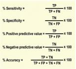
– Definitions of sensitivity, specificity, predictive values, and accuracy. TP: true positive – when the test is positive and the patient actually has the disease; FP: false positive – when the test is positive but a patient does not have the disease; TN: true negative – when the test is negative and the patient does not have the disease; FN: false negative – when the test is negative but the patient actually has the disease.
For the investigation of CAD, most studies have reported a sensitivity of ET between 61% and 73% (mean, 69%) and a specificity between 69% and 81% (mean, 75%). This variability is associated with differences in methodology and among studied populations.6, 323
The positive predictive value of ET is always higher in men, across all age groups, due to their higher prevalence of CAD.324
Women have a lower prevalence of CAD in general and, therefore, a greater prevalence of false-positives tests than men.325 In women, age has a greater influence on PPV: it is much lower in the young (age 35 to 50 years = 36%) compared to older women (age >65 years = 68%).326
In a cohort study comparing ET in both sexes, among women, the use of HRmax, duration of exercise, and ST-depression recovery time provided significant increases in PPV (from 47.8 to 61.5%) and NPV (88%).327
2.3. Pretest CVD Scores and Risk Factors
The administration of cardiovascular disease risk scores before ET aims to provide an individualized approach, predict possible complications (especially in intermediate and high-risk patients), and, consequently, allow a more contextualized analysis of ET findings. This assessment is recommended in adults and the elderly (typical age group: 40-75 years), and should be based on one of the following scores: European Society of Cardiology Systematic Coronary Risk Evaluation (SCORE2), American College of Cardiology/American Heart Association Atherosclerotic Cardiovascular Disease (ASCVD) algorithms, or Framingham Risk Score.328 - 330
In young adults (<40 years), evaluation and recording of classic risk factors – diabetes, smoking, dyslipidemia, stress, sedentary lifestyle, obesity, hypertension, and family history – are suggested.331
3. Clinical and Hemodynamic Responses to Exercise in the Adult Population
3.1. Clinical Responses
3.1.1. Exercise Tolerance
Exercise tolerance is recognized as the current best marker of life expectancy, regardless of age, sex, ethnicity, and comorbidities. During ET, it can be quantified objectively by the power generated in watts, by the duration of exercise, or by the metabolic equivalents of task (METs).332
Exercise tolerance can be quantified subjectively using the Borg or modified Borg Rating of Perceived Exertion scales (Figure 4), to measure the level of intensity of physical activity, tiredness, severity of dyspnea on exertion, and lower-limb fatigue.333, 334

Regardless of the presence of CVD, low exercise tolerance is associated with higher mortality rates and increased incidence of HF and CAD.332
Exercise intolerance (EI) is defined as impaired ability to perform physical activities due to the presence of symptoms such as dyspnea and/or fatigue.335
In both HFrEF and heart failure with preserved ejection fraction (HFpEF), EF is associated with worse quality of life, more frequent hospitalizations, and increased all-cause mortality.116, 336 In chronic diastolic heart failure, one of the main mechanisms of EI is chronotropic incompetence, which can be assessed by ET.337
In type 2 diabetes mellitus (T2DM), EI is also associated with chronotropic incompetence, both of which are associated with an increased risk of CVD and premature death.338
Figure 5 illustrates the main mechanisms and factors that contribute to the emergence and progression of EI.

– Mechanisms and factors associated with the emergence and progression of exercise intolerance and the role of ET. CAD: coronary artery disease; HF: heart failure; AF: atrial fibrillation; PAD: peripheral artery disease; ET: exercise test; HR: heart rate; ↓ METs: low cardiorespiratory fitness; ↑ Baseline HR: elevated baseline heart rate; ↓ HR Reserve: reduction of chronotropic reserve; ↓ HRrec: abnormal, slow, post-exercise recovery of HR.
ET and CPET are essential methods for diagnosing EI and monitoring physical training programs, aiming to document improvements in exercise tolerance and cardiopulmonary performance and consequent reductions in morbidity and mortality.339, 340 Physical training seems to be one of the only potentially effective and viable interventions for improving EI.338, 340
3.1.2. Cardiorespiratory Fitness/Functional Classification
Cardiorespiratory fitness (CRF)/functional classification by ET involves stratification of physical performance based on oxygen consumption, or uptake (VO2).
Maximal oxygen consumption (VO2max) expresses the highest amount of oxygen extracted from inspired air during dynamic exercise involving a large muscle mass. In submaximal ET, the highest VO2value obtained is more properly called the VO2peak.13, 134, 293
ET is considered maximal when:4
– Signs or symptoms of physical exhaustion develop.
– The patient is unable to continue.
– A Borg rating ≥18 is achieved.
– HR does not increase further even with increasing exercise intensity.
– The predicted HRmax is reached (always taking the previous items into account) or exceeded (≥110%).
In conventional ET, the VO2is estimated through formulas (indirect measurement), while in CPET, it is measured directly. Indirect measurement tends to overestimate VO2values due to the limitations of the original studies that generated the formulas.341, 342
Several formulas (“classic” and “novel”) are available for calculating predicted VO2peak, based on the type of ergometer, sex of the patient, protocol, whether the handrail was used for support, etc. (Annex 2).293, 343 - 348
VO2is most often expressed in mL/kg/min (mL.kg- 1 .min- 1 is also acceptable) It can also be expressed in MET and each MET corresponds to a VO2of 3.5 mL/kg/min.6
In the general population, VO2measured on a cycle ergometer is usually lower than on a treadmill. VO2max shows a progressive decline (on average, 8% to 10% per decade) after the age of 30. At age 60, the mean VO2max in men is approximately two-thirds that measured at age 20. When compared to men, women tend to have a lower VO2max due to lower hemoglobin, blood volume, stroke volume, and muscle mass.13, 134
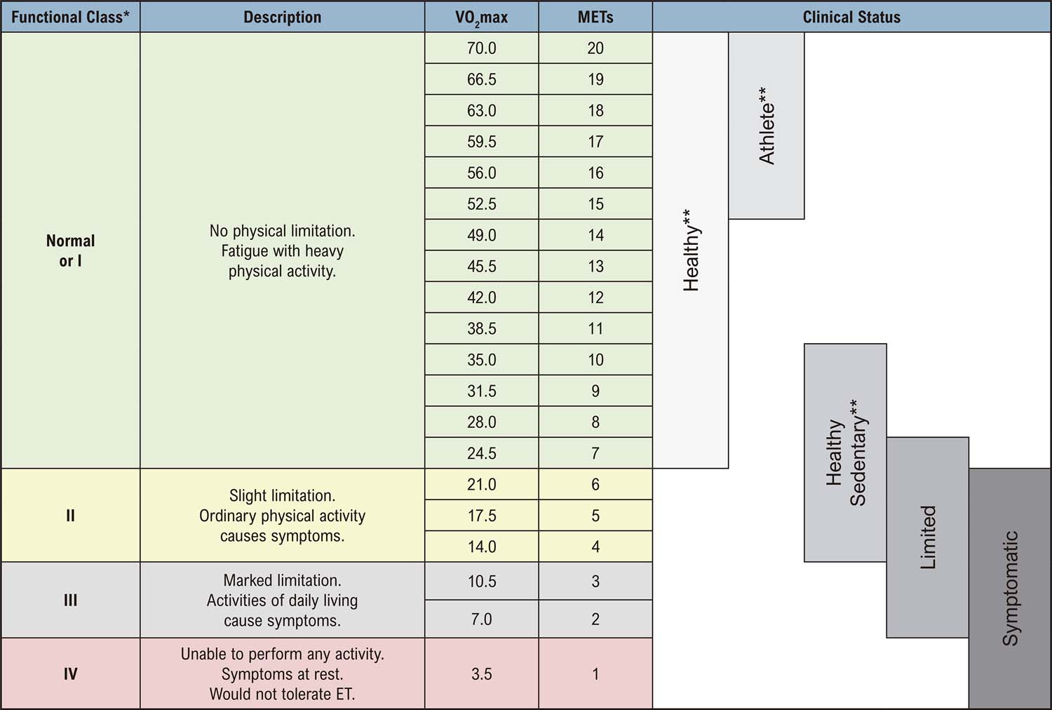
The New York Heart Association (NYHA) functional classification is used for diagnostic and prognostic purposes, contributes to therapeutic decision-making and exercise prescription, and is associated with VO2/METs achieved in ET (Figure 6).6

– VO 2 /MET values in relation to functional class and clinical status. 6,232 VO 2 max: maximum oxygen consumption; ET: exercise test; MET: metabolic equivalent of task. *New York Heart Association. **Depending on age and level of physical activity.
3.1.3. Symptoms
Symptoms during ET must be described in detail and correlated with clinical signs, hemodynamic response, and ECG findings. Any symptoms that lead to test cessation during the exercise phase must be described alongside the respective rating of perceived exertion (Borg or modified Borg) and the achieved performance (work load/METs).
The use of scales to quantify any symptoms of angina, dyspnea, and intermittent claudication (especially during CPET) is suggested. These scales must be visible to the patient and clearly explained to them before starting the test (Figure 7).13, 292, 349, 350

Chest pain should be described in terms of character, location, irradiation, aggravating/alleviating factors, duration, other concomitant symptoms, and the timing of pain onset and resolution during the ET. Clinicians are also advised to describe whether the pain was test-limiting; the initial HR, SBP, and double product; and respective ECG findings.
The occurrence of typical angina (i.e. progressing with increasing exertion) is by itself considered consistent with an ischemic response.6
In a study of 10,870 patients with symptom-limited ETs, typical angina pectoris was associated with an increased risk of mortality (RR: 2.7; 95% CI: 1.4-5.1; p<0.002) compared to non-anginal chest pain.351
The occurrence of dyspnea during ET is associated with a significant increase in all-cause mortality, but it is not associated with higher rates of ischemia compared to chest pain.352
In suspected PAD and evaluation of intermittent claudication, use of an intermittent claudication pain scale to quantify pain and its severity is recommended. Ideally, ET should continue until pain becomes intolerable and/or the patient can no longer walk (absolute claudication).353
The intermittent claudication scale ranges from 0 to 4 (see Figure 7) and can be used to quantify the progression of claudication during ET. The respective work load, duration of exercise, and the time point of pain resolution should be recorded.175
All other test-emergent symptoms (i.e. vertigo, dizziness, presyncope, headache) must be recorded, as well as the timing of occurrence, severity, duration, BP and HR at the time of onset, and any other information deemed useful to allow appropriate interpretation.
3.1.4. Inspection and Auscultation
Sweating and facial flushing are to be expected during intense physical exercise. Onset of skin pallor, accompanied or followed by diaphoresis, cyanosis, or tachypnea, denotes a pathological condition. Occurrence of any of these at low work loads and/or in combination with hemodynamic instability or ECG changes (ST deflection and complex arrhythmias) denotes even greater severity.1
Auscultation should be performed and compared before and after exercise in order to correlate it with any observed clinical signs. The presence of wheezing, rhonchi, or crackles before exercise may contraindicate the performance of ET; after exercise, they may indicate exercise-induced asthma or ventricular dysfunction.354, 355
On auscultation of new murmurs and/or worsening of pre-existing murmurs during exertion or recovery, their relationship with the cardiac cycle, location, quality or tone, intensity, and presence of ejection clicks should be described and recorded.356, 357
Exertion usually increases the intensity of regurgitant murmurs originating from the left heart chambers (mitral and aortic regurgitation).92, 94
Appearance of S3 on exertion in men over 40 years of age, in the presence of dyspnea or excess fatigue, may be associated with ventricular dysfunction.358 In adults and the elderly, the appearance of S3 soon after cessation of exercise is often associated with ventricular dysfunction, previous MI, and left bundle branch block.359
Adults and young athletes usually have S3 on baseline auscultation. If it appears during exercise in these populations, it is considered a physiological adaptation, and does not correlate with structural heart disease.360
3.2. Hemodynamic Responses
The following concepts are important to understanding the hemodynamic response to increasing exertion during ET:13, 134, 293
– Dromotropic reserve: increased speed of cardiac electrical impulse conduction.
– Inotropic reserve: capacity to increase ventricular function (efficiency of ventricular filling and emptying); evaluated by the behavior of SBP.
– Peripheral arterial resistance: capacity of peripheral vessels to adapt (vasodilate/vasoconstrict); evaluated by the behavior of DBP.
– Coronary reserve: capacity of the coronary network to adapt to increased blood flow in response to increased myocardial metabolic activity.
– Cardiac reserve: capacity of the heart to increase its output to compensate for the greater metabolic demand of the exercising skeletal musculature. It is influenced by all of the above parameters.
3.2.1. Heart Rate
Assessment of HR behavior allows evaluation of chronotropic response, chronotropic reserve, double product, and autonomic regulation, making it an important diagnostic and prognostic parameter.
3.2.1.1. Resting Heart Rate
The normal resting HR (RHR) range is 50 to 99 bpm, as measured by ECG at rest (sitting or supine). In the context of ET, RHH is a valuable parameter for the interpretation of HR response, autonomic regulation, and prognosis.361, 362
It is predictive of CAD, HF, AF, stroke and is associated with an increased risk of cardiovascular events, cardiac death, and death from all causes.363 - 365
Research has shown that, during ET, an RHR ≥80 bpm in the general population and ≥75 bpm in diabetics with stable CAD is associated with increased all-cause mortality.366
In a cohort of 56,634 individuals (49% women) without known CAD or AF, RHR ≥90 bpm was associated with a significant increase in all-cause mortality; in men, this was independent of physical fitness.367
3.2.1.2. Chronotropic Response
The normal chronotropic response to exercise consists of an increase in HR due to a decrease in vagal tone, followed by an increase in sympathetic tone and consequent adaptations of systemic vascular blood flow.368
The increase in HR follows the increase in work loads and usually presents a linear correlation with VO2alone between 50 and 90% of VO2max. In healthy adults, HR normally increases at a rate of ≈10 bpm per MET.134
Vagal withdrawal is responsible for the initial increase of 10 to 30 beats per minute (bpm), with further increments usually mediated by sympathetic activity. HR is responsible for most of the increase in cardiac output during exertion, particularly at higher loads (Figure 8).134, 299

– Heart rate response and autonomic adjustments during ET in adults. HRmax: maximal heart rate; Act.: activity; min: minute.
HR recovery (HRrec) during the first 30 to 60 seconds after exertion involves reactivation of the parasympathetic system and progressive inhibition of sympathetic activity. HRrec can also be influenced by the degree of venous return and the atrial baroreceptor response. Delayed HRrec is an important marker of autonomic dysfunction and risk of mortality (CV and all-cause) and major adverse cardiovascular events (MACE).369
The estimated maximum heart rate should not be used as the sole criterion for ET cessation or to assess the effectiveness of ET. There is generally a wide margin of error (wide dispersion of the mean) in formulas used to estimate this parameter, ranging from ±10 to 15 bpm. Women tend to have a lower estimated maximum heart rate.301, 370
Evaluation of chronotropic response through the estimated maximum heart rate is limited in patients on medications that interfere with autonomic modulation (antiarrhythmics and beta-blockers), in AF and atrial flutter, and in patients with rate-responsive pacemakers, ICDs, congenital AV block, etc. Major factors that interfere with the HRmax achieved during ET are listed in Table 29.
Table 30 lists variables which measure the HR response to ET, while Table 31 gives their definitions, criteria, and interpretations.
Particular Features of Chronotropic Response:
– In a cohort of 458 men (age 56±8.5; mean follow-up: 6 years) with CAD, an increase in HR during the first minute of the exercise phase of ET ≥12 bpm was strongly associated with cardiac death (RR: 15.6; 95% CI: 2.0-118.7; p<0.001) and nonfatal MI (RR: 5.0; 95% CI: 2.7-9.1; p<0.0001).379
– In a follow-up study of 306 patients with asymptomatic aortic stenosis (age 65±12 years, 33% women; mean follow-up duration 25 months), among severe cases, rapid early rise in HR (defined as reaching 85% HRmax or increase ≥ 50% HR at 3.5 METs) was associated with need for valve replacement (RR: 3.32; 95% CI: 2.03-5.45; p<0.001).374
3.2.2. Blood Pressure Response
Assessment of the blood pressure response (BPR) to exercise is an important diagnostic, prognostic, and cardiovascular risk stratification tool. BPR essentially depends on cardiac output, peripheral vascular resistance, and medication use.6, 380
In healthy adults, the increase in systolic blood pressure (SBP) is expected to be proportional to the increase in dynamic exercise load (corresponding to the inotropic response). As work load is maintained, the SBP stabilizes within 2 to 3 minutes.380, 381 The average increase in SBP is usually 10 mmHg/MET. In individuals who reach an exercise load >10 METs, the SBP increase is 6.2 mmHg/MET.6, 382 After cessation of exertion, SBP tends to decrease gradually due to the vagal response and rapid reduction in cardiac output. It generally returns to resting levels within the first 6 minutes of recovery, and may remain below pre-exercise levels for several hours (Figure 9).383

– Systolic and diastolic blood pressure response to ET in adult men and women. SBP: systolic blood pressure; DBP: diastolic blood pressure; min: minute.
During exertion, diastolic blood pressure (DBP) remains unchanged or may show minor fluctuations (±10 mmHg) due to the drop in peripheral arterial resistance. Occasionally, in healthy individuals, DBP sounds can be heard down to 0 mmHg during exercise without any pathologic significance. In such cases, use of phase IV Korotkoff sounds (abrupt muffling of sounds, which then become softer) to define DBP is recommended. At the onset of recovery, DBP may increase slightly or remain unchanged. It generally returns to resting levels within the first 6 minutes of recovery.384
ET should not be performed in patients with a resting SBP ≥180 and/or DBP >110 mmHg. When measuring with an aneroid sphygmomanometer, round values to the nearest 5 mmHg. When using automated BP devices, consider the actual absolute values.4, 6, 13
A hypertensive/exaggerated SBP response to exercise is defined as a maximum value ≥210 mmHg for men and ≥190 mmHg for women, regardless of protocol and/or ergometer.85, 385 - 387 In subjects who are normotensive at baseline, such an SBP response is associated with increased risk of future hypertension and cardiovascular events (Table 32).388, 389
Peak SBP on exertion becomes gradually higher values with advancing age. It is generally lower in women than in men, a difference that tends to disappear in older adults. Young women may present a plateau SBP response or even a slight drop in SBP at peak exercise, with no specific clinical significance.6, 87, 390
The DBP response is considered abnormal when there is a ≥15 mmHg increase and/or it exceeds >90 mmHg, considering normal pre-ET DBP values at rest.391 There is strong scientific evidence from Brazilian and international studies validating these criteria for normal SBP and DBP response to ET for the diagnosis of hypertension, assessment of treatment efficacy, association with CVD, and risk stratification.391 - 393
A hypertensive response to exercise (HRE) is more common in older adults and in individuals with hypertension, even when BP is well controlled at rest.394 In hypertensive patients, HRE is associated with a higher risk of future HF, LV hypertrophy, endothelial dysfunction, diastolic dysfunction, and cardiovascular events.392, 395, 396
In the absence of systemic arterial hypertension or other cardiovascular diseases, HRE is not a benign phenomenon; its main causes are endothelial dysfunction and stiffness of the major arteries. HRE is usually associated with functional and structural LV abnormalities, especially when accompanied by a rise in central BP.72, 305, 387, 397
Paradoxically, studies in patients with suspected or established CAD have shown that the occurrence of HRE was associated with less severe coronary lesions and lower mortality compared to those who had a normal BP response.398, 399
It is suggested that BP measurement be performed upon reaching a work load of 5 METs. SBP ≥150 mmHg is a discriminatory threshold for hypertension, alongside systolic hypertension on 24-hour ABPM and LV hypertrophy on echocardiography.400, 401
In a prospective cohort of 6,578 asymptomatic participants from the Lipid Research Clinics Prevalence Study (mean age 46 years; 45% women; mean follow-up duration, 20 years), among individuals who were normotensive or prehypertensive at rest, BP >180/90 mmHg during the second stage of the Bruce protocol was associated with risk of CVD mortality (SBP RR: 1.96; 95% CI: 1.40 to 2.74; p<0.001/DBP RR: 1.48; 95% CI:1.06 to 2.06; p=0.02).75
Pre-ET hypertension with a normal BP response to exercise is common in anxious patients, and is generally not associated with future development of hypertension.215, 402
SBP >250 mmHg and DBP ≥120 mmHg in normotensive individuals, or DBP ≥140 mmHg in hypertensive individuals, are considered criteria for test cessation.4, 6, 13
Any occurrence of hypotension/drop in BP during exercise, a rare event (incidence <2% to 6%), requires immediate intervention (cessation of exercise and placement of patient in the supine position) for safety reasons, as it portends acute risk of a MACE. The most common causes are: severe multivessel CAD with LV dysfunction; cardiomyopathies; LV outflow tract obstruction; increased vagal tone; hypovolemia; and arrhythmias. Its PPV is higher in men than in women (Figure 10).403

– Main blood pressure responses to ET and their repercussions. HTN: hypertension; CBF: cerebral blood flow; Hypertensive Resp.: hypertensive response; Orthostatic pos.: orthostatic position on the treadmill; BP: blood pressure, *Pretest normotension with a hypertensive response to exercise indicates risk of future hypertension. * 2 Associated with decreased adrenergic activity and histamine-mediated vasodilation. * 3 Associated with hyperthermia on exertion and pre-existing dehydration (including secondary to diuretics).
Hypotension and syncope during recovery may occur due to a variety of causes, ranging from physical exhaustion in an apparently healthy individuals to abnormal, pathologic responses, such as LV outflow tract obstruction (i.e. in AS and obstructive hypertrophic cardiomyopathy); dysautonomia, and negative vagal baroreflex regulation (see Figure 10).74, 386, 404, 405
Table 32 presents definitions of key terms, normal values and ranges, interpretation criteria, and implications regarding the BP response during ET.
3.2.3. Double Product
The double product (DP), or rate pressure product, expresses myocardial oxygen consumption (linear relationship with myocardial oxygen uptake and coronary blood flow). It is calculated by multiplying the HR by the SBP at any time during ET.6, 134, 423
DP is an important parameter in the assessment of anginal thresholds, ECG changes (ST-segment deviation and arrhythmias), cardiovascular efficiency, progression of cardiorespiratory fitness, and response to pharmacologic and interventional therapy. It has prognostic value independently of the presence of CAD and, therefore, serial measurement is recommended. A maximum DP (usually obtained at peak exertion during ET) <25,000 bpm.mmHg is associated with worse prognosis.115, 134, 423, 424
Factors that limit DP assessment include beta-blocker and antiarrhythmic therapy; uncontrolled hypertension; and atrial fibrillation and flutter with uncontrolled ventricular rate. It should not be calculated during tachyarrhythmias.134, 293, 299
4. ECG Responses 371
For proper analysis, description, and interpretation of ECG responses to ET, the following factors should be taken into account:
– Check proper electrode placement and attachment to minimize errors and artifacts.425, 426
– Follow the standard ECG reporting guidance of the Brazilian Society of Cardiology Guidelines on the Analysis and Issuance of Electrocardiographic Reports – 2022.295
– Use automated measurement systems for intervals, durations, and amplitudes of ECG waves and segments.427
– Consider the effects of any ECG filters applied (high, medium, low) for baseline stabilization and reduction of muscle and electrical artifacts. Use high-pass filters with a cutoff of at least 150 Hz in adults and adolescents. Filters with lower frequencies may interfere with capture of pacemaker spikes.295, 428, 429
– Review any automated measurements to rule out errors due to possible interference, artifacts, or abnormalities in the underlying tracing.427, 430
– Provide a detailed, contextualized description in the ECG record.
4.1. P Wave
4.1.1. Normal Responses
In adults, P waves on a resting ECG – in the presence of sinus rhythm – are positive in leads D1, D2, and aVF, with a mean electrical axis vector of 60° (range, 0° and 90°), maximum amplitude of 250 mV (2.5 mm), and duration ≤110 ms.295
During exercise, the following are normally observed (Figure 11):431

– Representation of the main responses of the P, Q, R, S, T, and U waves, PR interval, PR and ST-segments, and J point during the exertion stage of an ET. A. Normal response to exercise: P wave duration reduces and amplitude increases (peaked P wave); PRi shortens with increasing heart rate (HR); PR segment remains unchanged (at baseline); Q wave increases in amplitude; R wave presents a reduction in amplitude concomitantly with the increase of the S wave; J point maintains the same baseline level or exhibits is a slight depression; ST-segment remains at baseline or may exhibit rapid upsloping depression; T wave amplitude and morphology remain unchanged, with a rapid initial component and a slow final component (may exhibit a small decrease in amplitude and reduction in duration); U wave tends to remain positive at the onset of exercise and disappear as the HR increases. B. Abnormal response: P wave exhibits increased duration and reduced amplitude; PRi remains unchanged despite increasing HR; PR segment depression >0.5 mm; Q wave amplitude unchanged; R wave unchanged despite progressive exertion and S wave reduction; J-point depression with horizontal ST-segment depression (Y-point ≥1.0 mm); reduction in amplitude of the T wave (tends to be symmetrical); negative U wave. C. Abnormal response: Increased P wave duration, with notched morphology; prolongation of PRi as the HR increases; Q wave disappears; increasing R-wave amplitude with concomitant reduction of the S wave; J-point depression with downsloping ST-segment depression (Y-point ≥1.0 mm); peaked and symmetric T wave with increased amplitude.
– Gradual, linear increase in P wave amplitude as HR increases (in inferior leads in the frontal plane). This increase is, on average, 100 mV (1 mm).
– Preservation of the axis vector of the P wave.
– No change or, rarely, a minimal increase (≤20 ms) in duration.
Normal P wave response to recovery:
– In the first minute of recovery, there may be an additional increase in P wave amplitude (exceeding than seen during exertion), even as the HR declines. A progressive reduction in amplitude occurs thereafter, returning to baseline after the 6th minute.432
– P wave duration remains unchanged or, rarely, increases minimally (≤20 ms) until the 3rd minute.433
4.1.2. Abnormal Responses
The main abnormal P wave responses to exercise are:431, 434, 435
1) No change in P wave amplitude from baseline, especially regarding the negative component (change <0.25 mm). This increases sensitivity (69%) and specificity (78%) for the diagnosis of CAD.
2) Increased P wave duration. Is associated with left atrial pressure overload during exercise-induced ischemia.
3) Change in P wave morphology with increase in the amplitude of the terminal negative component in lead V1, at 50% of the maximum exercise time, is the single ECG change most predictive of CAD verified by myocardial perfusion imaging.
4) Dispersion of P wave duration combined with ST-segment depression increased ET sensitivity to 79% and PPV to 91%.436
4.2. PR Interval/Segment
4.2.1. Normal Responses
On a normal resting ECG, the PR segment (PRs) begins at the end of the P wave, ends with the beginning of the QRS complex, and is isoelectric. It serves as a bridge between atrial activation and ventricular activation, as well as atrial recovery, which is usually of very low amplitude and difficult to detect, as it occurs within the QRS. When sufficient amplification is used or in the presence of first-degree AV block, the atrial repolarization wave (Ta wave) can be visualized. The interval from beginning of the P wave to the end of the Ta wave is equivalent to the atrial QT interval.
The PR interval (PRi; start of P wave to start of QRS complex) normally measures 120 to 200 ms and is best determined in lead II.
The PRs serves as a baseline demarcation point for the assessment of changes in the ST-segment and wave amplitude. The line joining the PQ junctions (end of the PR segment and beginning of the QRS complex) is defined as the “baseline”, considering at least four successive complexes, at the same horizontal level and without artifacts. Computerized ECG analysis systems use automated algorithms which consider the end of the PRs as the isoelectric baseline.1, 295
During exertion, the PRs shortens and slopes slightly downwards in the inferior leads, while the PRi shortens to an extent directly proportional to the increase in HR.437
During the initial phase of recovery, the PRi and PRs are dependent on the degree of physical fitness of the patient. In sedentary men, the mean PRi is ≈110 ms; in athletes, ≈280 ms.438
4.2.2. Abnormal Responses
The main abnormal PRs responses during ET are:
– Even in normal individuals, a greater downward slope of the PRs may occur, attributable to exuberant atrial repolarization (negative Ta wave). If it persists in the initial period of ventricular repolarization, it will cause depression of the J point and ST-segment (upsloping; false-positive if in the inferior leads).152, 439
– Exercise-induced atrial infarction: PRs depression >0.5 mm, usually with ST-segment elevation (denoting associated ventricular infarction) and atrial arrhythmias.295, 440
– Pathologic PRi prolongation will be addressed in the section on atrioventricular blocks.
4.3. Q Wave
4.3.1. Normal Responses
On resting ECG, Q waves are considered normal when their duration is ≤30 ms and their amplitude is <0.2 mV (up to 0.4 mV in adolescents), which corresponds to <25% of the subsequent R wave. In lead III, Q wave duration can exceed 40 ms, but rarely reaches 50 ms. The presence of a Q wave in lead V1 is always pathological.295, 371
During exercise, the Q wave normally increases significantly in amplitude, particularly in CM5 configuration and in the lateral leads.441
4.3.2. Abnormal Responses
On resting ECG, the Q wave is considered abnormal when, in the absence of bundle branch block and/or a preexcitation syndrome, its duration is ≥40 ms and/or amplitude is greater than one-third of the adjacent R wave (in two or more leads from the same ventricular wall).
The main abnormal Q wave responses during ET are:
– Reduction in amplitude/disappearance of the Q wave during exercise or recovery, which may indicate septal ischemia and which, when associated with ST-segment depression, increases the PPV for CAD.441, 442
– Increased Q wave amplitude in the presence of ST-segment depression reduces the PPV, increasing the possibility of false positives.443
– Exercise-induced increase in Q wave duration (10±13 ms) in patients with single-vessel CAD and recent MI is associated with ischemia on thallium perfusion scan.444
– Transient Q waves can be seen in hypoglycemia, hyperkalemia, and asthma.
4.4. R Wave
4.4.1. Normal Responses
On resting ECG, the duration of R waves will depend on the lead and duration/pattern of the QRS complex. R-wave amplitude is variable; it increases progressively in the precordial leads and is generally <27 mm in V5 and V6.445
During exercise, the following phenomena usually occur:
– Reduction (2.6±1.1 mm) in average R-wave amplitude as maximal exertion is approached.446 A marked reduction is observed in the lateral leads (V5 and V6) at maximal exercise and in the first minute of recovery.
– As R waves decrease in amplitude, an increase in S waves is observed.115
4.4.2. Abnormal Responses
On resting ECG, patients with anterior wall MI exhibit reduced R-wave amplitude or absent R waves altogether.447, 448
The main abnormal R wave responses during ET are:
– Increase in R-wave amplitude resulting from changes in LV dimensions and myocardial ischemia.449 The presence and extent of reversible ischemia in patients with CAD correlates directly with increased amplitude of the R wave and ST-segment depression.450, 451
– R-vave amplitude increases significantly in the precordial leads during episodes of transmural ischemia.452
– Low-amplitude R waves (<10 mm) may occur with ST-segment depression of limited magnitude in relation to actual ischemia, thus interfering with the accuracy of ET for diagnosis of CAD.453, 454
4.5. S Wave
4.5.1. Normal Responses
On resting ECG, the S wave is commonly observed in leads I, II, III, aVF, V1, and V2 (where its amplitude is greater than that of the R wave); it is usually absent in V5 and V6. Under normal conditions, the S wave amplitude is <0.3 mV (30 mm).445
During ET, in the absence of intraventricular conduction disorders, the S wave usually:
– Tends to increase amplitude in inferolateral leads (particularly in aVF and V5; maximum 0.3-0.4 mV) with concomitant reduction of the R wave.115, 455
– In the first minute of recovery, the S wave amplitude remains unchanged or is slightly reduced. It returns to pretest range at 3 to 5 minutes of supine recovery.455
– The mean S wave duration shows a progressive reduction with increasing effort, regardless of intraventricular conduction disorders.456
4.5.2. Abnormal Responses
The major abnormal S wave responses to exercise are:
– Decreased S waves amplitude in patients with CAD are associated with subendocardial ischemia, even in the absence of ST-segment changes, increasing ET sensitivity.457
– In patients without intraventricular conduction disorders, unchanged S wave duration is usually associated with significant right coronary artery (RCA) or circumflex artery obstruction.456, 458
– A significant increase in duration (≈12.5±6 ms) is observed in patients with critical left anterior descending artery (LAD) obstruction, left anterior fascicular block, and right bundle branch block.456
4.6. QRS Duration
4.6.1. Normal Responses
The duration of QRS complexes generally decreases proportionally to the increase in HR during exercise (decrease ≈3.0 to 4.9 ms). Exceptionally, in healthy patients, there is no such change in duration.459
The variance in QRS complex duration (ΔQRSd) is a parameter that corresponds to the difference in QRS duration measured in aVF and V5 immediately upon cessation of effort (onset of recovery) as compared to the resting condition.459 The normal ΔQRSd is ≤3 ms.460
4.6.2. Abnormal Responses
Significance of ΔQRSd changes:
– A ΔQRSd >3 ms is considered positive for ischemia.460 In patients with CAD, this parameter is directly and significantly associated with the number of arteries with significant obstruction (4.8 ms = one artery, 7.8 ms = two arteries, 13.3 ms = three arteries; p<0.001) and regional wall motion abnormalities on radionuclide ventriculography (6.7 ms = one LV region, 13.5 ms = two LV regions, 21 ms = three regions; p<0.0001).461
– A ΔQRSd >3 ms improved the diagnostic accuracy of ET for CAD, regardless of ST-segment changes, compared to myocardial perfusion scan (sensitivity 93%, specificity 71%, PPV 86%).459
– A ΔQRSd >3 ms in women is more sensitive and specific than ST-T changes for the detection of ischemia.462, 463 A previous study showed that abnormal ΔQRSd in women increased sensitivity (91%), specificity (89%), and PPV (88%) of exercise test; in younger women (age 27-50 years), sensitivity was 80% and specificity 83%.462
– A ΔQRSd ≥15 ms in patients with ischemic heart disease predicted occurrence of severe ventricular arrhythmias (ventricular tachycardia or fibrillation) with a PPV of 73%. After surgical revascularization, these patients no longer had an abnormal ΔQRSd and arrhythmias were suppressed.464
4.7. High-frequency Assessment of QRS Fragmentation
4.7.1. Normal Responses
High-frequency assessment of QRS fragmentation, or high-frequency QRS (HF-QRS), is a special ECG filtering technique (usually between 150 and 250 Hz) that allows analysis of the high-frequency components of QRS complexes.465
The technique involves combining QRS complexes from the same lead (from a full 12-lead recording) or complexes from four leads (V3, V4, V5, and V6) to form a “precordial average complex”. The normalized root mean square (NRMS) and/or normalized peak amplitude (NAMP) of these complexes are then calculated. These parameters are generally evaluated on the resting ECG, at peak exertion, and during recovery.466
In healthy people, the HF-QRS is considered normal when the average NRMS values are >1 μV in all three stages of the ET (rest, peak exertion, and recovery) and an increase in NRMS is observed during and immediately after exercise (compared to the resting ECG).466, 467
4.7.2. Abnormal Responses
The HF-QRS is considered abnormal (positive for ischemia), regardless of changes in the ST-segment, when: there is an absolute reduction ≥1μV or a relative reduction ≥50% between the maximum and minimum NRMS values in ≥3 leads in patients who reached maximum exertion. In submaximal tests (≤85% of the predicted maximum HR), the relative reduction must be adjusted linearly, between 40% and 50%, according to the ratio between the actual maximum HR achieved and the predicted HR. HF-QRS has greater sensitivity, specificity, and PPV than ST-segment changes.468 - 470
Abnormal HF-QRS is usually found in post-MI patients with myocardial scarring, ischemia, or conduction delay due to non-homogeneous ventricular activation. It is a predictor of mortality and cardiac events in patients with CAD.467, 468
Abnormal HF-QRS demonstrated incremental diagnostic value when associated with ST-segment changes (PPV 80.4% vs. 74.9%; p<0.0001). Joint analysis of HF-QRS and ST-segment changes identified 92.3% of individuals with significant ischemia.471
Analysis of both HF-QRS and ST-segment depression demonstrated a 99% NPV for severe ischemic disease. On multivariate regression, abnormal HF-QRS was an independent risk factor for MACE at 2 years (RR: 2.8; 95% CI: 1.7-4.4; p<0.001).472
4.8. T Wave
4.8.1. Normal Responses
On the resting ECG, the T wave is rounded, asymmetrical, with a slower initial upsloping portion and a faster downsloping final portion, usually with the same polarity as the QRS and positive in almost all leads (always negative in aVR). Its duration ranges from 100 to 300 ms, and its amplitude is a maximum of 5 mm in peripheral leads and <15 mm in precordial leads, corresponding to approximately 10% to 30% of the total amplitude of the QRS complex that precedes it.473, 474
In the initial phase of exercise, there is a general decrease in T wave amplitude followed by an increase at higher exercise loads (returning to baseline), with a further increase in the initial phase of recovery.455
4.8.2. Abnormal Responses
When interpreting T wave changes, pre-existing diseases (especially hypertension, valvular heart disease, and CKD), risk factors and the pretest probability of CAD should be considered.
On resting ECG, the main T wave changes that may be associated with ischemia are:
– Positive, symmetric, pointed T waves followed by a U wave (positive or negative).475
– Negative, symmetric, pointed T waves in leads with a predominantly positive QRS (except III, aVR, and V1).115
– Biphasic pattern in the anterior thoracic leads (V1 to V3), usually associated with myocardial ischemia in unstable angina.
– Flat (usually nonspecific; may be associated with myocardial ischemia).
Abnormal T wave responses to exertion:
– Increase in amplitude (>2.5 mV), symmetric, in leads V2 to V4: in patients with chest pain, is associated with severe ischemia.115
– T wave pseudonormalization, corresponding to the presence of an inverted T wave ≥1 mm in any lead at rest which becomes positive at peak exertion; is generally associated with reversible, fixed defects on myocardial perfusion scintigraphy.476
– T wave pseudonormalization associated with U wave inversion in anterior thoracic leads is highly indicative of critical LAD stenosis.477
– T wave pseudonormalization in leads related to previous MI at low work loads has shown to be a sensitive, specific indicator for the presence of residual myocardial viability. Sensitivity and accuracy were higher for prior MI.478, 479
– In submaximal ET, performed in the early phase of stabilized unstable angina or post-MI without Q waves, T wave pseudonormalization (regardless of the occurrence of ST-segment depression) was predictive of survival at 6-month follow-up.480
– T wave pseudonormalization in populations with a low prevalence of CAD is a nondiagnostic finding.152
4.9. U Wave
4.9.1. Normal Responses
On resting ECG, the U wave is a low-amplitude, low-frequency deflection that follows the T wave (usually with the same polarity), with an amplitude proportional to that of the T wave (≈5% to 25%; mean: 0.33 mm). It is best observed in leads V2 and V3 (≤2 mm), has a mean duration of 221±73 ms (<50% of the preceding T wave), and is most commonly identified at HRs <95 bpm. It is seen in up to 50% of normal individuals. During exercise, duration, amplitude, and axis generally remain unchanged, but U waves become difficult to identify at HR >120 bpm due to encroachment of the T and P waves.481 - 483
4.9.2. Abnormal Responses
A negative U wave on resting ECG is considered abnormal and is often associated with mitral and/or aortic regurgitation, hypertension, and ischemic heart disease.484, 485 Increased amplitude may be associated with hypokalemia and medications (digitalis, amiodarone, and quinidine).483, 484, 486
The main abnormal U wave responses during ET are:
– At baseline (positive), increased exertional amplitude (≥0.5 mV) in precordial leads is associated with inferoposterior myocardial ischemia, and is a marker of significant circumflex coronary artery (CX) or RCA obstruction.487, 488
– Appearance of a positive U wave on exertion and/or during the first 3 minutes of recovery is often associated with CX or RCA obstruction.489
– Appearance of a negative U wave with amplitude ≥0.5 mV, persisting for at least 1 minute during and/or after exercise, is associated with severe LAD disease.487, 490
– Appearance of a negative U wave is a marker of a well-developed collateral circulation in patients with severe CAD or stable angina.487 In precordial leads, it is a marker of viable myocardium after MI.491
– Transient exercise-induced U wave inversion with amplitude ≥0.5 mV in anterior wall leads (V2 to V5), is associated with episodes of acute ischemia and severe LAD disease.492, 493
4.10. Early Repolarization
Early repolarization pattern (IRP) is seen in 1% to 13% of the general population.494, 495 In middle-aged individuals, it has been associated with increased risk of sudden cardiac death (SCD).496, 497
The criteria for diagnosis of early repolarization on resting ECG are:498, 499
1) QRS complex duration <120 ms.
2) End-QRS notch or slur on the downstroke of a prominent R wave. If there is notch, it must be completely above the baseline. The point of J-wave onset (Jo) must also be above the baseline (Figure 12).

– ST-segment elevation patterns, including early repolarization. ST-seg.: ST-segment; ms: milliseconds. *Exercise-induced ST-segment elevation (≥1.0 mm measured at 60 ms after the J-point). **In the early repolarization pattern, ST-segment elevation should be measured 100 ms after the Jt point, and the pattern of elevation (upsloping, horizontal, or descending) should also be assessed.
3) The peak of the J-point notch (Jp) must be ≥0.1 mV in two or more contiguous leads of a 12-lead ECG, except V1 through V3.500
ST-segment elevation should be measured 100 ms after the Jt point (termination of the J-point notch). In addition to the magnitude of elevation, the pattern should be described:
– “Early repolarization with upsloping ST-segment”, when the ST-segment is ascending (inclined upwards) and followed by a vertical T wave.
– “Early repolarization with horizontal or descending ST-segment”, when the ST-segment is horizontal or descending (inclined downwards).
Behavior and significance of ERP in ET:
– Common in young individuals. In this setting, usually reduces progressively with increasing exertion, and may disappear altogether at moderate loads. ERP with rapidly upsloping ST-segment elevation in the anterolateral leads has been reported in athletes.501
– Persistent ERP on exertion has been observed in symptomatic patients (aborted sudden cardiac death, sustained ventricular arrhythmia, and/or unexplained syncope).502
– During recovery, ERP resumes slowly, progressively, and, in approximately 30% of patients, occurs at the 5th minute.503, 504
High-risk markers verifiable on ET in patients with ERP:55, 494
– Exercise-induced polymorphic VT.
– Persistent horizontal and/or descending ST elevation during exercise in inferior or inferolateral leads is associated with risk of idiopathic VF and a threefold increase in risk of sudden arrhythmic death.500
4.11. ST-segment Elevation
Exercise-induced ST-segment elevation is defined as an ST-segment elevation ≥1.0 mm (≥0.10 mV) within 60 ms of the J-point, occurring in two or more leads, regardless of the presence of a Q wave (Figure 12). There may be reciprocal ST-segment depression (“mirror image”).505, 506
Exercise-induced ST-segment elevation is generally associated with severe myocardial ischemia (usually transmural), coronary artery spasm, Prinzmetal angina, left ventricular aneurysm, peri-infarction ischemia, and LV wall motion abnormalities.506, 507
The leads in which exercise-induced ST-segment elevation occur correlate with anatomic-vascular segments of the LV.506, 508, 509 Use of Myers’ topographic description of ischemic manifestations is advised:295
– Anteroseptal wall: leads V1, V2, V3.
– Anterior wall: leads V1, V2, V3, and V4.
– Localized anterior wall: leads V3, V4, or V3-V5.
– Anterolateral wall: leads V4 to V5, V6, I, and aVL.
– Extensive anterior wall: V1 to V6, I, and aVL.
– Lateral wall: leads V5 and V6.
– High lateral wall: I and aVL.
– Inferior wall: II, III, and aVF.
The terms “posterior wall” and “dorsal” should no longer be used due to current evidence that leads V7 through V9 refer to the lateral wall.
Thirty percent of patients with a past anterior wall MI and 15% of those with prior inferior wall MI exhibit exercise-induced ST-segment elevation in the leads involved, and its significance varies depending on additional ET findings.510, 511 The finding of exercise-induced ST-segment elevation in leads with abnormal Q waves may represent residual ischemia of the peri-infarct area (myocardial viability), ventricular dyskinesia, or akinetic LV wall motion.511, 512 However, it does not allow quantification of viable tissue, justifying additional investigation by imaging methods to ascertain whether therapeutic intervention is indicated.512, 513
Particular features of exercise-induced ST-segment elevation:
– Exercise-induced ST-segment elevation ≥0.2 mV (2 mm) in leads without Q waves mandates test cessation.
– Exercise-induced ST-segment elevation is more commonly associated with severe proximal obstruction than with coronary spasm in unobstructed arteries.514, 515
– Exercise-induced ST-segment elevation, with or without concomitant depression, was predictive of the presence, extent, and location of myocardial ischemia assessed by myocardial perfusion scintigraphy. Exercise-induced ST-segment elevation in lead V1 with ST-segment depression in aVR and V4-V6 was associated with left main coronary artery (LMCA) or proximal LAD stenosis.509
– Exercise-induced ST-segment elevation in lead aVR showed 100% sensitivity to detect LMCA stenosis (specificity 33.5%) and 94.3% sensitivity for LAD stenosis (specificity 26.6%). The combination of exercise-induced ST-segment elevation in both aVR and V1 reduced sensitivity (74.4% and 65.9%) but increased specificity (68.5% and 64.4%).508, 516
– Exercise-induced ST-segment elevation in lead aVR was associated with MACE in 33% of patients at 2-year follow-up.517
– In patients who have undergone successful single-vessel PCI without residual ischemia for treatment of MI, exercise-induced ST-segment elevation may be associated with impaired coronary microcirculation and lower myocardial viability.518
4.12. J Point and Upsloping Depression
The J point (where the end of the QRS joins the onset of the ST-segment) is usually depressed on exertion in the lateral wall leads, gradually returning to its pre-exercise tracing during recovery. J-point depression is more common in older adults and is generally not associated with CAD.519
Upsloping ST depression – J-point depression followed by rapidly ascending ST depression without Y-point depression (measured at 60 or 80 ms from the J-point) – is seen in 10% to 20% of normal subjects. It is not considered a diagnostic criterion for CAD (Figure 13).519

– ST-segment behavior and types of ST depression. ST-seg.: ST-segment. *Upsloping, horizontal, or downsloping: depending on the HR at the time of measurement, the Y point will be 60 or 80 ms from the J point. **Slow upsloping: measured at point Y within 80 ms of the J point.
4.13. Slow Upsloping, Horizontal, and Downsloping ST-segment Depression
Exercise-induced ST-segment depression is the most frequent ECG manifestation of myocardial (usually subendocardial) ischemia. The diagnostic accuracy of exercise-induced ST-segment depression will depend on age, sex, clinical characteristics, pre-existing cardiovascular diseases, prevalence of CAD, intensity of exertion, and HR achieved.277, 453, 520, 521
The following ST-segment changes during exercise and/or recovery are considered abnormal and suggestive of exercise-induced ischemia (Figure 13):
1) Horizontal (rectified) or downsloping (descending) depression ≥1 mm (measured at the Y point). Depending on the HR at the time of measurement, the Y point will be 60 or 80 ms from the J point (i.e. generally, in children/young people 60 ms and in adults/elderly 80 ms).6, 522, 523
2) Depression with slow upsloping morphology (apply pre-test CVD risk scores – see Section 2.3):
– ≥1.5 mm in individuals at moderate or high risk of CAD;
– ≥2 mm in individuals at low risk of CAD (measured at the Y point within 80 ms of the J point).524 - 527
Slow upsloping ST-segment depression during exercise has lower sensitivity, specificity, low PPV and, accordingly, yields more false-positive results compared to horizontal or downsloping depression.
Particular aspects regarding quantification and interpretation of exercise-induced ST-segment depression:
– Depression <1.0 mm does not meet criteria for myocardial ischemia (nonischemic).
– When there is ST-segment depression at rest, with the patient in the standing position, consider only any additional ST depression occurring during exercise.
– The presence of ST-segment depression ≥1.0 mm at rest weakens the association of any additional ST depression with obstructive CAD.528 However, in patients under investigation for chest pain, the presence of ST depression at rest did not interfere with the diagnostic accuracy and sensitivity of ET.529
– Factors that influence the magnitude of exercise-induced ST-segment depression and CAD severity: pretest probability; exercise capacity; timing of onset; work load at onset; duration and number of leads with depression; timing of normalization during recovery. The lower the work load and DP at which ST depression appears, the worse the prognosis and the greater the probability of multivessel CAD.522, 523, 530, 531
– In patients with early repolarization at rest, if exercise-induced ST-segment depression occurs, consider only those changes below baseline.
– In the presence of right bundle branch block, ignore secondary ST depression in leads V1, V2, and V3 regarding ischemia; analysis and interpretation of ST depression in other leads should follow the conventional pattern described in this section.
– Exercise-induced ST-segment depression with concomitant exertional angina or anginal equivalent increases the sensitivity of the ET; it is associated with severe CAD and worse prognosis.532
– Exercise-induced ST-segment depression ≥3 mm (0.3 mV) in addition to any resting depression in the presence of suspected or known CAD mandates test cessation.6, 209
– Downsloping ST-segment depression is usually associated with more severe ischemia when compared to horizontal ST-segment depression.533, 534
– Early normalization of abnormal ST-segment depression within the first minute of recovery has been associated with lower ischemic burden and higher probability of a “false-positive” ET for CAD.535, 536 However, persistence of ST-segment depression for >3 minutes after cessation of effort was associated with severe CAD. Recurrence of exercise-induced ST-segment depression after early normalization during recovery also indicates severe CAD.4, 299
– Exercise-induced ST-segment depression observed exclusively during recovery has the same diagnostic and prognostic accuracy as that observed during exertion.537 - 539 However, its occurrence exclusively in late recovery (after the 3rd minute) increases the probability of a false-positive test for CAD.538
Regarding ST-segment depression and risk stratification:
– Exercise-induced ST-segment depression in asymptomatic individuals of both sexes is generally associated with a greater probability of future coronary events (angina, MI, or cardiac death).184, 540, 541
– In a 19-year follow-up, exercise-induced ST-segment depression in patients with low physical capacity (<8 METs) yielded a relative risk of 4.8 (95% CI: 2.9-7.9; p=0.013) for sudden cardiac death.541
– In a 3.4-year follow-up of patients with resting ST-segment depression ≥1.0 mm, only additional exercise-induced ST-segment depression ≥2.0 mm had significant prognostic value for MI and sudden cardiac death.542
– In a prospective cohort of 11,605 patients (52.9% male, mean follow-up 6.7 years), the occurrence of exercise-induced ST-segment depression (horizontal or downsloping) was associated with a relative risk of 3.9 (95% CI: 2.7-5.7) of ACS in 1 year in those without typical angina, and a relative risk of 20.8 (95% CI: 13.9-31.3) in those with typical angina.543
– In a 5-year prospective follow-up of 366 peri/postmenopausal women (age 54.4±5.5 years) with low-to-intermediate Framingham Risk Scores, horizontal/downsloping exercise-induced ST-segment depression ≥1 mm was an independent risk factor for CV events (RR: 10.3; 95% CI: 1.9-61.4; p=0.007).544
Table 33 lists the main situations and conditions that interfere with assessment of ST-segment changes during ET regarding diagnosis of myocardial ischemia and CAD.
ET is considered nondiagnostic for CAD when 85% of the predicted HRmax is not achieved in the absence of ST-segment changes and/or angina (or anginal equivalent).4, 6
4.13.1. ST Hump Sign
The “ST hump sign” (STHS), also known as convex ST-segment depression, is attributed to exaggerated atrial repolarization waves with upsloping ST depression followed by a hump-like wave after the J point.545, 546 In asymptomatic individuals with no known heart disease, it has been associated with resting hypertension and hypertensive response to exercise. It is probably a “false-positive” finding for obstructive CAD, with a good prognosis.547
Particular features of STHS:
– Has been associated with LV diastolic dysfunction.546
– Has been considered a risk factor for sudden cardiac death in hypertrophic cardiomyopathy (5.3 years of follow-up).548
– In a cohort of 81 patients with hypertrophic cardiomyopathy (mean age 42 years; 30% women; follow-up 5.3 years), was found in 52% of patients, with a CV mortality rate of 19%.548
– In a cohort of 237 nonconsecutive patients (59% men, mean age 41 years), of which 130 had a STHS, it showed a strong correlation with LV diastolic dysfunction (in 88% of patients).546
Additional studies are needed to better define the clinical repercussions of STHS.
4.14. Normalization of ST-segment Changes
Repolarization changes present at rest (T wave inversion and ST-segment depression) may, during exertion, exhibit progressive reduction and even normalization (“pseudonormalization of the ST-segment”) during anginal episodes and in chronic CAD.549, 550 This may be related to vectors directed oppositely to areas of ischemia canceling out. It is an uncommon finding and should only be considered significant when associated with pain or anginal equivalent.549, 551
4.15. ST/HR Slope, ST/HR Index, ST/HR Loop, and ST/HR Hysteresis
The magnitude of ST-segment depression is associated with increases in exertion load, HR, and myocardial oxygen demand, especially in the presence of CAD. The correlation of ST-segment depression with HR behavior improves the accuracy of ET (PPV) for CAD diagnosis and risk stratification.423, 552
4.15.1. ST/HR Slope
The ST/HR slope is calculated using linear regression, by correlating the magnitude of ST-segment depressions (most frequently within 60 ms of the J point) in each lead individually (including CM5; excluding aVR, aVL, and V1), with the respective HR at each stage of the ET.553 Practical use of the ST/HR slope requires a ramp protocol with small increments, which is essential for adequate regression.554
An ST/HR slope >2.4 µV/bpm is considered abnormal; values >6 µV/bpm are suggestive of severe CAD (three-vessel CAD or LMCA involvement).553
Particular features of the ST/HR slope:
– Has shown 78% sensitivity, 93% specificity, and 89% accuracy in identifying three-vessel CAD or LMCA lesions.555
– At peak exertion, in patients with suspected CAD, an ST/HR slope ≥6.0 µV/bpm identified three-vessel CAD with 78% sensitivity, 97% specificity, a PPV of 93%, and 90% accuracy.556
4.15.2. ST/HR Index
The ST/HR index represents the ratio of ST-segment depression (ΔST) to change in HR (ΔHR) in the standing or sitting position. It does not require logistic regression or a smooth ramp protocol.557 The ST/HR index is deemed abnormal if >1.6 µV/bpm.553, 557, 558
ΔST: change in ST-segment depression from rest; ΔHR: change in HR
Note: 1 mm of voltage corresponds to 0.1 mV (millivolt) and 100 μV (microvolts)
Particular features of the ST/HR index:
– A cutoff value of 4.7 µV/bpm (±4.7) was associated with severe ischemia on myocardial perfusion scintigraphy (score ≥11; p<0.0001), with a sensitivity of 77% and specificity of 82%.559
– Its use has been shown to improve prediction of coronary events in asymptomatic high-risk men and in asymptomatic low-risk men and women.557
4.15.3. ST/HR Loop
The ST/HR loop is a continuous graph reflecting the behavior of ST-segment deflection (elevation or depression) in relation to the change in HR in lead V2 during exercise and at each minute of recovery. According to the direction of rotation of the ST/HR loop, patients can be divided into: clockwise rotation (with ST-segment deflection changing more quickly during recovery) or counterclockwise rotation (with more prolonged deflection during recovery).560
Particular features of the ST/HR loop:
– In post-MI patients, counterclockwise rotation of the ST/HR loop showed 88% sensitivity, 73% specificity, and 77% accuracy for myocardial ischemia in the peri-infarct area.561
– A sampling frequency <2 samples/min may impair performance in diagnosing CAD.562
4.15.4. ST/HR Hysteresis
ST/HR hysteresis measures the difference (in area) of ST depression between the exercise and recovery phases in relation to the corresponding HR. ST-segment amplitudes are measured in microvolts, 60 ms from the J point.563, 564 ST/HR hysteresis may provide greater diagnostic and prognostic accuracy compared to isolated analysis of ST deviation or the ST/HR index.563, 565, 566
Particular features of ST/HR hysteresis:
– Has shown 89% accuracy in detecting CAD.563
– A cutoff value of -15 µV provided the best diagnostic discrimination of CAD. In this study, an ST/HR slope of 2.4 µV/bpm and an ST/HR index of 1.6 µV/bpm were also helpful in the diagnosis of CAD.567
4.16. QT Interval, QTc, QT Hysteresis, and QT Dispersion
The QT interval (QTi) represents the ventricular electrical activity. It is directly related to HR (an increase in HR leads to shortening of the QTi) and is influenced by neurohumoral changes, including exertion.568, 569
Due to the variation of QTi with HR, correction of QTi for HR (QTc) is recommended:
On baseline ECG, QTc is usually corrected by Bazett’s formula for HR values between 60 and 90 bpm (Figure 14). When HR <60 bpm or >90 bpm, other formulas (such as the Fridericia, Framingham, and Hodges formulas) should be used instead. A technical limitation is the difficulty of accurately measuring the QTi in all phases of exercise, especially at higher HR values.6, 570, 571

On baseline ECG, the QTi is considered prolonged if >500 ms. QTc is normal up to ≤450 ms for men and ≤470 ms for women.295, 371, 572
Normally, the QTi begins to shorten at the onset of exertion. An increase in HR to 160 bpm shortens the QTi by 25% to 40%.573 However, in some individuals (usually women), paradoxical QTi prolongation may occur in the first few minutes of exercise.
QTc increases at the onset of exertion, followed by a progressive decrease as HR becomes elevated. During recovery, as the HR decreases, the QTc returns to its baseline pattern.574, 575
QTi dispersion (dQTi) is the difference between the longest and shortest QTi measured on all 12 ECG leads during a given ET phase (exercise or recovery).
QTc hysteresis (hQTc) – more appropriately, QT/RR hysteresis – is usually estimated using one of the following methods (Figure 14):576, 577
– The hQTc variance (∆hQTc) is estimated by the difference (in ms) between the QTi measured in a predetermined RR (generally 600 ms) during exercise and recovery.578
– Area of the hQTc loops (AhQTc), estimated by quantifying the difference in area (in ms) of the QT/RR loop adjusted separately for HR increase and decrease during exercise and recovery.579
Healthy women exhibit greater QTc variation during exercise and recovery than men, resulting in a higher HQTc.580
4.16.1. Abnormal Responses
– Absence of QTc reduction at peak exertion has been associated with exercise-induced ischemia. However, it cannot be used as the sole criterion for this diagnosis.581
– Patients with stable CAD and a normal ET (without inducible ischemia) have a higher risk of arrhythmia when their QTc at peak exertion increased significantly compared to baseline (from 381 ms to 447 ms; p<0.001).582
– In patients with ischemic ST-segment changes, QTc (by Bazett’s formula; OR: 1.051) and dQTi (OR: 1.117) measured in the second minute of recovery were independent predictors of critical CAD. On recovery, a QTc ≥404 ms or dQTi ≥37 ms increased sensitivity to 90%.583
– Exercise-induced QTc prolongation (QTc >440 ms by Bazett’s formula) can discriminate post-MI patients at high risk of sudden cardiac death.584
– QTc interval hysteresis (AhQTc method) ≥375 during exercise and recovery was an independent predictor of myocardial ischemia (OR: 1.61; 95% CI: 1.22-2.12; p=0.0008).585
– A QTc interval hysteresis (AhQTc method) of 11 ms showed 77.9% sensitivity, 85.2% specificity, a PPV of 87%, NPV of 75.4%, and accuracy superior to that of the Duke score for CAD detection.586
– In a study of 273 patients with no past MI (age 56± 9 years, both sexes), dQTi immediately after exercise ≥60 ms (OR: 2.60; p<0.01) was a significant predictor of CAD, regardless of sex or the presence of ST-segment depression.587
– QTc measurements at peak exertion and recovery (3-4 minutes) contribute to the identification of LQTS1, and are recommended for survivors of sudden cardiac death.70, 153, 588, 589
– Patients with LQTS1 showed a maximal increase in QTc at peak exercise (SD: ±21 to ±90 ms). QTc prolongation >30 ms in the third minute of recovery was associated with 75% positivity for LQTS1 on genetic testing.140
– Patients with LQTS1 showed progressive or persistent QTc prolongation with increasing HR on exertion. In LQTL2, there was maximal prolongation of the QTc at submaximal HR (50% of the predicted HRmax), with the QTc at peak exertion significantly lower than in LQTL1 (335±45 ms vs . 366±33 ms; p=0.01).590
– The isolated occurrence of QTc >480 ms in the 4th minute of recovery adds 1 point to the long QT syndrome risk score (Schwartz score).591 A screening algorithm combining QTc at rest and at 4 minutes of recovery showed a sensitivity of 94% and specificity of 90% to detect LQTS.592
It is recommended that QTi and QTc values always be checked on the baseline ECG. If within normal limits, they do not need to be recorded in the ET report. If abnormal or if ET is being performed to investigate the behavior of the QT interval or in sudden cardiac death survivors, the report must include the baseline ECG QTc values, the formula used for correction, the longest QTc value observed on exertion, the QTc on peak exertion, and the behavior of the QTc during recovery (recording the QTc at the 4th minute of recovery is mandatory).
4.17. Disorders of Atrioventricular Conduction, Intraventricular Conduction, and Impulse Formation
4.17.1. Atrioventricular Conduction Disorders
4.17.1.1. First-degree Atrioventricular (AV) Block
Defined as PR interval (PRi) prolongation >200 ms (at HRs between 50 and 90 bpm) on baseline ECG in adults. Prevalence varies with age: in healthy young adults (20 to 30 years old), it is 0.65% to 2%; in the general population, ≈4%; in those over 60, between 3% and 5%. It is usually asymptomatic and, in medium and long-term follow-up, has been associated with a slightly increased risk of CAD, HF, and AF.593 - 596 In high CV risk patients, it is strongly associated with ischemic stroke, MI, and CV death.593, 595, 597
In most cases, the PRi prolongation observed on the baseline ECG normalizes with exercise due to autonomic modulation. In healthy individuals, the PRi can reach up to 100 ms as HR increases.295, 598, 599
First-degree AV block (exercise-induced or persistent) is defined as a measured PRi greater than predicted for HR. PRi-for-HR prediction equations:
Patients with severe first-degree AV block (PRi ≥300 ms) on baseline ECG may develop a clinical picture similar to that of pacemaker syndrome during ET. Such patients are more likely to become symptomatic on light or moderate exertion due to failure of the PRi to adapt to exercise. PRi does not decrease appropriately as HR increases, causing excessive approximation of atrial systole to the preceding ventricular systole. Some of these symptomatic patients, particularly those with normal LV function, may benefit from pacemaker implantation with dual-chamber pacing (Class of Recommendation: IIa; Level of Evidence: B).601, 602
First-degree AV block can occur at the end of exercise or in recovery, particularly in occult AV node disease. It may be associated with medication use (digitalis, beta-blockers, some calcium channel blockers, etc.) or conditions that prolong AV conduction time (myocarditis, Chagas disease, etc.).439
The Finnish Cardiovascular Study (FINCAVAS) of 1,979 patients undergoing ET (mean follow-up 47 months) demonstrated that first-degree AV block in the 2nd minute of recovery was associated with risk of CV mortality (continuous RR: 1.29, p=0.006; dichotomous RR: 2.41, p=0.045).603
4.17.1.2. Type I Second-degree AV Block (Mobitz I)
Characterized by gradual slowing of AV conduction (Wenckebach phenomenon) with progressive lengthening of the PRi until AV conduction is blocked. The block frequency can be variable, and repetition of the cycle may occur.604
The presence of Mobitz I AV block on the baseline ECG in asymptomatic healthy individuals does not contraindicate ET, and normalization of AV conduction usually occurs. In asymptomatic patients with heart disease, the benefit of ET should be carefully weighed.605 - 607
Persistence or onset of Mobitz I AV block during exertion are considered test cessation criteria when presenting with: symptoms of low cardiac output or angina; increased number of blocked beats; or HR reduction with progression of exercise.6, 608 - 610
Mobitz I AV block with QRS complexes lasting ≥120 msec is associated with infranodal AV block in 30% to 40% of patients, having the same prognostic significance as seen in type II second-degree AV block, as both indicate severe disease of the His-Purkinje system.131
4.17.1.3. Type II Second-degree AV Block (Mobitz II)
In second-degree type II AV block (Mobitz II), sudden failure of AV conduction occurs, with the block located at or below the level of the His-Purkinje system.611
The presence of Mobitz II AV block on the baseline ECG is a contraindication for ET, because it is associated with severe disease in the cardiac conduction system and other heart diseases.606
Exercise-induced Mobitz II AV block is a test cessation criterion because it interferes with maintenance of cardiac output.612, 613 It is usually associated with CAD or aortic valve stenosis, and may progress to complete AV block.614, 615
4.17.1.4. Type 2:1 AV Block/Advanced or High-grade AV Block/Third-degree AV Block or Complete AV Block
In 2:1 AV block, for every two beats of atrial origin, one is conducted and depolarizes the ventricle, while the other is blocked, with maintenance of constant PP intervals (considering that blocked premature atrial contractions have been ruled out). Exercise-induced 2:1 AV block is unusual and is associated with a drop in cardiac output, possibly leading to dyspnea and syncope. Most patients have a history of symptoms, and ET may be indicated for further investigation and to distinguish nodal from infranodal block. Type 2:1 AV block may be preceded by a run of Mobitz I or II block. Type 2:1 AV block triggered by increased HR (including exertion) is usually associated with disease of the His-Purkinje system.612, 616 - 618
In advanced or high-grade AV block, there is AV conduction in fewer than half of the atrial beats, yielding a ratio of 3:1, 4:1, or higher. The presence of AV conduction is noted by the constant PR interval with each beat that generates a QRS complex. Most exercise-induced high-grade AV blocks are intra- or infra-Hisian.619, 620
In third-degree AV block, also known as complete AV block, there is complete dissociation between atrial and ventricular electrical activity, resulting in blocked P waves that do not depolarize the ventricles. A subsidiary pacemaker below the level of the block takes over ventricular rhythm. The frequency of the atrial rhythm is usually higher than that of the escape rhythm. Complete heart block of supra-Hisian origin presents with QRS complex of the ventricular escape rhythm similar to those of the baseline ECG, while in those of infra-Hisian origin, the escape QRS complexes are wide.621
Acquired complete heart block is a contraindication to ET because increased sympathetic activity without a corresponding effective increase in HR can result in complex ventricular arrhythmias and serious complications.
Exercise-induced complete heart block is uncommon, and may be associated with transient ischemia or severe degenerative disease of the conduction system. If it occurs, test cessation is mandatory.207, 208, 622
Congenital AV block has a prevalence of 1 per 15,000 to 20,000 live births (60% are women).623 ET can be used to help document symptoms, assess increased ventricular escape response, ascertain whether ectopy is present, and assess the hemodynamic repercussions of the block. Complete heart block located within the His-Purkinje system carries a worse prognosis. Occurrence of exercise-induced ventricular ectopy is associated with an increased risk of sudden death.624
ET can be performed in individuals with congenital AV block if there are no comorbidities (congenital or otherwise) that would jeopardize the safety of ET.
Particular aspects of ET in congenital complete heart block:
– Many patients may exhibit normal functional capacity. VO2max and HRmax prediction equations should not be used. Exercise-induced ectopy is frequent (50% to 70% of patients).625, 626
– The natural history of congenital complete heart block consists of a progressive decline in ventricular rates throughout life. On resting ECG, between the ages of 6 and 10 years, the average HR is 50 bpm; between 16 and 20 years, 45 bpm; and over age 40 years, 38 bpm. Among adults with congenital complete heart block, 8% had sudden cardiac death as the first symptom. Fatigue, dyspnea, dizziness, and exercise-induced ventricular ectopy accounted for 26.5% of pacemaker placements.627
4.17.2. Intraventricular Conduction Disorders
Intraventricular conduction disorders (blocks) may precede the ET or may develop or disappear with exertion, with various clinical repercussions and associations with heart diseases.
ECG criteria for diagnosing intraventricular conduction disorders are found in the Brazilian Society of Cardiology Guidelines on the Analysis and Issuance of Electrocardiographic Reports – 2022.295
4.17.2.1. Left Bundle Branch Block
Left bundle branch block (LBBB) is rare in patients aged <50 years and almost never occurs in those aged <35 years, suggesting that it is an acquired disorder secondary to CV disease. Population studies have shown that the prevalence increases steadily after 50 years of age (<1%), reaching 6% at age 80.295, 628, 629
4.17.2.1.1. Pre-existing Left Bundle Branch Block
LBBB on baseline ECG poses a challenge for the analysis of ST-segment depression, which is generally not associated with myocardial ischemia in this setting, thus reducing the specificity and accuracy of ET.628, 630 - 632 In normal, healthy individuals with LBBB, ST-segment depression during exercise can reach up to 10 mm. ET can still be performed to investigate symptoms, as analysis of the other test variables is not impaired.12, 628
In the presence of LBBB, use of the Sgarbossa diagnostic score is recommended in order to identify possible acute adverse events during ET:633, 634
– ST-segment elevation ≥1 mm concordant with QRS (in lead with predominantly positive QRS): score = 5.
– ST depressions ≥1 mm in V1, V2 or V3: score = 3.
– ST elevation ≥5 mm discordant with QRS (in a lead with predominantly negative QRS): score = 2.
Interpretation of the Sgarbossa score in ET:
– ≥3 points = acute myocardial infarction (AMI) in presence of LBBB.
– <3 and >0 points = cannot rule out AMI. In high-risk patients or those with symptoms, perform additional evaluation.
– 0 points = expected behavior of LBBB during ET.
To diagnose AMI in the presence of LBBB as a complication during ET, one can also use the modified Sgarbossa score, Barcelona algorithm, or Smith criteria.635 - 638
Particular aspects of ET in patients with pre-existing LBBB:
– Causes major hemodynamic effects, including asynchronous myocardial activation, impaired systolic and diastolic function, and reduced ejection fraction.639 - 641
– In dilated cardiomyopathy with LBBB, the VO2peak is significantly lower than predicted VO2max (≈33% lower; p<0.001). LBBB and QRS duration are predictors of poor exercise tolerance.642
– Disappearance of the LBBB during exercise is exceedingly rare situation, usually associated with temporary (lasting for days or months) or transient (lasting only seconds or hours) bundle branch block. It is considered a fortuitous observation and its true relation to exercise is uncertain. Such blocks may be associated with underlying heart disease or conditions such as hypertensive disease, rheumatic fever, pulmonary embolism, hyperkalemia, and thyrotoxicosis.643, 644
4.17.2.1.2. Exercise-induced Left Bundle Branch Block
Exercise-induced LBBB (EI-LBBB) occurs in approximately 0.4-0.5% of patients undergoing ET.202, 203, 645 Its mechanism remains uncertain, and may be associated with valvular heart disease, cardiomyopathy, congenital heart disease, primary defects of the conduction system, CAD, or even in patients with no detectable disease. A longitudinal study showed that CAD and HF were the most prevalent causes.203
Transient EI-LBBB can cause reversible left ventricular dyssynchrony and secondary changes in filling pattern.646, 647 In addition, patients with EI-LBBB are at increased risk of developing permanent LBBB, ventricular dysfunction, and, rarely, complete heart block requiring pacemaker implantation.203
The main clinical forms of EI-LBBB are:
1) Asymptomatic, both at onset and at resolution.
2) Abrupt onset of chest pain (usually localized and nonradiating) of varying severity (ranging from discomfort to severe pain), simultaneous with onset, and concomitant improvement upon disappearance of LBBB, with a normal ECG before and after LBBB. A minority of patients may experience pain relief even before the LBBB disappears.
3) Typical chest pain before the onset of LBBB, which does not improve with resolution of LBBB, is usually associated with CAD (especially if ST-segment depression precedes LBBB).643, 648
Painful LBBB syndrome consists of chest pain associated with EI-LBBB (as described in item 2, above) in a patient with normal ventricular function, with no other possible causes, and with an S/T wave amplitude ratio <1.8 (precordial and inferior wall leads). ET plays an essential role in its diagnosis.648 - 652
Particular aspects of EI-LBBB and associated abnormalities:
– In intermittent LBBB or after disappearance of EI-LBBB, deep, symmetric T wave inversions (in V1 to V4) commonly occur in normally conducted beats. They are a consequence of the LBBB itself, constituting an electrical phenomenon secondary to abnormal activation, and should not be interpreted as myocardial ischemia.653 - 657
– During the run of LBBB, the accuracy of ST-segment changes to detect myocardial ischemia is impaired.631
– Occurrence of ST-segment depression preceding the onset of LBBB is a valuable finding and its interpretation is not impaired; if it meets criteria for myocardial ischemia, it is usually associated with CAD.648
– Case reports and series have shown that the onset of LBBB at HR ≤125 bpm correlates strongly with the presence of obstructive CAD.649, 658 When EI-LBBB occurred at HR >125/min, normal coronary angiography and a better prognosis were generally observed.202, 659
– A cohort of 25 patients with EI-LBBB (among 16,500 ETs) identified a PPV of 72% for CAD and, when occurring at HR <120 bpm, it was associated with proximal LAD stenosis.660
– Painful LBBB syndrome and EI-LBBB at HR >125 bpm are generally not associated with obstructive CAD.648 - 652
– In a study of 9,318 patients (mean follow-up 6.9 years), only 20 patients had EI-LBBB. Those with CAD (60%) had a worse prognosis (increased risk of death and MI). The risk of developing permanent LBBB and complete heart block was similar in patients with and without CAD.648
4.17.2.2. Fascicular Blocks
Fascicular blocks, or hemiblocks, on baseline ECG do not preclude the analysis of exercise-induced changes in ventricular repolarization in terms of ischemia; however, they can decrease the accuracy of ET.661, 662
Exercise-induced left fascicular blocks are rare. The most accepted mechanism for its occurrence is ischemia-induced slow conduction in fibers of the left bundle and its hemifascicles, or in the myocardial fibers of Purkinje.663
Particular aspects of exercise-induced left fascicular blocks:
– They correlate strongly not only with the presence of CAD, but with extension and severity.661, 664 - 667
– Exercise-induced left posterior fascicular block (LPFB) has been associated with RCA or multivessel CAD.668
– Exercise-induced left anterior (LAFB) and left septal (LSFB) fascicular blocks are associated with LMCA or LAD disease.669
– Several case reports have documented the reversibility of exercise-induced LAFB after interventional treatment of coronary obstruction.664, 665, 670
4.17.2.3. Right Bundle Branch Block
4.17.2.3.1. Pre-existing Right Bundle Branch Block
Right bundle branch block (RBBB) occurs in 0.2% to 3% of the general population. Prevalence increases with age, and is higher in men (≈14.3% in men >80 years).671
Isolated RBBB is generally benign, except in certain heart diseases (i.e. cardiomyopathy, CAD, or HF), in which it is associated with increased CV mortality. RBBB is common in apparently healthy individuals.672 The main differential diagnosis of the ECG with RBBB is Brugada syndrome.673 - 675
RBBB on the baseline ECG invalidates the interpretation of ST changes on exertion, but only in leads V1 to V3. It is usually present at rest and increases with exertion, with no association with CAD. Quantification and interpretation of ST-segment depression in the other leads allows diagnosis of exercise-induced ischemia.645, 676, 677
Particular aspects of pre-existing RBBB:
– A study of 3,609 patients undergoing ET identified RBBB in 163 (4.5%), of whom 133 were followed up (36% went on to develop MI). ET had a sensitivity of 27%, specificity of 87%, and accuracy of 62% for CAD. During follow-up, an annual mortality rate of 10% was observed.678
– In a study of 23,026 patients without a diagnosis of CVD, 220 (0.96%) with RBBB had a higher all-cause mortality rate (RR: 1.5; 95% CI: 1.1-2.0; p=0.0058) and CV mortality rate (RR: 1.7; 95% CI: 1.1-2.8; p=0.0178). Patients with RBBB had lower exercise tolerance, slow HR recovery, and a higher prevalence of dyspnea.673
– In a cohort of 7,073 adults undergoing ET plus myocardial perfusion scintigraphy (mean follow-up 6.7 years), mortality was higher among those 190 patients with RBBB than in those with no such block (24% vs. 11%, respectively; RR: 1.5; 95% CI: 1.1-2.1; p=0.007), even after adjustment for exercise capacity, nuclear perfusion defects, and other risk factors. Incomplete RBBB has not been associated with mortality.679
– There are few case reports in the literature of pre-existing RBBB disappearing during exercise; the mechanism involved is unclear.680 - 682
4.17.2.3.2. Exercise-induced Right Bundle Branch Block
Exercise-induced RBBB (EI-RBBB) occurs in approximately 0.25% of patients undergoing ET, thus being less common than EI-LBBB. It is usually associated with CAD.152
Particular aspects of EI-RBBB:
– Analysis of ischemic changes during a run of EI-RBBB is similar to that described above for patients with pre-existing RBBB.
– Two cohorts evaluating patients with EI-RBBB identified a high prevalence of CAD in these patients, whether single or multivessel.645, 660
– In a cohort of 8,047 patients (mean follow-up 8.8 years), those 23 patients with EI-RBBB had a higher prevalence of CAD and HF and a higher risk of death.204
– In a cohort of 3,974 men (mean age 57.5 years; mean follow-up 5.9 years), 1.9% had EI-RBBB, which was associated with an increased risk of all-cause mortality (p<0.001).306
4.17.3. Disorders of Impulse Formation
Development of abnormal heart rhythms during ET is common in patients with and without CVD. Arrhythmias are often isolated, transient, episodic, and asymptomatic, and generally pose no risk of CV events. They usually exhibit great spontaneous and circadian variability, which hinders reproducibility.299
When abnormal heart rhythms occur, continuous ECG recording is suggested for diagnosis, quantification, and documentation of arrhythmias.
At rest, the main mechanisms involved in arrhythmogenesis are reentry, enhanced automaticity/triggered activity, and late/delayed afterdepolarizations. Other factors include electrolyte and pH abnormalities, hypoxia, hemodynamic factors (pre and afterload, LV wall distention, etc.), autonomic modulation, circulating catecholamines, drug interactions, and myocardial ischemia. Exercise can be a trigger due to the simultaneous withdrawal of vagal activity, increased sympathetic activity, changes in cardiac automaticity, and increased myocardial oxygen consumption. During recovery, there is a sudden resumption of vagal tone and other hemodynamic changes that can also precipitate arrhythmias (Figure 15).63, 133, 134, 142

– Main mechanisms and factors involved in arrhythmogenesis during ET. *Remain as potential mechanisms throughout all phases of the ET. ↑ = increase. O 2 : oxygen.
During the pretest interview, clinicians are advised to investigate factors that might precipitate or aggravate arrhythmias, such as physical exertion, excess intake of caffeine and alcohol, smoking, recreational drug use, and hyperthyroidism. If abnormal heart rhythms occur during ET, correlation with pretest data and possible exercise-related triggers (i.e. ischemia) is advised.63, 133, 134, 683
4.17.3.1. Ventricular Arrhythmias
ET is useful for investigating symptoms suggestive of arrhythmia, establishing the diagnosis, and assessing arrhythmia behavior (potentiation and suppression) and prognosis in selected patients.684
Premature ventricular contractions (PVCs) present as ectopic ventricular beats, premature in relation to the previous RR interval, usually with a post-extrasystolic pause. If there is no change in duration of the RR interval, the PVC is known as an interpolated PVC.63, 188, 371 PVCs are a common finding in clinical practice, increasing in frequency with age and CV disease. The prevalence in the apparently healthy population ranges from 1% to 4% at rest and 5% to 34% during ET. In patients with cardiomyopathy, the incidence of PVCs during ET can reach ≈90%.684, 685
PVCs can be classified according to:
– Morphology: monomorphic or polymorphic (more than one morphology).
– Relationship with the sinus rhythm and with other PVCs:
• Isolated: a single ventricular ectopic beat.
• Paired (pairs, doublet or couplet): two PVCs, with the same or different morphology, with a fixed or variable coupling interval.
• Triplet: three ventricular ectopic beats in a row. Considered equivalent to NSVT.
• Bigeminy: PVCs alternate with normal sinus rhythm at a 1:1 ratio, i.e. every other beat is a PVC. Occurs repetitively for short or prolonged runs.
• Trigeminy: PVCs alternate with normal sinus rhythm at a 1:2 ratio, i.e. every third beat is a PVC.
• Quadrigeminy: PVCs alternate with normal sinus rhythm at a 1:3 ratio, i.e. every fourth beat is a PVC.
– Frequency: The Lown-Wolf grading sstem allows quantification of PVCs, and is thus useful for defining severity. Classes I and II require Holter nonitoring:
0: Absence of PVCs.
I: <30 PVCs/hour.
II: ≥ 30 PVCs/hour.
III: Polymorphic PVCs.
IVa: Ventricular doublets.
IVb: Ventricular tachycardia (3 or more consecutive PVCs).
V: short coupling interval (R-on-T phenomenon).
In the general population, PVCs are defined as frequent when at least one PVC is observed on a baseline 12-lead ECG or ≥30 PVCs/h (Lown-Wolf class II) are observed on Holter monitoring. Frequent PVCs are associated with increased CV risk and mortality.63, 191
Classification according to morphology, QRS duration, and relationship with the sinus rhythm and with other PVCs can be done both on baseline ECG and during the ET.
Ventricular tachycardia (VT) corresponds to at least three successive ventricular beats with HR >100 bpm. It is classified according to:
– Morphology: Monomorphic VT (uniform morphology) or polymorphic VT (three or more morphologies).
– Duration: nonsustained (NSVT) if <30 seconds or sustained if ≥30 seconds.
– Symptoms and hemodynamic repercussions: absent or present.
If wide-QRS tachycardia is present (QRS >120 ms), algorithms (Brugada or Vereckei) may be necessary for the differential diagnosis of supraventricular tachycardia versus aberrant ventricular conduction.686
Torsades de pointes -type VT presents with wide, polymorphic QRS complexes, “twisting” around the baseline, preceded by long/short cycles, with a long QTc interval (congenital or secondary to drugs and electrolyte disturbances). It is usually self-limiting.
Bidirectional VT is VT with a RBBB (or, rarely, LBBB) morphology associated with alternating blockade of the anterior and posterior divisions (hemifascicles) of the left bundle branch. The “bidirectional” appearance is so named because a ventricular beat with a positive QRS complex is followed by another with a negative QRS morphology, and so on successively. It is usually associated with severe advanced cardiomyopathy, digitalis toxicity, and familial catecholaminergic polymorphic ventricular tachycardia.
Exercise-induced ventricular arrhythmias (EIVA) are defined as any PVCs or ventricular tachycardia emerging during exercise and recovery. When frequent, it is a marker of poor prognosis.687
Main definitions of frequent EIVA:
1) Occurrence of ≥7 PVCs/minute, ventricular bigeminy or trigeminy, doublets or triplets, ventricular tachycardia, ventricular flutter, torsades de pointes, or ventricular fibrillation.192
2) When, during any 30-second period of the ET, ventricular arrhythmias account for more than 10% of beats, or VT (defined as ≥3 consecutive PVCs) occurs.688, 689
When EIVA occurs, the ET report should include its manner of presentation and whether it was frequent (according to the above definitions).
Symptoms associated with EIVA generally consist of a “missed beat” or “rapid heartbeat” sensation (palpitations), chest discomfort, fatigue, and dizziness. Existing ejection murmurs may be exacerbated due to increased stroke volume and contractile force after compensatory pauses. In VTs, symptoms tend to be more frequent and intense. With sustained VT, hemodynamic repercussions are common, often associated with complaints of chest discomfort or pressure, typical chest pain, dyspnea, palpitations, diaphoresis, dizziness, nausea, presyncope, and syncope.690
Particular aspects of ventricular arrhythmias during ET:
– Frequent PVCs at any phase of the ET is associated with a higher risk of death from all causes and CV death, especially when PVCs occur during recovery, and is even higher if they occur during both exercise and recovery.191 - 194, 196, 689, 691
– Markers of poor prognosis: polymorphic PVCs; PVCs with LV outflow tract morphology; PVCs of increasing density on exertion; PVCs with short coupling interval.684, 692
– In a study with 302 patients (mean age 54 years, 152 men), 22% had frequent EIVAs associated with a higher occurrence of perfusion abnormalities and ST-segment depression, especially among men (67% vs. 38%; p<0.05).693
– In the apparently healthy population (including athletes), isolated PVCs at rest tend to decrease with exercise, and generally carry a good prognosis.694 - 696
– In athletes with EIVT (≥3 consecutive beats at ≥120 bpm, sustained or nonsustained), further investigation is recommended for diagnostic elucidation and risk stratification.136, 694, 695
– PVCs and EIVAs in familial arrhythmic conditions (i.e. long QT syndrome) and catecholamine-sensitive conditions (i.e. right ventricular outflow tract VT).63, 130, 188
4.17.3.2. Supraventricular Arrhythmias
Supraventricular arrhythmias are relatively common, often repetitive, occasionally persistent, and rarely fatal. The main precipitating factors are age (more common in older adults), sex (more common in females), and comorbidities (i.e. hypertension, valvular heart disease, and cardiomyopathies).697 - 700
An isolated supraventricular extrasystole (SVES) is a premature atrial contraction (PAC) or premature junctional contraction (PJC) that is followed by ventricular depolarization, with morphology and duration similar to those of the preceding sinus beats. Atrial tachycardia (AT) is an atrial rhythm originating in a region other than the sinus node, characterized by the presence of a P wave distinct from the normal sinus P wave, with an atrial rate >100 bpm. Variable AV conduction is common. Multifocal atrial tachycardia (MFAT) has the same characteristics as multifocal atrial rhythm, with an atrial rate >100 bpm.
Typical atrioventricular nodal reentry tachycardia (AVNRT) uses the AV node as a fundamental part of its circuit through the mechanism of nodal reentry. The atrial activation wave is generally within the QRS complex and is not observable. In cases of AVNRT with wide QRS, tachycardias of ventricular origin must be included in the differential diagnosis. In atypical AVNRT, the direction of activation is reversed (retrograde atrial conduction), with an RP interval longer than the PR interval.
Orthodromic atrioventricular reentry tachycardia (OAVRT) uses the normal conduction system in the antegrade direction and an accessory pathway in the retrograde direction. The QRS complex is narrow and the P wave is retrograde, with different morphologies possible (depending on the location of the accessory pathway) and an RP interval >80 ms.
Supraventricular tachycardias can be classified on the basis of the RP interval:
– Short RP’ (usually up to 120-140 ms): associated with typical AVNRT and anomalous bundle branch reentry tachycardia.
– Long RP’: associated with atypical AVNRT, OAVRT, and Coumel’s (or permanent junctional reentrant) tachycardia (in which reentry is caused by retrograde conduction through an accessory pathway).
Supraventricular arrhythmias may present with a wide QRS complex in the following situations:
– Aberrant conduction, in which a supraventricular stimulus (normal or extrasystolic) meets resistance to regional propagation in the conduction system, generating a QRS with bundle branch block morphology.
– Supraventricular tachycardia with aberrant conduction is a generic umbrella term for short- or long-RP’ supraventricular tachycardias.
Supraventricular arrhythmias (SVES, atrial fibrillation, atrial flutter, PSVT) are commonly exercise-induced. They occur in up to 10% of ETs of apparently healthy patients and in up to 25% of ETs of those with known or suspected CAD.189, 197
Isolated PACs are a common finding during ET, with an incidence of 4% to 18%. They are usually asymptomatic, even when exercise-induced. PACs do not correlate with myocardial ischemia or risk of CV/MI mortality.189, 197, 198
Isolated PACs, sinus arrhythmias with runs of sinus bradycardia, and wandering atrial pacemaker rhythm are relatively common at the onset of exertion and recovery, both in apparently healthy patients and in those with heart disease. PACs on baseline ECG tend to become progressively less frequent with exertion, and are generally benign.6, 142, 699 However, older adults with frequent PACs during exercise (>5/stage) are at increased risk of AF/flutter (RR: 15.23; 95% CI: 4.59-50.56; p<0.001).199
OAVRT is more common in middle-aged and elderly patients, while in adolescents, a similar prevalence of OAVRT and AVNRT is observed. Both are rare during ET, begin and end suddenly, cause HR of 150 to 250 bpm, and usually require adenosine for termination.129, 190
The incidence of manifest pre-excitation or WPW pattern in the general population is 0.1% to 0.3%, and exercise-induced PSVT (EI-PSVT) is exceedingly rare.129, 701, 702
The incidence of EI-PSVT ranges from 3.4% to 15% in patients with paroxysmal supraventricular arrhythmias.185, 197 It is more common in elderly men, is generally asymptomatic, nonsustained, occurs near peak exertion, and is not associated with exercise-induced ischemia or CV mortality.189 However, it is usually associated with palpitations, chest discomfort, malaise, dizziness, presyncope, and dyspnea. More rarely, sudden exercise intolerance, hypotension, frank syncope, signs of HF, and even shock may occur.129, 703 - 705
Particular aspects of supraventricular arrhythmias during ET:
– Individuals with EI-PSVT (i.e. a run of 3 or more consecutive heartbeats) had a higher risk of AF at a mean follow-up of 5.7 years (RR: 7.6; p<0.001).189
– During episodes of PSVT, ST-segment depression may occur and is generally not associated with myocardial ischemia.706, 707
– Exercise-induced supraventricular arrhythmias are most commonly associated with advanced age, COPD, recent alcohol intake, or excess caffeine intake.6
4.17.3.3. Atrial Fibrillation/Atrial Flutter
Atrial fibrillation (AF) is characterized by disorganized atrial electrical activity (“f” waves) with an atrial rate between 450 and 700 bpm and a variable ventricular response.371 At rest, consider rhythm and rate as follows:
– Slow ventricular response when HR ≤50 bpm.
– Strict (or adequate) rate control when HR is 51 to 89 bpm.
– Lenient (or inadequate) rate control when HR is 90 to 110 bpm.
– Rapid ventricular response when HR >110 bpm.295
AF classification criteria:119
– Paroxysmal: a single episode of AF that spontaneously converts to normal sinus rhythm within 7 days.
– Persistent: patient requires electrical or chemical cardioversion to restore sinus rhythm.
– Permanent or chronic: present for >6 months or when the patient and physician decide to no longer attempt to restore sinus rhythm.
Atrial flutter is characterized by organized atrial electrical activity (“F” waves), and is divided into:133, 371
– Type I (common or typical), with counterclockwise activation, an atrial rate between 240 and 340 bpm, and “F” waves with a sawtooth appearance (negative in the lower leads and positive in V1).
– Type II (atypical or unusual), with clockwise activation, an atrial rate between 340 and 430 bpm, varying degrees of AV block, and wide positive “F” waves in the inferior leads.
In AF and flutter, HR should be determined from a 6-second ECG tracing. Generally, exertion does not cause an increase in the frequency of atrial waves, and the increase in ventricular rate depends on AV conduction. In AF (persistent and chronic) and chronic atrial flutter, ET is useful in assessing symptoms and HR response, optimizing therapy, and informing exercise prescription/rehabilitation.708 - 712
Preferably, patients should continue to take any medications to control rhythm and/or ventricular rate and anticoagulants; these should not be withheld for ET.133
In patients with untreated permanent AF, the HR during an ET usually ranges from 90 to 170 bpm. AF with HR <60 bpm on resting ECG may be associated with AV node disease, sinus node dysfunction, or medications that affect HR (beta-blockers and antiarrhythmics).133
Patients with AF can safely be exercised to symptom-limited maximal exertion in the absence of other formal indications for test cessation. Drug control of HR is considered adequate when the chronotropic response is similar to that of patients in normal sinus rhythm.708 - 711 Marked increases in HR, reaching or exceeding submaximal HR as early as the first stage of exercise, as well as a HRpeak >110% of the predicted maximum HR (for age), require optimization of pharmacotherapy, as they are predictors of HF and reduce physical ability.159, 713 - 716 In patients with HF and permanent AF, ΔHR has been shown to be associated with exercise performance, morbidity, and mortality.158, 717, 718
In the presence of AF, assessment of ischemia by ET is impaired, as any changes in the ST-segment may be due to the arrhythmia itself (low PPV). However, absence of exercise-induced ST-segment depression confers a high NPV for ischemia.713, 719
Permanent AF, regardless of the underlying disease (HF, hypertension, ischemic heart disease, cardiomyopathy, or valvular heart disease), is associated with a lower VO2max than predicted for age.715, 717
Conversion to sinus rhythm has been shown to reduce HRpeak (≈40 bpm) and improve cardiorespiratory fitness (≈15%).720
In chronic atrial flutter, there is usually a change in the AV conduction pattern – with a reduction in the degree of blockade and an increase in HR – during exercise. Flutter-associated symptoms (fatigue, dyspnea, malaise) are all related to elevated HR. Although atrial flutter with 1:1 AV conduction is rare, recognizing it is essential, as it can precipitate rapid hemodynamic instability and syncope. At the onset of recovery, a transient type I second-degree AV block is usually seen, followed by a return to baseline ECG pattern. Ischemia cannot be assessed if atrial flutter is present.721 - 723
Exercise-induced AF and flutter are rare, each occurring in <1% of the population. During ET, there may be a switch from exercise-induced AF to exercise-induced atrial flutter or vice versa, as one arrhythmia can trigger the other.724 Both can occur in apparently healthy individuals, in rheumatic heart disease, hyperthyroidism, Wolff-Parkinson-White syndrome, and cardiomyopathy. Both tend to cause hemodynamic repercussions when there is a rapid ventricular response to exercise. In exercise-induced AF, assessment of ischemia is hampered by the low PPV, and in exercise-induced flutter, it is altogether impossible.201, 725
4.17.3.4. Bradyarrhythmias/Chronic Chronotropic Incompetence
A heterogeneous group of individuals have inappropriately low resting HR, and most are asymptomatic and unaware of this abnormality. Resting bradycardia is common in high-performance athletes, in whom it is associated with increased vagal tone. It may also be secondary to medications (antiarrhythmics and beta-blockers), intrinsic sinus node disease (generally due to ischemia), degenerative changes, atrial cardiomyopathy, and sinus node dysfunction.131, 726, 727
In this setting, the ET allows analysis of the chronotropic response to sympathetic stimuli, correlating any potential symptoms with bradycardia, which may play a decisive role in diagnosis and therapy (including the decision to implant a permanent pacemaker).131, 368
In patients with nonpathologic sinus bradycardia undergoing ET, a normal chronotropic response is observed, which characterizes these individuals as “vagotonic”. However, in patients with sinus bradycardia and a depressed chronotropic response, sinus node dysfunction (SND) is usually seen. Some patients with SND may achieve an appropriate HRpeak during exertion, but may exhibit very slow HR acceleration in the early stages of the protocol or rapid HR deceleration in the early recovery stage. SND can trigger symptoms of CHF and angina on exertion.133
In patients with nonpathologic sinus bradycardia undergoing ET, a normal chronotropic response is observed, which characterizes these individuals as “vagotonic”.
Patients with sinus node dysfunction (SND) undergoing ET may present with: sinus bradycardia at rest, with a depressed chronotropic response to exertion; rarely, very slow HR acceleration in the initial stages of the protocol, potentially even reaching the predicted HRmax; and very rarely, isolated rapid HR deceleration at the beginning of recovery despite absence of good physical conditioning. SND can trigger symptoms of CHF and angina on exertion.133
Some patients with exercise-induced sinus bradycardia may also experience exercise-induced syncope, with a profound drop in BP due to the Bezold-Jarisch reflex.205, 206
4.17.3.5. Inappropriate Sinus Tachycardia
Inappropriate sinus tachycardia syndrome (IST) is a chronic disorder characterized by:133, 728
– Increased HR (sinus rhythm) disproportionate to physiologic demand: resting daytime HR >100 bpm; mean HR on 24-hour Holter >90 bpm; exaggerated HR response to minimal physical exertion or emotional stress.
– Absence of other causes that would explain sinus tachycardia.
– On resting ECG, during sinus tachycardia, the P wave has an axis and morphology similar to those of regular sinus rhythm.
– Symptoms associated with tachycardia, such as palpitations, fatigue, dyspnea, exercise intolerance, and anxiety.
Most patients with IST are women aged ≈38±12 years. In a middle-aged population, the prevalence of IST (symptomatic or asymptomatic) is up to 1.2%. However, in post-COVID and chronic (long) COVID, this prevalence can reach up to 20% (age 40.1±10 years, 85% women, 83% mild COVID-19), despite absence of structural heart disease, a pro-inflammatory state, myocyte injury, or hypoxia.729, 730
The natural course and prognosis of IST are generally benign, and tachycardia-induced cardiomyopathy rarely occurs. An association with comorbid psychiatric conditions is not uncommon. The main differential diagnosis is with postural orthostatic tachycardia syndrome (POTS), a disorder of the autonomic nervous system in which an increase in HR >30 bpm or HR >120 bpm is observed in the first 10 minutes when standing (hence, orthostatic).731, 732
ET is useful in the evaluation of IST, as it usually shows an early, excessive increase in HR in response to minimal exertion (HR >130 bpm within the first 90 seconds of exercise with the Bruce protocol) and/or rapid achievement of HRmax. This HR response is different from that found in sedentary individuals, as there are associated symptoms.133, 733, 734
4.18. Indirect Metabolic Assessment
4.18.1. VO 2 /METs
Estimation of aerobic capacity should preferably be described using the amount of effort performed in METs or the respective estimated VO2max. The actual value achieved must be the one reported, as well as the percentage of the predicted value. It should not be expressed in number of minutes of exercise or stage of exercise achieved, as these terms hinder clinical interpretation and may vary greatly between protocols.735, 736
Calculation of VO2and its conversion to METs, predicted values for age and sex, and their prognostic significance were described elsewhere in this Guideline.
ET is indicated to determine exercise tolerance in apparently healthy individuals with CVD (ischemic heart disease, HF, cardiomyopathies, valvular heart diseases, arrhythmias, congenital heart diseases, PAD, etc.) and comorbidities (i.e. diabetes and COPD).
In adult patients with CVD, failure to achieve the following is considered indicative of exercise intolerance/poor physical performance:
– Work load of 5 METs (estimated aerobic capacity).737
– Aerobic capacity of 15.0 mmL/kg/min (measured directly by CPET).
Particular features of VO2/METs:
In adults, achieving <5 METs (in women) or <7 METs (in men) during an exercise test was considered a high-risk marker; this is no longer true. Each individual should be evaluated individually, in relation to age, physical conditioning, and comorbidities.735, 738
– Several studies have shown that patients who achieve ≥10 METs on ET, especially older adults, have low rates of adverse cardiac events and mortality, regardless of exercise-induced ischemia.257, 739 - 742
– For every 1 MET increase in work load, there was an 18% reduction in cardiac events in men aged ≥65 years and a 14% reduction in those aged <65 years.742
4.18.2. Functional Aerobic Impairment (FAI)
Functional aerobic impairment (FAI) is the percent difference between the VO2maximum actually achieved (estimated or measured) and the predicted VO2(for age, sex, and activity level), calculated using regression equations.
The FAI is a measure that expresses percent deficit, i.e. the percentage of impaired functional aerobic capacity:743
Legend (age in years):
– VO2peak: estimated by ET or measured by CPET.
– Calculation of VO2max predicted (mL/kg/min):
1) For sedentary subjects
• Women: 42.3 – (0.356 × age)
• Men: 57.8 – (0.445 × age)
2) For active subjects
• Women: 42.9 – (0.312 × age)
• Men: 69.7 – (0.612 × age)
FAI values can be interpreted as follows (Table 34):
– >26%: the higher the FAI value, the greater the impairment of the patient’s functional capacity.
– 0% to 26%: there is no significant impairment considering the patient’s age and sex.
– Negative values: the patient exceeded the predicted VO2. This is commonly observed in apparently healthy and active subjects, especially athletes.
Its main indications are:
– In serial ET, to quantify the progression of aerobic capacity (worsening or improving).
– In athletes, to quantify the progression of improvement in functional capacity.
– To estimate cardiorespiratory fitness for age. FAI with a positive value denotes that the patient has a capacity below that expected for age, and vice versa.745
– Objective evolution of cardiorespiratory fitness for physical training and rehabilitation programs.29, 746, 747
4.18.3. Myocardial Aerobic Impairment (MAI)
Myocardial aerobic impairment (MAI), also known as left ventricular impairment (LVI), is a measure that expresses the percentage of compromised left ventricular myocardial capacity to respond to the demands of physical activity, based on HR and SBP responses to exercise. The MAI calculation formula is:748
Legend:
– Maximum DP (double product) achieved = HRpeak × SBPmax.
– Maximum predicted DP in sedentary patients:
• Women: [354 – (0.48 × age)] × 100
• Men: [438 – (1.59 × age)] × 100
– Predicted DP for active men:
Maximum predicted DP = [364 – (0.58 × age)] × 100
Note: age in years. MAI interpretation is impaired when there is a drop in intra-exercise HR and/or hypotension/drop in intra-exercise SBP (see Tables 31 and 32).
Particular features of the MAI:
– Used in serial ET for longitudinal follow-up after interventions in patients with CAD and valvular heart disease.749, 750
– A cohort of 104 patients undergoing PCI and 38 status post successful CABG who underwent ET (pre- and post-intervention; 2-year follow-up) found significant improvement in the MAI in both groups (PCI: from 20.2±17.8% to 9.9±15.8%; CABR: from 31.9±21.7% to 9.9±19.3%).751
4.19. Post-test Risk Scores and ET Prognostic Variables
Application of post-test scores reduces interpretation biases, improves diagnostic accuracy, allows estimation of prognosis, provides cost-effective strategies for managing CVD, and assists non-specialist physicians in the interpretation of ET results.752 - 755
Scores are developed, validated, and applied in specific populations. The choice of score must be based on the characteristics of each patient, and the clinician is advised to record the rationale for score selection in the ET report.
4.19.1. Duke Score
The Duke score is among those most widely used for the diagnostic evaluation of severe CAD and the prediction of morbidity and mortality.756, 757 It is indicated for symptomatic patients of both sexes with suspected CAD, aged between 45 and 75 years.758, 759 Application of the Duke score has significant limitations in asymptomatic, low-risk patients; after CABG; and after recent MI.760, 761
Calculation of the Duke score requires only 3 ET variables: magnitude of the ST-segment deviation, exercise tolerance time, and whether exertional angina occurred. The equation is:762
Legend:
1) Exercise time: duration of exercise in minutes (Bruce protocol). If another protocol is used, convert the achieved METs to the corresponding Bruce protocol value to determine the time in minutes.
2) ST deviation: depression or elevation of the ST-segment, measured in millimeters.
3) Angina: zero, if no angina; 1 point, if angina occurred during exertion; 2 points if the angina was test-limiting.
The final Duke score ranges from ≥ +15 points to ≤ -25 points, allowing risk classification:
– High: score ≤ -11 points, indicates a 5-year survival of ≈67% and annual mortality ≥5%.
– Intermediate: ranges from -10 to +4 points, indicates a 5-year survival of ≈90%.
– Low: ≥ +5 points, indicates a 5-year survival of ≈97% and annual mortality ≤1%.756, 757
Particular features of the Duke score:
– In the original score development study, carried out in men, 74% of those classified as high-risk had three-vessel occlusive CAD or an LMCA lesion on coronary angiography.762
– A study of 976 women and 2,249 men who underwent ET and coronary angiography found that women and men differed in terms of Duke score (1.6 vs . -0.3; p<0.0001), prevalence of CAD (32% vs. 72%; p<0.001), and 2-year mortality (1.9% vs. 4.9%; p<0.0001). The Duke score performed better to rule out CAD in women, especially in those classified as low risk.763
– In a prospective study with 603 patients followed for 2 years, patients with normal MPI and a low-to-intermediate-risk Duke score had fewer nonfatal MIs, whereas high-risk patients had a higher risk of MI and CV death.764
– 6,251 patients from the GISSI-2 study underwent ET 1 month after MI and were stratified by the Duke score. At 6-month follow-up, mortality rates by Duke risk groups were: low risk = 0.6%; intermediate risk = 1.8% (RR: 2.50; 95% CI:1.47-12.59; p=0.0001); and high risk = 3.4% (RR: 5.13; 95% CI: 3.61-15.55; p=0.0001).765
4.19.2. Athens Score/QRS Score
The Athens QRS score is a post-test score for the diagnostic evaluation of multivessel CAD that can be used in both sexes. It should not be used in patients with bundle branch blocks (right or left), left ventricular hypertrophy, ventricular pre-excitation, or ventricular bigeminy.766 - 768
Exercise-induced changes in the amplitude of Q, R, and S waves are useful for the diagnosis of CAD.769 In 1990, these were compiled into a score dubbed the “Athens QRS score”, now also simply called the QRS score.770
It is calculated by averaging the amplitude of the Q, R, and S waves in three consecutive QRS complexes in leads aVF and V5, at rest and immediately after exercise. QS complexes should be treated as either a Q wave or an S wave. The QRS score is calculated by the formula:767, 769, 770
ΔR = mean R at rest – mean R at peak exertion.
ΔQ = mean Q at rest – mean Q at peak exertion.
ΔS = mean S at rest – mean S at peak exertion.
In patients without CAD, a QRS score >5 mm is considered normal.
In ET, the QRS score is considered abnormal in the following situations:
– A QRS score ≤ +5 mm predicts the presence of obstructive CAD, regardless of ST-segment changes, with sensitivity ranging from 75% to 86% and specificity from 73% to 79%. The QRS score is associated with CAD severity; the lower the score, the greater the probability of significant multivessel coronary stenosis.770, 771
– In women, a QRS score <5 mm in the presence of ischemic ECG response to exercise increased sensitivity from 59% to 80%, specificity from 40% to 94%, accuracy from 50% to 87%, and reduced false positives from 60% to 6%.772
– A QRS score < +5 mm has greater diagnostic capacity for restenosis when compared to ST-segment depression in patients 6 months after single-vessel PCI (sensitivity = 80%, specificity = 89%, PPV = 77%) and in ischemia 1 year after CABG (sensitivity = 75%, specificity = 86%, PPV = 62%).766, 773
– A QRS score ≤ -3 mm predicts multivessel coronary stenosis 1 month after MI.774
– A QRS score < -4 mm in patients with CAD was an independent predictor of cardiac mortality (RR: 11.7; 95% CI: 2.5-55.4; p=0.002).768
4.19.3. Raxwal and Morise Score
The Raxwal and Morise score is a post-ET score to assess the probability of CAD in patients of both sexes, symptomatic or asymptomatic. Its calculation involves a sum of points, as shown in Table 35.775, 776
CAD probability classification according to Raxwal and Morise scores:
– Low probability: 0 to 39 points.
– Intermediate probability: 40 to 60 points.
– High probability: >60 points.
A study with 4,640 patients (mean age 50 years, 53% males) with no known CAD undergoing ET to assess chest pain found that the Raxwal and Morise scores and the Duke score adequately stratified patients into low, intermediate, and high risk categories (p<0.00001). In this study, the Raxwal and Morise scores had the best prognostic value for all-cause mortality.776
5. Test Cessation Criteria
Overall, test cessation should be driven by symptoms, physical examination findings, cardiovascular, respiratory, and ECG variables, failure of ECG monitoring, and any other conditions considered to pose a risk of serious intercurrent events (Table 36).
6. ET Reporting
Immediately after the end of the recovery phase, the physician performing the ET shall analyze and interpret all pre-ET data, symptoms, normal and abnormal physical examination findings, ECG variables, measurements and recordings obtained, intercurrent events, scores, and information of prognostic relevance. Based on this information, the ET report must be written, containing the minimum requirements listed in this section and divided as follows:
1) Description of general ET data.
2) Observed, measured, and recorded data.
3) Descriptive report of the ET.
4) Conclusions.
5) ECG recordings.
6.1. General Information
The ET report should begin with a general description of the patient, their health condition, indications for ET, and the ergometer/protocol selected:
1) Patient identification: name, sex, weight, height, BMI, medical record number in the ET system (number or code assigned to the ET).
2) Patient’s health condition: current medications (note whether the requesting physician has complied with the instructions to withhold medication); risk factors for CVD; pre-test risk score.
3) Indication for the ET and/or corresponding ICD code.
4) Ergometer, exercise protocol, and ECG recording system (12 or 13 leads, electrode position). In step protocols, a detailed description of all variables should be provided: duration (minimum, average, and maximum); load (initial and final); and/or speed (initial and final) and grade (initial and final).
5) Additional optional notes or remarks considered relevant, such as results of previous ETs, pacemaker/ICD model if present, etc.
6.2. Observed, Measured, and Recorded Data
After the general data section, the report must present the data obtained at all phases of the ET (rest, exercise, and recovery):
1) Tables containing SBP and DBP measurements (with respective HRs and associated symptoms) and ECG records (with HR, METs, BP, and DP at the time of recording).
2) The use of graphs to evaluate BP (systolic and diastolic), HR, and ST-segment behavior during the test is recommended.
3) Table containing information about any ET-emergent symptoms and their timing of onset and improvement (noting the BP, HR, and DP at the time if possible).
4) Data obtained on: HR at rest; predicted HRmax; actual achieved HRmax or HRpeak; FAI; double product; score (numeric or interpretative) on the perceived exertion scale (Borg or modified Borg); VO2/METs (predicted, achieved, and percentage); functional classification.
5) Use of a post-test risk score is recommended. A caption or legend for interpretation of the score result should be included.
6) Any other measurements, data, tables, and graphs deemed relevant: behavior of ECG variables such as QTi, QTc, QRS score, etc.; scales used to quantify angina, dyspnea, and intermittent claudication; pacemaker and/or ICD behavior.
7) Additional tests that can add diagnostic and prognostic value to the ET: oximetry; ankle-brachial index; blood biomarkers.
6.3. Descriptive Report
A systematic, descriptive and interpretive report of the ET should be written, briefly presenting the behavior of the data and variables obtained during all phases of the test. The information necessary for proper description and interpretation of an ET is contained in this Guideline.
The descriptive report must contain:
1) Relevant data from the patient’s pre-test interview (targeted history and review of systems) and physical examination (general and specific).
2) A brief note on how well the patient adapted to the ergometer and protocol.
3) The timing of and reason(s) for test cessation.
4) A brief description of clinical responses (symptoms, signs, physical examination findings, exercise tolerance) and their possible interpretations. If an adverse event occurs, a detailed report thereof must be provided.
5) Description of the hemodynamic responses (HR, BP, DP, and other relevant parameters) and interpretation of results.
6) Description of ECG responses. When abnormal, include their respective interpretations:
– Waves and intervals: P wave, PR interval, Q wave, R wave, S wave, QRS complex, T wave, U wave, QT interval, QTc.
– ST-segment: depression, elevation, pseudonormalization, or absence of changes. Describe timing of onset, the greatest magnitude reached, if there were associated symptoms, timing of normalization, and whether criteria for ischemia were met.
– Normal atrioventricular conduction or AV conduction disorder (pre-existing or exercise-induced).
– Intraventricular conduction or IV conduction disorders (pre-existing or exercise-induced).
– Arrhythmias: timing of occurrence, any associated symptoms, hemodynamic repercussions, behavior (changes in density and complexity at all phases of the ET) and timing of resolution (if resolved).
7) Interpretive description of indirect metabolic assessment: VO2/METs, FAI, functional classification, etc.
8) Any comments deemed pertinent regarding pre-test risk score data. If using a post-test risk score (optional) and ET prognostic variables (if applicable), include any relevant comments regarding those as well.
9) Any noteworthy results obtained regarding the following conditions:
– Patient’s specific clinical picture (pre-existing diseases, physical and psychological limitations).
– Effect and interference of any current medications.
– Limitations to/interference with interpretation of the ET variables.
Including exercise prescriptions in the ET report, based on the physical performance achieved during the test, is discouraged. Exercise prescription is the sole responsibility of the referring physician.
Suggestions:
– Include a glossary of terms and abbreviations found in the ET report, to facilitate understanding.
– When relevant, include interpretation of and comparison with any previous ETs.
– Use an exercise test system/software that facilitates preparation of the descriptive report by presenting in an orderly manner all information that might be included and using pre-programmed language to standardize reports, while still allowing editing and individualized descriptions.
– Include a supplemental report on any additional tests performed at the time of the ET (i.e. laboratory tests, biomarker measurements).
6.4. Conclusion
The conclusion must present, in a concise and clear manner, information related to the indication for performing the test and any abnormal parameters relevant to diagnosis and prognosis. These include:
1) Clinical response.
2) BP response, based on the concepts presented in Table 32.
3) Chronotropic response, based on the concepts presented in Table 31.
4) Arrhythmias.
5) ECG response, including ventricular repolarization.
6) Functional classification.
7) Limitations to/interference with interpretation of the ET variables.
Use of the following expressions is not recommended: “positive test”, “negative test”, or “inconclusive test”. These expressions are vague, generally restricted to a single variable (ST-segment), and disregard the nuanced interpretation of multiple parameters/variables needed for adequate diagnosis and prognosis.
6.5. ECG Recording
The ET report needs to contain ECG recordings that demonstrate the ECG progression across all phases (rest, exercise, and recovery) and correspond to the pertinent information in the descriptive report. It is suggested that recordings be obtained in 12 or 13 leads and that continuous rhythm recording (generally in lead II) be performed for documentation of arrhythmias. Each ECG record must describe the ET phase, the velocity and amplitude of the tracing, the HR and, if available, the measured BP.
Recording of automatically calculated complexes is optional. Avoid in case of artifacts, large oscillation of the isoelectric baseline, and ventricular arrhythmias, all of which interfere with automated calculation.
One suggestion is to incorporate into the report a table containing any automated measurements of waves, intervals, and ECG segments, as long as they are relevant and consistent with the ET diagnosis.
7. Other Diagnostic Tests Performed Simultaneously with or in Addition to ET
7.1. Ankle-brachial Index
Calculation of the ankle-brachial index (ABI) is a noninvasive test for the diagnosis and follow-up of peripheral artery disease (PAD) of the lower extremities. ABI is a strong marker of atherosclerosis, functional impairment, cardiovascular risk, and mortality.778, 779 It can identify patients at risk of complications in the lower extremities, and can thus inform optimized preventive management of these patients.780
ABI may be performed:
– At rest, during a specialized clinical examination. It is recommended in patients with clinical suspicion or physical examination findings consistent with PAD (Class of Recommendation: I; Level of Evidence: B), i.e. lower-limb pain on exertion; intermittent claudication; loss of pulses and/or onset of leg bruits; and failure of wound healing on the lower extremities. It is also recommended in patients at risk of PAD: those with CAD or atherosclerotic obstruction of arteries elsewhere in the body (i.e. carotid; subclavian); chronic kidney disease; HF; asymptomatic men and women aged >65 years, or <65 years if at high risk for CVD, or >50 years if with family history of PAD.780 - 785
– Post-exertion, as part of a treadmill test (post-exercise ABI), it is useful for establishing the diagnosis of PAD in symptomatic patients when the ABI at rest is normal or borderline abnormal. ABI allows objective quantification of functional limitations attributable to symptoms, as well as improved risk stratification. Indications are listed in Table 37.780, 781, 786 - 788
7.1.1. Method of ABI Measurement
When performed as part of a treadmill ET, ABI measurement comprises two phases: at rest and post-exercise.
7.1.1.1. Resting ABI
The resting ABI is measured with the patient in the supine position with the head and heels supported, at a comfortable ambient temperature (19-22°C), after 5 to 10 minutes of rest. Systolic blood pressure measurements are performed in all four limbs using continuous-wave Doppler ultrasound (5-10 MHz):780, 781, 791
– The cuff size should be suitable for the circumference and width of the limbs (i.e. cover at least 40% of the limb circumference)
– The cuff should be placed just above the ankle (avoiding areas with wounds), with its lower edge 2 cm above the uppermost point of the medial malleolus
– Ultrasound transmission gel should be applied over the Doppler transducer, which is then placed over the wrist area, at an angle of 45° to 60°. The transducer is moved until the clearest sound of arterial flow is detected. The cuff is then progressively inflated to 20 mmHg above the pressure level at which the sound disappears. It should be deflated slowly to detect the reappearance of the sound, and the corresponding SBP recorded.
– Measure and record SBP in the brachial arteries of both arms and in the posterior and anterior tibial arteries of both ankles (or dorsalis pedis of the same limb if the pulse is absent in one of the tibial arteries).796
– To calculate the ABI, use the highest pressure found among the measurements of the arteries of each leg.
The ABI for each leg is calculated by dividing the highest ankle SBP by the arm SBP on the same side.780 ABI values must be recorded to two decimal places.781
ABI: ankle-brachial index; SBP: systolic blood pressure (measured by Doppler ultrasound). Numerator: highest systolic blood pressure measured in one of the arteries (posterior and anterior tibial, or dorsalis pedis of the same limb if the pulse is absent in one of the tibial arteries).
Denominator: highest brachial artery pressure measured in both arms.
Note: record which ankle artery had the highest SBP, as the measurement for calculation of post-exercise ABI should be performed on the same artery.
Automatic blood pressure measurement devices are, in most cases, invalid for measuring ankle SBP, and generally overestimate BP values when low.
The ABI measured at rest must be interpreted according to the purpose of the test:
– For diagnosis of PAD, interpret each leg separately (one resting ABI per leg).
– For CV risk stratification, consider the lowest ABI of the two legs.
Resting ABI may be classified as:780, 781
– Abnormal (low): ≤0.90. In symptomatic patients, this is a diagnostic criterion for PAD with >50% stenosis and is associated with an increased risk of coronary events, CV mortality, and all-cause mortality at 10 years.781, 797
– Borderline low: 0.91-0.99. PAD cannot be ruled out. If there are symptoms suspicious for exercise-induced ischemia of the lower limbs, post-exercise ABI measurement is advised.791
– Normal: 1.00-1.40.
– High: >1.40. Represents arterial stiffening (intima-media calcification), leading to incompressibility of the arteries. This is more prevalent in older adults (especially those with diabetes or CKD) and has a lower sensitivity for the diagnosis of PAD, but is associated with a higher risk of CV events and mortality.778
Resting ABI performs reasonably well for diagnosis of PAD, with a sensitivity of 61% to 73% and a specificity of 83% to 96%.790, 798 Sensitivity is lower in patients with advanced diabetes and/or CKD, due to the frequent occurrence of arterial calcification in these populations.785, 791
7.1.1.2. Post-exercise ABI
In healthy patients, treadmill exercise leads to a progressive increase in SBP in the central circulation and upper limbs. However, due to physiologic vasodilation of the lower-limb muscles, there is a decrease in SBP at the ankle level and, consequently, a slight decrease in the overall post-exercise ABI (on average, 5% of the resting ABI value). In patients with PAD, a >20% reduction occurs.791, 799
At 1 to 2 minutes of recovery, the lower-limb SBP increases rapidly, returning to pre-exercise levels. Return to at least 90% of the resting ABI value in the first 3 minutes of recovery rules out PAD with 94% specificity.791, 799
In occlusive PAD (typically of proximal vessels), ankle pressure decreases more markedly with exertion, and the time taken to return to resting ABI values is prolonged, proportionally to the severity of PAD.791
7.1.2. Patient Preparation and Measurement Technique
In addition to the general instructions for ET, it is recommended that patients:
1) Wear clothes that allow easy exposure of the arms and legs for cuff placement and BP measurement.
2) Refrain from wearing shorts or pants that are tight-fitting at the legs or made of compression fabrics, such as elastane (Spandex®/Lycra®): fitness shorts, training shorts, “run-dry” or “quick-dry” shorts, etc.
A baseline SBP measurement is obtained in the arms and ankles at rest.
1) The ET must necessarily be performed on a treadmill.
2) A fixed-load protocol must be used, with a walking speed of 3.2 km/h (2 mph), 10% to 12.5% grade, and maximum duration of 5 minutes (so as not to impair assessment of ABI recovery time).801 For patients with significant limitations, the speed and/or grade may be reduced.
3) The test should be terminated early if the patient complains of lower-limb pain and/or absolute claudication (preventing further exercise).
4) There must be no active recovery.
5) All clinical, hemodynamic, and ECG parameters, cardiorespiratory fitness, and scores must be evaluated as recommended elsewhere in this Guideline.
6) All symptoms must be recorded. Use of a scale of perceived exertion (Borg or modified Borg) and an intermittent claudication scale is recommended.
7) After test cessation, recovery should be passive, with the patient in the supine position. SBP should be immediately measured in both arms and both ankles, on the sides that read the highest SBP for calculation of the resting ABI. SBP measurements and ABI calculation should be repeated bilaterally at least 3 times and/or until lower-limb SBP returns to the baseline value. All SBP values obtained should be recorded, alongside the respective measurement times and any associated symptoms. Post-exercise ABI is calculated according to the following equation:
ABI: ankle-brachial index; SBP: systolic blood pressure (measured by Doppler ultrasound).
Note: ankle artery with the highest SBP used to calculate resting ABI. Measure in both ankles and calculate post-exercise ABI for the two limbs. Denominator: highest brachial artery pressure measured in both arms.
Calculation of the percent change in ABI in each limb (individually) can be calculated through the following equation:
The criteria for diagnosing PAD have not yet been fully standardized. It is recommended that the following be considered “abnormal” test results consistent with a diagnosis of PAD:780, 781, 791, 799
– Post-exercise ABI <0.90 (70% to 88% sensitivity and 72% accuracy for PAD)788, 791, 795, 802 or
– A >20% drop in post-exercise ABI values compared to resting values for the same limb (67% sensitivity for risk stratification of mortality and CV events)800, 802, 803 or
– A drop >30 mmHg in ankle SBP in relation to the baseline SBP value.
One limitation of post-exercise ABI is when the resting ABI is abnormally high due to an incompressible ankle artery (SBP >250 mmHg), which makes it impossible to adequately detect PAD by the post-exercise measurement.
Particular features of post-exercise ABI:
– One study demonstrated that, in individuals with normal resting ABI, the addition of post-exercise ABI identified 25% more lesions with significant stenoses (>75%).802
– A study of 619 patients (mean age 64.2 years; 64% men) with suspected PAD, normal resting ABI, and post-exercise ABI <0.90 showed a sensitivity of 81.7%, specificity of 94.7%, PPV of 84.8%, and NPV of 94.4%. A percent change in ABI >20% showed 70.4% sensitivity, 83.4% specificity, 47.2% PPV, and 93% NPV.787
7.2. Noninvasive Oximetry
Noninvasive oximetry (pulse oximetry) is a technique that aims to continuously monitor tissue oxygenation. It contributes to the detection of hypoxia and hemodynamic instability, both at rest and during exertion, which could go unnoticed on clinical evaluation alone. Noninvasive oximetry is considered a medical procedure, and as such is covered in the Hierarchical Brazilian Classification of Medical Procedures (code 4.14.01.51-4).804
A conventional pulse oximeter continuously, indirectly, and transcutaneously monitors the oxygen saturation of hemoglobin in arterial blood (SpO2) through spectrophotometric methods. The determination of SpO2is based on the average of a series of measurements compared to an internal calibration curve (SpO2values <70% are unreliable).805, 806
Multi-wavelength pulse oximeters allow measurement of carbon monoxide (CO) bound to hemoglobin (Co-oximetry) and determination of carboxyhemoglobin (CO2Hb) and methemoglobin (MetHb) concentrations, better defining potential causes of hypoxia/hypoxemia.807
The oximeter also records the plethysmographic waveform, which corresponds to the pulsatile signals associated with each heartbeat. The plethysmographic waveform plays an important role in assessing signal quality and interpreting saturation data (Figure 16):808

– Oximeter plethysmographic waveform behavior. Normal (A), in low perfusion (B), and in the presence of artifacts (movement [C] and noise [D]).
– When normal, the waveform shows a clear dicrotic notch synchronized with each heartbeat.
– In low-perfusion conditions, the waveform is typically sinusoidal, with low amplitude and no notches (but still synchronized with the heartbeat).
– The presence of arrhythmias can make the waves appear irregular and, in case of PVCs, the waveform may exhibit reduced amplitude.
– Artifacts (noise or movement) lead to an irregularly shaped wave, with multiple notches and no clear relationship with the heartbeat.
The plethysmographic waveform bears a resemblance to invasive blood pressure tracings, but is not directly analogous to either blood pressure or cardiac output.
Pulse oximetry can be performed during all phases of ET (rest, exercise, and recovery), as is already standard in CPET. It is suitable for pediatric and adult patients alike.
The addition of oximetry increases the safety of ET and allows a more accurate assessment of symptoms of dyspnea and fatigue in specific populations. The indication to perform pulse oximetry in association with ET does not modify its original indications and degrees of recommendation.809
General indications for adding pulse oximetry to ET include: patients complaining of dyspnea on exertion; lung diseases; cardiomyopathies and other heart diseases; HF; valvular heart disease; and post-COVID condition.228, 810 Key specific indications are listed in Table 38.
7.2.1. Equipment
The pulse oximeter should ideally be capable of measuring light absorption at various wavelengths, determining both O2saturation and CO-oximetry, and recording the FR and plethysmographic waveform. Prefer devices that have been calibrated and validated to obtain measurements during physical exertion. The oximeter can be a standalone tabletop model or integrated into the ET system/cart. It should preferably be capable of communicating with the computer/cart software, allowing export of the data necessary for inclusion in the ET report.804
Oximetry sensors must be appropriately sized for the patient (pediatric vs. adult), and the fixation method must be appropriate for the selected body site (i.e. fingertipe, earlobe). The sensor cable must be flexible, sized appropriately to allow patient movement, and capable of withstanding strain.814 It is recommended that SpO2behavior, FR, and the plethysmographic waveform be monitored continuously throughout the ET (whether on the computer/cart or directly on the oximeter display). The portable fingertip oximeters so widely used in clinical practice are not recommended for ET/CPET.
7.2.2. Procedures for Noninvasive Oximetry 804
– Explain and guide the patient through the oximetry procedure.
– Make sure that the area where the sensor will be placed is clean.
– When using an earlobe sensor, remove all ear jewelry that could interfere with proper sensor placement and fit.
– When using a fingertip sensor (preferably on the index finger), remove any nail polish and hand jewelry. Do not place the sensor on the limb where BP is to be monitored.
– Make sure that the sensor is properly attached (neither too loose nor too tight).
– Turn on the oximeter and wait for its self-calibration and initial measurement.
– Wait for the oximeter to detect pulse and FR and calculate the oxygen saturation. If artifacts or signal fluctuations occur, reposition the sensor and/or switch to another body site (i.e. finger to earlobe).
During the exercise phase of the ET:815
– When using a fingertip sensor, discourage the patient from grasping the treadmill/ergometer handrail too tightly, as this introduces noise.
– If frequent artifacts (noise and/or movement) are recorded, adjust the sensor and, if necessary, reposition it.
– If markedly altered values are obtained on sequential measurements (i.e. not caused by artifacts), examine for symptoms, measure RF, and perform pulmonary auscultation to correlate with the oximetry findings.816
7.2.3. Interpretation of Findings
FR should be interpreted according to the patient’s age group and work load achieved. The plethysmographic waveform should be interpreted during monitoring as described earlier in this section.
Regarding baseline SpO2evaluation (at rest, on ambient air), consider the following classification: ≥97% – normal lung function; 96-91% – mild to moderate abnormal lung function; ≤90% – hypoxemia/hypoxia.
Baseline SpO2values <94% are usually found in smokers and in patients with chronic lung disease, chronic heart disease, or HF. In the event of hypoxia, assess the patient’s symptoms and clinical condition and determine whether test cessation would be warranted.219
In asymptomatic, apparently healthy adults, SpO2fluctuations between 1% and 4% are to hbe expected during exertion. This fluctuation is expected to disappear in the first few minutes of recovery.
A ≥5% drop in SpO2has been associated with baseline conditions (male sex, older age, smoking), in addition to lower HRmax and VO2.816
In both pediatric and adult patients, severe hypotension, low cardiac output, vasoconstriction, and hypothermia reduce the pulsatile volume of blood in the extremities, causing desaturation.817 - 819
In the pediatric population, progressive desaturation (<90%) or a 10-point drop in saturation at rest with concomitant symptoms mandates test cessation.820
In some situations and conditions, pulse oximetry may yield unreliable readings (Table 39).817
7.3. Biomarkers and Other Laboratory Tests
Performing laboratory tests and measuring biomarkers in addition to ET/CPET has specific indications. When these will be done concomitantly, it is recommended that the procedure be performed at a hospital-level facility. Measurement of blood lactate and arterial blood gases are the most frequently performed laboratory tests.
In patients with suspected stable CAD, ultra-high sensitivity cardiac troponin T (u-hscTnT) has demonstrated clinical utility due to its predictive value both alone as well as when performed in addition to ET for the diagnosis of CAD.821 - 824 A u-hscTnT value >6.0 ng/L at rest was predictive of CAD (RR: 2.55; 95% CI: 1.40-4.65; p=0.002) and, when elevated during ET, increased the accuracy to 0.71, demonstrating its value as a marker of MACE risk.822, 825
Figure 17 illustrates other potential biomarkers that have been studied in the last decade. The body of scientific evidence on the utility, reproducibility, and applicability of biomarkers continues to grow, as do prospects for their incorporation into clinical practice.821, 826 - 830

– Biomarkers with potential for adjunctive use in ET/CPET. Resp.: response; S.: system; NS: nervous system; ANP: atrial natriuretic peptide; BNP: B-type natriuretic peptide; NT-proBNP: inactive N-terminal fragment of B-type natriuretic peptide; GDF-15: growth differentiation factor-15; SST2: interleukin-1 receptor protein; u-hscTnI: ultra-high sensitivity cardiac troponin-I; CK-Mb: creatine kinase-MB fraction; TRAIL: tumor necrosis factor-related apoptosis-inducing ligand; MR-proADM: midregional pro-adrenomedullin; LDL: low-density lipoprotein; proET-1: endothelin 1 precursor peptides; NGAL: neutrophil gelatinase associated lipocalin; NAG: N-acetyl-beta-D-glucosaminidase; FGF23: fibroblast growth factor 23; CRP: C-reactive protein; TNF-α: tumor necrosis factor alpha; Lp-PLA2: lipoprotein-associated phospholipase A2 (platelet activating factor); APO-1: apolipoprotein A-1; TIMP-1: tissue inhibitor of metalloproteinase 1; MMP: matrix metalloproteinases; IL-6: interleukin-6.
Laboratory tests may be performed:
– Outside of the context of ET, often in combination or as part of routine screening/testing.
– During the pretest interview, to assess the patient’s baseline status.
– During exercise, which requires placement of a venous and/or arterial line during the pretest period and adequate setup for sample collection. The purpose is to assess the acute effect of exercise on the biomarkers of interest.
– During recovery, to evaluate the late effects of exercise or confirm return to baseline condition.
– During two or more ET phases (pretest and exercise; pretest and recovery; exercise and recovery; at all three phases).
Some of these biomarkers are widely used in clinical practice; others still lack scientific evidence to support their clinical use.
7.3.1. Blood Lactate
Measurement of serum lactate levels is one of the best available indicators to assess cell metabolism, both in patients with comorbidities and in athletes. When added to ET, serum lactate measurement can optimize the assessment of conditions such as CAD, COPD, chronic renal failure, and multiple sclerosis. It also contributes to optimal exercise prescription, particularly in the context of competitive sports and rehabilitation (Table 40).831, 832
In patients with comorbidities, despite the complexity of the biochemical pathways related to blood lactate kinetics, serum lactate levels have proven to be a better prognostic predictor than variables derived from tissue oxygenation and oxygen consumption. In these patients, part of the increase in serum lactate may be associated with hypoxemia.833
The normal resting blood lactate concentration is <2 mmol/L (18 mg/dL). Patients who complete an ET with progressive, incremental effort load until voluntary exhaustion exhibit significantly increased serum lactate (≈8-10 mmol/L). Exercise is considered maximal when the serum lactate concentration in adults is >9 mmol/L (healthy individuals) or >5 mmol/L (with comorbidities).834, 835 However, it is during recovery (between 3 and 8 minutes) that the highest lactate levels are observed (≈15-25 mmol/L).836
In sports medicine, serum lactate measurement (which may be combined with CPET) has been most often indicated for serial performance assessment, adjustment of training loads, and determination of lactate threshold. Suggestions:
– Perform protocol on a cycle ergometer with an initial load of 40 W and load increments of 40 W/4 min.835
– If possible, use a protocol and ergometer seeking to simulate the sport practiced by the subject (this includes allowing exercise to continue for longer periods than usual).837 - 839
– Percentile tables for serum lactate level according to the exercise intensity achieved and/or sport practiced by the patient require validation in the Brazilian population.
In competitive athletes, additional serum lactate analyses can be carried out, i.e. behavior of the serum lactate curve, recovery pattern, maximum lactate concentration in steady state, and minimum lactate test, among others.840, 841
7.3.2. Arterial Blood Gas Analysis
Arterial blood gas analysis (ABG) is an invasive diagnostic test that measures blood pH, partial pressures of carbon dioxide (PaCO2or pCO2) and oxygen (PaO2), bicarbonate ion (HCO3) and oxyhemoglobin saturation. Its key purpose is evaluation of acid-base balance.
Baseline (resting) ABG allows assessment of the adequacy of ventilation, acid-base balance, and oxygenation; evaluation of the patient’s response to therapy; diagnostic investigation; and monitoring of the severity and progression of cardiorespiratory and metabolic diseases. General indications for ABG in ET/CPET include:842, 843
– More accurate assessment of dyspnea.
– Differential diagnosis of hypoxemia.
– Conditions in which the addition of ABG will aid in the diagnosis and increase the safety and accuracy of assessment. Examples: advanced lung diseases (COPD and emphysema); in combination with resting spirometry.
– In high-performance athletes (especially masters athletes).
The indication to perform ABG in addition to ET/CPET does not modify its original indications and degrees of recommendation (Table 40).
Blood samples for ABG can be obtained through:844
– An arterial line in an upper limb (generally in the radial artery) which is not being used for BP measurement. The sampling technique should follow standard procedures for ABG collection, followed by a heparin flush in order to allow further collection from the same line if necessary.
– Arterialized earlobe blood is not recommended, as it correlates poorly with PaO2and PaCO2.845, 846
The following ABG parameters can be used as criteria to ascertain whether an ET/CPET was maximal:847, 848
– pH: <7.25.
– Base excess: <9 mmol/L (healthy individuals), <5 mmol/L (with comorbidities).134, 849
Arterial blood gas analysis is necessary to calculate the dead space to tidal volume ratio (VD/DT) corrected for mechanical dead space during CPET. This correction produces quantitatively and qualitatively different results, which can have a major impact on the interpretation of V/Q mismatch.850 - 852
Particular aspects of ABG when performed in addition to ET/CPET:
– In HF, there may be a progressive increase in PaCO2with exercise, due to poorly ventilated lung area. In severe HF, an increase in VD/DT and PaCO2in the absence of hypoxemia it associated with massive V/Q mismatch.850, 853
– In HFpEF, V/Q mismatch and worsening of gas exchange efficiency may occur, a reflected by an increase in VD/DT.850, 851
– In smokers, a high VD/DT ratio (with compensatory increases in minute ventilation) is associated with a greater degree of dyspnea and exercise intolerance.854
8. Particular Aspects of ET Performance and Interpretation in Specific Clinical Conditions
8.1. Dextrocardia/Situs Inversus
Dextrocardia (DxC) is a congenital anomaly in which the heart is positioned in the right side of the chest, with mirror-image inversion of its chambers ( situs inversus ). The prevalence of DxC ranges from 1 in 6,000 to 1 in 35,000 births. When there is associated transposition of the abdominal organs, it is known as situs inversus totalis .865, 866
It is generally asymptomatic, but may be associated with congenital heart malformations. The most common are atrioventricular discordance with right ventricular outflow tract obstruction in situs solitus and double-outlet right ventricle with outflow tract obstruction in situs inversus .867
DxC should not be confused with dextroposition of the heart, a situation in which there is no inversion of the cardiac chambers and the heart is simply shifted into the right side of the chest (due to, i.e. pneumonectomy or a large left pleural effusion). Dextroposition does not require any modification to the usual ECG lead placement for ET.868
DxC is usually diagnosed in childhood, and is usually reported by the patient during the pre-ET interview. Its recognition on physical examination (auscultation of heart sounds and apex in the right side of the chest) and/or resting ECG helps prevent potential diagnostic errors.
DxC should be suspected if, a negative P wave, QRS and T wave are seen in lead I on resting ECG. In such an event, the clinician should rule out an inadvertent transposition of the upper limb electrodes and verify the progression of R-wave amplitude in the precordial leads (in DxC, the R waves do not increase in amplitude from V1 to V6).869
In patients known to have DxC, the following measures are recommended:
1) Document the resting ECG with the electrodes in the classic positions, considering that:
1.1) In situs inversus, the following phenomena are observed:870, 871
– Inverted P wave in leads I and aVL and a positive P wave in aVR due to the mirrored (left) location of the sinus node.
– Inverted ventricular activation with negative QRS complex and negative T wave in lead I. The right precordial leads mirror the left precordial leads of a normal heart.
– Q waves are present in the right precordial leads, due to right-left septal depolarization secondary to mirror inversion of the heart.
1.2) In situs solitus, the following are observed:870, 872
– Normal progression of atrial depolarization, regardless of cardiac location (right or left side of the chest).
– Counterclockwise ventricular depolarization, usually with Q waves in lead I, aVL, and left precordial leads (due to appropriate septal depolarization).
1.3) In situs ambiguus and right atrial isomerism (heterotaxy), P waves can have different origins, as they represent the activity of bilateral atrial pacemakers. However, the P wave axis may still be normal if the right sinus node is the dominant pacemaker. Due to the absence of a functional sinus node, patients with left atrial isomerism have an ectopic atrial pacemaker with a generally abnormal P wave. With age, this ectopic pacemaker undergoes progressive deceleration, and most patients require permanent pacemaker implantation.
2) In ET, ECG monitoring must be adjusted for each type of DxC:
2.1) In situs inversus, both the limb electrodes and the precordial leads must be reversed:
– In the position of V2 (4th intercostal space, on the left parasternal line), place electrode V1 (V1R).
– In the position of V1 (4th intercostal space, on the right parasternal line), place electrode V2 (V2R).
– V3R: between electrodes V2R and V4R.
– V4R: in the 5th right intercostal space, on the right midclavicular line.
– V5R: in the 5th right intercostal space, on the anterior axillary line.
– V6R: in the 5th right intercostal space, on the midaxillary line.
– The arm and leg electrodes are to be reversed (right to left and vice versa).
– In 13-lead systems, lead CM5 (manubrium) is placed as usual.
– It is mandatory that the clinician note in the report the inversion of the electrodes and add the letter “R” or “D” (for right or dexter ) after each lead.
2.2) In situs solitus with DxC, the precordial lead electrodes must be inverted (V1 to V6 placed over the right side of the chest, as in situs inversus ), while the limb electrodes should remain unchanged.
2.3) In situs ambiguus, no change in the conventional ECG electrode placement is recommended.872
ET performed with the electrodes in the positions recommended above allows appropriate evaluation of all ECG variables of interest and their respective interpretations.
The prevalence of arrhythmias in patients with DxC is significantly higher than in the general population (RR: 2.60; 95% CI: 1.67-4.06; p<0.001), with the most common being atrial fibrillation or flutter (RR: 3.06; 95% CI: 1.02-9.18; p=0.046).873
Close attention should be paid to the possibility of cardiac malformations associated with DxC and their possible interference with the hemodynamic, functional, and electrocardiographic responses to ET.870, 874, 875
8.2. Chagas Disease/Chagas Cardiomyopathy
Chagas disease (ChD) remains a serious public health problem worldwide. Appropriate measures are required for its diagnosis, treatment, and follow-up. In Latin America, an estimated ≈6 million people are infected, of whom 30% to 40% may have the cardiac form of the disease, with a mortality rate of ≈24.5 per 1,000 patients/year.876, 877
ChD has a broad spectrum of clinical presentations: the indeterminate, clinically inapparent form; Chagas cardiomyopathy (ChC), with cardiac involvement, ECG changes, and positive serology for Trypanosoma cruzi ; and dilated ChC, with HF and ventricular dysfunction. Cardiac arrhythmias (arrhythmic syndrome) may be the sole manifestation of ChC, often occurring in combination with HF and/or thromboembolic events.878, 879
ET is useful in all forms of ChD, not least due to the frequent presence of comorbidities. General indications for ET in ChD:879, 880
– Assessment of functional capacity, chronotropic response, and BP behavior.881 - 883
– Assessment of symptoms, including chest pain.884
– Assessment of comorbidities (i.e. hypertension, CAD, etc.).
– Detection and evaluation of arrhythmias (suspected or known) and atrioventricular conduction disorders.885
– Risk stratification for sudden death (including as part of the Rassi score criteria).878, 886, 887
– Serial evaluation, including as part of a rehabilitation program, to inform exercise prescription.888
– Disability/worker’s compensation and/or work capacity assessment.889
Particular aspects of ET in ChD:
– On resting ECG, no single electrocardiographic finding is pathognomonic; multiple changes are common, including those resulting from comorbidities.890
– Resting ECG changes potentially associated with poor prognosis: pathologic Q waves (mimicking prior MI), low-voltage QRS complexes, and primary T wave changes.886, 891
– Functional capacity is relatively preserved in the indeterminate form and in chronic ChC, even in the presence of ECG changes and depressed ventricular function.892, 893 Functional impairment is a predictor of decline in cardiovascular function, including in the indeterminate form and in the early stages of ChC. In dilated ChC, VO2peak is an independent predictor of death.883, 885, 894
– The exercise protocol should be adapted to the patient’s functional class, which includes the use of attenuated protocols as necessary.
– Decreased chronotropic response and/or chronotropic incompetence are considered manifestations of autonomic dysfunction secondary to ChD. This dysautonomia is generally associated with the density and lethality of ventricular arrhythmias.895, 896
– Patients with indeterminate ChD and isolated Chagas gastrointestinal disease may still have chronotropic incompetence or exercise-induced ventricular arrhythmias.881, 897
– Exercise-induced ST-segment changes may be secondary to LV apical aneurysm, especially in ChD chest pain syndrome. However, these abnormalities may also be inherent to ChD (acute or chronic).898
– Occurrence of precordial pain with ischemic ST changes on an interpretable ECG has a PPV of 100% for obstructive CAD.884
– Baseline ECG changes associated with intraventricular conduction disorders generally make it impossible to properly assess ischemia in the ST-segment.884
– Changes in heart rhythm on baseline ECG are predictors of severity/functional class in ChC, while exercise-induced changes correlate with CV mortality.899, 900
– NSVT, especially in patients with LV dysfunction, is a predictor of sudden cardiac death.877, 901, 902
– Exercise-induced ventricular arrhythmia or increased ventricular arrhythmia density as compared to baseline ECG (>10%) are associated with risk of CV death.877, 903
– Sudden death in ChC, usually precipitated by exertion, can be caused by ventricular tachycardia or fibrillation, asystole, or complete AV block.904
– For exercise prescription and serial evaluation in the context of a rehabilitation program, ET should be performed without discontinuing or withholding any usual medications. This is especially important for patients on negative chronotropic agents, such as beta-blockers, digitalis, or antiarrhythmics, to mimic the condition they will be in during physical training sessions. It is suggested that monitoring of oxygen saturation and cardiometabolic variables (i.e. CPET) be performed as well, to better quantify possible limitations.905
– For exercise prescription and serial evaluation in the context of a rehabilitation program, ET should be performed without discontinuing or withholding any usual medications (including negative chronotropic agents), to mimic the patient’s condition during physical training sessions.888
8.3. Peripheral Artery Disease
Peripheral artery disease (PAD) is characterized by atherosclerotic lesions of the arteries of the lower limbs that cause reduced blood flow, intermittent claudication (IC), and exercise-induced ischemic muscle pain, relieved by rest (usually within 10 minutes).175, 780 Atypical manifestations such as cramps and exercise limitation may occur without any symptoms clearly associated with the lower limbs.906 It is estimated that, worldwide, PAD affects more than 200 million people, and its presentation and natural history are influenced by sex, age, and ethnicity. PAD frequently occurs in smokers and patients with diabetes.907
IC occurs in 7.5% to 33% of these patients, progressively undermining walking ability, functional capacity, and quality of life. ABI at rest is the main test for diagnosing and establishing the severity of PAD, regardless of symptoms.780, 908
Patients with a suspected or confirmed PAD are at increased risk of obstructive arterial disease elsewhere in the body, especially in the subclavian, coronary, and renal arteries. Patients with both PAD and CAD experience a worse natural history of PAD and increased CV and all-cause mortality.175, 176, 909
ET is generally indicated in PAD for assessment of claudication/lower-limb pain, quantification of ischemia is present, evaluation of functional capacity, risk stratification, exercise prescription, and optimization of therapy.175 - 177, 781, 801
Particular aspects of ET in PAD:
1) Follow pretest orientations closely, especially regarding smoking cessation and avoiding physical exertion before the test.
2) In the pre-ET phase, a vascular examination should be carried out, including palpation of lower-extremity pulses, auscultation for femoral murmurs, and inspection of the legs and feet. Pulses should be evaluated and classified according to amplitude: 0 = absent (no palpable pulse); 1 = decreased (barely palpable pulse); 2 = normal; 3 = increased. Particular attention should be paid to the presence of acute or chronic cutaneous lesions, especially in patients with diabetes (diabetic foot).781
3) An attenuated protocol is recommended:
– Fixed load (no speed or grade increments), at a of 2 mph (3.2 km/h) and 10% to 12% incline. This protocol may not allow accurate assessment of individuals with PAD and severe functional limitation, due to difficulty in maintaining effort at such a marked incline.
– Or, an incremental protocol, either step (i.e. Naughton) or ramp (with speed adjusted to ensure a small incremental load and little or no incline). When using a ramp protocol, the ET report should include the initial and final speeds and grades, as well as the planned load in METs/min, to allow later reproducibility of the protocol.
4) The patient should be instructed to hold onto the treadmill handrail if necessary. Whether the patient does so should be recorded in the report.
5) The test should cease when the patient developed exercise-limiting claudication and/or maximal tolerated pain in the lower limbs. The test may also be terminated due to other CV symptoms (i.e. chest pain, dyspnea, etc.) or any other parameters that would contraindicate continuation of exercise.906
6) Any symptoms should be recorded, as well as the timing of onset, affected body part (buttock, thigh, calf, other), and timing of improvement. Special attention should be paid to recording the time points of onset of any claudication (initial claudication) and of claudication that limits/prevents further walking (absolute claudication). Use of a scale of perceived exertion (Borg or modified Borg) and an intermittent claudication scale (see Figure 7) is recommended.
7) During recovery, the vascular examination should be repeated (see item 2), and the patient cleared or discharged only after returning to baseline condition.
8) The report must include the maximum pain- and/or claudication-free walking distance, the distance walked until onset of initial and absolute claudication, METs achieved, and the patient’s functional capacity.
Depending on the patient’s condition and the clinician’s judgment, a post-exercise ABI test, simultaneously or in addition to the ET, may be deemed necessary. The indications, methods, and interpretation of this test are described in Part 2, Section 7.1 of this Guideline.
Particular aspects of ET in PAD:
– Table 41 lists the main differential diagnoses of exertional lower-limb pain and claudication unrelated to PAD.
– Studies have shown that, in patients with PAD, ET variables are useful for risk stratification of MACE and CV mortality.803, 910
– Decreased functional capacity on ET is a strong predictor of long-term mortality in patients with PAD, surpassing all classical risk factors (including smoking and HF) for risk stratification purposes.177, 911
– Studies in patients with PAD who underwent ET for exercise prescription or optimization of physical exercise programs showed improvement in cardiovascular health (BP response, HR, HR recovery, and functional capacity) which correlated with improvement in walking performance.781, 912
8.4. Parkinson’s Disease
Parkinson’s disease (PD) is the second most common neurodegenerative disorder worldwide, affecting 0.4% of people aged <40 years and about 1.6% of those aged ≥65. It is characterized by classic motor dysfunctions (bradykinesia, rigidity, rest tremor) and reduced parasympathetic and sympathetic activity, both at rest and during exertion.913, 914
Autonomic and cardiovascular dysfunctions are common in PD and precede motor dysfunction by at least a decade. As PD progresses, patients usually experience fatigue, reduced functional capacity, and increasingly poor quality of life. The presence of Lewy bodies in the hypothalamus and sympathetic and parasympathetic centers impairs autonomic modulation, chronotropism, inotropism, and peripheral vascular resistance.915, 916
In PD, abnormal BP regulation can manifest as:
– Orthostatic hypotension (OH), affecting ≈50% of patients.917, 918
– Supine hypertension (SH), defined by an SBP >140 mmHg and/or DBP >90 mmHg when measured after 5 minutes of rest in the supine position; it affects 79% of patients and increases the incidence of target-organ damage (risk factor for stroke and MACE).915, 919
– Presence of both SH and OH is common (95%).
Particular aspects of ET in PD:
1) Particular attention is needed to the possibility of SH and OH in the pretest phase.
2) The protocol must be carefully individualized. Use of attenuated protocols is suggested for patients with greater physical limitation.
3) In patients with symptomatic OH and/or a history of exertional syncope, ET is best performed on a cycle ergometer to reduce the risk of falls.
4) The patient should be encouraged to hold onto the treadmill handrail or cycle ergometer handlebars, not least to reduce tremor.
5) In early or mild PD, there may be no significant functional limitation or hemodynamic impairment.167, 920
6) Gait behavior during ET should be recorded, including whether any freezing episodes (when walking and other voluntary movements may suddenly cease) occurred. The characteristic gait of PD involves festination, with small, shuffling steps, a decrease in cadence rate and walking speed, and abnormal range of motion.921
7) Patients with PD and OH generally have a reduced increase in BP in response to exercise, and may have exercise-induced hypotension. This BP response, which is associated with reduced vasoconstriction in the peripheral vasculature, can accentuate metabolic derangements and tissue/cerebral hypoperfusion, causing greater fatigue and lower exercise tolerance.920, 922
8) In PD without OH but with reduced functional capacity, lower SBP peak, lower HRpeak, and depressed chronotropic response are generally present.167, 914, 923
Physical exercise constitutes a relevant nonpharmacologic, preventive and therapeutic approach to delay the development of PD, control PD symptoms (motor and otherwise) once established, and reduce the risk of CV events.921, 924, 925 In these patients, ET is indicated for assessment of cardiorespiratory fitness and medical clearance/exercise prescription, including in the context of cardiovascular rehabilitation.926, 927
8.5. Valvular Heart Disease
ET plays a relevant role in the evaluation of patients with valvular heart disease (VHD), contributing to the investigation of symptoms, optimization of therapy, indication of invasive treatment, and medical clearance for physical activity/exercise prescription (including rehabilitation and sports).93, 94, 240
Patients with VHD tend to gradually and imperceptibly reduce their level of physical activity (sedentary lifestyle), making exercise-induced symptoms difficult to notice. In these patients, ET plays a fundamental role in confirming truly asymptomatic status.17, 928
In mild VHD, ET is generally well tolerated and safe. In some specific scenarios, however, the risk of complications and adverse events may be increased, thus requiring adoption of specific preventive measures (i.e. performance in hospital or a hospital-level facility, use of an attenuated protocol) or even contraindicating ET altogether.
Relative and absolute contraindications have been described in Part 1, Section 2.3 of this Guideline. ET in a hospital setting with cardiology support available is recommended in asymptomatic patients with severe valve involvement (stenosis or regurgitation), in patients with multiple-valve disease (moderate/severe), and in those with congenital heart disease.
Adaptations to ET methodology for patients with VHD:13, 112, 293
– The pre-test history and review of systems should focus on the etiology, severity, and course of VHD, current medications, presence of symptoms, and potential relative and absolute contraindications.
– Absolute contraindications to ET: clear indication for valve surgery, uncontrolled hypertension, complex arrhythmias, or systemic disease with exercise intolerance.14, 929
– The physical examination, in addition to cardiac and pulmonary auscultation, should include a search for thrills and palpation of peripheral pulses.
– The ET protocol should consider the patient’s functional capacity and avoid large increments in work load (attenuated protocol).930
– In serial exercise test, the protocol last used should preferably be repeated, to allow comparison of symptoms, hemodynamic response, and functional capacity.
– The ET must be symptom-limited, and recovery should preferable be active.
– Close observation, characterization, and detailed description of any ET-emergent signs and symptoms is mandatory, with particular emphasis on fatigue, dyspnea, dizziness, chest pain, pallor, and diaphoresis, all of which are criteria for therapeutic intervention and prognostic markers.931
The following subsections will address particular aspects related to mitral and aortic valve disease, due to their widespread incidence and the availability of specific scientific evidence regarding ET in these settings.
8.5.1. Aortic Stenosis
The main etiologies of aortic stenosis (AS) are rheumatic, degenerative (or atherosclerotic), and congenital heart disease. It is one of the most common forms of acquired valvular heart disease; ≈3% of the Brazilian population aged >75 has severe AS.96, 103
The classic symptom triad is angina, syncope, and dyspnea. Immediate surgical intervention is mandatory after symptom onset, due to worsening prognosis and a median survival of up to 2 years if untreated (if not associated with HF). Sudden cardiac death is the most fearsome complication, but rarely occurs in truly asymptomatic patients.
ET can be performed safely in asymptomatic or mildly symptomatic patients with AS.934 It is indicated to elucidate questionable symptoms, assess functional capacity, confirm absence of symptoms, and confirm decreased exercise tolerance. A normal ET indicates a very low probability of symptom onset and/or complications at 6 to 12 month follow-up.97
The incidence of abnormal TE depends on the severity of AS, ranging from 25% to 65%. Half of patients with asymptomatic severe AS will have an abnormal ET.108, 936, 937
Brazilian and international guidelines consider the following ET findings as indications for surgical aortic valve replacement:92 - 94, 109
– Exercise-induced symptoms clearly attributable to aortic stenosis.
– Drop in blood pressure during exercise.
– Reduced exercise tolerance/functional capacity.
Particular aspects of ET in AS:
– The resting ECG in severe AS is usually (in 85% of cases) consistent with LVH, associated with secondary changes in ventricular repolarization. ST depression (>1 mm) is seen in approximately two-thirds of patients, even those with mild/moderate AS. Other changes include left atrial overload; left bundle branch block; and AF, especially in hypertensive older adults.112, 115
– Approximately one-third of patients asymptomatic at baseline develop symptoms on exertion, which indicates a high probability of developing symptoms at rest and/or complications within 12 months of ET.935, 938
– Occurrence of dyspnea at high work loads with rapid improvement during recovery, as well as dyspnea in patients with comorbid COPD (especially older adults), may be nonspecific findings.112
– Vertigo, presyncope, and syncope increase the PPV for development of symptoms at rest at 1-year follow-up.937
– In patients with AS, ET has no utility for detection of CAD (≈20% have asymptomatic obstructive CAD without ST depression). Half of patients exhibit exercise-induced ST-segment depression (horizontal or downsloping, >2 mm), which a high-risk marker but generally not associated with CAD.934, 938
– It is common for patients to reach the submaximal HR predicted for age, but without an appropriate increase in cardiac output (usually only 50%).939
– Early increase in HR (85% of predicted HRmax or ≥50% increase in HR in the first 6 minutes) in patients with severe AS is associated with need for valve replacement (RR: 3.21; 95% CI: 1.70-6.08; p<0.001) and, in moderate AS, all-cause mortality (RR: 16.02; 95% CI: 1.83-140.02; p=0.012).374, 938
– The BP response to ET is considered abnormal when there is a drop during exercise or an increase in SBP <20 mmHg; either is associated with greater incidence of symptoms.240, 930, 938
– A hypertensive response to exercise is associated with elevated SBP at rest, greater LV mass, and increased arterial stiffness. However, it is not associated with a higher incidence of symptoms or reduced functional capacity.940, 941
– Exercise-induced complex ventricular arrhythmias mandate ET cessation, are considered a positive ET, and are a marker of poor prognosis. Ventricular arrhythmias during recovery have limited correlation with the severity and prognosis of AS.939, 942
– Low functional capacity in asymptomatic severe AS is associated with increased mortality.943
8.5.2. Aortic Regurgitation
Aortic regurgitation (AR) may result from a primary anomaly, such as a bicuspid aortic valve (in young patients), or from degeneration (in older adults). It may also occur secondary to rheumatic heart disease (the leading cause) or aortic root dilatation (in chronic hypertension, Marfan syndrome, etc.). In the Framingham Heart Study, the overall prevalence was 4.9%, with 0.5% of participants having moderate or severe AR.104, 944
Most patients remain asymptomatic for decades. In asymptomatic patients with normal LV systolic function, mortality is <0.2% per year. With depressed systolic function, most develop symptoms (mean rate >25% per year) and require surgical intervention within 2 to 3 years. Dyspnea, angina, or HF are markers of poor prognosis: mortality is >10% per year in patients with angina and >20% in those with HF. Other predictors of adverse outcomes are age, LV end-systolic volume, and cardiorespiratory fitness. Onset of symptoms, even if mild, is sufficient to indicate surgical intervention, regardless of LV function.102, 105, 945
ET is performed to elucidate questionable symptoms. Rapid progression of ventricular enlargement or decline in functional capacity over the course of serial ETs is also a reason to consider surgical intervention.97, 946
Particular aspects of ET in AR:
– Resting ECG may be normal early in the disease or show evidence of LV hypertrophy. Initially, with LV volume overload, prominent Q waves develop in leads I, aVL, and V3 to V6. As AR progresses, there is a decrease in Q waves and a progressive increase in the overall amplitude of the QRS complexes.112, 947
– Exercise-induced ST-segment depression (>1.0 mm) is common, and generally is not associated with obstructive CAD.948
– Exercise-induced ventricular arrhythmias are also relatively common and correlate significantly with the degree of LV hypertrophy and dysfunction.949
– Dyspnea at low work loads in patients with normal systolic function is exceedingly rare. In patients with exercise-induced symptoms, other signs of LV dysfunction should be sought carefully.950
– Asymptomatic individuals with moderate to severe AR can engage in high-intensity physical exercise, and even those with severe AR can perform moderate-intensity exercise, provided that the LV and aorta are not enlarged and EF is >50%.951, 952
8.5.3. Mitral Stenosis
The most common cause of mitral stenosis (MS) worldwide is rheumatic fever. Isolated MS is twice as common in women as in men. Other causes of MS are rare and include congenital anomalies, radiation exposure to the chest, mucopolysaccharidosis, left atrial myxoma, and mitral annular calcification secondary to aging. Patients often present with exercise intolerance and right HF due to the development of post-capillary pulmonary hypertension.92, 93, 240, 953
ET is useful for elucidating questionable symptoms or manifestations discordant with the severity of MS; it discloses symptoms in up to 46% of patients with moderate-to-severe MS previously considered asymptomatic. It also allows assessment of fatigue and cardiorespiratory fitness in significant MS (valve area ≤1.5 cm2 ).931, 954
Particular aspects of ET in MS:
– Must be performed with patient on all current medications (including digoxin and beta-blockers).
– Resting ECG usually shows left atrial enlargement, atrial extrasystoles, or atrial fibrillation (intermittent or persistent). In MS with severe and/or long-term PAH, right ventricular hypertrophy, right axis deviation, and RBBB may be found.112
– An exaggerated HR response to exercise is common, due to the reduction in stroke volume (especially if AF is present). Exercise-induced ST-segment depression may occur, usually without any association with obstructive CAD.955
– Exercise-induced ventricular arrhythmias have been demonstrated to occur in 60% of patients with MS (complex arrhythmias in 20%). Incidence and complexity were not associated with stenosis severity.956
– Dyspnea is the most frequent symptom and has prognostic value. Exercise tolerance correlates well with lesion severity. In severe MS, the increase in pulmonary pressure with exertion can lead to pulmonary congestion.106, 957
– In these patients, exercise-induced chest pain is usually associated with inadequate SBP elevation (blunted inotropic response) and is unrelated to CAD.106, 240
8.5.4. Mitral Regurgitation
Primary mitral regurgitation (MR) may occur as the result of a structural abnormality of the mitral valve (as in rheumatic heart disease), valve degeneration (i.e. in myxomatous disease and fibroelastic deficiency), or after endocarditis. Secondary (functional) MR is usually due to dilated cardiomyopathy, ischemic heart disease, or MI. It affects ≈24 million people worldwide, with wide variation across countries.958, 959
In MR, ET is useful for assessment of symptoms and cardiorespiratory fitness (especially if symptoms are questionable) and, when symptom-limited, helps indicate surgical intervention. Exercise-induced symptoms are associated with disease severity. In asymptomatic patients with preserved cardiorespiratory fitness, valve repair can be postponed safely.98 - 100
Severe MR leads to high left atrial pressures, secondary PAH, and eccentric LV hypertrophy with systolic dysfunction. LV dysfunction usually precedes moderate-to-severe MR, with exercise-induced dyspnea and exercise intolerance.960, 961
MR secondary to papillary muscle ischemia is usually associated with circumflex or right coronary artery disease. In ischemic or dilated cardiomyopathy, LV remodeling and mitral valve deformation may lead to MR, which carries a worse prognosis in this setting.962, 963
Even patients with mild symptoms can decompensate quickly. Onset of symptoms indicates that LV compensatory mechanisms are overloaded.963, 964
Particular aspects of ET in MR:
– Resting ECG in mild MR usually shows T wave flattening or inversion in the inferior leads. In moderate/severe MR, left atrial overload is usually observed. LV hypertrophy occurs in ≈⅓ of patients and right ventricular hypertrophy in ≈15%; AF is common.965
– Exercise-induced repolarization changes preclude use of ET for investigation of CAD, especially if LV hypertrophy and ST-segment changes in the baseline ECG are present. Even with a normal baseline ECG, exercise-induced ST-segment depression usually occurs; this is not associated with CAD nor related to clinical/functional deterioration.966
– Frequent, complex ventricular arrhythmias (couplets, non-sustained VT) usually occurs during exercise and recovery.967
– Reaching HRmax and/or experiencing a ≥29 bpm drop in HR in the first minute of recovery are associated with a low risk of cardiac events.966
– A depressed SBP response to exercise is common. In more severe cases, BP may drop during exercise due to a reduction in cardiac output; this mandates immediate test cessation.931
– Cardiorespiratory fitness is reduced in ≈20% of asymptomatic patients with severe MR, and portends a worse prognosis.962, 968
8.5.5. Mitral Valve Prolapse
Mitral valve prolapse (MVP) is a valvular heart disease with genetic predisposition resulting in myxomatous changes in the mitral valve leaflets. Its prevalence is around 2% to 3%, and the natural history is benign in most cases.969, 970
MVP can be primary (“nonsyndromic”) or secondary (“syndromic”) to connective tissue disorders (Marfan syndrome, Loeys-Dietz syndrome, Ehlers-Danlos syndrome, osteogenesis imperfecta, pseudoxanthoma elasticum, and osteoarthritis). It can also be seen in hypertrophic cardiomyopathy.971
MVP can lead to progressive mitral regurgitation due to loss of leaflet apposition, and is accordingly the main cause of surgical mitral valve replacement due to mitral regurgitation in developed countries. Chronic mitral regurgitation is often associated with pulmonary hypertension with subsequent right HF, increased risk of arrhythmias (including AF), thromboembolic events, and infective endocarditis.95, 969, 971
Young women with mitral leaflet thickening and/or prolapse may have an increased predisposition to complex arrhythmias and arrhythmogenic sudden cardiac death (arrhythmogenic MVP), which is the most feared complication. The baseline ECG usually shows negative T waves in the inferior wall and a RBBB pattern.970, 972
MVP is a relatively common finding in athletes, in whom it follows a generally benign course. The presence of moderate/severe mitral regurgitation and ventricular arrhythmia are markers of athletes at higher risk.973, 974
Patients with idiopathic MVP often complain of palpitations or exertional tachycardia. They may also present with dyspnea, fatigue, reduced exercise tolerance, presyncope, syncope, and sequelae of stroke.
Risk markers for sudden death in MVP are: presyncope/syncope; Barlow’s disease (myxomatous degeneration of collagen tissues); family history of sudden death; moderate/severe mitral regurgitation; and ventricular arrhythmias.
ET is useful in MVP for assessment of symptoms, determination of exercise tolerance, detection of exertional arrhythmias, and medical clearance for physical activity/exercise prescription.
Particular aspects of ET in MVP:
– Baseline cardiac auscultation may be normal or reveal s mid-systolic click, late systolic or mid-systolic murmur (due to mitral regurgitation). Systolic murmur may be present only when the patient is standing.975
– Baseline ECG is normal in most patients. Nonspecific T wave and ST-segment changes may occur, especially in the inferior leads. In patients with chronic mitral regurgitation, patterns consistent with LA and LV hypertrophy may be observed.976
– Major abnormalities seen during exertion:
1) Reduced exercise tolerance, usually with a pattern analogous to neurocirculatory asthenia (characterized by palpitations, weakness, shortness of breath, labored breathing, and other subjective complaints, even with light physical exertion).977
2) Exercise-induced ST-segment depression that strongly mimics the ischemic pattern of obstructive CAD, usually in patients with associated MR. In MVP, exercise-induced ST-segment depression has low sensitivity and specificity for CAD (false-positive ET).978, 979
3) Exercise-induced ventricular arrhythmias, with right bundle branch block pattern or complex arrhythmias (polymorphic PVCs, ventricular couplets, and/or non-sustained VT) are risk markers for suspected arrhythmogenic MVP.974, 980
8.6. ET after Myocardial Revascularization
ET in the follow-up of patients who have undergone myocardial revascularization, whether through coronary artery bypass grafting (CABG) surgery or percutaneous coronary intervention (PCI), allows:6, 13, 27, 134
– Elucidation of symptoms.45, 981
– Determination of cardiorespiratory fitness/exercise tolerance.
– Assessment of response to pharmacotherapy.
– Risk stratification/prognosis.982
– Assessment of arrhythmia density and complexity.983
– Medical clearance for return to work and disability/work capacity assessment.
– Medical clearance for exercise prescription (including CV rehabilitation).17, 29
– Assessment of restenosis (post-PCI), graft stenosis/occlusion (post-CABG), and/or progression of CAD. In these cases, ET is not indicated for periodic or routine evaluation of asymptomatic patients unless other specific clinical indications are present.984 - 986
Important points to be considered prior to performing ET in this setting:6, 13, 293
– Whether symptoms persist despite complete revascularization.
– Whether revascularization was partial or complete.
– Whether ventricular dysfunction/HF persisted and/or developed after revascularization, particularly after MI.987
– Need to withhold or discontinue medication.
– Patient’s level of physical activity before and after revascularization.
– Serial comparative analysis of hemodynamic and ECG findings in pre-CABG or pre-PCI ET/CPET, if available.
8.6.1. ET after Percutaneous Coronary Intervention
Depending on the time elapsed since PCI, ET has several indications with recognized value in follow-up of clinical outcome and CAD progression:6, 13, 988
– At 1-3 months post-PCI: investigation of new symptoms; assessment of residual ischemic load after incomplete PCI; optimization of drug therapy; medical clearance/exercise prescription (including cardiac rehabilitation); risk stratification; and disability/worker’s compensation and/or work capacity assessment.982, 989, 990
– At 3-6 months post-PCI: investigation of ischemic manifestations due to possible restenosis/reocclusion; evaluation of patients who have undergone PCI for myocardial bridging; optimization of therapy.50, 991, 992
– At 6-24 months post-PCI: assessment of symptoms; in patients at high risk of further CV events and progression of CAD, for optimization of therapy (including nonpharmacologic interventions).50, 984, 993
– >24 months post-PCI: follow-up of CAD progression and restenosis, which should be performed serially in high-risk patients.994
Particular aspects of ET after PCI:
– ET should always be symptom-limited, not least because it plays a fundamental role in managing patients during cardiac rehabilitation program (≥2 weeks after hospital discharge).29
– Absence of angina does not always mean complete revascularization. Exertional angina suggests residual ischemia and/or restenosis.50, 995
– DP <25,000 bpm.mmHg suggests poor outcome or left ventricular dysfunction. Comparison with findings on pre-PCI ET, if available, is essential. Factors such as intraprocedural MI, progression of CAD, development of collateral circulation, hypertension, and current medications can all interfere with DP.996
– ST-segment normalization after PCI is the main indicator of successful revascularization. In the early post-PCI stage (1 to 6 months), the recurrence of ST-segment changes observed in pre-PCI ET does not mean restenosis. However, persistence of pre-PCI changes combined with a decrease in DP (or reversal of ET to ischemic findings after normalization) is suggestive of poor prognosis. At >6 months post-PCI, if CAD is suspected, a positive ET may reflect partial or complete stent occlusion, presence of residual lesions, progression of atherosclerosis or development of new atherosclerotic lesions, and even areas of myocardium showing post-acute ischemic adaptation.997
– At >2 years post-PCI, interpretation of exercise-induced ST-segment depression may still be limited if there is multivessel CAD, incomplete revascularization, history of MI, or pre-existing ECG changes (LVH and LBBB). On ET of patients with single-vessel and multivessel disease, a sensitivity of 46% and specificity of 77% (for >50% occlusion) and sensitivity of 50% and specificity of 84% (for >70% occlusion) were observed.998, 999
– Prolongation of the QRS complex at peak exertion has shown to be a good predictor of coronary events (at 23±9 months).1000
– Most patients increase their exercise tolerance time (ETT) after PCI, which does not always translate to complete or sufficient revascularization. Reduction of ETT over serial exercise tests is usually associated with restenosis and/or CAD progression.996, 1001
8.6.2. ET after Coronary Artery Bypass Grafting
ET after CABG allows assessment of symptoms and cardiorespiratory fitness, optimization of therapy, and medical clearance/prescription of physical activities (including CV rehabilitation).27, 29
There are two stages at which ET may be indicated after CABG: early (<6 months), to assess the immediate results of revascularization; and late (>6 months), to assess the progression of response to treatment. In the first 5 years after CABG, ET is not indicated for serial evaluation of asymptomatic patients unless another specific indication exists.1002, 1003
Early ET after CABG should not be performed before approximately 3 months. The aim is to:115, 1004
– Ensure that sternal fixation – a common source of chest pain/discomfort and difficulty breathing, and, thus, a confounding factor – is stable.
– Cardiocirculatory and microcirculatory adaptations such as correction of tachycardia, blood volume, hematocrit, excess catecholamines, and coronary reserve are well established.
– Earlier ET is safe, but has limited clinical applicability due to the risk of false-positive results.
In ET performed after complete, successful CABG:1005
– Reduction and/or even normalization of exercise-induced ST-segment depression may occur.
– In up to 30% of patients, exercise-induced ST-segment depression may occur without graft obstruction/occlusion and/or new coronary lesion.
– Occluded grafts may not cause exercise-induced ST-segment depression consistent with ischemia.
– In the early post-CABG phase (<6 months), exercise-induced ST-segment depression suggests graft thrombosis. In the chronic post-CABG phase, it is associated with atherosclerosis and/or progression of CAD.1006
In graft occlusions, the following may occur:
– Decreased exercise tolerance time, even without associated ECG changes.1007
– Increased DP due to elevated HRmax.
– DP <25,000 bpm.mmHg after CABG is suggestive of graft obstruction and/or left ventricular dysfunction.
Particular aspects of post-CABG ET:
– The improvement of anginal symptoms after CABG decreases over time, due to loss of graft patency and/or CAD progression. Five years after CABG with complete revascularization, angina has returned in ≈30% of patients.1008
– In a study of serial ET follow-up of 435 post-CABG with complete revascularization, there was significant improvement in angina, ETT, and exercise-induced ST-segment depression in the first year of follow-up, with improvements persisting into the sixth year.1009
– ETT and maximal DP were highest in the first 3 years of follow-up.1009, 1010
Part 3 – Cardiopulmonary Exercise Test
1. Introduction
Cardiopulmonary exercise test (CPET) is a diagnostic method that allows simultaneous analysis of clinical, hemodynamic, electrocardiographic, and ventilatory parameters and gas concentrations in expired air. The indications for CPET are given in Part 1, Section 2.4.17, 219, 229, 293, 1011
CPET incorporates real-time measurement of the volume of expired air (VE) by air flow and volume sensors (pneumotachograph, Pitot tube, turbine flowmeter) and the fractions of expired oxygen (FEO2) and carbon dioxide (FECO2) by a gas analyzer, allowing collection of multiple variables of clinical interest (Figure 18). CPET has diagnostic and prognostic value, excellent cost-effectiveness, and is relevant both in clinical practice and in research settings.17, 29, 219, 737, 1012, 1013

– CPET equipment: air flow and volume sensors, O 2 and CO 2 analyzers, ECG, stethoscope/sphygmomanometer, and specific hardware/software.
2. Exercise Physiology Applied to CPET
All physical activity consumes energy. This requires uninterrupted resynthesis of adenosine triphosphate (ATP), which in skeletal muscle occurs through three metabolic pathways:
– Anaerobic alactic or ATP-creatine phosphate (CP) system: provides energy immediately, but for a very short time. During extremely intense activity, cellular stores of ATP-CP are depleted within seconds, which is the limiting factor.
– Lactic anaerobic system or anaerobic glycolysis: provides short-term energy, for a short time. In high-intensity activities lasting few minutes, it generates incremental amounts of waste lactic acid, which leads to limiting metabolic acidosis.
– Aerobic or oxidative system: requires O2consumption (VO2). Provides an “unlimited” long-term source of energy, allowing sustained mild to moderate physical exertion at the expense of macronutrient metabolism.229, 293, 1011
These three metabolic pathways function in an integrated, interdependent manner, and the recovery of anaerobic pathways is dependent on aerobic metabolism (Figure 19).

– Integrated, interdependent participation of anaerobic and aerobic metabolic pathways in physical exertion.
When performing CPET, one must take into account that, during the transition from rest to exertion, all three metabolic pathways (aerobic, alactic anaerobic, and lactic anaerobic) participate concomitantly. Only after the mitochondrial activation period does the aerobic pathway become predominant.
From the first ventilatory threshold (VT1), also known as the anaerobic or lactate threshold (Figure 20), the lactic anaerobic pathway becomes increasingly active (moderate- to high-intensity exercise), with metabolic acidosis compensated by respiratory alkalosis. From the second ventilatory threshold (VT2) onward, also known as the respiratory compensation point (RCP), incrementally limiting metabolic acidosis occurs due to the impossibility of ventilatory/respiratory compensation, culminating in physical exhaustion.219, 229, 293, 1011

– Anaerobic threshold (VT1 or lactate threshold) as a function of exercise intensity (% of VO 2 max) in sedentary and trained individuals.
The anaerobic threshold (AT) has prognostic value in patients with HF, and is relevant for performance assessment and exercise prescription in cardiovascular rehabilitation and sports.1014 As exercise tolerance increases with adaptation to training, the AT shifts to the right, approaching the VO2max .
3. Pulmonary Ventilation, Expired Gas Analysis, and Derived Variables
3.1. Pulmonary Ventilation
3.1.1. Baseline Spirometry
Baseline spirometry measures the volume and flows derived from maximal inspiratory and expiratory maneuvers.
The minimal parameters for the appropriate interpretation of lung function tests when performed to assess ventilatory disorders (obstructive, restrictive, and mixed) are: vital capacity (VC); forced expiratory volume in one second (FEV1); FEV1/FVC ratio; total lung capacity (TLC); and residual volume (RV). Normal values of these parameters for white and black Brazilian populations are shown in Table 42.1015 - 1017
The variables most widely used in clinical practice are:
– Vital capacity (VC): corresponds to the largest volume of air mobilized during maximal expiration, which can be obtained through forced maneuvers (forced vital capacity, FVC) or slow maneuvers (slow vital capacity, SVC; far less common). The FVC corresponds to the volume obtained in a single maximum inspiration followed by maximal forced expiration. During exertion, both in sedentary and active individuals, despite large increases in VE, VT only exceptionally exceeds 60% of FVC. FVC varies with age, BMI, and sex (4-5 L in men and 3-4 L in women).
– FEV1: volume of air exhaled in the first second during an FVC maneuver. It is one of the most clinically useful variables, aiding in the diagnosis of obstructive ventilatory disorders.
– Forced mid-expiratory flow (FEF25-75): represents the mean forced expiratory flow obtained during an FVC maneuver, at 25% to 75% of FVC.
– Peak expiratory flow (PEF) rate: corresponds to maximal air flow during an FVC maneuver.
– Flow-volume loop: graphic representation of the flow and volume generated during an FVC maneuver. This plays an important role in the diagnosis of certain respiratory conditions, as it allows visual identification of obstructive and restrictive patterns, cutoff of inspiratory or expiratory flows, and assessment of responses to bronchodilator administration.
3.1.2. Ergospirometry
Minute ventilation (VE), measured in L/min, corresponds to the total volume of air that ventilates the respiratory tract, including dead space (VD). It is calculated as:
3.1.3. Ventilatory Reserve
In healthy young adults at rest, the FR is usually ≈12 breaths/min and the VT ≈0.5 L/min, resulting in a VE = 6 L/min (0.5 L/min × 12 = 6 L/min). During strenuous physical exertion, the LV usually increases 17 to 20-fold, and the FR to 35-45 breaths/min. Exceptionally, the FR may exceed 50 breaths/min for a VT >2 L/min (Figure 21).

– Increase in VE is initially dependent almost exclusively on VT, which soon plateaus; then becomes dependent solely on the increase in FR.
Ventilatory reserve (VR) reflects the relationship between maximal exercise ventilation (VEmax) and maximal voluntary ventilation (MVV) at rest (both in L/min; VR = [MVV–VEmax/MVV]x100). Its normal range is 20% to 40%, which corresponds on average to 3,000 mL in men and 2,100 mL in women. MVV is assessed by breathing in an out as deeply and quickly as possible for 15 seconds; the result is extrapolated to what would be obtained if the maneuver persisted for 1 minute. The normal range is 35 to 40 times the FEV1. VR measurement contributes to the differential diagnosis of dyspnea.1011, 1017, 1018
Prediction equations:
MMV: maximal voluntary ventilation
FEV1: forced expiratory volume in one second
VR: ventilatory reserve
VEmax: maximal exercise ventilation
The increase in VTduring exertion occurs due to the additional use of inspiratory (VRInsp) and expiratory (VRExp) reserve volumes, which can reach, respectively, 75% to 85% of TLC and 40% of TLC. These volumes are determinants of VEmax, which, under normal conditions, is <70% of MVV (resulting in a VR between 20% and 40%). It bears stressing that during strenuous exercise, particularly by athletes, a higher percentage of VR can be mobilized.219, 229, 293, 1011
3.2. Oxygen Consumption
Oxygen consumption (VO2) corresponds to the difference between the O2concentration in inspired air and the concentration in expired air. It reflects the amount of O2consumed in metabolic activity (Figure 22). VO2can be expressed in absolute form (L/min) or, preferably, relative to body weight most often expressed in mL/kg/min (mL.kg- 1 .min- 1 is also acceptable). These values can be converted into metabolic equivalents of task (1 MET = 3.5 mL/kg/min VO2).

– Oxygen consumption (VO 2 ) in absolute values (L/min), relative values (mL/kg/min), and metabolic equivalents of task (METs) during a maximal CPET. A characteristic VO 2 curve plateauing at peak exertion indicates that the patient has reached VO 2 max.
The highest VO2value obtained in CPET can be defined as VO2max or VO2peak:
– VO2max when, despite an increase in the exercise load, a VO2plateau is reached, with no further increase or only a slight increase (<50 mL/min or 2.1 mL/kg/min) (Figure 22).
– VO2peak when the highest value achieved at the end of an exhaustive effort occurs in the absence of a plateau in VO2(Figure 23).219, 229, 293, 1011

– Oxygen consumption (VO 2 ) in absolute values* and workload in watts (W) during CPET. A VO 2 curve with no plateau at peak exertion indicates that the patient has not reached VO 2 max. (This image is purely illustrative: in practice, VO 2 is usually presented in values relative to body weight.)
VO2max is considered the gold standard for determination of cardiorespiratory fitness (CRF).1012
3.3. Carbon Dioxide Production
Carbon dioxide production (VCO2), which in CPET is derived from FECO2, is usually expressed in L/min, and is rarely used alone. However, the variables derived from its measurement, such as the respiratory quotient (RQ) and the ventilatory equivalent of CO2(VE/VCO2), have great clinical utility.219, 229, 293, 1011
3.4. Ventilatory Thresholds
The ventilatory thresholds are metabolic transition points observed on VE, VO2, VCO2curves and their derived variables. VTs can be expressed indirectly, in relation to metabolism (i.e. VO2in mL/min or L/min or in % of VO2max) or to cardiovascular demand (i.e. VO2in mL/bpm) – see Figures 20 and 23.219, 229, 293, 1011
The identification of ventilatory thresholds in patients with CVD (such as HF and CAD) has prognostic value, contributes to optimization of pharmacologic therapy, and optimizes the exercise prescription both for cardiovascular rehabilitation and for physical training of apparently healthy individuals, particularly competitive athletes engaged in predominantly aerobic activities.17, 219, 229, 293, 1011, 1014, 1016
3.4.1. First Ventilatory Threshold
The first ventilatory threshold (VT1), also known as the anaerobic threshold (AT) or lactate threshold (LT), reflects the acceleration of the rate of sustained lactate buildup in the bloodstream, identifying the transition to a stage of increasing and incremental participation of anaerobic lactic metabolism, but at which metabolic acidosis is still compensated by respiratory alkalosis (until the second ventilatory threshold, VT2, is reached). Its direct measurement is performed by measuring blood lactate (in mmol/L or mEq/L), while indirect measurement is based on CPET parameters; VT1 can be observed at the point when the VE/VO2(Figure 24A) and VCO2/VO2curves begin to dissociate (Figure 24B).219, 229, 293, 1011

3.4.2. Second Ventilatory Threshold
The second ventilatory threshold (VT2), also known as the respiratory compensation point (RCP) or onset of blood lactate accumulation (OBLA), is the point at which decompensation of metabolic acidosis occurs. It is represented by a second inflection in the VE and CO2curves in relation to the VO2curve. It corresponds to the transition from very high intensity exercise to maximal exercise, with severe, incremental decompensation of metabolic acidosis, rapidly progressing to physical exhaustion. From VT2 onward, there is also a disproportionate increase in VE in relation to VCO2, i.e. dissociation of the VE and VCO2curves, with an increase in the VE/VCO2slope. At this point, in patients with HF, a VE/VCO2slope ≥34 is indicative of poor prognosis (Figure 25).219, 229, 293, 1011

– Second ventilatory threshold (VT2), derived from the ventilatory equivalent of CO 2 (VE/VCO 2 ), which is abnormal in heart failure (HF) and normal in healthy individuals. The VE/VCO 2 slope is a predictor of mortality and hospitalization in HF, indicating worse prognosis if ≥34.
3.5. Respiratory Quotient
The respiratory quotient (RQ) or respiratory exchange ratio (RER) is the ratio between VCO2and VO2. It allows identification of exercise intensity and of which macronutrient is being consumed to generate energy:
– During initial CPET loads, it is approximately 0.72 (almost exclusively fat).
– At VT1, it is usually ≈0.82 (≈40% fat, ≈60% carbohydrate).
– At VT2, it is approximately 1.00 (almost exclusively carbohydrates).
At higher intensities and close to peak exertion, the VCO2surpasses the VO2, making the RQ incrementally >1.00. A RQ ≥1.10 is considered a sign of near exhaustion or exhaustion, allowing the test to be considered maximal.219, 229, 293, 1011, 1012
3.6. Ventilatory Equivalents of Oxygen and Carbon Dioxide
The ventilatory equivalents of O2(VE/VO2) and of CO2(VE/VCO2) indicate, respectively, the VE required to consume 1 L/min of O2and produce/dispose of 1 L/min of CO2. During progressive exertion, the VE/VO2ratio decreases up to VT1, at which point it progressively increases, with positive inflections at VT1 and VT2. The VE/VCO2slope decreases up to VT2, then increases thereafter.
These parameters can be used to assess cardiorespiratory efficiency, help identify thresholds, and contribute to diagnosis, prognosis, optimization of therapy, and exercise prescription in various clinical scenarios, especially ischemic heart disease and HF (Figure 26).219, 229, 293, 1011

– Ventilatory equivalents of oxygen (VE/VO 2 ) and carbon dioxide (VE/VCO 2 ) and their relationships to workload and blood lactate curve. Also shown are the ventilatory thresholds (VT1 and VT2) and cardiorespiratory optimal point (COP).
The VE/VCO2curve is considered a predictor of mortality and/or hospitalization, especially in patients with HF and COPD. The higher the ratio, the worse the prognosis (see Figure 25).
3.7. End-tidal Oxygen and Carbon Dioxide Pressures
The end-tidal partial pressure of oxygen (PETO2) is obtained from the measurement of FEO2in expired air by the gas analyzer. It is usually measured in mmHg (based on Dalton’s law, which states that the total pressure of a gaseous mixture is equal to the sum of the partial pressures of its component gases).
At rest and at sea level, normal PETO2is ≈100 mmHg. It decreases transiently at the start of exercise (due to a disproportionate increase in VO2in relation to VE), and then increases 10 to 30 mmHg until maximum exertion (Figure 27).

– End-tidal partial pressures of oxygen (PETO 2 ) and carbon dioxide (PETCO 2 ), with determination of ventilatory thresholds (VT1 and VT2).
PETO2, both at rest and during exertion, has been shown to correlate with 4 CPET parameters that are usually abnormal in patients with LV dysfunction: VO2peak; anaerobic threshold (VT1); delta VO2to delta work rate (ΔVO2/ΔWR) ratio; and increased slope of the VE/VCO2curve (a marker of ventilation/perfusion mismatch).
PETO2is higher in patients with impaired cardiopulmonary function, with VT1 being the metabolic state that presents the best correlation.1019
The end-expiratory partial pressure of carbon dioxide (PETCO2) is derived from measurement of FECO2, and is usually reported in mmHg. It reflects the alveolar and arterial partial pressure of carbon dioxide (PaCO2). Normally, at rest and at sea level, the alveolar CO2pressure (PCO2) is ≈40 mmHg, rising 3-8 mmHg on exertion. It reaches its peak at VT2 and then decreases until maximum exertion. Like PETO2, PETCO2correlates with VO2peak, VT1, ∆VO2/∆WR, and increased slope of the VE/VCO2curve (see Figure 27).1019
PETCO2measured at VT1 (the time point of greatest metabolic stability) correlates with cardiac output (CO) and, in patients with chronic HF, reflects disease severity. Its measurement may be compromised by acute hyperventilation, increased dead space (due to emphysema and other lung diseases), and in rapid shallow breathing.1019, 1020
In exercise-induced right-to-left shunting, there is an abrupt, sustained increase in PETCO2with a concomitant decrease in PETO2, coinciding with an increase in VCO2/VO2and oxygen desaturation.1021, 1022
3.8. Oxygen Pulse
The oxygen pulse (OP or O2pulse) is obtained by dividing the VO2by the heart rate (VO2/HR) and is given in mL/kg/min/bpm. It reflects the amount of O2transported with each cardiac systole and is directly related to stroke volume, allowing assessment of LV function. In myocardial ischemia, LV dysfunction can be detected by a plateau (early flattening) or decreasing (downsloping) OP response (Figure 28 A and B).219, 229, 1013, 1023

– Abnormal oxygen pulse morphology due to myocardial ischemia. A = decreasing; B = flattening.
In the ORBITA study, among the CPET parameters tested, only OP plateau objectively detected the severity of myocardial ischemia as diagnosed on dobutamine stress echocardiography.1013
3.9. Delta VO 2 to Delta Work Rate (ΔVO 2 /ΔWR) Ratio
The ΔVO2/ΔWR ratio provides a physiological measure of the work rate, HR, and VO2at which myocardial ischemia develops, allowing its diagnosis, quantification, and, if possible, reversal with treatment. It is most easily determined on a cycle ergometer with a ramp protocol, and is expressed in mL/min/W.219, 229, 293, 1011, 1024
In healthy adults, the ratio is linear from the onset of exercise to peak exertion, and its value is ≈10 mL/min/W.1023, 1024 In exercise-induced ischemia, as LV dysfunction occurs, the ∆VO2/∆WR curve flattens from the ischemic threshold onward (Figure 29).1023, 1024

3.10. Cardiorespiratory Optimal Point
The cardiorespiratory optimal point (COP) corresponds to the nadir of the VE/VO2curve (Figure 26). It is an easy-to-use submaximal variable. It reflects cardiorespiratory efficiency and has predictive value for cardiovascular and all-cause mortality, alone or combined with VO2max.1025, 1026
3.11. Oxygen Uptake Efficiency Slope (OUES)
The oxygen uptake efficiency slope (OUES) is a highly reproducible variable, even when derived from submaximal VO2values. It reflects a nonlinear relationship of the ventilatory response to exertion, generated by a logarithmic regression between VO2and VE. In simple terms, the OUES can be said to represent the absolute increase in VO2corresponding to a 10-fold increase in VE.1014, 1027, 1028
where a = reference value of OUES (calculated by the formulas presented in the text)
b = intercept on the curve
VE = minute ventilation
In 2012, Sun et al.1028 presented a series of formulas to predict the OUES:
– For men: 1.178 – (age × 0.032) + (0.023 × height [cm]) + (0.008 × weight [kg])
– For women: 0.61 – (age × 0.032) + (0.023 × height [cm]) + (0.008 × weight [kg])
The steeper the adjusted VO2slope, the greater the OUES and, therefore, the greater the efficiency of O2uptake. Patients with severe HF have lower ventilatory efficiency, with low OUES values (Figure 30).1014, 1027 - 1029

– Oxygen uptake efficiency slope (OUES) in subjects with good (healthy) and low heart failure efficiency.
OUES has prognostic value as a marker of event risk in HF, even as a standalone variable.1014 Other studies have also documented the prognostic value of OUES compared to other CPET variables.1027, 1029
3.12. Exercise Oscillatory Ventilation
Exercise oscillatory ventilation (EOV) is an abnormal, reproducible phenomenon, easily recognizable on submaximal CPET. It is characterized by cyclic fluctuation of VE (anoscillatory ventilatory pattern >15% of mean resting VE) and expired gas kinetics that persists >60% of exercise duration.229, 1030 - 1034
EOV is considered a marker of severity and worse prognosis in HF, especially when it occurs early and the cycle lasts >1 minute (Figure 31).229, 1026, 1035

– Illustration of exercise oscillatory ventilation in the CPET of a patient with severe heart failure with reduced ejection fraction (HFrEF).
3.13. Oxygen Uptake Recovery Delay
Oxygen uptake recovery delay (VO2RD) is an easily recognizable parameter which can contribute to prognosis, optimization of pharmacotherapy, and exercise prescription for cardiovascular rehabilitation and sports training. T½ corresponds to the time required for VO2to decrease 50% from VO2peak. It is shorter in well-trained individuals, while its increasea is associated with worse prognosis in patients with HF (Figure 32).1011, 1036 - 1038

– Oxygen kinetics during exercise and recovery, with their respective T½ values, in a healthy individual and a patient with HFrEF.
3.14. Circulatory Power and Ventilatory Power
Circulatory power (CP) is the product of peak systolic blood pressure (SBP peak) times VO2max or VO2peak. Ventilatory power (VP) is the product of SBPpeak divided by VE/VCO2. Both have prognostic value in HFrEF, regardless of circulatory power. Combined assessment of both improves risk stratification for major outcomes (death, ventricular assist device, heart transplant).1039
3.15. Reference Ranges for CPET Variables
Whenever possible, measurements obtained during CPET (Figure 33) should be presented relative to their respective reference (predicted) values or ranges; this is essential for proper interpretation and conclusions. Suggested reference values are given in Tables 43 and 44.

– CPET measurements and variables and key inter-relationships. The system/software integrates information and returns multiple variables of clinical interest based on ventilatory (via flow transducers and inspired air volume), metabolic (via expired gas analysis: LV, FEO 2, FECO 2 ), and hemodynamic (ECG, HR, BP) measurements. From these measurements, multiple variables are derived, which can then be analyzed graphically. VO 2 : oxygen consumption; VCO 2 : CO 2 production; VE: minute ventilation; EOV: exercise oscillatory ventilation; VR: ventilatory reserve; MVV: maximal voluntary ventilation; VT1: first ventilatory threshold; VT2: second ventilatory threshold; PETO 2 : end-tidal partial pressure of oxygen; PETCO 2 : end-tidal partial pressure of carbon dioxide; VO 2 max: maximum oxygen consumption; %VO 2 max:percentage of maximum oxygen consumption; HR: heart rate; RF: respiratory frequency; WL: work load in watts; VO2RD: oxygen uptake recovery delay; OUES: oxygen uptake efficiency slope; T½: time required for a 50% drop from VO 2 peak in recovery; ΔVO 2 /ΔWR: delta VO 2 to delta work load ratio; COP: cardiorespiratory optimal point; OP: oxygen pulse; RQ: respiratory quotient.
4. Equipment and Methods
4.1. Ergometers
The ergometers most commonly used in CPET are the treadmill and the bicycle or cycle ergometer.
In the general population, the treadmill has some advantages over the cycle ergometer, such as greater familiarity with the activity (walking vs. cycling), use of a larger muscle mass, and work against gravity, which causes greater stress on the cardiorespiratory system and, consequently, a higher VO2max/VO2peak (≈5-15% greater). One limitation of cycle ergometers is the difficulty or inaccuracy in determining the workload in watts.1040
Cycle ergometers can be mechanically or electromagnetically braked, with the latter being preferred as they allow automatic increments of workload with small changes in cycling cadence (40-70 rpm). Advantages of cycle ergometers over treadmills include a lower risk of falls and fewer artifacts, facilitating ECG recording and BP measurement.1040
When assessing high-performance athletes, one would ideally use the ergometer closest to their habitual sporting activity, i.e. a cycle ergometer for cyclists and a treadmill for runners. One should also consider the facility’s experience, the athlete’s familiarity, and whether specialty ergometers (i.e. row, tank, and ski ergometers) are available.1011, 1040
4.2. Airflow or Volume Transducers
Only certified equipment that allows full assessment of the key parameters proposed in this Guideline and by scientific societies such as the Brazilian Societies of Cardiology and Pulmonology, the European Respiratory Society, and the American Thoracic Society should be used.
Airflow or volume transducers available:
– Pneumotachograph: flow transducer that measures the pressure differential of ventilated air through a low-resistance membrane.
– Pitot tube: flowmeter that measures the pressure differential between ports oriented in the direction of air flow.
– Turbine flowmeter: volume transducer in which an ultralight turbine coupled to an optoelectronic revolution counter is placed in the path of the ventilatory flow.
4.3. Gas Analyzers
Two main types of gas analyzers are available:
1) Mass spectrometer: considered the gold standard, is able to analyze all collected gases. Its high cost restricts widespread use, especially in commercial systems.
2) Single gas analyzers:
– CO2analyzer: based on the absorption of CO2molecules in the infrared range.
– O2analyzers: may be paramagnetic, which take advantage of the effect of O2molecules in a magnetic field, or electrochemical, in which reactions between O2and a suitable substrate at high temperatures are measured by a sensor.
4.4. Measurements of Gas Exchange
VO2and VCO2are defined as the difference between the inspired and expired volumes of these gases. VO2equation:
VInsp: inspired air volume, calculated on the premise that the expired and inspired fraction of nitrogen remains constant;
VExp: expired air volume; Time: time (in minutes) over which the measurement is performed; FIO2: concentration of O2 in inspired air; FEO2: concentration of O2in expired air.
Methods for measuring gas exchange:
– Mixing chamber: these systems contain a two-way breathing valve and chamber for continuous measurement of O2and CO2concentrations. They are more accurate when using fixed-load protocols. For incremental protocols, their accuracy is similar to that of breath-by-breath systems.
– Breath-by-breath: the most widely used method. O2and CO2concentrations are measured near the mouthpiece (50 to 100 samples/min). To avoid measurement errors, breath-by-breath systems require corrections for steam saturation, temperature, atmospheric pressure, and the delay between sampling and actual arrival of the gas at the analyzer.
4.5. Calibration, Quality Control, and Sanitization
Calibration and quality control procedures must follow the device manufacturer’s recommendations regarding method, frequency, and regularity.
– Volumetric and gas calibrations should be performed routinely.
– Volumetric calibration: using a 3-liter syringe (volume variations of up to 3% are considered acceptable).
– Gas calibration: performed with ambient air and a compressed mixture of CO2and O2. Breath-by-breath systems require additional calibration for sampling delay and gas arrival at the analyzer.1041
Sanitization must be a routine process. In addition to the methodological aspects of ET (see in Part 2, Section 1.1.3), it must provide for constant sanitizing of providers’ hands, surfaces, and CPET equipment, in line with institutional protocols and relevant recommendations from health authorities.1042
Calibration and quality control aim to ensure the quality and reproducibility of test results. However, multiple factors can interfere with this, including changes in clinical status, patient motivation, and patient adaptation curve.
4.6. Protocols
Validated protocols can be divided into two categories:
– Incremental: step or ramp (the latter is most recommended for clinical practice, whether with a cycle ergometer or treadmill).
– Fixed-load: used in athletes and in patients with known lung disease, for evaluation of flow-volume loops and dynamic hyperinflation.1043
The protocols and criteria for cessation of exercise are the same as for ET, described elsewhere in this Guideline.
4.7. Data Analysis Software
CPET software should:
– Integrate/correlate ET variables with ventilatory (ergospirometry) data and gas analyzer output, allowing determination of variables relevant to CPET.
– Allow numeric and graphic visualization of data.
– Allow clear demarcation of the anaerobic threshold, the respiratory compensation point, and maximal exertion.
– Allow flow-volume loops to be plotted at rest and during exercise.
– Present the results in an orderly and clear manner, including, whenever possible, the respective reference ranges or values.1044
4.8. Guidelines for Patients When Scheduling CPET
The same recommendations apply as for ET (listed in Part 2, Section 1.1.6 of this Guideline). However, a CPET-specific informed consent form is mandatory.
5. CPET in Selected Specific Situations
5.1. Heart Failure
A classic indication for CPET is the selection of patients with end-stage HF for heart transplantation (class IB evidence): when on beta-blockers, VO2peak <12.0 mL/kg/min; in those intolerant to beta-blockers, VO2peak <mL/kg/min.1045, 1046
Determination of ventilatory thresholds allows optimal prescription of aerobic exercise, particularly in the rehabilitation of patients with HF.17, 29
CPET variables useful for risk stratification and optimization of therapy in patients with HF (Table 45):
1) VO2max/peak is an excellent prognostic marker of mortality.1045 - 1048
2) Weber’s classification based on VO2max/peak and increasing mortality rates: A (VO2>20 mL/kg/min), B (16-20 mL/kg/min), C (10-15 mL/kg/min), or D (<10 mL/kg/min).1049
3) VE/VCO2slope >34 is an independent predictor of worse prognosis in HF.1047, 1048
4) Arena et al. risk classification by VE/VCO2for events (mortality, transplantation, or left ventricular assist device implantation) by ventilatory class (VClass): VClass-I: ≤29; VClass-II: 30.0-35.9; VClass-III: 36.0-44.9; VClass-IV: ≥45.0. Event-free survival for subjects in VClass-I, II, III, and IV, respectively: 97.2%, 85.2%, 72.3%, and 44.2% (p<0.001).1050
5) CPET score: based on a combination of test variables with a score >15 associated with a high risk of events: VE/VCO2curve >34 = 7 points; HR drop <6 bpm in the 1st minute of recovery = 4 points; OUES <1.47 L/min = 3 points; VO2peak ≤14 mL/kg/min = 2 points; resting PETCO2<33 mmHg = 2 points.1014, 1021, 1022, 1030
6) Other prognostic variables: VO2VT1, OUES, oxygen pulse, EOV, and COP.1014, 1019, 1020, 1025, 1026, 1032, 1033, 1051 - 1056
5.2. Coronary Artery Disease
In stable coronary artery disease, CPET essentially enables:
– Diagnosis, discrimination of pathophysiological aspects, and determination of the severity of exercise-induced ischemia.737, 1012, 1013, 1023 - 1029, 1035, 1060 - 1068, 1069
– Determination of cardiorespiratory fitness (CRF) by direct measurement of VO2peak/VO2max (the gold standard).737, 1012, 1069, 1070
– Contributes to the selection and possible optimization of therapeutic interventions.1013, 1062, 1063, 1065, 1066, 1068
– Individualized, optimized prescription of aerobic exercise for CV rehabilitation.
CPET variables with diagnostic utility and prognostic significance in CAD:
– Oxygen pulse, VE/VCO2curve, PETCO2and PETO2, ∆VO2/∆WR all allow detection of exercise-induced myocardial ischemia, conferring greater sensitivity and specificity to CPET compared to ET.1013, 1023, 1024, 1027 - 1029, 1035, 1061 - 1068, 1071
– Prognostic variables: cardiorespiratory fitness (VO2max/VO2peak); VO2at anginal threshold; COP.737, 1012, 1025, 1026, 1069, 1070
– Persistent or transient LV dysfunction may occur, with ventilation/perfusion (V/Q) mismatch due to reduced pulmonary blood flow with adequate ventilation. In LV dysfunction, PETCO2(at VT1), PETO2(at VT1 and at peak exertion), and their indices and ratios – VO2peak, VT1, ∆VO2/∆WR, and increased VE/VCO2slope – are abnormal.1019, 1020, 1027, 1029, 1035, 1060, 1061
5.3. Hypertrophic Cardiomyopathy
When performing CPET in patients with hypertrophic cardiomyopathy (HCM), bear in mind that (Table 46):
– VO2max or VO2peak (when RQ ≥1.10 is not reached) have prognostic value.17, 29, 1012, 1072, 1073
– CPET can identify patients with exercise-induced LV outflow tract obstruction, particularly by the oxygen pulse, allowing optimization of therapy and adjustment of exercise intensity for CV rehabilitation.219, 1021, 1072, 1073
5.4. Valvular Heart Disease
In valvular heart disease, particularly in aortic stenosis (AS), CPET contributes to evaluation of clinical and functional impact and helps inform treatment decisions.939, 1078
Specifically in AS:
– Prognosis worsens with the presence of stress-induced symptoms.
– CPET is relevant for improving diagnosis and prognosis and differential diagnosis of limited exercise capacity, especially in sedentary individuals, those with exercise intolerance, or patients with multiple comorbidities.
– Maximal CPET should be restricted to asymptomatic or questionably symptomatic patients; it is absolutely contraindicated in indisputably symptomatic patients.1079, 1080
5.5. Lung Diseases
5.5.1. Chronic Obstructive Pulmonary Disease
The severity of chronic obstructive pulmonary disease (COPD) can be established by symptoms and lung function test (spirometry) data. However, resting spirometry does not allow determination of the severity of exercise intolerance.1081
An inability to increase ventilation sufficiently to allow adequate levels of gas exchange (ventilatory limitation or constraint) is characteristic of obstructive pulmonary conditions, but it can also occur in restrictive diseases (interstitial and chest-wall diseases). A ventilatory reserve (VR) <15% at peak exertion, especially when the RQ <1.00, establishes the diagnosis of ventilatory limitation.337, 1081 - 1083
In COPD, VO2peak is the best marker of cardiorespiratory fitness (CRF), i.e. of when the patient has exercised to their limit. The possibility low ACR associated with high ventilatory demand and severe lower-limb fatigue should also be considered.1081
Dynamic hyperinflation is one of the factors that can cause intolerable dyspnea during exercise. As respiratory flow increases during exercise, air trapping may occur, with a progressive increase in residual volume and, consequently, a reduction in inspiratory capacity (IC). This often occurs in conjunction with a reduction in tidal volume (VT), indicating that ventilatory mechanics have reached their limits. In CPET, dynamic hyperinflation can be better observed when periodic analyses of the flow-volume curve are performed alongside IC measurement during exercise, especially when there is a discrepancy between symptom severity and the degree of airway obstruction.1081, 1082
Dynamic hyperinflation, one of the ventilatory mechanisms that can cause intolerable dyspnea on exertion, results from increased respiratory flow and air trapping with progressive increase in VR and reduction in VT, with a consequent reduction in IC. On CPET, dynamic hyperinflation can be observed through periodic analyses of the flow-volume curve alongside IC measurement, showing a discrepancy between symptom intensity and the degree of airway obstruction.1081, 1082
5.5.2. Pulmonary Vascular Disease
Pulmonary arterial hypertension (PAH), defined as a mean pulmonary arterial pressure (mPAP) ≥25 mmHg, has dyspnea on exertion as its earliest symptom.1084 Resting mPAP is often normal in the early stages of pulmonary vascular disease (PVD), becoming abnormal when more than 50% of the pulmonary circulation is obstructed, leading to delayed diagnosis of PAH.246, 1085 - 1092 Identification of PAH by CPET can be performed using an invasive method (the gold standard) or a noninvasive method (Table 47).
The invasive method, available only in a select few institutions worldwide, requires placement of a pulmonary artery catheter for direct measurement of mPAP. Its reference value is determined arbitrarily as >30 mmHg. This method is not recommended for routine use in the early detection of PVD.1092 - 1094
The noninvasive method is useful in the evaluation of patients with dyspnea of unclear etiology and/or suspected PVD, considering that:
– The VE/VCO2slope at VT1 and at peak exertion are very high in PAH.1092
– Low PETCO2values at rest and during exercise, with an increase in PETO2, have been associated with PAH due to V/Q ratio mismatch (reduced pulmonary blood flow despite adequate ventilation).
– VE/VCO2>37 and PETCO2<30 mmHg at COP may indicate PVD, in the absence of acute hyperventilation.
– Exceptionally low PETCO2values (<20 mmHg) suggest PAH as a cause of exertional dyspnea.1091
When CPET is used to assess severity and response to therapy in established PAH:
– In idiopathic PAH, VO2peak <10.4 mL/kg/min and SBP peak <120 mmHg portend a worse prognosis (Table 48).246
– VO2>15.0 mL/kg/min indicates a good prognosis.1086, 1093
– VE/VCO2≥54 at VT1 and ≥62 at maximum exertion indicate worse prognosis/shorter survival.1089
– A flatter VE/VCO2curve (higher values) is observed in PAH caused by chronic pulmonary thromboembolism (PTE). In the early stages of PTE, VE/VCO2values are not associated with functional class or severity.1088
5.6. Differential Diagnosis of Dyspnea
CPET is an important tool in the differential diagnosis of exertional dyspnea or exercise-induced dyspnea (EID), particularly of pulmonary and cardiac causes.219, 229, 1018, 1100, 1101 Poor cardiorespiratory fitness, common to the probable etiologies of EID, is not a good isolated parameter for differential diagnosis.219, 229 - 231, 1018, 1100 - 1104
Parameters used in the differential diagnosis of EID:219, 229 - 231, 1018, 1100 - 1104
– Ventilatory efficiency assessed through VE/VO2and VE/VCO2, ventilatory reserve, and dead space/tidal volume ratio (VD/DT).
– Analysis of the flow-volume loop.
– Arterial oxygen saturation, which contributes to the detection of derangements in lung diffusion.
In lung diseases, EID is associated with high ventilatory equivalents, reduced VR, increased VD/DT ratio, reduced PETCO2, and oxygen desaturation, observed concomitantly or separately.219, 229 - 231, 1018, 1100 - 1104
Regarding the ventilatory limitations of EID (Table 49):219, 229 - 231, 1018, 1100 - 1104
– Pre-CPET spirometry allows detection and characterization of ventilatory disorders (obstructive, restrictive, and mixed).
– VT1 is often not detected (with RQ <1.0).
– Upon cessation of effort due to ventilatory limitation, low VO2and HRpeak are generally observed.
– A decrease in arterial oxygen saturation >5% (measured by pulse oximetry) indicates limitation of pulmonary etiology.
– The ΔVO2/ΔWR and VO2/HR (oxygen pulse) ratios are normal, except when there is comorbid PAH with cardiac output involvement.
– On post-exercise spirometry performed within 30 minutes after peak exertion, a reduction in FEV1 ≥15% is indicative of exercise-induced bronchospasm.
Differential diagnosis of EID secondary to HF:219, 229, 1014, 1018 - 1020, 1028 - 1033, 1035, 1037 - 1039, 1048 - 1056, 1060, 1061, 1105
– Elevated VE/VCO2and reduced VO2max correlate with the severity of HF.
– Preserved ventilatory reserve, often without arterial oxygen desaturation, is indicative of EID of cardiac etiology. Complex congenital heart diseases are an exception, as desaturation is common in this setting.
– Increased VD/VT ratio and presence of exercise oscillatory ventilation (EOV) are markers of worse prognosis in HF.
Cardiac and pulmonary causes may overlap, generating mixed patterns, reflecting secondary effects of the underlying disorder or of comorbid disorders.
Some patients develop dyspnea and/or exercise intolerance as sequelae of COVID-19, including in the chronic form of the disease (“long COVID”). In a post-COVID-19 context, other sequelae should also be investigated: pulmonary fibrosis, pulmonary thromboembolism, myocarditis, ventricular diastolic and/or systolic dysfunction, and myopathy.1106, 1107
5.7. Athletes and Exercise Enthusiasts
Athletes are a heterogeneous population. Their activities range from high-workload aerobics (i.e. marathon runners) to primarily technical sports involving little physical demand (i.e. target shooting).1106
CPET is primarily aimed at:
– Direct determination of VO2, which is the gold standard for assessing cardiorespiratory fitness.
– Determination of ventilatory thresholds (VT1 and VT2), enabling individualized, optimized prescription of aerobic training.219, 229, 293, 1011
5.8. Cardiorespiratory Rehabilitation
When prescribing exercise for patients undergoing cardiac rehabilitation, the following key aspects should be taken into account:
– Cardiorespiratory fitness should be determined through VO2max.737, 1012, 1069, 1070
– Ventilatory thresholds (VT1 and VT2) should be ascertained for individualized definition of the optimal aerobic training zone.17, 219, 229, 293, 1011
– Claudication distances (initial and absolute), determined on a treadmill ergometer.
– In PAD, use of a cycle ergometer will allow better assessment of cardiopulmonary fitness.
– Ischemic threshold and its clinical repercussions (angina, arrhythmias, hypotension), to allow fine-tuning of therapy and exercise intensity.17, 29
– In patients with a history of MI leading to LV dysfunction, rehabilitation increases PETCO2, which is associated with improved cardiac output.1019, 1020
6. Interpretation and Preparation of the CPET Report
The interpretation and description of all ET variables must be incorporated into the CPET report, as set out in this Guideline.
Clinicians are advised to include the following in their reports:
– Detailed description of the behavior of CPET variables, particularly those found to be abnormal and/or pertinent to the reason for requesting a CPET.
– Reference ranges for the variables of interest, preferably with data from the population to which the patient belongs if available (age, sex, ethnicity, level of daily physical activity, etc.).
– Graphs and tables of the variables of interest and their respective interpretations, based on the indication for CPET and possible diagnoses.
– Information regarding risk stratification/prognosis.
Considering that exercise prescription is the responsibility of the attending (i.e. referring) physician, automatic clearance for such prescription based exclusively on CPET findings is not recommended.
Part 4 – Exercise Test Combined with Cardiac Imaging Methods
1. Cardiovascular Stress Combined with Cardiac Imaging Methods
According to the Update of the Brazilian Guideline on Nuclear Cardiology, the basic principle of combining cardiovascular stress with myocardial perfusion images and echocardiography consists of creating heterogeneity in blood flow between vascular territories irrigated by normal coronary arteries and those irrigated by arteries with significant obstructive lesions.1109, 1110
ET and pharmacologic stress, combined with MPI and echocardiography, are used to assess heterogeneity in regional blood flow, and the resulting images show similar sensitivity and specificity for detection of CAD.8, 1111, 1112
1.1. Myocardial Perfusion Imaging
The two existing cardiovascular stress modalities (physical exercise and pharmacological vasodilation) have shown similar sensitivity and specificity for the detection of CAD by analysis of perfusion images.9Figure 34 presents an algorithm to guide selection of the stress method to be employed.
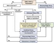
– Choice of stress modality to be combined with myocardial perfusion scintigraphy for the diagnosis of coronary artery disease. LBBB: left bundle branch block; ET: exercise test; WPW: Wolff-Parkinson-White; HR: heart rate; MPS: myocardial perfusion scintigraphy. * 1 Functional capacity to perform estimated daily physical activities >5 METs and ability to perform the necessary effort on whichever ergometer is available; * 2 Alternative: regadenoson (not yet available in Brazil); * 3 May be performed at the discretion of the requesting/referring physician. * 4 As this is a diagnostic test, a new myocardial perfusion scan under pharmacologic stress (dipyridamole or adenosine) or hybrid stress must be performed to ensure accuracy.
1.1.1. Physical Stress Methods (Exercise Test)
Physical stress, through ET or CPET, adds diagnostic and prognostic value to imaging methods by addressing clinical, hemodynamic, and electrocardiographic parameters. To obtain the best results, the methodological specificities of each imaging modality, discussed below, must be considered and adopted.9, 762, 1111
1.1.1.1. Contraindications to Physical Stress in MPI
The contraindications to physical stress in this setting are the same as for conventional exercise test, described in Part 1, Section 2.3 of this Guideline.
1.1.1.2. Guidelines for Patients when Scheduing MPI with Physical Stress
Preparation for the procedure involves:
– 3-hour fast with a light meal before the test.
– Methylxanthine (caffeine)-containing foods, beverages, and drugs should be withheld for at least 12 hours before the test, as some tests may need to be converted to pharmacologic or hybrid stress and would thus require vasodilator administration (Appendix 3).
– Discontinuation of drugs that may interfere with ET/CPET (especially antiarrhythmics and antianginal drugs, i.e. beta-blockers, calcium channel blockers, and nitrates) should follow the recommendations in Part 2, Section 1.1.6 of this Guideline, and is delegated to the requesting/referring physician.1111
– Patients are advised to bring the reports of any recently performed ET/CPET, to inform better selection of the effort protocol and allow comparison between tests.
– The other orientations for ET described in Part 2 of this Guideline all apply.
1.1.1.3. Methodological Aspects of Physical Stress in MPI
Before ET, peripheral venous access in one of the upper extremities must be obtained for injection of the radiopharmaceutical.6 This injection should be performed at peak exercise, which may not correspond to the predicted HRmax; the indication and purpose of the test (diagnosis or assessment of response to therapy) must be taken into account (Class of recommendation - Level of evidence: IIb-C).1111
Following injection of the radiopharmaceutical, the patient should be encouraged to continue exercise at the same load for one additional minute, to improve myocardial uptake of the radiotracer. If the same load cannot be maintained, the clinician should attempt to reduce the speed and/or grade of the ergometer instead of abrupt cessation.9
Images should be acquired after the end of the exercise phase:
– When using MIBI-99MTc (used in most services in Brazil), 30 to 60 minutes after cessation of stress.
– When using Thallium-201, acquisition should start no later than 10 to 15 minutes after cessation of stress, to prevent significant redistribution. Initial or distribution images should be acquired after the radioisotope injection (single injection), and late (redistribution) images, 3 to 4 hours later.
Radiation exposure of staff performing ET varies widely depending on the facility’s volume and preventive measures in place, but the effective dose received by physicians performing ET generally remains below acceptable limits. Clinicians are advised to familiarize themselves with and adopt all procedures recommended by the Brazilian National Nuclear Energy Commission for MPI.
1.1.1.4. Interpretation of ET Results in MPI
All diagnostic and prognostic markers described in this Guideline regarding conventional ET apply to the interpretation of combined ET/MPI, and must be included in the final report. The percent predicted HRmax at the time of injection of the radiopharmaceutical must be included in the report to allow analysis and interpretation of MPI.
1.1.2. Methodological Aspects of Pharmacologic Stress in MPI
Pharmacologic stress tests may be performed with: primary vasodilators (adenosine and dipyridamole), which cause redistribution of coronary flow; or drugs that promote increased myocardial oxygen consumption, such as dobutamine and atropine, which cause effects similar to those observed in dynamic exercise with increased myocardial workload.
Pharmacologic stress tests are preferred: in patients with physical limitations to exercise due to musculoskeletal or neuromuscular conditions; in those patients on drugs that interfere with myocardial oxygen consumption (MVO2), when ET is performed for diagnostic purposes; those with low functional capacity; in compensated HF; in LBBB, when an artificial pacemaker is present; and in patients with other contraindications to physical exercise.
1.1.2.1. Vasodilators
1.1.2.1.1. Dipyridamole
Dipyridamole acts by inhibiting the enzyme adenosine deaminase, which degrades endogenous adenosine, in addition to blocking adenosine reuptake by the cell membrane, with consequent increases in extracellular adenosine concentration leading to coronary and systemic vasodilation. Its biological half-life is approximately 45 minutes. The recommended dose for MPI is 0.56 milligrams per kilogram (mg.kg- 1 ) per minute, up to a maximum dose of 60 mg, administered intravenously over the course of 4 minutes, diluted in 50 mL of saline solution. Dipyridamole can be injected manually, without an infusion pump. The radiopharmaceutical is administered through the same intravenous (IV) line, 2 to 4 minutes after completion of the dipyridamole infusion (hyperemia or maximum vasodilation).9, 1109 - 1111
Adverse effects of vasodilators occur in 50% of patients receiving dipyridamole and in 80% of those receiving adenosine. With dipyridamole, these manifestations are reversed upon administration of intravenous aminophylline, at a dose of 1 to 2 mg.kg- 1 up to 240 mg, 2 minutes after radiotracer injection. Vasodilators (sensitivity and specificity ≈80-90%) and physical stress (sensitivity and specificity ≈85-90%) have comparable accuracy for the detection of CAD.1113, 1114
1.1.2.1.2. Adenosine
Adenosine induces coronary vasodilation by specific activation of A2Areceptors in the cell membrane, resulting in a 4- to 5-fold increase in coronary flow. The usual dose is 140 μg/kg/min, given via continuous infusion over 6 minutes. Use of an infusion pump is mandatory. During the third minute of infusion, the radiopharmaceutical is injected via another peripheral venous line (due to the ultra-short plasma half-life of 2 to 10 seconds), and the adenosine infusion is continued for a further 3 minutes.
When patients are markedly symptomatic or show ischemic changes at the start of the adenosine infusion, a shorter 4-minute infusion protocol can be used instead, which has been shown to be able to detect ischemia and CAD. In this protocol, the radiopharmaceutical is injected during the second minute and the adenosine infusion continued for a further 2 minutes.
Clinical status and ECG, BP, and HR must be monitored continuously during pharmacologic stress testing with vasodilators, and for a few minutes afterwards.
Preparation for the procedure:
– 3-hour fast.
– Avoid intake of any food, beverage, or other product containing methylxanthines (competitive adenosine receptor inhibitors), including coffee, tea, chocolate, energy drinks, soft drinks or other caffeinated beverages, caffeine-containing medications (analgesics, cold and flu medicines, muscle relaxants, nonsteroidal anti-inflammatory drugs) for 12 hours before the test.
– The aforementioned foods, beverages, and medications must be avoided for at least 12 hours1111 before the test if adenosine will be used and for at least 24 hours if dipyridamole will be used.9
– Theophylline should be discontinued at least 12 hours before the test.
– When using adenosine, two IVs must be placed: one for adenosine infusion and another for injection of the radiopharmaceutical. When using dipyridamole, only one IV line is required.
– An infusion pump is mandatory for administration of adenosine and optional for administration of dipyridamole.
– Aminophylline (1 to 2 mg.kg- 1, i.e. a 72 to 240 mg dose) should be readily available to reverse any serious side effects of vasodilator stress. In case of dipyridamole-induced adverse effects, aminophylline should be administered 2 minutes after the radiotracer injection.
Contraindications:1110, 1111, 1115
1) Relative: history of reactive airway disease with no exacerbations or episodes for at least 3 months; sinus node dysfunction; marked sinus bradycardia; severe bilateral carotid artery disease.
2) Absolute: active bronchospasm; status asthmaticus; recent (<3 months) episodes of reactive airway disease; second- or third-degree AV block (unless an artificial pacemaker is in place); systolic hypotension (<90 mmHg); recent (<2 months) stroke or TIA; recent (<24 hours) use of dipyridamole, if the test is to be performed with adenosine.
1.1.2.2. Drugs that Increase Myocardial Oxygen Consumption
These drugs are an alternative for patients who cannot undergo ET or pharmacologic stress with dipyridamole or adenosine. The most commonly used agent is dobutamine, which acts predominantly on beta-1 (ß-1) adrenergic receptors, with inotropic and chronotropic stimulation, and to a lesser extent on beta-2 (ß-2) receptors, with a peripheral vasodilation response dependent on the infused dose. It increases cardiac output, HR, and SBP, leading to an increase in MVO2and, consequently, to coronary vasodilation.
Dobutamine is administered via intravenous infusion pump at an initial dose of 5-10 μg/kg/min for 3 minutes, followed by incremental doses of 20 μg/kg/min and 30 μg/kg/min up to a maximum of 40 μg/kg/min.1116 In patients who do not reach submaximal HR and have no evidence of ischemia, intravenous atropine can be added at a dose of 0.25 to 2 mg.1110 If necessary, handgrip maneuvers (i.e. squeezing a tennis ball) can be added as well. Early use of atropine after the first phase of dobutamine infusion has proven to be safe and reduce infusion time and patient complaints during stress, without altering the diagnostic accuracy of the test.1117
The radiotracer is injected at HRpeak, and dobutamine infusion should be continued for 1 minute thereafter.
Throughout the test, clinical signs and symptoms should be monitored and ECG, BP, and HR recorded continuously. For reversal of adverse effects, short-acting beta-blockers (i.e. metoprolol or esmolol) can be injected intravenously after the first minute of radiotracer injection.
Preparation for the procedure:
– 3-hour fast.
– Withhold any beta-blockers for 48 to 72 hours before dobutamine stress.
– Peripheral venous access for infusion of dobutamine and radiotracer.
– Dobutamine should only be administered via infusion pump.
– Metoprolol (5 mg) should be readily available to reverse any serious side effects of dobutamine. However, it is contraindicated in patients with history of severe bronchospasm and in those with COPD. In these patients, esmolol (single dose, 100 and 200 mg), a cardioselective beta-blocker, is preferred.
Contraindications:
1) Relative: abdominal aortic aneurysm (>5cm diameter), presence of thrombi in the left ventricle, LVEF <25% (increased risk of ventricular arrhythmias).
2) Absolute : cardiac arrhythmias (sustained or paroxysmal atrial fibrillation or ventricular tachycardia), severe aortic stenosis, hypertrophic obstructive cardiomyopathy, systolic hypotension (<90 mmHg) or hypertension (>200 mmHg), unstable angina or recent MI, aortic aneurysm or dissection, symptomatic cerebrovascular insufficiency, presence of ICD, alterations in potassium metabolism.9
1.1.3. Hybrid Stress Methods
These are ideal for patients who have limited exercise capacity or are on medications that prevent the HR response to exercise. Hybrid stress is not recommended in patients with LBBB, WPW syndrome, or an artificial pacemaker.
The combination of vasodilators with low-workload exercise (i.e. up to the second stage of the Bruce protocol or slight fatigue) has been shown to reduce subdiaphragmatic (hepatic) activity and improve image quality.1118 It also helps reduce the occurrence and severity of adverse effects seen with dipyridamole and adenosine.
In in selected patients, a hybrid stress protocol can consist of applying the first stage of the modified Bruce protocol (1.7 mph, 0% grade) for 4 to 6 minutes after dipyridamole infusion. The radiopharmaceutical is then injected during exercise, which must continue for another 2 minutes to allow adequate myocardial uptake, reducing side effects and improving image quality. Regarding preparation for the procedure, the same guidance applies as for exercise- and vasodilator-based protocols.
1.1.4. Novel Drugs
The selective A2Areceptor agonist regadenoson, which promotes coronary vasodilation, has been shown to induce adequate coronary hyperemia with a lower severity of systemic effects (such as chest pain and AV blockade). Regadenoson, which is not yet available in Brazil, has a short biological half-life (2 to 4 minutes), which shortens test time and minimizes and limits the duration of adverse effects. It can be used in patients with COPD or asthma.1119
The recommended dose of regadenoson is 0.4 mg IV (no adjustment for weight required), injected into a peripheral vein followed by a 5-mL flush of saline solution; the radiopharmaceutical should be administered 10 to 20 seconds later.1120
1.2. Stress Echocardiography
The different stress modalities used for stress echocardiography have been compared elsewhere in this Guideline, including their physiological effects, hemodynamic responses, and contraindications (see Table 18). In most adult patients capable of exercising, exercise stress echocardiography (ESE) is considered the modality of choice for assessment of myocardial ischemia.
1.2.1. Methodology 8,260,265,1121,1122
First, a baseline echocardiogram should be obtained at rest to assess cardiac structure (chamber sizes and wall thickness), heart valves and their gradients, regional wall motion, and global ventricular function.
Baseline echocardiography also aims to investigate potential causes for the patient’s symptoms (i.e. aortic dissection) and to identify any condition that might make stress unsafe (i.e. severe valve disease in a symptomatic patient). Under these circumstances, the possibility of postponing or canceling the stress test altogether should be carefully evaluated.
The acquisition, analysis, and interpretation of echocardiographic data (baseline and stress) should follow the recommendations of international organizations260, 265, 1121 and relevant Brazilian Society of Cardiology position statements and guidelines: Position Statement on Indications of Echocardiography in Adults;8 Position Statement on Indications for Echocardiography in Fetal and Pediatric Cardiology and Congenital Heart Disease of the Adult;1121 and Standardization of Examination Equipment and Techniques for Performing Echocardiographic Examinations ( Normatização dos Equipamentos e Técnicas de Exame para Realização de Exames Ecocardiográficos ).1122
Images obtained at rest will be compared with those obtained during and after stress. Obtaining data across multiple cardiac cycles at peak stress increases the accuracy of interpretation. Continuous recording of resting and stress images is recommended.
In treadmill tests, images obtained at rest and immediately after exercise should be compared side by side (in quad screen format). In pharmacologic stress tests, images obtained at peak stress should be compared with resting, low-dose, and pre-peak or early-recovery images (also using the quad screen format).
Echocardiographic images are generally obtained from parasternal long-axis, short-axis, and apical two–and four-chamber views. In some cases, the subcostal and apical long-axis views may be used. Other views and maneuvers may be necessary on a case by case basis.
Development of wall thickening under stress in segments which are markedly hypokinetic or akinetic at rest is indicative of myocardial viability. For proper determination of viability, myocardial thickening must be observed carefully at lower levels of stress, to avoid a rapid increase in HR and associated ischemia.
Abnormal changes in diastolic function may precede abnormalities in systolic wall motion. When detection of ischemia is the primary objective of a stress test, the clinician is advised to record diastolic parameters close to peak effort or after assessment of regional wall motion.
Color Doppler assessment of mitral regurgitation on baseline echocardiography and during peak exertion may allow detection of ischemic mitral regurgitation.
Stress echocardiography is as accurate as positron emission tomography in detecting reversible dysfunction in patients with hibernating myocardium.
1.2.1.1. Exercise Stress Methods8, 1123
Just as conventional ET protocols, exercise stress echocardiography can be performed on a treadmill or cycle ergometer. The most widely used treadmill-based method is the Bruce protocol, with echocardiographic images being acquired at rest and immediately upon completion of exercise.
If using a cycle ergometer, the patient can remain vertical (bicycle or conventional cycle ergometer) or supine (supine cycle ergometer or in-bed cycle ergometer). On a supine cycle ergometer with lateral rotation, the patient pedals in an inclined position, allowing better adaptation and yielding higher-quality echocardiographic images. The most widely used cycle ergometer protocol, the Åstrand test, starts with a load of 25W that is increased progressively in 25W increments every 2 or 3 minutes (Figure 35).

– Representation of an exercise stress echocardiogram performed on a bicycle/cycle ergometer. BP: blood pressure; ECG: electrocardiogram; ECHO: echocardiogram; min: minute.
Skin preparation and electrode placement for ECG monitoring are as those standardized for ET. The leads may be displaced slightly up and down in case they interfere with the acoustic windows needed to perform echocardiography.
A 12-lead ECG should be recorded at rest and every 2 minutes (or at every stage) throughout the test (during exertion and recovery). One ECG lead should be continuously displayed on the echocardiograph monitor to provide a reference of ST-segment changes and arrhythmias.
BP should be measured at rest, at each stage of the exercise protocol, and during recovery.
Analysis and interpretation of HR, BP, scores (pre- and post-test), ECG recordings, and cardiorespiratory fitness parameters are similar to those for ET. Correlation of these data with echocardiographic findings is recommended.
Although some facilities terminate the test when 85% of the predicted HRmax for age is reached, all are advised to continue the test until physical exhaustion (Borg scale ≥18) or until absolute criteria for test cessation arise. Reaching the predicted HRmax increases the sensitivity of the test and may reveal abnormalities that would only occur at a high enough workload.8, 260, 1123
1.2.1.2. Pharmacologic Stress Methods
1.2.1.2.1. Dobutamine260, 1123
Pharmacologic stress with dobutamine (or dobutamine plus atropine) is the most widely used alternative modality for assessing myocardial ischemia when a patient is unable to exercise.1124
Dobutamine is usually administered in graded doses, starting at 5 μg/kg/min, with increments at 3-minute intervals to 10, 20, 30, and 40 μg.kg- 1 .min- 1 . Dobutamine doses of 20 or 30 μg.kg- 1 .min- 1 generally allow reaching target HR (85% of maximum predicted HR for age) more quickly and with fewer side effects, especially if HR is not increasing as expected.
Infusion of dobutamine at a low initial dose (2.5 μg.kg- 1 .min- 1 ) may facilitate recognition of myocardial viability in echocardiographically abnormal segments.
In beta-blocked patients and/or those in whom the predicted HRmax is not reached, the sensitivity of DSE for detection of ischemia is reduced. In such cases, atropine can be administered to achieve the target HR (0.25 to 0.50 mg, at 1-minute intervals, as needed; maximum cumulative dose 2.0 mg). To avoid side effects, including central nervous system toxicity, the lowest possible dosage should be used.1125
In older adults, patients with a small body habitus, and in those who are already close to the predicted HRmax, the use of atropine in a dose of 0.25 mg is recommended. A total dose <1.0 mg is also recommended in patients with neuropsychiatric conditions, patients with liver disease, or those with a BMI <24 kg/m2 .
Pharmacologic stress should be terminated upon achieving target HR, hypotension, LV wall motion abnormalities (new or worsening), complex arrhythmias, severe hypertension, and intolerable symptoms, among other endpoints.
1.2.1.2.2. Vasodilators260, 1123
Pharmacologic stress testing with vasodilators (dipyridamole or adenosine) induces an increase in coronary flow, and should preferably be used to assess myocardial perfusion.1126 It can also be performed to assess ischemia and myocardial viability. Pharmacologic stress echocardiography with vasodilators has a lower sensitivity for CAD detection than ESE or DSE.
Vasodilating agents are contraindicated in patients with reactive airway obstruction, unstable or complicated acute coronary syndrome, severe cardiac arrhythmias (VT and AV block), or severe hypotension.
Dipyridamole is usually given in doses of up to 0.84 mg.kg- 1 over 6 to 10 minutes. Administering atropine or performing a handgrip exercise at peak infusion increases the sensitivity of the test.1127
Adenosine in conjunction with contrast-enhanced echocardiography can also be used to assess myocardial perfusion. The adenosine infusion rate is 140 μg.kg- 1 .min- 1 over 4 to 6 minutes. Adenosine has a shorter half-life and, therefore, a shorter duration of action than dipyridamole.
1.2.1.3. Ultrasound Enhancing Agents
Three microbubble ultrasound enhancing agents (UEAs) are commercially available worldwide: Sonovue ®, Definity ®, and Optison ®. UEAs are used to improve endocardial border delineation, image quality, and CAD detection by stress echocardiography. Management (whether by bolus or continuous infusion) aims to opacify the entire LV cavity, without swirling artifacts in the apex or attenuation of basal segments due to acoustic shadowing. UEAs can also be used as myocardial perfusion markers.
Use of these agents in nonpregnant adults is safe; anaphylactic reactions may occur, but are very rare. UEA administration is not recommended in pregnancy. Specific administration protocols are given in the 2018 ASE guidelines.1128
-
Guideline Development: Department of Exercise Test, Exercise, Nuclear Cardiology, and Cardiovascular Rehabilitation (DERC), Brazilian Society of Cardiology (SBC)SBC Clinical Practice Guidelines Committee: Carisi Anne Polanczyk (Coordinator), Humberto Graner Moreira, Mário de Seixas Rocha, Jose Airton de Arruda, Pedro Gabriel Melo de Barros e Silva – Period 2022-2023Note: These guidelines are for information purposes and should not replace the clinical judgment of a physician, who must ultimately determine the appropriate treatment for each patient.
References
-
1 Meneghelo RS, Araújo CG, Stein R, Mastrocolla LE, Albuquerque PF, Serra SM et a. III Diretrizes da Sociedade Brasileira de Cardiologia sobre Teste Ergométrico. Arq Bras Cardiol. 2010;95(5 Suppl 1):1-26. https://doi.org/10.1590/S0066-782X2010000800001
» https://doi.org/10.1590/S0066-782X2010000800001 -
2 Andrade J, Brito FS, Vilas-Boas F, Castro I, Oliveira JA, Guimarães JI, et al. II Diretrizes da Sociedade Brasileira de Cardiologia Sobre Teste Ergométrico. Arq Bras Cardiol. 2002;78 (suppl 2). https://doi.org/10.1590/S0066-782X2002000800001
» https://doi.org/10.1590/S0066-782X2002000800001 -
3 Guazzi M, Arena R, Halle M, Piepoli MF, Myers J, Lavie CJ. 2016 Focused Update: Clinical Recommendations for Cardiopulmonary Exercise Testing Data Assessment in Specific Patient Populations. Circulation. 2016;133(24):e694-711. https://doi.org/10.1161/CIR.0000000000000406
» https://doi.org/10.1161/CIR.0000000000000406 -
4 Marcadet D-M, Pavy B, Bosser G, Claudot F, Corone S, Douard H, et al. French Society of Cardiology guidelines on exercise tests (part 1): Methods and interpretation. Arch Cardiovasc. Dis. 2018;111(12):782-90. https://doi.org/10.1016/j.acvd.2018.05.005
» https://doi.org/10.1016/j.acvd.2018.05.005 -
5 Smarz K, Jaxa-Chamiec T, Chwyczko T, Glówczynska R, Jegier A, Niedoszytko P, et al. Cardiopulmonary exercise testing in adult cardiology - expert opinion of the Working Group of Cardiac Rehabilitation and Exercise Physiology of the Polish Cardiac Society. Kardiol Pol. 2019; 77(7-8):730-56. https://doi.org/10.33963/KP.14889
» https://doi.org/10.33963/KP.14889 -
6 Fletcher GF, Ades PA, Kligfield P, Arena R, Balady GJ, Bittner VA, et al. Exercise Standards for Testing and Training: A Scientific Statement From the American Heart Association. Circulation. 2013;128(8):873-934. https://doi.org/10.1161/CIR.0b013e31829b5b44
» https://doi.org/10.1161/CIR.0b013e31829b5b44 - 7 Conselho Federal de Medicina. Resolução 2.221 de 23/11/2018. Homologa a Portaria CMF n.1/2018, que atualiza a relação de especialidades e áreas de atuação médicas aprovadas pela Comissão Mista de Especialidades. Diário Oficial da União. Brasília. 24 janeiro de 2019.
-
8 Barberato SH, Romano MMD, Beck AL de S, Rodrigues ACT, Almeida ALC de, Assunção BMBL, et al. Position Statement on Indications of Echocardiography in Adults - 2019. Arq Bras Cardiol. 2019;113(1):135-81. https://doi.org/10.5935/abc.20190129
» https://doi.org/10.5935/abc.20190129 -
9 Mastrocola LE, Amorim BJ, Vitola JV, Brandão SCS, Grossman GB, Lima R de SL, et al. Atualização da Diretriz Brasileira de Cardiologia Nuclear - 2020: Arq Bras Cardiol. 2020;114(2):325-429. https://doi.org/10.36660/abc.20200087
» https://doi.org/10.36660/abc.20200087 -
10 Min JK, Gilmore A, Jones EC, Berman DS, Stuijfzand WJ, Shaw LJ, et al. Cost-effectiveness of diagnostic evaluation strategies for individuals with stable chest pain syndrome and suspected coronary artery disease. Clin Imaging. 2017;43:97-105. https://doi.org/10.1016/j.clinimag.2017.01.015
» https://doi.org/10.1016/j.clinimag.2017.01.015 -
11 Carmo PB, Magliano CA, Rey HCV, Camargo GC, Trocado LFL, Gottlieb I. Análise da Custo-Efetividade da Angiotomografia Coronariana no SUS, em Comparação com Outros Métodos Não Invasivos na Suspeita de DAC Estável. Arq Bras Cardiol. 2021;118(3):578-85. https://doi.org/10.36660/abc.20201050
» https://doi.org/10.36660/abc.20201050 -
12 Gibbons RJ, Balady GJ, BrickerT, Chaitman B, Fletcher GF, Froelicher VF, et al. ACC/AHA 2002 Guideline update for exercise testing: summary article: a report of the American College of Cardiology/American Heart Association Task Force on Practice Guidelines (Committee to Update the 1997 Exercise Testing Guidelines. Circulation. 2002;106(14):1883-92. https://doi.org/10.1161/01.CIR.0000034670.06526.15
» https://doi.org/10.1161/01.CIR.0000034670.06526.15 - 13 American College of Sports Medicine, Liguori G, Feito Y, Fountaine C, Roy B, editors. ACSM's guidelines for exercise testing and prescription. 11th ed. Philadelphia: Wolters Kluwer; 2021. 541p. ISBN:1975150198.
-
14 Marcadet DM, Pavy B, Bosser G, Claudot F, Corone S, Douard H, et al. French Society of Cardiology guidelines on exercise tests (part 2): Indications for exercise tests in cardiac diseases. Arch Cardiovasc Dis. 2019;112(1):56-66. https://doi.org/10.1016/j.acvd.2018.07.001
» https://doi.org/10.1016/j.acvd.2018.07.001 -
15 Mezzani A, Hamm LF, Jones AM, McBride PE, Moholdt T, Stone JA, et al. Aerobic exercise intensity assessment and prescription in cardiac rehabilitation: a joint position statement of the European Association for Cardiovascular Prevention and Rehabilitation, the American Association of Cardiovascular and Pulmonary Rehabilitation and the Canadian Association of Cardiac Rehabilitation. Eur J Prev Cardiol. 2013;20(3):442-67. https://doi.org/10.1177/2047487312460484
» https://doi.org/10.1177/2047487312460484 -
16 Hansen D, Abreu A, Ambrosetti M, Cornelissen V, Gevaert A, Kemps H, et al. Exercise intensity assessment and prescription in cardiovascular rehabilitation and beyond: why and how: a position statement from the Secondary Prevention and Rehabilitation Section of the European Association of Preventive Cardiology. Eur J Prev Cardiol. 2022;29(1):230-45. https://doi.org/10.1093/eurjpc/zwab007
» https://doi.org/10.1093/eurjpc/zwab007 -
17 Pelliccia A, Sharma S, Gati S, Bäck M, Börjesson M, Caselli S, et al. 2020 ESC Guidelines on sports cardiology and exercise in patients with cardiovascular disease. Eur Heart J. 2021;42(1):17-96. https://doi.org/10.1093/eurheartj/ehaa605
» https://doi.org/10.1093/eurheartj/ehaa605 -
18 Guazzi M, Wilhelm M, Halle M, Van Craenenbroeck E, Kemps H, de Boer RA, et al. Exercise testing in heart failure with preserved ejection fraction: an appraisal through diagnosis, pathophysiology and therapy - A clinical consensus statement of the Heart Failure Association and European Association of Preventive Cardiology of the European Society of Cardiology. Eur J Heart Fail. 2022;24(8):1327-45. https://doi.org/10.1002/ejhf.2601
» https://doi.org/10.1002/ejhf.2601 -
19 Martignon C, Pedrinolla A, Ruzzante F, Giuriato G, Laginestra FG, Bouça-Machado R, et al. Guidelines on exercise testing and prescription for patients at different stages of Parkinson's disease. Aging Clin Exp Res. 2021;33(2):221-46. https://doi.org/10.1007/s40520-020-01612-1
» https://doi.org/10.1007/s40520-020-01612-1 -
20 Oliveira GMM, Brant LCC, Polanczyk CA, Malta DC, Biolo A, Nascimento BR, et al. Estatística Cardiovascular - Brasil 2021. Arq Bras Cardiol. 2022;118(1):115-373. https://doi.org/10.36660/abc.20211012
» https://doi.org/10.36660/abc.20211012 -
21 American Board of Internal Medicine Foundation, American Society of Nuclear Cardiology. Stress Tests for Chest Pain. Choosing Wisely 2017. [Accessed June 11, 2022] Available from: https://www.choosingwisely.org/patient-resources/stress-tests-for-chest-pain/
» https://www.choosingwisely.org/patient-resources/stress-tests-for-chest-pain/ -
22 Prasad DS, Kabir Z, Revathi Devi K, Peter PS, Das BC. Prevalence and RIsk factors for Silent Myocardial ischemia (PRISM): A clinico observational study in patients of type 2 diabetes. Indian Heart J. 2019;71(5):400-5. https://doi.org/10.1016/j.ihj.2019.12.002
» https://doi.org/10.1016/j.ihj.2019.12.002 -
23 Pereira WVC, Vancea DMM, de Andrade Oliveira R, de Freitas YGPC, Lamounier RN, Silva Júnior WS, et al. 2022: Position of Brazilian Diabetes Society on exercise recommendations for people with type 1 and type 2 diabetes. Diabetol Metab Syndr. 2023;15(1):2. https://doi.org/10.1186/s13098-022-00945-3
» https://doi.org/10.1186/s13098-022-00945-3 -
24 Bourque JM, Beller GA. Value of exercise ECG for risk stratification in suspected or known CAD in the era of advanced imaging technologies. JACC Cardiovasc Imaging. 2015;8(11):1309-21. https://doi.org/10.1016/j.jcmg.2015.09.006
» https://doi.org/10.1016/j.jcmg.2015.09.006 -
25 Gulati M, Levy PD, Mukherjee D, Amsterdam E, Bhatt DL, Birtcher KK, et al. 2021 AHA/ACC/ASE/CHEST/SAEM/SCCT/SCMR Guideline for the Evaluation and Diagnosis of Chest Pain: A Report of the American College of Cardiology/American Heart Association Joint Committee on Clinical Practice Guidelines. Circulation. 2021;144(22):e368-e454. https://doi.org/10.1161/CIR.0000000000001029
» https://doi.org/10.1161/CIR.0000000000001029 -
26 Cesar L, Ferreira J, Armaganijan D, Gowdak L, Mansur A, Bodanese L, et al. Guideline For Stable Coronary Artery Disease. Arq Bras Cardiol. 2014;103(2 Suppl 2):1-56. https://doi.org/10.5935/abc.2014S004
» https://doi.org/10.5935/abc.2014S004 -
27 Knuuti J, Wijns W, Saraste A, Capodanno D, Barbato E, Funck-Brentano C, et al. 2019 ESC Guidelines for the diagnosis and management of chronic coronary syndromes. Eur Heart J. 2020;41(3):407-77. https://doi.org/10.1093/eurheartj/ehz425
» https://doi.org/10.1093/eurheartj/ehz425 -
28 Rozanski A, Berman D. Optimizing the Assessment of Patient Clinical Risk at the Time of Cardiac Stress Testing. JACC Cardiovasc Imaging. 2020;13(2 Pt 2):616-23. https://doi.org/10.1016/j.jcmg.2019.01.038
» https://doi.org/10.1016/j.jcmg.2019.01.038 -
29 Carvalho T, Milani M, Ferraz AS, Silveira AD, Herdy AH, Hossri CAC, et al. Diretriz Brasileira de Reabilitação Cardiovascular - 2020. Arq Bras Cardiol. 2020;114(5):943-87. https://doi.org/10.36660/abc.20200407
» https://doi.org/10.36660/abc.20200407 -
30 Visseren FLJ, Mach F, Smulders YM, Carballo D, Koskinas KC, Bäck M, et al. 2021 ESC Guidelines on cardiovascular disease prevention in clinical practice. Eur Heart J. 2021;42(34):3227-337. https://doi.org/10.1093/eurheartj/ehab484
» https://doi.org/10.1093/eurheartj/ehab484 -
31 Fihn SD, Blankenship JC, Alexander KP, Bittl JA, Byrne JG, Fletcher BJ, et al. 2014 ACC/AHA/AATS/PCNA/SCAI/STS Focused Update of the Guideline for the Diagnosis and Management of Patients With Stable Ischemic Heart Disease: A Report of the American College of Cardiology/American Heart Association Task Force on Practice Guidelines, and the American Association for Thoracic Surgery, Preventive Cardiovascular Nurses Association, Society for Cardiovascular Angiography and Interventions, and Society of Thoracic Surgeons. Circulation. 2014;130(19):1749-67. https://doi.org/10.1161/CIR.0000000000000095
» https://doi.org/10.1161/CIR.0000000000000095 -
32 Yiadom MYAB, Kosowsky JM. Management Strategies for Patients with Low-Risk Chest Pain in the Emergency Department. Curr Treat Options Cardiovasc Med. 2011;13(1):57-67. https://doi.org/10.1007/s11936-010-0108-3
» https://doi.org/10.1007/s11936-010-0108-3 -
33 Nicolau JC, Feitosa GS, Petriz JL, Furtado RH, Précoma DB, Lemke W, et al. Diretrizes da Sociedade Brasileira de Cardiologia sobre Angina Instável e Infarto Agudo do Miocárdio sem Supradesnível do Segmento ST - 2021. Arq Bras Cardiol. 2021;117(1):181-264. https://doi.org/10.36660/abc.20210180
» https://doi.org/10.36660/abc.20210180 -
34 Ghorayeb N, Stein R, Daher DJ, Silveira AD, Ritt LEF, Santos DFP, et al. The Brazilian Society of Cardiology and Brazilian Society of Exercise and Sports Medicine Updated Guidelines for Sports and Exercise Cardiology - 2019. Arq Bras Cardiol. 2019 112(3):326-68. https://doi.org/10.5935/abc.20190048
» https://doi.org/10.5935/abc.20190048 -
35 Gulati M, Levy PD, Mukherjee D, Amsterdam E, Bhatt DL, Birtcher KK, et al. 2021 AHA/ACC/ASE/CHEST/SAEM/SCCT/SCMR Guideline for the Evaluation and Diagnosis of Chest Pain. J Am Coll Cardiol. 2021;78(22):e187-285. https://doi.org/10.1016/j.jacc.2021.07.053
» https://doi.org/10.1016/j.jacc.2021.07.053 -
36 O'Gara PT, Kushner FG, Ascheim DD, Casey DE, Chung MK, de Lemos JA, et al. 2013 ACCF/AHA Guideline for the Management of ST-Elevation Myocardial Infarction: A Report of the American College of Cardiology Foundation/American Heart Association Task Force on Practice Guidelines. Circulation. 2013;127(4) e362-425. https://doi.org/10.1161/CIR.0b013e3182742cf6
» https://doi.org/10.1161/CIR.0b013e3182742cf6 -
37 Klimis H, Ferkh A, Brown P, Zecchin R, Altman M, Thomas L. Determinants of Exercise Capacity Following ST-Elevation Myocardial Infarction (STEMI). J Cardiovasc Dev Dis. 2021;8(11):140. https://doi.org/10.3390/jcdd8110140
» https://doi.org/10.3390/jcdd8110140 -
38 Lachman S, Terbraak MS, Limpens J, Jorstad H, Lucas C, Scholte op Reimer W, et al. The prognostic value of heart rate recovery in patients with coronary artery disease: A systematic review and meta-analysis. Am Heart J May 2018;199:163-9. https://doi.org/10.1016/j.ahj.2018.02.008
» https://doi.org/10.1016/j.ahj.2018.02.008 -
39 Virani SS, Newby LK, Arnold SV, Bittner V, Brewer LC, Demeter SH, et al. 2023 AHA/ACC/ACCP/ASPC/NLA/PCNA Guideline for the Management of Patients With Chronic Coronary Disease: A Report of the American Heart Association/American College of Cardiology Joint Committee on Clinical Practice Guidelines. Circulation 2023;148. https://doi.org/10.1161/CIR.0000000000001168
» https://doi.org/10.1161/CIR.0000000000001168 -
40 Marx N, Federici M, Schütt K, Müller-Wieland D, Ajjan RA, Antunes MJ, et al. 2023 ESC Guidelines for the management of cardiovascular disease in patients with diabetes. European Heart Journal 2023:ehad192. https://doi.org/10.1093/eurheartj/ehad192
» https://doi.org/10.1093/eurheartj/ehad192 -
41 Dun Y, Wu S, Cui N, Thomas RJ, Olson TP, Zhou N, et al. Screening for Asymptomatic Coronary Artery Disease via Exercise Stress Testing in Patients With Type 2 Diabetes Mellitus: A Systematic Review and Meta-Analysis. Front Cardiovasc Med. 2021 Nov 1;8:770648. https://doi.org/10.3389/fcvm.2021.770648
» https://doi.org/10.3389/fcvm.2021.770648 -
42 Reynolds HR, Diaz A, Cyr DD, Shaw LJ, Mancini GBJ, Leipsic J, et al. Ischemia With Nonobstructive Coronary Arteries. JACC Cardiovasc Imaging. 2023;16(1):63-74. https://doi.org/10.1016/j.jcmg.2022.06.015
» https://doi.org/10.1016/j.jcmg.2022.06.015 -
43 Bhatia LC, Singal R, Jain P, Mishra N, Mehra V. Detection of Silent Myocardial Ischaemia in Asymptomatic Diabetic Patients during Treadmill Exercise Testing. High Blood Press Cardiovasc Prev. 2012;19(3):137-42. https://doi.org/10.1007/BF03262463
» https://doi.org/10.1007/BF03262463 -
44 Beijk MA, Vlastra WV, Delewi R, van de Hoef TP, Boekholdt SM, Sjauw KD, et al. Myocardial infarction with non-obstructive coronary arteries: a focus on vasospastic angina. Neth Heart J. 2019;27(5):237-45. https://doi.org/10.1007/s12471-019-1232-7
» https://doi.org/10.1007/s12471-019-1232-7 -
45 Montalescot G, Sechtem U, Achenbach S, Andreotti F, Arden C, Budaj A, et al. (Task Force Members) 2013 ESC Guidelines on the management of stable coronary artery disease: The Task Force on the management of stable coronary artery disease of the European Society of Cardiology. Eur Heart J. 2013;34(38):2949-3003. https://doi.org/10.1093/eurheartj/eht296
» https://doi.org/10.1093/eurheartj/eht296 -
46 Aizenberg DJ. Cardiovascular Testing in Asymptomatic Patients. Med Clin North Am. 2016;100(5):971-9. https://doi.org/10.1016/j.mcna.2016.04.004
» https://doi.org/10.1016/j.mcna.2016.04.004 -
47 Jonas DE, Reddy S, Middleton JC, Barclay C, Green J, Baker C, et al. Screening for Cardiovascular Disease Risk With Resting or Exercise Electrocardiography: Evidence Report and Systematic Review for the US Preventive Services Task Force. JAMA. 2018;319(22):2315-28. https://doi.org/10.1001/jama.2018.6897
» https://doi.org/10.1001/jama.2018.6897 -
48 Timmis A, Raharja A, Archbold RA, Mathur A. Validity of inducible ischaemia as a surrogate for adverse outcomes in stable coronary artery disease. Heart. 2018;104(21):1733-8. https://doi.org/10.1136/heartjnl-2018-313230
» https://doi.org/10.1136/heartjnl-2018-313230 -
49 Lee SH, Choi KH, Lee JM, Shin D, Hwang D, Kim HK, et al. Residual functional SYNTAX score by quantitative flow ratio and improvement of exercise capacity after revascularization. Catheter Cardiovasc Interv. 2021;97(4):E454-66. https://doi.org/10.1002/ccd.29118
» https://doi.org/10.1002/ccd.29118 -
50 Kini V, Parks M, Liu W, Waldo SW, Ho PM, Bradley SM, et al. Patient Symptoms and Stress Testing After Elective Percutaneous Coronary Intervention in the Veterans Affairs Health Care System. JAMA Netw Open. 2022;5(6):e2217704. https://doi.org/10.1001/jamanetworkopen.2022.17704
» https://doi.org/10.1001/jamanetworkopen.2022.17704 -
51 Kaw R, Nagarajan V, Jaikumar L, Halkar M, Mohananey D, Hernandez AV, et al. Predictive Value of Stress Testing, Revised Cardiac Risk Index, and Functional Status in Patients Undergoing Noncardiac Surgery. J Cardiothorac Vasc Anesth. 2019;33(4):927-32. https://doi.org/10.1053/j.jvca.2018.07.020
» https://doi.org/10.1053/j.jvca.2018.07.020 -
52 Gualandro D, Yu P, Caramelli B, Marques A, Calderaro D, Fornari L, et al. 3rd Guideline For Perioperative Cardiovascular Evaluation on the Brazilian Society of Cardiology. Arq Bras Cardiol. 2017;109(3 Supl 1):1-104. https://doi.org/10.5935/abc.20170140
» https://doi.org/10.5935/abc.20170140 -
53 Würzburger L, Wiech P, Rossi VA, Neunhäuserer D, Caselli S, Schmied CM, et al. Hypertensive Response to Exercise in Athletes: Unremarkable Finding or Relevant Marker for Future Cardiovascular Complications? Int J Hypertens.Nov 1;2022:8476751. https://doi.org/10.1155/2022/8476751
» https://doi.org/10.1155/2022/8476751 -
54 Zimarino M, Montebello E, Radico F, Gallina S, Perfetti M, Iachini Bellisarii F, et al. ST segment/heart rate hysteresis improves the diagnostic accuracy of ECG stress test for coronary artery disease in patients with left ventricular hypertrophy. Eur J Prev Cardiol. 2016;23(15):1632-9. https://doi.org/10.1177/2047487316655259
» https://doi.org/10.1177/2047487316655259 -
55 Refaat MM, Hotait M, Tseng ZH. Utility of the Exercise Electrocardiogram Testing in Sudden Cardiac Death Risk Stratification: Utility of Exercise ECG Testing in SCD Risk Stratification. Ann Noninvasive Electrocardiol. 2014;19(4):311-8. https://doi.org/10.1111/anec.12191
» https://doi.org/10.1111/anec.12191 -
56 Myung E, Camocardi EC, Domingos Neto J, Murta GA, Sanches V, Nozaki D, et al. Diretriz Técnica da ANAMT(DT04):acidentes,morte súbita,síncope e trabalho:rastreamento por eletrocardiograma. Rev Bras Med Trab.2018;16(2):246-52. https://doi.org/10.5327/Z1679443520180270
» https://doi.org/10.5327/Z1679443520180270 -
57 Sipilä K, Tikkakoski A, Alanko S, Haarala A, Hernesniemi J, Lyytikäinen L-P, et al. Combination of low blood pressure response, low exercise capacity and slow heart rate recovery during an exercise test significantly increases mortality risk. Ann Med. 2019;51(7-8):390-6. https://doi.org/10.1080/07853890.2019.1684550
» https://doi.org/10.1080/07853890.2019.1684550 -
58 Minkkinen M, Nieminen T, Verrier RL, Leino J, Lehtimäki T, Viik J, et al. Prognostic capacity of a clinically indicated exercise test for cardiovascular mortality is enhanced by combined analysis of exercise capacity, heart rate recovery and T-wave alternans. Eur J Prev Cardiol. 2015;22(9):1162-70. https://doi.org/10.1177/2047487314557190
» https://doi.org/10.1177/2047487314557190 -
59 Forman DE, Arena R, Boxer R, Dolansky MA, Eng JJ, Fleg JL, et al. Prioritizing Functional Capacity as a Principal End Point for Therapies Oriented to Older Adults With Cardiovascular Disease: A Scientific Statement for Healthcare Professionals From the American Heart Association. Circulation. 2017;135(16):e894-918. https://doi.org/10.1161/CIR.0000000000000483
» https://doi.org/10.1161/CIR.0000000000000483 -
60 Salokari E, Laukkanen JA, Lehtimaki T, Kurl S, Kunutsor S, Zaccardi F, et al. The Duke treadmill score with bicycle ergometer: Exercise capacity is the most important predictor of cardiovascular mortality. Eur J Prev Cardiol. 2019;26(2):199-207. https://doi.org/10.1177/2047487318804618
» https://doi.org/10.1177/2047487318804618 -
61 Chou R, Arora B, Dana T, Fu R, Walker M, Humphrey L. Screening asymptomatic adults with resting or exercise electrocardiography: a review of the evidence for the U.S. Preventive Services Task Force. Ann Intern Med. 2011;155(6):375-85. https://doi.org/10.7326/0003-4819-155-6-201109200-00006
» https://doi.org/10.7326/0003-4819-155-6-201109200-00006 -
62 Greenland P, Alpert JS, Beller GA, Benjamin EJ, Budoff MJ, Fayad ZA, et al. 2010 ACCF/AHA Guideline for Assessment of Cardiovascular Risk in Asymptomatic Adults. J Am Coll Cardiol. 2010;56(25):e50-103. https://doi.org/10.1016/j.jacc.2010.09.001
» https://doi.org/10.1016/j.jacc.2010.09.001 -
63 Al-Khatib SM, Stevenson WG, Ackerman MJ, Bryant WJ, Callans DJ, Curtis AB, et al. 2017 AHA/ACC/HRS Guideline for Management of Patients With Ventricular Arrhythmias and the Prevention of Sudden Cardiac Death: A Report of the American College of Cardiology/American Heart Association Task Force on Clinical Practice Guidelines and the Heart Rhythm Society. Circulation. 2018;138(13):e272-e391. https://doi.org/10.1161/CIR.0000000000000549
» https://doi.org/10.1161/CIR.0000000000000549 -
64 Kanaley JA, Colberg SR, Corcoran MH, Malin SK, Rodriguez NR, Crespo CJ, et al. Exercise/Physical Activity in Individuals with Type 2 Diabetes: A Consensus Statement from the American College of Sports Medicine. Med Sci Sports Exerc. 2022;54(2):353-68. https://doi.org/10.1249/MSS.0000000000002800
» https://doi.org/10.1249/MSS.0000000000002800 -
65 Cosentino F, Grant PJ, Aboyans V, Bailey CJ, Ceriello A, Delgado V, et al. 2019 ESC Guidelines on diabetes, pre-diabetes, and cardiovascular diseases developed in collaboration with the EASD. Eur Heart J. 2020;41(2):255-323. https://doi.org/10.1093/eurheartj/ehz486
» https://doi.org/10.1093/eurheartj/ehz486 -
66 Mundwiler J, Schüpbach U, Dieterle T, Leuppi JD, Schmidt-Trucksäss A, Wolfer DP, et al. Association of Occupational and Leisure-Time Physical Activity with Aerobic Capacity in a Working Population. PLoS One. 2017;12(1):e0168683. https://doi.org/10.1371/journal.pone.0168683
» https://doi.org/10.1371/journal.pone.0168683 -
67 Smilowitz NR, Berger JS. Perioperative Cardiovascular Risk Assessment and Management for Noncardiac Surgery: A Review. JAMA. 2020;324(3):279-90. https://doi.org/10.1001/jama.2020.7840
» https://doi.org/10.1001/jama.2020.7840 -
68 Budts W, Pieles GE, Roos-Hesselink JW, Sanz de la Garza M, D'Ascenzi F, Giannakoulas G, et al. Recommendations for participation in competitive sport in adolescent and adult athletes with Congenital Heart Disease (CHD): position statement of the Sports Cardiology & Exercise Section of the European Association of Preventive Cardiology (EAPC), the European Society of Cardiology (ESC) Working Group on Adult Congenital Heart Disease and the Sports Cardiology, Physical Activity and Prevention Working Group of the Association for European Paediatric and Congenital Cardiology (AEPC). Eur Heart J. 2020;41(43):4191-9. https://doi.org/10.1093/eurheartj/ehaa501
» https://doi.org/10.1093/eurheartj/ehaa501 -
69 Moorman AJ, Dean LS, Yang E, Drezner JA. Cardiovascular Risk Assessment in the Older Athlete. Sports Health. 2021;13(6):622-9. https://doi.org/10.1177/19417381211004877
» https://doi.org/10.1177/19417381211004877 -
70 Stiles MK, Wilde AAM, Abrams DJ, Ackerman MJ, Albert CM, Behr ER, et al. 2020 APHRS/HRS expert consensus statement on the investigation of decedents with sudden unexplained death and patients with sudden cardiac arrest, and of their families. Heart Rhythm.2021;18(1):e1-e50. https://doi.org/10.1016/j.hrthm.2020.10.010
» https://doi.org/10.1016/j.hrthm.2020.10.010 -
71 Jae SY, Franklin BA, Choo J, Choi Y-H, Fernhall B. Exaggerated exercise blood pressure response during treadmill testing as a predictor of future hypertension in men: a longitudinal study. Am J Hypertens. 2015;28(11):1362-7. https://doi.org/10.1093/ajh/hpv036
» https://doi.org/10.1093/ajh/hpv036 -
72 Kurl S, Laukkanen JA, Rauramaa R, Lakka TA, Sivenius J, Salonen JT. Systolic Blood Pressure Response to Exercise Stress Test and Risk of Stroke. Stroke. 2001;32(9):2036-41. https://doi.org/10.1161/hs0901.095395
» https://doi.org/10.1161/hs0901.095395 -
73 Lewis GD, Gona P, Larson MG, Plehn JF, Benjamin EJ, O'Donnell CJ, et al. Exercise Blood Pressure and the Risk of Incident Cardiovascular Disease (from the Framingham Heart Study). Am J Cardiol. 2008;101(11):1614-20. https://doi.org/10.1016/j.amjcard.2008.01.046
» https://doi.org/10.1016/j.amjcard.2008.01.046 -
74 Schultz MG, La Gerche A, Sharman JE. Blood pressure response to exercise and cardiovascular disease. Curr Hypertens Rep. 2017;19(11):89. https://doi.org/10.1007/s11906-017-0787-1
» https://doi.org/10.1007/s11906-017-0787-1 -
75 Weiss SA, Blumenthal RS, Sharrett AR, Redberg RF, Mora S. Exercise Blood Pressure and Future Cardiovascular Death in Asymptomatic Individuals. Circulation. 2010;121(19):2109-16. https://doi.org/10.1161/CIRCULATIONAHA.109.895292
» https://doi.org/10.1161/CIRCULATIONAHA.109.895292 -
76 Caselli S, Serdoz A, Mango F, Lemme E, Vaquer Seguì A, Milan A, et al. High blood pressure response to exercise predicts future development of hypertension in young athletes. Eur Heart J. 2019;40(1):62-8. https://doi.org/10.1093/eurheartj/ehy810
» https://doi.org/10.1093/eurheartj/ehy810 -
77 Bacon SL, Lavoie KL, Campbell TS, Fleet R, Arsenault A, Ditto B. The role of ischaemia and pain in the blood pressure response to exercise stress testing in patients with coronary heart disease. J Hum Hypertens. 2006;20(9):672-8. https://doi.org/10.1038/sj.jhh.1002043
» https://doi.org/10.1038/sj.jhh.1002043 -
78 Kones R. Recent advances in the management of chronic stable angina I: Approach to the patient, diagnosis, pathophysiology, risk stratification, and gender disparities. Vasc Health Risk Manag. 2010 Aug 9;6:635-56. https://doi.org/10.2147/VHRM.S7564
» https://doi.org/10.2147/VHRM.S7564 -
79 Grotle A-K, Stone AJ. Exaggerated exercise pressor reflex in type 2 diabetes: Potential role of oxidative stress. Auton Neurosci. 2019 Dec;222:102591. https://doi.org/10.2147/VHRM.S7564
» https://doi.org/10.2147/VHRM.S7564 -
80 Schultz MG, Hordern MD, Leano R, Coombes JS, Marwick TH, Sharman JE. Lifestyle Change Diminishes a Hypertensive Response to Exercise in Type 2 Diabetes. Med Sci Sports Exerc. 2011;43(5):764-9. https://doi.org/10.1249/MSS.0b013e3181fcf034
» https://doi.org/10.1249/MSS.0b013e3181fcf034 -
81 Kokkinos P, Faselis C, Sidossis L, Zhang J, Samuel IBH, Ahmed A, et al. Exercise blood pressure, cardiorespiratory fitness and mortality risk. Prog Cardiovasc Dis. 2021 Aug;67:11-7. https://doi.org/10.1016/j.pcad.2021.01.003
» https://doi.org/10.1016/j.pcad.2021.01.003 -
82 Perçuku L, Bajraktari G, Jashari H, Bytyçi I, Ibrahimi P, Henein MY. The exaggerated systolic hypertensive response to exercise associates cardiovascular events: a systematic review and meta-analysis. Pol Arch Intern Med. 2019;129(12):855-63. https://doi.org/10.20452/pamw.15007
» https://doi.org/10.20452/pamw.15007 -
83 Riaz H, Alashi A, Lang R, Seballos R, Feinleib S, Sukol R, et al. Exaggerated blood pressure response on exercise treadmill testing and longer term outcomes in primary prevention. Eur Heart J Qual Care Clin Outcomes. 2020;6(4):343-4. https://doi.org/10.1093/ehjqcco/qcaa043
» https://doi.org/10.1093/ehjqcco/qcaa043 -
84 Brunel P, Baschiera F, Cifkova R. Exercise testing in hypertensive patients for assessing the cardiovascular protective potential of antihypertensive drugs. Ther Adv Cardiovasc Dis. 2013;7(2):99-108. https://doi.org/10.1177/1753944713476363
» https://doi.org/10.1177/1753944713476363 -
85 Mizuno R, Fujimoto S, Saito Y, Yamazaki M. Clinical importance of detecting exaggerated blood pressure response to exercise on antihypertensive therapy. Heart. 2016;102(11):849-54. https://doi.org/10.1136/heartjnl-2015-308805
» https://doi.org/10.1136/heartjnl-2015-308805 -
86 Schultz MG, Currie KD, Hedman K, Climie RE, Maiorana A, Coombes JS, et al. The Identification and Management of High Blood Pressure Using Exercise Blood Pressure: Current Evidence and Practical Guidance. Int J Environ Res Public Health. 2022;19(5):2819. https://doi.org/10.3390/ijerph19052819
» https://doi.org/10.3390/ijerph19052819 -
87 Sharman JE, La Gerche A. Exercise blood pressure: clinical relevance and correct measurement. J Hum Hypertens. 2015;29(6):351-8. https://doi.org/10.1038/jhh.2014.84
» https://doi.org/10.1038/jhh.2014.84 -
88 Moore MN, Climie RE, Otahal P, Schultz MG. Exercise blood pressure and cardiovascular disease risk: a systematic review and meta-analysis of cross-sectional studies. J Hypertens. 2021;39(12):2395-402. https://doi.org/10.1097/HJH.0000000000002962
» https://doi.org/10.1097/HJH.0000000000002962 -
89 Cornelissen VA, Verheyden B, Aubert AE, Fagard RH. Effects of aerobic training intensity on resting, exercise and post-exercise blood pressure, heart rate and heart-rate variability. J Hum Hypertens. 2010;24(3):175-82. https://doi.org/10.1038/jhh.2009.51
» https://doi.org/10.1038/jhh.2009.51 -
90 Hietanen H, Pääkkönen R, Salomaa V. Ankle blood pressure as a predictor of total and cardiovascular mortality. BMC Cardiovasc Disord. 2008 Feb 12;8:3. https://doi.org/10.1186/1471-2261-8-3
» https://doi.org/10.1186/1471-2261-8-3 -
91 de Liefde II, Hoeks SE, van Gestel YRBM, Klein J, Verhagen HJM, van Domburg RT, et al. Prognostic value of hypotensive blood pressure response during single-stage exercise test on long-term outcome in patients with known or suspected peripheral arterial disease. Coron Artery Dis. 2008;19(8):603-7. https://doi.org/10.1097/MCA.0b013e328316e9ed
» https://doi.org/10.1097/MCA.0b013e328316e9ed -
92 Tarasoutchi F, Montera MW, Ramos AI ,Sampaio RO, Rosa VEE, Accorsi TAD, et al. Atualização das Diretrizes Brasileiras de Valvopatias - 2020. Arq Bras Cardiol. 2020;115(4):720-75. https://doi.org/10.36660/abc.20201047
» https://doi.org/10.36660/abc.20201047 -
93 Otto CM, Nishimura RA, Bonow RO, Carabello BA, Erwin JP, Gentile F, et al. 2020 ACC/AHA Guideline for the Management of Patients With Valvular Heart Disease: A Report of the American College of Cardiology/American Heart Association Joint Committee on Clinical Practice Guidelines. Circulation. 2021;143(5):e72-e227. https://doi.org/10.1161/CIR.0000000000000923
» https://doi.org/10.1161/CIR.0000000000000923 -
94 Vahanian A, Beyersdorf F, Praz F, Milojevic M, Baldus S, Bauersachs J, et al. 2021 ESC/EACTS Guidelines for the management of valvular heart disease. Eur Heart J. 2022;43(7):561-632. https://doi.org/10.1093/eurheartj/ehab395
» https://doi.org/10.1093/eurheartj/ehab395 -
95 Gati S, Malhotra A, Sharma S. Exercise recommendations in patients with valvular heart disease. Heart. 2019;105(2):106-10. https://doi.org/10.1136/heartjnl-2018-313372
» https://doi.org/10.1136/heartjnl-2018-313372 -
96 Abdelghani M, Sardar S, Hamada A. Asymptomatic severe aortic stenosis: contemporary evaluation and management. Heart Views. 2022;23(1):16-21. https://doi.org/10.4103/heartviews.heartviews_34_22
» https://doi.org/10.4103/heartviews.heartviews_34_22 -
97 Bonow RO, Leon MB, Doshi D, Moat N. Management strategies and future challenges for aortic valve disease. Lancet. 2016;387(10025):1312-23. https://doi.org/10.1016/S0140-6736 (16)00586-9
» https://doi.org/10.1016/S0140-6736 (16)00586-9 -
98 Lancellotti P, Magne J. Stress testing for the evaluation of patients with mitral regurgitation. Curr Opin Cardiol. 2012;27(5):492-8. https://doi.org/10.1097/HCO.0b013e3283565c3b
» https://doi.org/10.1097/HCO.0b013e3283565c3b -
99 Dulgheru R, Marchetta S, Sugimoto T, Go YY, Girbea A, Oury C, et al. Exercise Testing in Mitral Regurgitation. Prog Cardiovasc Dis. 2017;60(3):342-50. https://doi.org/10.1016/j.pcad.2017.10.004
» https://doi.org/10.1016/j.pcad.2017.10.004 -
100 Naji P, Griffin BP, Barr T, Asfahan F, Gillinov AM, Grimm RA, et al. Importance of Exercise Capacity in Predicting Outcomes and Determining Optimal Timing of Surgery in Significant Primary Mitral Regurgitation. J Am Heart Assoc. 2014;3(5):e001010. https://doi.org/10.1161/JAHA.114.001010
» https://doi.org/10.1161/JAHA.114.001010 -
101 Pierard LA, Dulgheru R. Exercise Testing and Stress Imaging in Aortic Valve Disease. Curr Treat Options Cardiovasc Med. 2017;19(7):54. https://doi.org/10.1007/s11936-017-0551-5
» https://doi.org/10.1007/s11936-017-0551-5 -
102 Khanji MY, Ricci F, Galusko V, Sekar B, Chahal CAA, Ceriello L, et al. Management of aortic stenosis: a systematic review of clinical practice guidelines and recommendations. Eur Heart J - Qual Care Clin Outcomes. 2021;7(4):340-53. https://doi.org/10.1093/ehjqcco/qcab016
» https://doi.org/10.1093/ehjqcco/qcab016 -
103 Généreux P, Stone GW, O'Gara PT, Marquis-Gravel G, Redfors B, Giustino G, et al. Natural History, Diagnostic Approaches, and Therapeutic Strategies for Patients With Asymptomatic Severe Aortic Stenosis. J Am Coll Cardiol. 2016;67(19):2263-88. https://doi.org/10.1016/j.jacc.2016.02.057
» https://doi.org/10.1016/j.jacc.2016.02.057 -
104 Akinseye OA, Pathak A, Ibebuogu UN. Aortic Valve Regurgitation: A Comprehensive Review. Curr Probl Cardiol 2018;43(8):315-34. https://doi.org/10.1016/j.cpcardiol.2017.10.004
» https://doi.org/10.1016/j.cpcardiol.2017.10.004 -
105 Kusunose K, Agarwal S, Marwick TH, Griffin BP, Popovic ZB. Decision Making in Asymptomatic Aortic Regurgitation in the Era of Guidelines: Incremental Values of Resting and Exercise Cardiac Dysfunction. Circ Cardiovasc Imaging. 2014;7(2):352-62. https://doi.org/10.1161/CIRCIMAGING.113.001177
» https://doi.org/10.1161/CIRCIMAGING.113.001177 -
106 Laufer-Perl M, Gura Y, Shimiaie J, Sherez J, Pressman GS, Aviram G, et al. Mechanisms of Effort Intolerance in Patients With Rheumatic Mitral Stenosis. JACC Cardiovasc Imaging. 2017;10(6):622-33. https://doi.org/10.1016/j.jcmg.2016.07.011
» https://doi.org/10.1016/j.jcmg.2016.07.011 -
107 Galusko V, Ionescu A, Edwards A, Sekar B, Wong K, Patel K, et al. Management of mitral stenosis: a systematic review of clinical practice guidelines and recommendations. Eur Heart J - Qual Care Clin Outcomes. 2022;8(6):602-18. https://doi.org/10.1093/ehjqcco/qcab083
» https://doi.org/10.1093/ehjqcco/qcab083 -
108 Çelik M, Milojevic M, Durko AP, Oei FBS, Mahtab EAF, Bogers AJJC. Asymptomatic Patients with Severe Aortic Stenosis and the Impact of Intervention. J Cardiovasc Dev Dis. 2021;8(4):35. https://doi.org/10.3390/jcdd8040035
» https://doi.org/10.3390/jcdd8040035 -
109 Sevilla T, Revilla-Orodea A, San Román JA. Timing of Intervention in Asymptomatic Patients with Aortic Stenosis. Eur Cardiol Rev. 2021 Sep;16:e32. https://doi.org/10.15420/ecr.2021.11
» https://doi.org/10.15420/ecr.2021.11 -
110 Lewey J, Andrade L, Levine LD. Valvular Heart Disease in Pregnancy. Cardiol Clin. 2021;39(1):151-61. https://doi.org/10.1016/j.ccl.2020.09.010
» https://doi.org/10.1016/j.ccl.2020.09.010 -
111 Regitz-Zagrosek V, Roos-Hesselink JW, Bauersachs J, Blomström-Lundqvist C, Cífková R, De Bonis M, et al. 2018 ESC Guidelines for the management of cardiovascular diseases during pregnancy. Eur Heart J. 2018;39(34):3165-241. https://doi.org/10.1093/eurheartj/ehy340
» https://doi.org/10.1093/eurheartj/ehy340 - 112 Otto CM, Bonow RO, editors . Valvular heart disease: a companion to Braunwald's heart disease. 5th ed. Philadelphia(PA):Elsevier; 2021. ISBN:9780323546331.
-
113 Fleisher LA, Fleischmann KE, Auerbach AD, Barnason SA, Beckman JA, Bozkurt B, et al. 2014 ACC/AHA Guideline on Perioperative Cardiovascular Evaluation and Management of Patients Undergoing Noncardiac Surgery: A Report of the American College of Cardiology/American Heart Association Task Force on Practice Guidelines. Circulation. 2014;130(24):e278-333. https://doi.org/10.1161/CIR.0000000000000106
» https://doi.org/10.1161/CIR.0000000000000106 -
114 Henri C, Piérard LA, Lancellotti P, Mongeon F-P, Pibarot P, Basmadjian AJ. Exercise Testing and Stress Imaging in Valvular Heart Disease. Can J Cardiol. 2014;30(9):1012-26. https://doi.org/10.1016/j.cjca.2014.03.013
» https://doi.org/10.1016/j.cjca.2014.03.013 - 115 Libby P, Bonow R, Mann D, Tomaselli G, Bhatt D, Solomon S, editors. Braunwald's heart disease: a textbook of cardiovascular medicine. 12th ed. Philadelphia: Elsevier; 2021. ISBN-13:978-0323824675.
-
116 Salzano A, De Luca M, Israr MZ, Crisci G, Eltayeb M, Debiec R, et al. Exercise Intolerance in Heart Failure with Preserved Ejection Fraction. Heart Fail Clin. 2021;17(3):397-413. https://doi.org/10.1016/j.hfc.2021.03.004
» https://doi.org/10.1016/j.hfc.2021.03.004 -
117 Domínguez E, Palau P, Núñez E, Ramón JM, López L, Melero J, et al. Heart rate response and functional capacity in patients with chronic heart failure with preserved ejection fraction: Heart rate response and functional capacity in patients with chronic HfpEF. ESC Heart Fail. 2018;5(4):579-85. https://doi.org/10.1002/ehf2.12281
» https://doi.org/10.1002/ehf2.12281 -
118 Paolillo S, Agostoni P, De Martino F, Ferrazzano F, Marsico F, Gargiulo P, et al. Heart rate during exercise: mechanisms, behavior, and therapeutic and prognostic implications in heart failure patients with reduced ejection fraction. Heart Fail Rev. 2018;23(4):537-45. https://doi.org/10.1007/s10741-018-9712-1
» https://doi.org/10.1007/s10741-018-9712-1 -
119 Keteyian SJ, Ehrman JK, Fuller B, Pack QR. Exercise Testing and Exercise Rehabilitation for Patients With Atrial Fibrillation. J Cardiopulm Rehabil Prev. 2019;39(2):65-72. https://doi.org/10.1097/HCR.0000000000000423
» https://doi.org/10.1097/HCR.0000000000000423 -
120 Rowin EJ, Maron BJ, Olivotto I, Maron MS. Role of Exercise Testing in Hypertrophic Cardiomyopathy. JACC Cardiovasc Imaging. 2017;10(11):1374-86. https://doi.org/10.1016/j.jcmg.2017.07.016
» https://doi.org/10.1016/j.jcmg.2017.07.016 -
121 Elliott PM, Anastasakis A, Borger MA, Borggrefe M, Cecchi F, Charron P, et al. 2014 ESC Guidelines on diagnosis and management of hypertrophic cardiomyopathy: The Task Force for the Diagnosis and Management of Hypertrophic Cardiomyopathy of the European Society of Cardiology (ESC). Eur Heart J. 2014;35(39):2733-79. https://doi.org/10.1093/eurheartj/ehu284
» https://doi.org/10.1093/eurheartj/ehu284 -
122 Mitrani RD, Alfadhli J, Lowery MH, Best TM, Hare JM, Fishman J, et al. Utility of exercise testing to assess athletes for post COVID-19 myocarditis. Am Heart J Plus. 2022;14:100125. https://doi.org/10.1016/j.ahjo.2022.100125
» https://doi.org/10.1016/j.ahjo.2022.100125 -
123 Montera MW, Marcondes-Braga FG, Simões MV, Moura LAZ, Fernandes F, Mangine S, et al. Diretriz de Miocardites da Sociedade Brasileira de Cardiologia - 2022. Arq Bras Cardiol. 2022;119(1):143-211. https://doi.org/10.36660/abc.20220412
» https://doi.org/10.36660/abc.20220412 -
124 Carvalho VO, Bocchi EA, Guimarães GV. Aerobic exercise prescription in adult heart transplant recipients: a review: Aerobic Exercise Prescription in Adult Heart Transplant. Cardiovasc Ther. 2011;29(5):322-6. https://doi.org/10.1111/j.1755-5922.2010.00175.x
» https://doi.org/10.1111/j.1755-5922.2010.00175.x -
125 Kourek C, Karatzanos E, Nanas S, Karabinis A, Dimopoulos S. Exercise training in heart transplantation. World J Transplant. 2021;11(11):466-79. https://doi.org/10.5500/wjt.v11.i11.466
» https://doi.org/10.5500/wjt.v11.i11.466 -
126 Maron BJ, Udelson JE, Bonow RO, Nishimura RA, Ackerman MJ, Estes NAM, et al. Eligibility and Disqualification Recommendations for Competitive Athletes With Cardiovascular Abnormalities: Task Force 3: Hypertrophic Cardiomyopathy, Arrhythmogenic Right Ventricular Cardiomyopathy and Other Cardiomyopathies, and Myocarditis: A Scientific Statement From the American Heart Association and American College of Cardiology. Circulation. 2015;132(22):e273-80. https://doi.org/10.1161/CIR.0000000000000239
» https://doi.org/10.1161/CIR.0000000000000239 -
127 Pelliccia A, Solberg EE, Papadakis M, Adami PE, Biffi A, Caselli S, et al. Recommendations for participation in competitive and leisure time sport in athletes with cardiomyopathies, myocarditis, and pericarditis: position statement of the Sport Cardiology Section of the European Association of Preventive Cardiology (EAPC). Eur Heart J. 2019;40(1):19-33. https://doi.org/10.1093/eurheartj/ehy730
» https://doi.org/10.1093/eurheartj/ehy730 -
128 Fihn SD, Gardin JM, Abrams J, Berra K, Blankenship JC, Dallas AP, et al. 2012 ACCF/AHA/ACP/AATS/PCNA/SCAI/STS Guideline for the Diagnosis and Management of Patients With Stable Ischemic Heart Disease: A Report of the American College of Cardiology Foundation/American Heart Association Task Force on Practice Guidelines, and the American College of Physicians, American Association for Thoracic Surgery, Preventive Cardiovascular Nurses Association, Society for Cardiovascular Angiography and Interventions, and Society of Thoracic Surgeons. Circulation. 2012;126(25):e354-471. https://doi.org/10.1161/CIR.0b013e318277d6a0
» https://doi.org/10.1161/CIR.0b013e318277d6a0 -
129 Page RL, Joglar JA, Caldwell MA, Calkins H, Conti JB, Deal BJ, et al. 2015 ACC/AHA/HRS Guideline for the Management of Adult Patients With Supraventricular Tachycardia: Executive Summary: A Report of the American College of Cardiology/American Heart Association Task Force on Clinical Practice Guidelines and the Heart Rhythm Society. Circulation. 2016;133(14):e471-505. https://doi.org/10.1161/CIR.0000000000000310
» https://doi.org/10.1161/CIR.0000000000000310 -
130 Priori SG, Blomström-Lundqvist C, Mazzanti A, Blom N, Borggrefe M, Camm J, et al. 2015 ESC Guidelines for the management of patients with ventricular arrhythmias and the prevention of sudden cardiac death: The Task Force for the Management of Patients with Ventricular Arrhythmias and the Prevention of Sudden Cardiac Death of the European Society of Cardiology (ESC)Endorsed by: Association for European Paediatric and Congenital Cardiology (AEPC). Eur Heart J. 2015;36(41):2793-867. https://doi.org/10.1093/eurheartj/ehv316
» https://doi.org/10.1093/eurheartj/ehv316 -
131 Glikson M, Nielsen JC, Kronborg MB, Michowitz Y, Auricchio A, Barbash IM, et al. 2021 ESC Guidelines on cardiac pacing and cardiac resynchronization therapy. Eur Heart J. 2021;42(35):3427-520. https://doi.org/10.1093/eurheartj/ehab364
» https://doi.org/10.1093/eurheartj/ehab364 -
132 Silka MJ, Shah MJ, Silva JA, Balaji S, Beach C, Benjamin M, et al. 2021 PACES Expert Consensus Statement on the Indications and Management of Cardiovascular Implantable Electronic Devices in Pediatric Patients: Executive Summary. Heart Rhythm. 2021; 18(11):1925-50. https://doi.org/10.1016/j.hrthm.2021.07.051
» https://doi.org/10.1016/j.hrthm.2021.07.051 - 133 Issa ZF, Miller JM, Zipes D. Clinical arrhythmology and electrophysiology: a companion to Baunwald's heart disease. 3rd edition. Philadelphia:Elsevier; 2018. ISBN-13:978-0325223561.
- 134 Thomas GS, Wann LS, Ellestad MH, editors . Ellestad's stress testing: principles and practice . Sixth edition. New York (NY): Oxford University Press; 2018. ASIN:B07QH3MR17.
-
135 Finocchiaro G, Barra B, Molaro S, Zampieri M, Monje-Garcia L, Evans C, et al. Prevalence and clinical correlates of exercise-induced ventricular arrhythmias in arrhythmogenic right ventricular cardiomyopathy. Int J Cardiovasc Imaging. 2022;38(2):389-96. https://doi.org/10.1007/s10554-021-02395-w
» https://doi.org/10.1007/s10554-021-02395-w -
136 Crescenzi C, Panattoni G, Stazi A, Martino A, Sgueglia M, De Ruvo E, et al. Ventricular arrhythmias and risk stratification of cardiac sudden death in athletes. Minerva Cardioangiol. 2020;68(2):110-22. https://doi.org/10.23736/S0026-4725.20.05178-6
» https://doi.org/10.23736/S0026-4725.20.05178-6 -
137 Fischbach PS, Frias PA, Strieper MJ, Campbell RM. Natural History and Current Therapy for Complete Heart Block in Children and Patients with Congenital Heart Disease. Congenit Heart Dis. 2007;2(4):224-34. https://doi.org/10.1111/j.1747-0803.2007.00106.x
» https://doi.org/10.1111/j.1747-0803.2007.00106.x -
138 Magalhaes LP, Guimaraes ICB, Melo SL, Mateo EIP, Andalaft RB, Xavier LFR, ]et al. Diretriz de Arritmias Cardíacas em Crianças e Cardiopatias Congênitas SOBRAC e DCC - CP. Arq Bras Cardiol. 2016;107(1 Suppl 3):1-58. https://doi.org/10.5935/abc.20160103
» https://doi.org/10.5935/abc.20160103 -
139 Epstein AE, DiMarco JP, Ellenbogen KA, Estes 3rd NA, Freedman RA, et al. ACC/AHA/HRS 2008 Guidelines for Device-Based Therapy of Cardiac Rhythm Abnormalities: A Report of the American College of Cardiology/American Heart Association Task Force on Practice Guidelines (Writing Committee to Revise the ACC/AHA/NASPE 2002 Guideline Update for Implantation of Cardiac Pacemakers and Antiarrhythmia Devices): Developed in Collaboration With the American Association for Thoracic Surgery and Society of Thoracic Surgeons. Circulation. 2008;117(21):e350-408. https://doi.org/10.1161/CIRCUALTIONAHA.108.189742
» https://doi.org/10.1161/CIRCUALTIONAHA.108.189742 -
140 Horner JM, Horner MM, Ackerman MJ. The diagnostic utility of recovery phase QTc during treadmill exercise stress testing in the evaluation of long QT syndrome. Heart Rhythm. 2011;8(11):1698-704. https://doi.org/10.1016/j.hrthm.2011.05.018
» https://doi.org/10.1016/j.hrthm.2011.05.018 -
141 Yang Y, Lv T, Li SY, Liu P, Gao QG, Zhang P. Utility of Provocative Testing in the Diagnosis and Genotyping of Congenital Long QT Syndrome: A Systematic Review and Meta-Analysis. J Am Heart Assoc. 2022;11(14):e025246. https://doi.org/10.1161/JAHA.122.025246
» https://doi.org/10.1161/JAHA.122.025246 - 142 Zipes DP, Jalife J, Stevenson WG, editors. Cardiac electrophysiology: from cell to bedside. 7th ed. Philadelphia(PA):Elsevier; 2018. ISBN:978-0-323-447-33-1.
-
143 Martinelli Filho M, Zimerman L, Sociedade Brasileira de Cardiologia. Diretrizes Brasileiras de Dispositivos Cardíacos Eletrônicos Implantáveis (DCEI). Arq Bras Cardiol. 2007;89(6):e210-37. https://doi.org/10.1590/S0066-782X2007001800017
» https://doi.org/10.1590/S0066-782X2007001800017 -
144 Leong KMW, Ng FS, Roney C, Cantwell C, Shun-Shin MJ, Linton NWF, et al. Repolarization abnormalities unmasked with exercise in sudden cardiac death survivors with structurally normal hearts. J Cardiovasc Electrophysiol. 2018;29(1):115-26. https://doi.org/10.1111/jce.13375
» https://doi.org/10.1111/jce.13375 -
145 Kim C, Jung H, Choi HE, Kang SH. Cardiac Rehabilitation After Acute Myocardial Infarction Resuscitated From Cardiac Arrest. Ann Rehabil Med. 2014;38(6):799-804. https://doi.org/10.5535/arm.2014.38.6.799
» https://doi.org/10.5535/arm.2014.38.6.799 -
146 Stephens P. Sudden cardiac death in the young: the value of exercise testing. Cardiol Young. 2017;27(S1):S10-8. https://doi.org/10.1017/S1047951116002171
» https://doi.org/10.1017/S1047951116002171 -
147 Masrur S, Memon S, Thompson PD. Brugada Syndrome, Exercise, and Exercise Testing: Brugada syndrome and exercise. Clin Cardiol. 2015;38(5):323-6. https://doi.org/10.1002/clc.22386
» https://doi.org/10.1002/clc.22386 -
148 Morita H, Asada ST, Miyamoto M, Morimoto Y, Kimura T, Mizuno T, et al. Significance of Exercise-Related Ventricular Arrhythmias in Patients With Brugada Syndrome. J Am Heart Assoc. 2020;9(23):e016907. https://doi.org/10.1161/JAHA.120.016907
» https://doi.org/10.1161/JAHA.120.016907 -
149 Brubaker PH, Kitzman DW. Chronotropic Incompetence: Causes, Consequences, and Management. Circulation. 2011;123(9):1010-20. https://doi.org/10.1161/CIRCULATIONAHA.110.940577
» https://doi.org/10.1161/CIRCULATIONAHA.110.940577 -
150 Kallergis EM, Nakou ES, Simantirakis EN, Vardas PE. Chronotropic incompetence presenting as sudden sinus slowing during exercise stress test: the pathophysiological mechanisms. Clin Res Cardiol. 2015;104(7):611-3. https://doi.org/10.1007/s00392-015-0830-7
» https://doi.org/10.1007/s00392-015-0830-7 -
151 Smart Na,Clark H, Brubaker P, Witte KK, Jamil H, Gierula J, et al. Effects of chronotropic incompetence on exercise capacity in people with heart failure versus age-matched controls. Heart Fail Rev. 2022;27(3):795-809. https://doi.org/10.1007/s10741-021-10081-1
» https://doi.org/10.1007/s10741-021-10081-1 -
152 Fletcher GF, Balady GJ, Amsterdam EA, Chaitman B, Eckel R, Fleg J, et al. Exercise Standards for Testing and Training: A Statement for Healthcare Professionals From the American Heart Association. Circulation. 2001;104(14):1694-740. https://doi.org/10.1161/hc3901.095960
» https://doi.org/10.1161/hc3901.095960 -
153 Chan CH, Hu YF, Chen PF, Wu IC, Chen SA. Exercise Test for Patients with Long QT Syndrome. Acta Cardiol Sin. 2022;38(2):124-33. https://doi.org/10.6515/ACS.202203_38 (2).20211101A.
» https://doi.org/10.6515/ACS.202203_38 -
154 Patel TM, Kamande SM, Jarosz E, Bost JE, Hanumanthaiah S, Berul CI, et al. Treadmill exercise testing improves diagnostic accuracy in children with concealed congenital long QT syndrome. Pacing Clin Electrophysiol. 2020;43(12):1521-8. https://doi.org/10.1111/pace.14085
» https://doi.org/10.1111/pace.14085 -
155 Karlsson D, Engvall J, Ando AA, Aneq MÅ. Exercise testing for long-term follow-up in arrhythmogenic right ventricular cardiomyopathy. J Electrocardiol. 2017;50(2):176-83. https://doi.org/10.1016/j.jelectrocard.2016.10.011
» https://doi.org/10.1016/j.jelectrocard.2016.10.011 -
156 Eberly L, Garg L, Vidula M, Reza N, Krishnan S. Running the Risk: Exercise and Arrhythmogenic Cardiomyopathy. Curr Treat Options Cardiovasc Med. 2021;23(10):64. https://doi.org/10.1007/s11936-021-00943-0
» https://doi.org/10.1007/s11936-021-00943-0 -
157 Cerrone M. Exercise: a risky subject in arrhythmogenic cardiomyopathy. J Am Heart Assoc. 2018;7(12):e009611. https://doi.org/10.1161/JAHA.118.009611
» https://doi.org/10.1161/JAHA.118.009611 -
158 Elshazly MB, Wilkoff BL, Tarakji K, Wu Y, Donnellan E, Abi Khalil C, et al. Exercise Ventricular rates, cardiopulmonary exercise performance, and mortality in patients with heart failure with atrial fibrillation. Circ Heart Fail. 2021;14(2):e007451. https://doi.org/10.1161/CIRCHEARTFAILURE.120.007451
» https://doi.org/10.1161/CIRCHEARTFAILURE.120.007451 -
159 Aihara K, Kato Y, Suzuki S, Arita T, Yagi N, Semba H, et al. Prognostic value of the heart rate profile during exercise in patients with atrial fibrillation. Eur J Prev Cardiol. 2018;25(15):1634-41. https://doi.org/10.1177/2047487318797398
» https://doi.org/10.1177/2047487318797398 -
160 Satoh A, Niwano S, Niwano H, Kamiya K, Kishihara J, Aoyama Y, et al. Prediction of Inappropriate implantable cardioverter-defibrillator therapies through parameters obtained in a simple exercise stress test. Int Heart J. 2012;53(5):276-81. https://doi.org/10.1536/ihj.53.276
» https://doi.org/10.1536/ihj.53.276 -
161 Kooiman KM, Knops RE, Olde Nordkamp L, Wilde AAM, de Groot JR. Inappropriate subcutaneous implantable cardioverter-defibrillator shocks due to T-wave oversensing can be prevented: Implications for management. Heart Rhythm. 2014;11(3):426-34. https://doi.org/10.1016/j.hrthm.2013.12.007
» https://doi.org/10.1016/j.hrthm.2013.12.007 -
162 Pichara NL, Sacilotto L, Scanavacca MI, Cardoso AF, Soares BMAF, Falcochio PPPF, et al. Evaluation of a new treadmill exercise protocol to unmask type 1 Brugada electrocardiographic pattern: can we improve diagnostic yield? Europace 2023;25:euad157. https://doi.org/10.1093/europace/euad157
» https://doi.org/10.1093/europace/euad157 - 163 Durstine JL, Moore GE, Painter PL, S. Roberts American College of Sports Medicine, editors. ACSM's exercise management for persons with chronic diseases and disabilities. 4th ed. Champaign, IL: Human Kinetics; 2016. ISBN-13:9781450434140.
-
164 Brothers JA, McBride MG, Seliem MA, Marino BS, Tomlinson RS, Pampaloni MH, et al. Evaluation of Myocardial Ischemia After Surgical Repair of Anomalous Aortic Origin of a Coronary Artery in a Series of Pediatric Patients. J Am Coll Cardiol. 2007;50(21):2078-82. https://doi.org/10.1016/j.jacc.2007.06.055
» https://doi.org/10.1016/j.jacc.2007.06.055 -
165 Caselli S, Ferreira D, Kanawati E, Di Paolo F, Pisicchio C, Attenhofer Jost C, et al. Prominent left ventricular trabeculations in competitive athletes: A proposal for risk stratification and management. Int J Cardiol. 2016;223:590-5. https://doi.org/10.1016/j.ijcard.2016.08.272
» https://doi.org/10.1016/j.ijcard.2016.08.272 -
166 Feng YS, Yang SD, Tan ZX, Wang MM, Xing Y, Dong F, et al. The benefits and mechanisms of exercise training for Parkinson's disease. Life Sci. 2020;245:117345. https://doi.org/10.1016/j.lfs.2020.117345
» https://doi.org/10.1016/j.lfs.2020.117345 -
167 Werner WG, DiFrancisco-Donoghue J, Lamberg EM. Cardiovascular Response to Treadmill Testing in Parkinson Disease. J Neurol Phys Ther. 2006;30(2):68-73. https://doi.org/10.1097/01.NPT.0000282570.78544.00
» https://doi.org/10.1097/01.NPT.0000282570.78544.00 -
168 Connes P, Machado R, Hue O, Reid H. Exercise limitation, exercise testing and exercise recommendations in sickle cell anemia. Clin Hemorheol Microcirc. 2011;49(1-4):151-63. https://doi.org/10.3233/CH-2011-1465
» https://doi.org/10.3233/CH-2011-1465 -
169 Ahner MM, Motta SB da, Franzoni L. Podemos Realizar o Teste de Esforço Máximo em Esteira em Indivíduos com Doença Falciforme? Arq Bras Cardiol. 2022;118(3):576-7. https://doi.org/10.36660/abc.20220036
» https://doi.org/10.36660/abc.20220036 -
170 Araújo C, Resende MBS, Tupinambás JT, Dias RCTM, Barros FC, Vasconcelos MCM, et al. Testes Ergométricos em Pacientes com Anemia Falciforme: Segurança, Viabilidade e Possíveis Implicações no Prognóstico. Arq Bras Cardiol. 2022;118(3):565-75. https://doi.org/10.36660/abc.20200437
» https://doi.org/10.36660/abc.20200437 -
171 Allen SK, Brown V, White D, King D, Hunt J, Wainwright J, et al. Multimodal Prehabilitation During Neoadjuvant Therapy Prior to Esophagogastric Cancer Resection: Effect on Cardiopulmonary Exercise Test Performance, Muscle Mass and Quality of Life: a Pilot Randomized Clinical Trial. Ann Surg Oncol. 2022;29(3):1839-50. https://doi.org/10.1245/s10434-021-11002-0
» https://doi.org/10.1245/s10434-021-11002-0 -
172 Hijazi Y, Gondal U, Aziz O. A systematic review of prehabilitation programs in abdominal cancer surgery. Int J Surg. 2017;39:156-62. https://doi.org/10.1016/j.ijsu.2017.01.111
» https://doi.org/10.1016/j.ijsu.2017.01.111 -
173 Ha D, Mazzone PJ, Ries AL, Malhotra A, Fuster M. The Utility of Exercise Testing in Patients with Lung Cancer. J Thorac Oncol. 2016;11(9):1397-410. https://doi.org/10.1016/j.jtho.2016.04.021
» https://doi.org/10.1016/j.jtho.2016.04.021 -
174 Campbell KL, Winters-Stone KM, Wiskemann J, May AM, Schwartz AL, Courneya KS, et al. Exercise Guidelines for Cancer Survivors: Consensus Statement from International Multidisciplinary Roundtable. Med Sci Sports Exerc. 2019;51(11):2375-90. https://doi.org/10.1249/MSS.0000000000002116
» https://doi.org/10.1249/MSS.0000000000002116 -
175 Birkett ST, Harwood AE, Caldow E, Ibeggazene S, Ingle L, Pymer S. A systematic review of exercise testing in patients with intermittent claudication: A focus on test standardisation and reporting quality in randomised controlled trials of exercise interventions. PLoS One. 2021;16(5):e0249277. https://doi.org/10.1371/journal.pone.0249277
» https://doi.org/10.1371/journal.pone.0249277 -
176 Lamberti N, López-Soto PJ, Guerzoni F, Napoli N, Gasbarro V, Zamboni P, et al. Changes in exercise capacity and risk of all-cause mortality in patients with peripheral artery disease: a 10-year retrospective cohort study. Intern Emerg Med. 2020;15(2):289-98. https://doi.org/10.1007/s11739-019-02176-3
» https://doi.org/10.1007/s11739-019-02176-3 -
177 Leeper NJ, Myers J, Zhou M, Nead KT, Syed A, Kojima Y, et al. Exercise capacity is the strongest predictor of mortality in patients with peripheral arterial disease. J Vasc Surg. 2013;57(3):728-33. https://doi.org/10.1016/j.jvs.2012.07.051
» https://doi.org/10.1016/j.jvs.2012.07.051 -
178 Herzog CA, Natwick T, Li S, Charytan DM. Comparative Utilization and Temporal Trends in Cardiac Stress Testing in U.S. Medicare Beneficiaries With and Without Chronic Kidney Disease. JACC Cardiovasc Imaging. 2019;12(8 Pt 1):1420-6. https://doi.org/10.1016/j.jcmg.2018.04.012
» https://doi.org/10.1016/j.jcmg.2018.04.012 -
179 Silva-Filho A, Azoubel L, Barroso R, Carneiro E, Dias-Filho C, Ribeiro R, et al. A Case-control Study of Exercise and Kidney Disease: Hemodialysis and Transplantation. Int J Sports Med. 2019;40(3):209-17. https://doi.org/10.1055/a-0810-8583
» https://doi.org/10.1055/a-0810-8583 -
180 Ehrman JK, Fernandez AB, Myers J, Oh P, Thompson PD, Keteyian SJ. Aortic Aneurysm: Diagnosis,Mamagement, Exercise Testing, and Training. J Cardiopulm Rehabil Prev. 2020;40(4):215-23. https://doi.org/10.1097/HCR.0000000000000521
» https://doi.org/10.1097/HCR.0000000000000521 -
181 Gäverth J, Parker R, MacKay-Lyons M. Exercise Stress Testing After Stroke or Transient Ischemic Attack: A Scoping Review. Arch Phys Med Rehabil. 2015;96(7):1349-1359.e12. https://doi.org/10.1016/j.apmr.2015.03.005
» https://doi.org/10.1016/j.apmr.2015.03.005 -
182 Dua JS, Cooper AR, Fox KR, Graham Stuart A. Exercise training in adults with congenital heart disease: feasibility and benefits. Int J Cardiol. 2010;138(2):196-205. https://doi.org/10.1016/j.ijcard.2009.01.038
» https://doi.org/10.1016/j.ijcard.2009.01.038 -
183 Bredy C, Ministeri M, Kempny A, Alonso-Gonzalez R, Swan L, Uebing A, et al. New York Heart Association (NYHA) classification in adults with congenital heart disease: relation to objective measures of exercise and outcome. Eur Heart J Qual Care Clin Outcomes. 2018;4(1):51-8. https://doi.org/10.1093/ehjqcco/qcx031
» https://doi.org/10.1093/ehjqcco/qcx031 -
184 Laukkanen JA, Makikallio TH, Rauramaa R, Kurl S. Asymptomatic ST-segment depression during exercise testing and the risk of sudden cardiac death in middle-aged men: a population-based follow-up study. Eur Heart J. 2009;30(5):558-65. https://doi.org/10.1093/eurheartj/ehn584
» https://doi.org/10.1093/eurheartj/ehn584 -
185 Berent R, Auer J, von Duvillard SP, Sinzinger H, Schmid P. Komplikationen bei der Ergometrie. Herz. 2010;35(4):267-72. https://doi.org/10.1007/s00059-011-3449-0
» https://doi.org/10.1007/s00059-011-3449-0 -
186 Saini V, Graboys TB, Towne V, Lown B. Reproducibility of exercise-induced ventricular arrhythmia in patients undergoing evaluation for malignant ventricular arrhythmia. Am J Cardiol. 1989;63(11):697-701. https://doi.org/10.1016/0002-9149 (89)90254-3
» https://doi.org/10.1016/0002-9149 (89)90254-3 -
187 Verdile L, Maron BJ, Pelliccia A, Spataro A, Santini M, Biffi A. Clinical significance of exercise-induced ventricular tachyarrhythmias in trained athletes without cardiovascular abnormalities. Heart Rhythm. 2015;12(1):78-85. https://doi.org/10.1016/j.hrthm.2014.09.009
» https://doi.org/10.1016/j.hrthm.2014.09.009 -
188 Cronin EM, Bogun FM, Maury P, Peichl P, Chen M, Namboodiri N, et al. 2019 EHRA/APHRS/LAHRS expert consensus statement on catheter ablation of ventricular arrhythmias. J Arrhythmia. 2019;35(3):323-484. https://doi.org/10.1002/joa3.12185
» https://doi.org/10.1002/joa3.12185 -
189 Maurer MS, Shefrin EA, Fleg JL. Prevalence and prognostic significance of exercise-induced supraventricular tachycardia in apparently healthy volunteers. Am J Cardiol. 1995;75(12):788-92. https://doi.org/10.1016/S0002-9149 (99)80412-3
» https://doi.org/10.1016/S0002-9149 (99)80412-3 -
190 Yeh SJ, Lin FC, Wu D. The mechanisms of exercise provocation of supraventricular tachycardia. Am Heart J. 1989;117(5):1041-9. https://doi.org/10.1016/0002-8703 (89)90860-0
» https://doi.org/10.1016/0002-8703 (89)90860-0 -
191 Ataklte F, Erqou S, Laukkanen J, Kaptoge S. Meta-Analysis of Ventricular Premature Complexes and Their Relation to Cardiac Mortality in General Populations. Am J Cardiol. 2013;112(8):1263-70. https://doi.org/10.1016/j.amjcard.2013.05.065
» https://doi.org/10.1016/j.amjcard.2013.05.065 -
192 Frolkis JP, Pothier CE, Blackstone EH, Lauer MS. Frequent Ventricular Ectopy after Exercise as a Predictor of Death. N Engl J Med. 2003;348(9):781-90. https://doi.org/10.1056/NEJMoa022353
» https://doi.org/10.1056/NEJMoa022353 -
193 Refaat MM, Gharios C, Moorthy MV, Abdulhai F, Blumenthal RS, Jaffa MA, et al. Exercise-Induced Ventricular Ectopy and Cardiovascular Mortality in Asymptomatic Individuals. J Am Coll Cardiol. 2021;78(23):2267-77. https://doi.org/10.1016/j.jacc.2021.09.1366
» https://doi.org/10.1016/j.jacc.2021.09.1366 -
194 Kim J, Kwon M, Chang J, Harris D, Gerson MC, Hwang S-S, et al. Meta-Analysis of Prognostic Implications of Exercise-Induced Ventricular Premature Complexes in the General Population. Am J Cardiol. 2016;118(5):725-32. https://doi.org/10.1016/j.amjcard.2016.06.007
» https://doi.org/10.1016/j.amjcard.2016.06.007 -
195 Lindow T, Ekström M, Brudin L, Hedman K, Ugander M. Prognostic implications of structural heart disease and premature ventricular contractions in recovery of exercise. Sci Rep. 2022;12(1):10265. https://doi.org/10.1038/s41598-022-14535-w
» https://doi.org/10.1038/s41598-022-14535-w -
196 Iqbal M, Putra ICS, Kamarullah W, Pranata R, Achmad C, Karwiky G, et al. Revisiting exercise-induced premature ventricular complexes as a prognostic factor for mortality in asymptomatic patients: A systematic review and meta-analysis. Front Cardiovasc Med 2022;9:949694. https://doi.org/10.3389/fcvm.2022.949694
» https://doi.org/10.3389/fcvm.2022.949694 -
197 Bunch TJ, Chandrasekaran K, Gersh BJ, Hammill SC, Hodge DO, Khan AH, et al. The prognostic significance of exercise-induced atrial arrhythmias. J Am Coll Cardiol. 2004;43(7):1236-40. https://doi.org/10.1016/j.jacc.2003.10.054
» https://doi.org/10.1016/j.jacc.2003.10.054 -
198 Beckerman J, Wu T, Jones S, Froelicher VF. Exercise Test-Induced Arrhythmias. Prog Cardiovasc Dis. 2005;47(4):285-305. https://doi.org/10.1016/j.pcad.2005.02.011
» https://doi.org/10.1016/j.pcad.2005.02.011 -
199 Hwang JK, Gwag HB, Park SJ, On YK, Kim JS, Park KM. Frequent atrial premature complexes during exercise: A potent predictor of atrial fibrillation. Clin Cardiol. 2018;41(4):458-64. https://doi.org/10.1002/clc.22895
» https://doi.org/10.1002/clc.22895 -
200 Younis A, Nof E, Israel A, Goldenberg I, Sabbag A, Glikson M, et al. Relation of Atrial Premature Complexes During Exercise Stress Testing to the Risk for the Development of Atrial Fibrillation in Patients Undergoing Cardiac Rehabilitation. Am J Cardiol. 2018;122(3):395-9. https://doi.org/10.1016/j.amjcard.2018.04.023
» https://doi.org/10.1016/j.amjcard.2018.04.023 -
201 Villanueva-Benito I, Solla-Ruiz I, Pardo-Fresno M, Escribano-Arias JL. Flutter auricular con conducción auriculoventricular 1:1 desencadenada por el esfuerzo. Rev Port Cardiol. 2011;30(7-8):683-4. https://doi.org/10.1016/S0870-2551 (11)70008-7
» https://doi.org/10.1016/S0870-2551 (11)70008-7 -
202 Grady TA. Prognostic Significance of Exercise-Induced Left Bundle-Branch Block. JAMA. 1998;279(2):153-6. https://doi.org/10.1001/jama.279.2.153
» https://doi.org/10.1001/jama.279.2.153 -
203 Stein R, Ho M, Oliveira CM, Ribeiro JP, Lata K, Abella J, et al. Bloqueio completo do ramo esquerdo esforço-induzido: prevalência e prognóstico. Arq Bras Cardiol. 2011;97(1):26-32. https://doi.org/10.1590/S0066-782X2011005000054
» https://doi.org/10.1590/S0066-782X2011005000054 -
204 Stein R, Nguyen P, Abella J, Olson H, Myers J, Froelicher V. Prevalence and Prognostic Significance of Exercise-Induced Right Bundle Branch Block. Am J Cardiol. 2010;105(5):677-80. https://doi.org/10.1016/j.amjcard.2009.10.050
» https://doi.org/10.1016/j.amjcard.2009.10.050 -
205 Sakai C, Kawasaki T, Sugihara H, Matoba S. The Bezold-Jarisch reflex in a patient with coronary spastic angina. Ann Noninvasive Electrocardiol. 2020;25(6):e12759. https://doi.org/10.1111/anec.12759
» https://doi.org/10.1111/anec.12759 - 206 Gündüz R, Payzin S, Kayikçioglu M, Hasdemir C. Sinus node deceleration during exercise stress testing: Bezold-Jarisch reflex versus sinus node ischemia. Anadolu Kardiyol Derg AKD Anatol J Cardiol. 2004;4(2):181-3. PMID: 15165957.
-
207 Yandrapalli S, Harikrishnan P, Ojo A, Vuddanda VLK, Jain D. Exercise induced complete atrioventricular block: Utility of exercise stress test. J Electrocardiol. 2018;51(1):153-5. https://doi.org/10.1016/j.jelectrocard.2017.09.001
» https://doi.org/10.1016/j.jelectrocard.2017.09.001 -
208 Yamazaki S, Kato T, Ushimaru S, Yokoi H, Mani H. Exercise-induced Atrioventricular Block with Coronary Artery Stenosis that Appeared Five Years after Bypass Surgery. Intern Med. 2018;57(3):363-6. https://doi.org/10.2169/internalmedicine.9398-17
» https://doi.org/10.2169/internalmedicine.9398-17 -
209 Podrid PJ, Graboys TB, Lown B. Prognosis of Medically Treated Patients with Coronary-Artery Disease with Profound ST-Segment Depression during Exercise Testing. N Engl J Med. 1981;305(19):1111-6. https://doi.org/10.1056/NEJM198111053051903
» https://doi.org/10.1056/NEJM198111053051903 -
210 Pinkstaff S, Peberdy MA, Kontos MC, Finucane S, Arena R. Quantifying Exertion Level During Exercise Stress Testing Using Percentage of Age-Predicted Maximal Heart Rate, Rate Pressure Product, and Perceived Exertion. Mayo Clin Proc. 2010;85(12):1095-100. https://doi.org/10.4065/mcp.2010.0357
» https://doi.org/10.4065/mcp.2010.0357 -
211 Ebner N, Jankowska EA, Ponikowski P, Lainscak M, Elsner S, Sliziuk V, et al. The impact of iron deficiency and anaemia on exercise capacity and outcomes in patients with chronic heart failure. Results from the Studies Investigating Comorbidities Aggravating Heart Failure. Int J Cardiol. 2016 Feb 15;205:6-12. https://doi.org/10.1016/j.ijcard.2015.11.178
» https://doi.org/10.1016/j.ijcard.2015.11.178 -
212 Bird JG, Morant K, Al-Souri D, Scott CG, Padang R, McCully RB, et al. Impact of Anemia on Exercise and Pharmacologic Stress Echocardiography. J Am Soc Echocardiogr 2020;33(9):1067-76. https://doi.org/10.1016/j.echo.2020.04.003
» https://doi.org/10.1016/j.echo.2020.04.003 -
213 Wowdzia JB, Davenport MH. Cardiopulmonary exercise testing during pregnancy. Birth Defects Res. 2021;113(3):248-64. https://doi.org/10.1002/bdr2.1796
» https://doi.org/10.1002/bdr2.1796 -
214 Campos MSB, Buglia S, Colombo CS, Buchler RD, Brito ASX de, Mizzaci CC, et al. Posicionamento sobre Exercícios Físicos na Gestação e no Pós-Parto - 2021. Arq Bras Cardiol. 2021;117(1):160-80. https://doi.org/10.36660/abc.20210408
» https://doi.org/10.36660/abc.20210408 -
215 Barroso WKS, Rodrigues CIS, Bortolotto LA, Mota-Gomes MA, Brandão AA, Feitosa AD de M, et al. Brazilian Guidelines of Hypertension - 2020. Arq Bras Cardiol. 2021;116(3):516-658. https://doi.org/10.36660/abc.20201238
» https://doi.org/10.36660/abc.20201238 -
216 Zhioua R, Ammous I, Errais K, Zbiba W, Ben Younes N, Lahdhiri I, et al. Frequency, Characteristics, and Risk Factors of Late Recurrence of Retinal Detachment. Eur J Ophthalmol. 2008;18(6):960-4. https://doi.org/10.1177/112067210801800617
» https://doi.org/10.1177/112067210801800617 -
217 Sultan ZN, Agorogiannis EI, Iannetta D, Steel D, Sandinha T. Rhegmatogenous retinal detachment: a review of current practice in diagnosis and management. BMJ Open Ophthalmol. 2020;5(1):e000474. https://doi.org/10.1136/bmjophth-2020-000474
» https://doi.org/10.1136/bmjophth-2020-000474 -
218 Pereira WVC, Vancea DMM, Oliveira R A, Freitas YGP, Lamounier DRN, Zagury RL, et al. Atividade física e exercício no DM1. Diretriz da Sociedade Brasileira de Diabetes. Edição 2023. https://doi.org/10.29327/557753.2022-6
» https://doi.org/10.29327/557753.2022-6 -
219 Balady GJ, Arena R, Sietsema K, Myers J, Coke L, Fletcher GF, et al. Clinician's Guide to Cardiopulmonary Exercise Testing in Adults: A Scientific Statement From the American Heart Association. Circulation. 2010;122(2):191-225. https://doi.org/10.1161/CIR.0b013e3181e52e69
» https://doi.org/10.1161/CIR.0b013e3181e52e69 -
220 Stickland MK, Neder JA, Guenette JA, O'Donnell DE, Jensen D. Using Cardiopulmonary Exercise Testing to Understand Dyspnea and Exercise Intolerance in Respiratory Disease. Chest. 2022;161(6):1505-16. https://doi.org/10.1016/j.chest.2022.01.021
» https://doi.org/10.1016/j.chest.2022.01.021 -
221 Glaab T, Taube C. Practical guide to cardiopulmonary exercise testing in adults. Respir Res. 2022;23(1):9. https://doi.org/10.1186/s12931-021-01895-6
» https://doi.org/10.1186/s12931-021-01895-6 -
222 Leclerc K. Cardiopulmonary exercise testing: a contemporary and versatile clinical tool. Cleve Clin J Med. 2017;84(2):161-8. https://doi.org/10.3949/ccjm.84a.15013
» https://doi.org/10.3949/ccjm.84a.15013 -
223 Malhotra R, Bakken K, D'Elia E, Lewis GD. Cardiopulmonary Exercise Testing in Heart Failure. JACC Heart Fail. 2016;4(8):607-16. https://doi.org/10.1016/j.jchf.2016.03.022
» https://doi.org/10.1016/j.jchf.2016.03.022 -
224 Heidenreich PA, Bozkurt B, Aguilar D, Allen LA, Byun JJ, Colvin MM, et al. 2022 AHA/ACC/HFSA Guideline for the Management of Heart Failure. J Am Coll Cardiol. 2022;79(17):e263-421. https://doi.org/10.1016/j.jacc.2021.12.012
» https://doi.org/10.1016/j.jacc.2021.12.012 -
225 Parshall MB, Schwartzstein RM, Adams L, Banzett RB, Manning HL, Bourbeau J, et al. An Official American Thoracic Society Statement: Update on the Mechanisms, Assessment, and Management of Dyspnea. Am J Respir Crit Care Med. 2012;185(4):435-52. https://doi.org/10.1164/rccm.201111-2042ST
» https://doi.org/10.1164/rccm.201111-2042ST -
226 Singh I, Joseph P, Heerdt PM, Cullinan M, Lutchmansingh DD, Gulati M, et al. Persistent Exertional Intolerance After COVID-19. Chest. 2022;161(1):54-63. https://doi.org/10.1016/j.chest.2021.08.010
» https://doi.org/10.1016/j.chest.2021.08.010 -
227 Besnier F, Bérubé B, Malo J, Gagnon C, Grégoire C-A, Juneau M, et al. Cardiopulmonary Rehabilitation in Long-COVID-19 Patients with Persistent Breathlessness and Fatigue: The COVID-Rehab Study. Int J Environ Res Public Health. 2022;19(7):4133. https://doi.org/10.3390/ijerph19074133
» https://doi.org/10.3390/ijerph19074133 -
228 Durstenfeld MS, Sun K, Tahir P, Peluso MJ, Deeks SG, Aras MA, et al. Use of Cardiopulmonary Exercise Testing to Evaluate Long COVID-19 Symptoms in Adults: A Systematic Review and Meta-analysis. JAMA Netw Open. 2022;5(10):e2236057. https://doi.org/10.1001/jamanetworkopen.2022.36057
» https://doi.org/10.1001/jamanetworkopen.2022.36057 -
229 Mezzani A. Cardiopulmonary Exercise Testing: Basics of Methodology and Measurements. Ann Am Thorac Soc. 2017;14(Supplement 1):S3-S11. https://doi.org/10.1513/AnnalsATS.201612-997FR
» https://doi.org/10.1513/AnnalsATS.201612-997FR -
230 Irewall T, Bäcklund C, Nordang L, Ryding M, Stenfors N. High Prevalence of Exercise-induced Laryngeal Obstruction in a Cohort of Elite Cross-country Skiers. Med Sci Sports Exerc. 2021;53(6):1134-41. https://doi.org/10.1249/MSS.0000000000002581
» https://doi.org/10.1249/MSS.0000000000002581 -
231 Weiler JM, Brannan JD, Randolph CC, Hallstrand TS, Parsons J, Silvers W, et al. Exercise-induced bronchoconstriction update - 2016. J Allergy Clin Immunol. 2016;138(5):1292-1295.e36. https://doi.org/10.1016/j.jaci.2016.05.029
» https://doi.org/10.1016/j.jaci.2016.05.029 -
232 Rohde LEP, Montera MW, Bocchi EA, Clausell NO, Albuquerque DC de, Rassi S, et al. Diretriz Brasileira de Insuficiência Cardíaca Crônica e Aguda. Arq Bras Cardiol. 2018; 111(3):436-539. https://doi.org/10.5935/abc.20180190
» https://doi.org/10.5935/abc.20180190 -
233 Mueller S, Haller B, Feuerstein A, Winzer EB, Beckers P, Haykowsky MJ, et al. Peak O2-pulse predicts exercise training-induced changes in peak VO2in heart failure with preserved ejection fraction. ESC Heart Fail. 2022;9(5):3393-406. https://doi.org/10.1002/ehf2.14070
» https://doi.org/10.1002/ehf2.14070 -
234 Myers J, Arena R, Cahalin LP, Labate V, Guazzi M. Cardiopulmonary Exercise Testing in Heart Failure. Curr Probl Cardiol. 2015;40(8):322-72. https://doi.org/10.1016/j.cpcardiol.2015.01.009
» https://doi.org/10.1016/j.cpcardiol.2015.01.009 -
235 Mehra MR, Canter CE, Hannan MM, Semigran MJ, Uber PA, Baran DA, et al. The 2016 International Society for Heart Lung Transplantation listing criteria for heart transplantation: A 10-year update. J Heart Lung Transplant. 2016;35(1):1-23. https://doi.org/10.1016/j.healun.2015.10.023
» https://doi.org/10.1016/j.healun.2015.10.023 -
236 Pavy B, Iliou MC, Vergès Patois B, Brion R, Monpère C, Carré F, et al. French Society of Cardiology guidelines for cardiac rehabilitation in adults. Arch Cardiovasc Dis. 2012;105(5):309-28. https://doi.org/10.1016/j.acvd.2012.01.010
» https://doi.org/10.1016/j.acvd.2012.01.010 -
237 Ezzatvar Y, Izquierdo M, Núñez J, Calatayud J, Ramírez-Vélez R, García-Hermoso A. Cardiorespiratory fitness measured with cardiopulmonary exercise testing and mortality in patients with cardiovascular disease: A systematic review and meta-analysis. J Sport Health Sci. 2021;10(6):609-19. https://doi.org/10.1016/j.jshs.2021.06.004
» https://doi.org/10.1016/j.jshs.2021.06.004 -
238 Akinci Özyürek B, Savas Bozbas S, Aydinalp A, Bozbas H, Ulubay G. Value of cardiopulmonary exercise testing in the diagnosis of coronary artery disease. Tuberk ve Toraks. 2019;67(2):102-7. https://doi.org/10.5578/tt.68200
» https://doi.org/10.5578/tt.68200 -
239 Domanski O, Richardson M, Coisne A, Polge A-S, Mouton S, Godart F, et al. Cardiopulmonary exercise testing is a better outcome predictor than exercise echocardiography in asymptomatic aortic stenosis. Int J Cardiol. 2017;227:908-14. https://doi.org/10.1016/j.ijcard.2016.10.070
» https://doi.org/10.1016/j.ijcard.2016.10.070 -
240 Baumgartner H, Falk V, Bax JJ, De Bonis M, Hamm C, Holm PJ, et al. 2017 ESC/EACTS Guidelines for the management of valvular heart disease. Eur Heart J. 2017;38(36):2739-91. https://doi.org/10.1093/eurheartj/ehx391
» https://doi.org/10.1093/eurheartj/ehx391 -
241 Mantegazza V, Apostolo A, Hager A. Cardiopulmonary Exercise Testing in Adult Congenital Heart Disease. Ann Am Thorac Soc. 2017;14(Supplement_1):S93-101. https://doi.org/10.1513/AnnalsATS.201611-876FR
» https://doi.org/10.1513/AnnalsATS.201611-876FR -
242 Diller G-P, Dimopoulos K, Okonko D, Li W, Babu-Narayan SV, Broberg CS, et al. Exercise Intolerance in Adult Congenital Heart Disease: Comparative Severity, Correlates, and Prognostic Implication. Circulation. 2005;112(6):828-35. https://doi.org/10.1161/CIRCULATIONAHA.104.529800
» https://doi.org/10.1161/CIRCULATIONAHA.104.529800 -
243 Müller J, Heck PB, Ewert P, Hager A. Noninvasive Screening for Pulmonary Hypertension by Exercise Testing in Congenital Heart Disease. Ann Thorac Surg. 2017;103(5):1544-9. https://doi.org/10.1016/j.athoracsur.2016.09.038
» https://doi.org/10.1016/j.athoracsur.2016.09.038 -
244 Olivotto I, Maron BJ, Montereggi A, Mazzuoli F, Dolara A, Cecchi F. Prognostic value of systemic blood pressure response during exercise in a community-based patient population with hypertrophic cardiomyopathy. J Am Coll Cardiol. 1999;33(7):2044-51. https://doi.org/10.1016/S0735-1097 (99)00094-7
» https://doi.org/10.1016/S0735-1097 (99)00094-7 -
245 Monserrat L, Elliott PM, Gimeno JR, Sharma S, Penas-Lado M, McKenna WJ. Non-sustained ventricular tachycardia in hypertrophic cardiomyopathy. J Am Coll Cardiol. 2003;42(5):873-9. https://doi.org/10.1016/S0735-1097 (03)00827-1
» https://doi.org/10.1016/S0735-1097 (03)00827-1 -
246 Galiè N, Humbert M, Vachiery J-L, Gibbs S, Lang I, Torbicki A, et al. 2015 ESC/ERS Guidelines for the diagnosis and treatment of pulmonary hypertension: The Joint Task Force for the Diagnosis and Treatment of Pulmonary Hypertension of the European Society of Cardiology (ESC) and the European Respiratory Society (ERS)Endorsed by: Association for European Paediatric and Congenital Cardiology (AEPC), International Society for Heart and Lung Transplantation (ISHLT). Eur Heart J. 2016;37(1):67-119. https://doi.org/10.1093/eurheartj/ehv317
» https://doi.org/10.1093/eurheartj/ehv317 -
247 Konstantinides SV, Meyer G, Becattini C, Bueno H, Geersing G-J, Harjola V-P, et al. 2019 ESC Guidelines for the diagnosis and management of acute pulmonary embolism developed in collaboration with the European Respiratory Society (ERS). Eur Heart J. 2020;41(4):543-603. https://doi.org/10.1093/eurheartj/ehz405
» https://doi.org/10.1093/eurheartj/ehz405 -
248 Foulkes SJ, Howden EJ, Haykowsky MJ, Antill Y, Salim A, Nightingale SS, et al. Exercise for the Prevention of Anthracycline-Induced Functional Disability and Cardiac Dysfunction: The BREXIT Study. Circulation. 2023;147(7):532-45. https://doi.org/10.1161/CIRCULATIONAHA.122.062814
» https://doi.org/10.1161/CIRCULATIONAHA.122.062814 -
249 Howden EJ, Bigaran A, Beaudry R, Fraser S, Selig S, Foulkes S, et al. Exercise as a diagnostic and therapeutic tool for the prevention of cardiovascular dysfunction in breast cancer patients. Eur J Prev Cardiol. 2019;26(3):305-15. https://doi.org/10.1177/2047487318811181
» https://doi.org/10.1177/2047487318811181 -
250 Goodyear SJ, Yow H, Saedon M, Shakespeare J, Hill CE, Watson D, et al. Risk stratification by pre-operative cardiopulmonary exercise testing improves outcomes following elective abdominal aortic aneurysm surgery: a cohort study. Perioper Med. 2013;2(1):10. https://doi.org/10.1186/2047-0525-2-10
» https://doi.org/10.1186/2047-0525-2-10 -
251 Hendel RC, Berman DS, Di Carli MF, Heidenreich PA, Henkin RE, Pellikka PA, et al. ACCF/ASNC/ACR/AHA/ASE/SCCT/SCMR/SNM 2009 Appropriate Use Criteria for Cardiac Radionuclide Imaging. J Am Coll Cardiol. 2009;53(23):2201-29. https://doi.org/10.1016/j.jacc.2009.02.013
» https://doi.org/10.1016/j.jacc.2009.02.013 -
252 2014 ESC/EACTS Guidelines on myocardial revascularization: The Task Force on Myocardial Revascularization of the European Society of Cardiology (ESC) and the European Association for Cardio-Thoracic Surgery (EACTS)Developed with the special contribution of the European Association of Percutaneous Cardiovascular Interventions (EAPCI). Eur Heart J. 2014;35(37):2541-619. https://doi.org/10.1093/eurheartj/ehu278
» https://doi.org/10.1093/eurheartj/ehu278 -
253 2014 ESC/ESA Guidelines on non-cardiac surgery: cardiovascular assessment and management: The Joint Task Force on non-cardiac surgery: cardiovascular assessment and management of the European Society of Cardiology (ESC) and the European Society of Anaesthesiology (ESA). Eur Heart J. 2014;35(35):2383-431. https://doi.org/10.1093/eurheartj/ehu282
» https://doi.org/10.1093/eurheartj/ehu282 -
254 Wolk MJ, Bailey SR, Doherty JU, Douglas PS, Hendel RC, Kramer CM, et al. ACCF/AHA/ASE/ASNC/HFSA/HRS/SCAI/SCCT/SCMR/STS 2013 Multimodality Appropriate Use Criteria for the Detection and Risk Assessment of Stable Ischemic Heart Disease. J Am Coll Cardiol. 2014;63(4):380-406. https://doi.org/10.1016/j.jacc.2013.11.009
» https://doi.org/10.1016/j.jacc.2013.11.009 -
255 Nicolau J, Timerman A, Marin-Neto J, Piegas L, Dornas C, Franci A, et al. Diretrizes da Sociedade Brasileira de Cardiologia sobre Angina Instável e Infarto Agudo do Miocárdio sem Supradesnível do Segmento ST (II Edição, 2007) - Atualização 2013/2014. Arq Bras Cardiol. 2014;102(3 Suppl 1):1-61. https://doi.org/10.5935/abc.2014S001
» https://doi.org/10.5935/abc.2014S001 -
256 Amsterdam EA, Kirk JD, Bluemke DA, Diercks D, Farkouh ME, Garvey JL, et al. Testing of Low-Risk Patients Presenting to the Emergency Department With Chest Pain: A Scientific Statement From the American Heart Association. Circulation. 2010;122(17):1756-76. https://doi.org/10.1161/CIR.0b013e3181ec61df
» https://doi.org/10.1161/CIR.0b013e3181ec61df -
257 Duvall WL, Savino JA, Levine EJ, Hermann LK, Croft LB, Henzlova MJ. Prospective evaluation of a new protocol for the provisional use of perfusion imaging with exercise stress testing. Eur J Nucl Med Mol Imaging. 2015;42(2):305-16. https://doi.org/10.1007/s00259-014-2864-x
» https://doi.org/10.1007/s00259-014-2864-x - 258 Chalela WA, Camargo EE, Marin-Neto JA, Meneghetti JC. [Actualization of the Guideline of the Sociedade Brasileira de Cardiologia on Nuclear Cardiology]. Arq Bras Cardiol. 2006;86 (Suppl 1):10-35. PMID: 16838682.
-
259 Schinkel AFL, Bax JJ, Poldermans D, Elhendy A, Ferrari R, Rahimtoola SH. Hibernating Myocardium: Diagnosis and Patient Outcomes. Curr Probl Cardiol. 2007;32(7):375-410. https://doi.org/10.1016/j.cpcardiol.2007.04.001
» https://doi.org/10.1016/j.cpcardiol.2007.04.001 -
260 Pellikka PA, Arruda-Olson A, Chaudhry FA, Chen MH, Marshall JE, Porter TR, et al. Guidelines for Performance, Interpretation, and Application of Stress Echocardiography in Ischemic Heart Disease: From the American Society of Echocardiography. J Am Soc Echocardiogr. 2020;33(1):1-41.e8. https://doi.org/10.1016/j.echo.2019.07.001
» https://doi.org/10.1016/j.echo.2019.07.001 -
261 Sicari R, Nihoyannopoulos P, Evangelista A, Kasprzak J, Lancellotti P, Poldermans D, et al. Stress Echocardiography Expert Consensus Statement--Executive Summary: European Association of Echocardiography (EAE) (a registered branch of the ESC). Eur Heart J. 2008;30(3):278-89. https://doi.org/10.1093/eurheartj/ehn492
» https://doi.org/10.1093/eurheartj/ehn492 -
262 Romano MMD, Branco M, Moreira HT, Schmidt A, Kisslo J, Maciel BC. Appropriate use of echocardiography and relation to clinical decision making in both inpatients and outpatients in a developing country. Echocardiography. 2018;35(1):9-16. https://doi.org/10.1111/echo.13725
» https://doi.org/10.1111/echo.13725 -
263 Qian L, Xie F, Xu D, Porter TR. Long-term prognostic value of stress myocardial perfusion echocardiography in patients with coronary artery disease: a meta-analysis. Eur Heart J Cardiovasc Imaging. 2021;22(5):553-62. https://doi.org/10.1093/ehjci/jeaa026
» https://doi.org/10.1093/ehjci/jeaa026 -
264 Kusunose K. Clinical application of stress echocardiography for valvular heart disease. J Med Ultrason. 2020;47(1):81-9. https://doi.org/10.1007/s10396-019-00958-y
» https://doi.org/10.1007/s10396-019-00958-y -
265 Lancellotti P, Pellikka PA, Budts W, Chaudhry FA, Donal E, Dulgheru R, et al. The clinical use of stress echocardiography in non-ischaemic heart disease: recommendations from the European Association of Cardiovascular Imaging and the American Society of Echocardiography. Eur Heart J Cardiovasc Imaging. 2016;17(11):1191-229. https://doi.org/10.1093/ehjci/jew190
» https://doi.org/10.1093/ehjci/jew190 -
266 Mandoli GE, Pastore MC, Vasilijevaite K, Cameli P, D'Ascenzi F, Focardi M, et al. Speckle tracking stress echocardiography: A valuable diagnostic technique or a burden for everyday practice? Echocardiography. 2020;37(12):2123-9. https://doi.org/10.1111/echo.14894
» https://doi.org/10.1111/echo.14894 -
267 Lee TH, Marcantonio ER, Mangione CM, Thomas EJ, Polanczyk CA, Cook EF, et al. Derivation and Prospective Validation of a Simple Index for Prediction of Cardiac Risk of Major Noncardiac Surgery. Circulation. 1999;100(10):1043-9. https://doi.org/10.1161/01.CIR.100.10.1043
» https://doi.org/10.1161/01.CIR.100.10.1043 -
268 Guidelines for Assessing and Managing the Perioperative Risk from Coronary Artery Disease Associated with Major Noncardiac Surgery. Ann Intern Med. 1997;127(4):309-12. https://doi.org/10.7326/0003-4819-127-4-199708150-00011
» https://doi.org/10.7326/0003-4819-127-4-199708150-00011 -
269 Pinho C, Grandini PC, Gualandro DM, Calderaro D, Monachini M, Caramelli B. Multicenter study of perioperative evaluation for noncardiac surgeries in Brazil (EMAPO) Clinics. 2007;62(1):17-22. https://doi.org/10.1590/S1807-59322007000100004
» https://doi.org/10.1590/S1807-59322007000100004 -
270 Harb SC, Griffin BP. Mitral Valve Disease: a Comprehensive Review. Curr Cardiol Rep. 2017;19(8):73. https://doi.org/10.1007/s11886-017-0883-5
» https://doi.org/10.1007/s11886-017-0883-5 -
271 Pibarot P, Dumesnil JG, Jobin J, Cartier P, Honos G, Durand L-G. Hemodynamic and physical performance during maximal exercise in patients with an aortic bioprosthetic valve. J Am Coll Cardiol 1999;34(5):1609-17. https://doi.org/10.1016/S0735-1097 (99)00360-5
» https://doi.org/10.1016/S0735-1097 (99)00360-5 -
272 Gersh BJ, Maron BJ, Bonow RO, Dearani JA, Fifer MA, Link MS, et al. 2011 ACCF/AHA Guideline for the Diagnosis and Treatment of Hypertrophic Cardiomyopathy. J Am Coll Cardiol. 2011;58(25):e212-60. https://doi.org/10.1016/j.jacc.2011.06.011
» https://doi.org/10.1016/j.jacc.2011.06.011 -
273 Harada T, Kagami K, Kato T, Ishii H, Obokata M. Exercise Stress Echocardiography in the Diagnostic Evaluation of Heart Failure with Preserved Ejection Fraction. J Cardiovasc Dev Dis. 2022;9(3):87. https://doi.org/10.3390/jcdd9030087
» https://doi.org/10.3390/jcdd9030087 -
274 Zile MR, Kjellstrom B, Bennett T, Cho Y, Baicu CF, Aaron MF, et al. Effects of Exercise on Left Ventricular Systolic and Diastolic Properties in Patients With Heart Failure and a Preserved Ejection Fraction Versus Heart Failure and a Reduced Ejection Fraction. Circ Heart Fail. 2013;6(3):508-16. https://doi.org/10.1161/CIRCHEARTFAILURE.112.000216
» https://doi.org/10.1161/CIRCHEARTFAILURE.112.000216 -
275 Picano E, Pellikka PA. Stress echo applications beyond coronary artery disease. Eur Heart J. 2014;35(16):1033-40. https://doi.org/10.1093/eurheartj/eht350
» https://doi.org/10.1093/eurheartj/eht350 -
276 Cotrim C, Almeida AR, Miranda R, Almeida AG, Cotrim H, Picano E, et al. Stress-Induced Intraventricular Gradients in Symptomatic Athletes During Upright Exercise Continuous Wave Doppler Echocardiography. Am J Cardiol. 2010;106(12):1808-12. https://doi.org/10.1016/j.amjcard.2010.07.053
» https://doi.org/10.1016/j.amjcard.2010.07.053 -
277 Banerjee A, Newman DR, Van den Bruel A, Heneghan C. Diagnostic accuracy of exercise stress testing for coronary artery disease: a systematic review and meta-analysis of prospective studies: Exercise stress testing and coronary artery disease. Int J Clin Pract. 2012;66(5):477-92. https://doi.org/10.1111/j.1742-1241.2012.02900.x
» https://doi.org/10.1111/j.1742-1241.2012.02900.x - 278 Conselho Federal de Medicina. Resolução No 2.153/2016. Altera o anexo da resolução CFM n.2056/2013 e dispõe a nova redação do manual de vistoria de fiscalização da medicina no Brasil. Diario Oficial Da União. Brasilia, de 18-09.2017- Seção 1.
-
279 Bernoche C, Timerman S, Polastri TF, Giannetti NS, Siqueira AW da S, Piscopo A, et al. Atualização da Diretriz de Ressuscitação Cardiopulmonar e Cuidados Cardiovasculares de Emergência da Sociedade Brasileira de Cardiologia - 2019. Arq Bras Cardiol. 2019;113(3):449-663. https://doi.org/10.5935/abc.20190203
» https://doi.org/10.5935/abc.20190203 -
280 Merchant RM, Topjian AA, Panchal AR, Cheng A, Aziz K, Berg KM, et al. Part 1: Executive Summary: 2020 American Heart Association Guidelines for Cardiopulmonary Resuscitation and Emergency Cardiovascular Care. Circulation. 2020;142(16 Suppl 2): S337-S357. https://doi.org/10.1161/CIR.0000000000000918
» https://doi.org/10.1161/CIR.0000000000000918 - 281 Brasil. Ministério da Saúde. Lei No 8.080/1990.Lei Orgânica da Saúde. Dispõe sobre as condições para a promoção, proteção e recuperação da saúde, a organização e o fechamento dos serviços correspondentes e dá outras prioridades. Diario Oficial da União. Brasilia, 20/09/1990.
- 282 Conselho Federal de Medicina. Resolução do Conselho Federal de Medicina No 1.821/2007. Aprova as normas técnicas concernentes a digitalização e o uso dos sistemas informatizados dos documentos dos prontuários dos pacientes.Autorização e eliminação do papel e troca de informação em saúde.informação identificada. Diario Oficial da União. Brasília, 23/11/2007.
- 283 Brasil. Ministério da Saúde. Lei No 13.787/2018. Dispõe sobre a digitalização e a utilização de sistemas informatizados para a guarda e armazenamento, manuseio do prontuário do paciente, Diario Oficial da União. Brasília, 28/12/2018.
- 284 Brasil. Presidência, Lei No 13.709/2018 - Dispõe sobre a proteção de dados pessoais e altera a Lei n.12.965 de 23 de abril de 2014. Diário Oficial da União. Brasilia, 14/08/2018.
- 285 Conselho Federal de Medicina. Recomendação CFM No 1/2016. Dispõe sobre o processo de obtenção de consentimento livre e esclarecido na assistência médica. Diario Oficial da União. Brasilia, 21/01/2016.
- 286 Conselho Federal de Medicina. Resolução n.2.221/2018 que homologa a Portaria CME n.1/2018, que atualiza a relação de especialidades e áreas de atuação médicas aprovadas pela Comissão Mista de Especialidades. Diario Oficial da União, 24/01/2019. Seção 1, pg 67.
-
287 Conselho Federal de Medicina. Resolução No 2.021, de 20 de junho de 2013. A realização do teste ergométrico é ato médico,devendo ser feito, em todas as suas etapas por medico habilitado,apto a attenderas ocorrebcias cardiovasculares sendo falta de etica sua delegação para outros profissionais da saúde. Disponível em: www.semesp.org.br/legislacao/resolucao-cfm n.2.021 de 20/06/2013
» www.semesp.org.br/legislacao/resolucao-cfm n.2.021 de 20/06/2013 -
288 No M, Kwak H-B. Effects of environmental temperature on physiological responses during submaximal and maximal exercises in soccer players. Integr Med Res. 2016;5(3):216-22. https://doi.org/10.1016/j.imr.2016.06.002
» https://doi.org/10.1016/j.imr.2016.06.002 -
289 Valtonen RIP, Kiviniemi A, Hintsala HE, Ryti NRI, Kenttä T, Huikuri HV, et al. Cardiovascular responses to cold and submaximal exercise in patients with coronary artery disease. Am J Physiol Regul Integr Comp Physiol. 2018;315(4):R768-76. https://doi.org/10.1152/ajpregu.00069.2018
» https://doi.org/10.1152/ajpregu.00069.2018 -
290 Zhao J, Lorenzo S, An N, Feng W, Lai L, Cui S. Effects of heat and different humidity levels on aerobic and anaerobic exercise performance in athletes. J Exerc Sci Fit. 2013;11(1):35-41. https://doi.org/10.1016/j.jesf.2013.04.002
» https://doi.org/10.1016/j.jesf.2013.04.002 - 291 Papathanasiou G, Georgakopoulos D, Papageorgiou E, Zerva E, Michalis L, Kalfakakou V, et al. Effects of smoking on heart rate at rest and during exercise, and on heart rate recovery, in young adults. Hellenic J cardiol.2013;may-Jun;64(3):168-77. PMID: 23685653.
-
292 Myers J, Arena R, Franklin B, Pina I, Kraus WE, McInnis K, et al. Recommendations for Clinical Exercise Laboratories: A Scientific Statement From the American Heart Association. Circulation. 2009;119(24):3144-61. https://doi.org/10.1161/CIRCULATIONAHA.109.192520
» https://doi.org/10.1161/CIRCULATIONAHA.109.192520 - 293 Wasserman K, Hansen JE, Sue DY, Stringer W, Sietsema KE, Sion X-G, et al.. Principles of exercise testing and interpretation: including pathophysiology and clinical applications. 5th ed. Philadelphia: Wolters Kluwer Health/Lippincott Williams & Wilkins; 2012. ISBN-13:978-16009138998.
-
294 Rasmussen K, Engby B, Bagger JP, Henningsen P. Exercise Stress Testing in Angina Patients with Positive Response to Hyperventilation Testing: Influence of the Coronary Artery Tone. Acta Med Scand.2009;220(2):109-15. https://doi.org/10.1111/j.0954-6820.1986.tb02738.x
» https://doi.org/10.1111/j.0954-6820.1986.tb02738.x -
295 Samesima N, God EG, Kruse JCL, Leal MG, Pinho C, França FF de AC, et al. Diretriz da Sociedade Brasileira de Cardiologia sobre a Análise e Emissão de Laudos Eletrocardiográficos - 2022. Arq Bras Cardiol.2022;119(4):638-80. https://doi.org/10.36660/abc.20220623
» https://doi.org/10.36660/abc.20220623 -
296 Puurtinen M, Nieminen T, Kähönen M, Lehtimäki T, Lehtinen R, Nikus K, et al. Value of leads V4R and CM5 in the detection of coronary artery disease during exercise electrocardiographic test: V4R and CM5 in detection of CAD. Clin Physiol Funct Imaging. 2010;30(4):308-12. https://doi.org/10.1111/j.1475-097X.2010.00942.x
» https://doi.org/10.1111/j.1475-097X.2010.00942.x - 297 Mastrocolla LE, Brito AX, Brito FS, Castro I, Godoy M, Alfieri RG, et al. Consenso Nacional de Ergometria. Arq Bras Cardiol. 1995;65(2):191-211.
-
298 Mason RE, Likar I. A new system of multiple-lead exercise electrocardiography. Am Heart J. 1966;71(2):196-205. https://doi.org/10.1016/0002-8703 (66)90182-7
» https://doi.org/10.1016/0002-8703 (66)90182-7 - 299 Froelicher VF, Myers J. Manual of exercise testing. Philadelphia, PA: Mosby; 2007. ISBN: 349-0323033024.
- 300 Karvonen MJ,Kentala E,Mustala O. The effects of training on heart rate; a longitudinal study. Ann Med Exp Biol Fenn. 1957;35(3):307-15. PMID:13470504.
-
301 Tanaka H, Monahan KD, Seals DR. Age-predicted maximal heart rate revisited. J Am Coll Cardiol. 2001;37(1):153-6. https://doi.org/10.1016/S0735-1097 (00)01054-8
» https://doi.org/10.1016/S0735-1097 (00)01054-8 -
302 Calvert AF, Bernstein L, Bailey IK. Physiological Responses to Maximal Exercise in a Normal Australian Population-Comparative Values in Patients with Anatomically Defined Coronary Artery Disease. Aust N Z J Med. 1977;7(5):497-506. https://doi.org/10.1111/j.1445-5994.1977.tb03371.x
» https://doi.org/10.1111/j.1445-5994.1977.tb03371.x -
303 Arena R, Myers J, Kaminsky LA. Revisiting age-predicted maximal heart rate: Can it be used as a valid measure of effort? Am Heart J. 2016;173:49-56. https://doi.org/10.1016/j.ahj.2015.12.006
» https://doi.org/10.1016/j.ahj.2015.12.006 - 304 Shookster D, Lindsey B, Cortes N, Martin JR. Accuracy of Commonly Used Age-Predicted Maximal Heart Rate Equations. Int J Exerc Sci. 2020;13(7):1242-50. PMID: 33042384; PMCID: PMC7523886.
-
305 Kim D, Ha JW. Hypertensive response to exercise: mechanisms and clinical implication. Clin Hypertens. 2016;22:17. https://doi.org/10.1186/s40885-016-0052-y
» https://doi.org/10.1186/s40885-016-0052-y -
306 Prakash M, Myers J, Froelicher VF, Marcus R, Do D, Kalisetti D, et al. Diagnostic exercise tests on 4000 consecutive men. Am Heart J. 2001;142(1):127-35. https://doi.org/10.1067/mhj.2001.115795
» https://doi.org/10.1067/mhj.2001.115795 -
307 Mieres JH, Heller GV, Hendel RC, Gulati M, Boden WE, Katten D, et al. Signs and Symptoms of Suspected Myocardial Ischemia in Women: Results from the What is the Optimal Method for Ischemia Evaluation in WomeN? Trial. J Womens Health. 2011;20(9):1261-8. https://doi.org/10.1089/jwh.2010.2595
» https://doi.org/10.1089/jwh.2010.2595 -
308 Del Buono MG, Arena R, Borlaug BA, Carbone S, Canada JM, Kirkman DL, et al. Exercise Intolerance in Patients With Heart Failure. J Am Coll Cardiol. 2019;73(17):2209-25. https://doi.org/10.1016/j.jacc.2019.01.072
» https://doi.org/10.1016/j.jacc.2019.01.072 -
309 Morris MJ, Woods JT, McLaughlin CW. Exertional Dyspnea and Excessive Dynamic Airway Collapse. Immunol Allergy Clin North Am. 2018;38(2):325-32. https://doi.org/10.1016/j.iac.2018.01.006
» https://doi.org/10.1016/j.iac.2018.01.006 -
310 Abidov A, Rozanski A, Hachamovitch R, Hayes SW, Aboul-Enein F, Cohen I, et al. Prognostic Significance of Dyspnea in Patients Referred for Cardiac Stress Testing. N Engl J Med. 2005;353(18):1889-98. https://doi.org/10.1056/NEJMoa042741
» https://doi.org/10.1056/NEJMoa042741 -
311 Bulthuis Y, Drossaers-Bakker W, Oosterveld F, van der Palen J, van de Laar M. Arm Crank Ergometer is Reliable and Valid for Measuring Aerobic Capacity During Submaximal Exercise. J Strength Cond Res. 2010;24(10):2809-15. https://doi.org/10.1519/JSC.0b013e3181e31242
» https://doi.org/10.1519/JSC.0b013e3181e31242 -
312 Zagatto AM, Papoti M, Gobatto CA. Comparação entre ergômetros específico e convencionais na determinação da capacidade aeróbia de mesatenistas. Rev Bras Med Esporte. 2009;15(3):204-8. https://doi.org/10.1590/S1517-86922009000300008 .
» https://doi.org/10.1590/S1517-86922009000300008 - 313 Lang PB, Latin RW, Berg KE, Mellion MB. The accuracy of the ACSM cycle ergometry equation. Med Sci Sports Exerc. 1992;24(2):272-6. PMID:1549019.
-
314 Myers J, Do D, Herbert W, Ribisl P, Froelicher VF. A nomogram to predict exercise capacity from a specific activity questionnaire and clinical data. Am J Cardiol. 1994;73(8):591-6. https://doi.org/10.1016/0002-9149 (94)90340-9
» https://doi.org/10.1016/0002-9149 (94)90340-9 -
315 Silva OB, Saraiva LCR, Sobral Filho DC. Teste ergométrico em crianças e adolescentes: maior tolerância ao esforço com o protocolo em rampa. Arq Bras Cardiol. 2007;89(6). https://doi.org/10.1590/S0066-782X2007001800007
» https://doi.org/10.1590/S0066-782X2007001800007 -
316 Silva OB, Sobral Filho D.. Uma nova Proposta para Orientar a Velocidade e Inclinação no Protocolo em Rampa na Esteira Ergométrica. Arq Bras Cardiol. 2003;81(1):42-53. https://doi.org/10.1590/S1517-86922012000600004 .
» https://doi.org/10.1590/S1517-86922012000600004 -
317 Foster C, Crowe AJ, Daines E, Dumit M, ]Green MA, Lettau S, et al. Predicting functional capacity during treadmill testing independent of exercise protocol. Med Amp Sci Sports Amp Exerc. 1996;28(6):752-6. https://doi.org/10.1097/00005768-199606000-00014
» https://doi.org/10.1097/00005768-199606000-00014 -
318 Balady GJ, Weiner DA, Rose L, Ryan TJ, Erario M. Physiologic responses to arm ergometry exercise relative to age and gender. J Am Coll Cardiol. 1990;16(1):130-5. https://doi.org/10.1016/0735-1097 (90)90469-6
» https://doi.org/10.1016/0735-1097 (90)90469-6 - 319 Mitropoulos A, Gumber A, Crank H, Klonizakis M. Validation of an Arm Crank Ergometer Test for Use in Sedentary Adults. J Sports Sci Med. 2017;16(4):558-64. PMID: 29238257; PMCID: PMC5721187.
-
320 Diamond GA. Reverend Bayes' silent majority. An alternative factor affecting sensitivity and specificity of exercise electrocardiography. Am J Cardiol. 1986;57(13):1175-80. https://doi.org/10.1016/0002-9149 (86)90694-6
» https://doi.org/10.1016/0002-9149 (86)90694-6 -
321 Diamond GA, Forrester JS. Analysis of Probability as an Aid in the Clinical Diagnosis of Coronary-Artery Disease. N Engl J Med.1979;300(24):1350-8. https://doi.org/10.1056/NEJM197906143002402
» https://doi.org/10.1056/NEJM197906143002402 -
322 Pryor DB. Value of the History and Physical in Identifying Patients at Increased Risk for Coronary Artery Disease. Ann Intern Med. 1993;118(2):81-90. https://doi.org/10.7326/0003-4819-118-2-199301150-00001
» https://doi.org/10.7326/0003-4819-118-2-199301150-00001 -
323 Zou KH, O'Malley AJ, Mauri L. Receiver-Operating Characteristic Analysis for Evaluating Diagnostic Tests and Predictive Models. Circulation. 2007;115(5):654-7. https://doi.org/10.1161/CIRCULATIONAHA.105.594929
» https://doi.org/10.1161/CIRCULATIONAHA.105.594929 -
324 Daugherty SL, Magid DJ, Kikla JR, Hokanson JE, Baxter J, Ross CA, et al. Gender differences in the prognostic value of exercise treadmill test characteristics. Am Heart J. 2011;161(5):908-14. https://doi.org/10.1016/j.ahj.2011.01.021
» https://doi.org/10.1016/j.ahj.2011.01.021 -
325 Kohli P, Gulati M. Exercise Stress Testing in Women: Going Back to the Basics. Circulation. 2010;122(24):2570-80. https://doi.org/10.1161/CIRCULATIONAHA.109.914754
» https://doi.org/10.1161/CIRCULATIONAHA.109.914754 -
326 Levisman JM, Aspry K, Amsterdam EA. Improving the Positive Predictive Value of Exercise Testing in Women for Coronary Artery Disease. Am J Cardiol. 2012;110(11):1619-22. https://doi.org/10.1016/j.amjcard.2012.07.027
» https://doi.org/10.1016/j.amjcard.2012.07.027 -
327 Wong YK. Improving the positive predictive value of exercise testing in women. Heart. 2003;89(12):1416-21. https://doi.org/10.1136/heart.89.12.1416
» https://doi.org/10.1136/heart.89.12.1416 -
328 . SCORE2 working group and ESC Cardiovascular risk collaboration, Hageman S, Pennells L, Ojeda F, Kaptoge S, Kuulasmaa K, et al. SCORE2 risk prediction algorithms: new models to estimate 10-year risk of cardiovascular disease in Europe. Eur Heart J. 2021;42(25):2439-54. https://doi.org/10.1093/eurheartj/ehab309
» https://doi.org/10.1093/eurheartj/ehab309 -
329 Arnett DK, Blumenthal RS, Albert MA, Buroker AB, Goldberger ZD, Hahn EJ, et al. 2019 ACC/AHA Guideline on the Primary Prevention of Cardiovascular Disease: A Report of the American College of Cardiology/American Heart Association Task Force on Clinical Practice Guidelines. Circulation. 2019;140(11) e596-646. https://doi.org/10.1161/CIR.0000000000000678
» https://doi.org/10.1161/CIR.0000000000000678 -
330 Coutinho RQ, Montarroyos UR, Barros IML de, Guimarães MJB, Leão APD, Costa LOBF, et al. Exercise Testing, Family History, and Subclinical Atherosclerosis Markers for Cardiovascular Risk Reclassification in Middle-Aged Women. Int J Cardiovasc Sci. 2021;34(4):383-92. https://doi.org/10.36660/ijcs.20200414
» https://doi.org/10.36660/ijcs.20200414 -
331 Précoma DB, Oliveira GMM de, Simão AF, Dutra OP, Coelho-Filho OR, Izar MC de O, et al. Updated Cardiovascular Prevention Guideline of the Brazilian Society of Cardiology - 2019. Arq Bras Cardiol. 2019; 113(4):787-891. https://doi.org/10.5935/abc.20190204
» https://doi.org/10.5935/abc.20190204 -
332 McCoy J, Bates M, Eggett C, Siervo M, Cassidy S, Newman J, et al. Pathophysiology of exercise intolerance in chronic diseases: the role of diminished cardiac performance in mitochondrial and heart failure patients. Open Heart. 2017;4(2):e000632. https://doi.org/10.1136/openhrt-2017-000632
» https://doi.org/10.1136/openhrt-2017-000632 - 333 Borg GA. Psychophysical bases of perceived exertion. Med Sci Sports Exerc. 1982;14(5):377-81. PMID: 7154893.
-
334 Amorim H, Cadilha R, Parada F, Rocha A. Progression of aerobic exercise intensity in a cardiac rehabilitation program. Rev Port Cardiol. 2019;38(4):281-6. https://doi.org/10.1016/j.repc.2018.07.009
» https://doi.org/10.1016/j.repc.2018.07.009 -
335 Kitzman DW, Groban L. Exercise Intolerance. Cardiol Clin. 2011;29(3):461-77. https://doi.org/10.1016/j.ccl.2011.06.002
» https://doi.org/10.1016/j.ccl.2011.06.002 -
336 Nayor M, Houstis NE, Namasivayam M, Rouvina J, Hardin C, Shah RV, et al. Impaired Exercise Tolerance in Heart Failure With Preserved Ejection Fraction. JACC Heart Fail. 2020;8(8):605-17. https://doi.org/10.1016/j.jchf.2020.03.008
» https://doi.org/10.1016/j.jchf.2020.03.008 -
337 Palange P, Ward SA, Carlsen K-H, Casaburi R, Gallagher CG, Gosselink R, et al. Recommendations on the use of exercise testing in clinical practice. Eur Respir J. 2006;29(1):185-209. https://doi.org/10.1183/09031936.00046906
» https://doi.org/10.1183/09031936.00046906 -
338 Keytsman C, Dendale P, Hansen D. Chronotropic Incompetence During Exercise in Type 2 Diabetes: Aetiology, Assessment Methodology, Prognostic Impact and Therapy. Sports Med 2015;45(7):985-95. https://doi.org/10.1007/s40279-015-0328-5
» https://doi.org/10.1007/s40279-015-0328-5 -
339 Pandey A, Shah SJ, Butler J, Kellogg DL, Lewis GD, Forman DE, et al. Exercise Intolerance in Older Adults With Heart Failure With Preserved Ejection Fraction. J Am Coll Cardiol. 2021;78(11):1166-87. https://doi.org/10.1016/j.jacc.2021.07.014
» https://doi.org/10.1016/j.jacc.2021.07.014 -
340 Tucker WJ, Angadi SS, Haykowsky MJ, Nelson MD, Sarma S, Tomczak CR. Pathophysiology of Exercise Intolerance and Its Treatment With Exercise-Based Cardiac Rehabilitation in Heart Failure With Preserved Ejection Fraction. J Cardiopulm Rehabil Prev. 2020;40(1):9-16. https://doi.org/10.1097/HCR.0000000000000481
» https://doi.org/10.1097/HCR.0000000000000481 -
341 Kokkinos P, Kaminsky LA, Arena R, Zhang J, Myers J. New Generalized Equation for Predicting Maximal Oxygen Uptake (from the Fitness Registry and the Importance of Exercise National Database). Am J Cardiol. 2017;120(4):688-92. https://doi.org/10.1016/j.amjcard.2017.05.037
» https://doi.org/10.1016/j.amjcard.2017.05.037 -
342 Kokkinos P, Kaminsky LA, Arena R, Zhang J, Myers J. A new generalized cycle ergometry equation for predicting maximal oxygen uptake: The Fitness Registry and the Importance of Exercise National Database (FRIEND). Eur J Prev Cardiol. 2018;25(10):1077-82. https://doi.org/10.1177/2047487318772667
» https://doi.org/10.1177/2047487318772667 - 343 Jones NL. Clinical exercise testing. 4th ed. Philadelphia: Saunders; 1997. ISBN:072166511X.
-
344 Rossi JM, Tebexreni AS, Alves ANF, Abreu FB, Nishio PA, Thomazi MC, et al. Categorias de Aptidão Física Baseadas no VO2max em População Brasileira com Suposto Alto Nível Socioeconômico e sem Cardiopatia Estrutural. Arq Bras Cardiol. 2020;115(3):468-77. https://doi.org/10.36660/abc.20190189
» https://doi.org/10.36660/abc.20190189 -
345 Myers J, Kaminsky LA, Lima R, Christle JW, Ashley E, Arena R. A Reference Equation for Normal Standards for VO2Max: Analysis from the Fitness Registry and the Importance of Exercise National Database (FRIEND Registry). Prog Cardiovasc Dis. 2017;60(1):21-9. https://doi.org/10.1016/j.pcad.2017.03.002
» https://doi.org/10.1016/j.pcad.2017.03.002 -
346 Almeida AEM, Stefani C M, Nascimento JA, Almeida NM, Santos A C, Ribeiro JP, et al. An Equation for the Prediction of Oxygen Consumption in a Brazilian Population. Arq Bras Cardiol. 2014;103(4):299-307. https://doi.org/10.5935/abc.20140137
» https://doi.org/10.5935/abc.20140137 -
347 Souza e Silva CG, Kaminsky LA, Arena R, Christle JW, Araújo CGS, Lima RM, et al. A reference equation for maximal aerobic power for treadmill and cycle ergometer exercise testing: Analysis from the FRIEND registry. Eur J Prev Cardiol. 2018;25(7):742-50. https://doi.org/10.1177/2047487318763958
» https://doi.org/10.1177/2047487318763958 -
348 Milani M, Milani JGPO, Cipriano GFB, Castro I, Cipriano Junior G. Reference Standards for Cardiorespiratory Fitness in Brazil: a pooled analysis and overview of heterogeneity in national and international studies. J Cardiopulm Rehabil Prev. 2022; 42(5):366-72. https://doi.org/10.1097/HCR.0000000000000690
» https://doi.org/10.1097/HCR.0000000000000690 -
349 Myers JN. Perception of chest pain during exercise testing in patients with coronary artery disease: Med Sci Sports Exerc. 1994;26(9):1082-6. https://doi.org/10.1249/00005768-199409000-00003
» https://doi.org/10.1249/00005768-199409000-00003 -
350 Mahler DA, Horowitz MB. Clinica evaluation of exertional dyspnea. Clin Chest Med. 1994;15(2):259-69. https://doi.org/10.1016/S0272-5231 (21)01072-8
» https://doi.org/10.1016/S0272-5231 (21)01072-8 -
351 Jones CR, Pothier CE, Blackstone EH, Lauer MS. Prognostic importance of presenting symptoms in patients undergoing exercise testing for evaluation of known or suspected coronary disease. Am J Med. 2004;117(6):380-9. https://doi.org/10.1016/j.amjmed.2004.06.004
» https://doi.org/10.1016/j.amjmed.2004.06.004 -
352 Argulian E, Agarwal V, Bangalore S, Chatterjee S, Makani H, Rozanski A, et al. Meta-Analysis of Prognostic Implications of Dyspnea Versus Chest Pain in Patients Referred for Stress Testing. Am J Cardiol. 2014;113(3):559-64. https://doi.org/10.1016/j.amjcard.2013.10.019
» https://doi.org/10.1016/j.amjcard.2013.10.019 - 353 Audat G, Harbonnier M,, Leftheriotis G, Abraham P. Comparison of reported symptoms to those produced by treadmill testing in patients with claudication suspected of arterial origin. Int Angiol J Int Union Angiol. 2014;33(4):379-83. PMID: 25056170.
-
354 Campisi ES, Schneiderman JE, Owen B, Moraes TJ, Campisi P. Exercise-induced laryngeal obstruction: Quality initiative to improve assessment and management. Int J Pediatr Otorhinolaryngol. 2019;127:109677. https://doi.org/10.1016/j.ijporl.2019.109677
» https://doi.org/10.1016/j.ijporl.2019.109677 -
355 Røksund OD, Heimdal J-H, Clemm H, Vollsæter M, Halvorsen T. Exercise inducible laryngeal obstruction: diagnostics and management. Paediatr Respir Rev. 2017;21:86-94. https://doi.org/10.1016/j.prrv.2016.07.003
» https://doi.org/10.1016/j.prrv.2016.07.003 -
356 Sato Y, Kawasaki T, Honda S, Harimoto K, Miki S, Kamitani T, et al. Third and Fourth Heart Sounds and Myocardial Fibrosis in Hypertrophic Cardiomyopathy. Circ J. 2018;82(2):509-16. https://doi.org/10.1253/circj.CJ-17-0650
» https://doi.org/10.1253/circj.CJ-17-0650 -
357 Sakai C, Kawasaki T, Kawamata H, Harimoto K, Shiraishi H, Matoba S. Absent fourth heart sound as a marker of adverse events in hypertrophic cardiomyopathy with sinus rhythm. Ann Noninvasive Electrocardiol. 2022; 27(3):e12932. https://doi.org/10.1111/anec.12932
» https://doi.org/10.1111/anec.12932 -
358 Mehta NJ, Khan IA. Third heart sound: genesis and clinical importance. Int J Cardiol. 2004;97(2):183-6. https://doi.org/10.1016/j.ijcard.2003.05.031
» https://doi.org/10.1016/j.ijcard.2003.05.031 -
359 Tavel ME. The appearance of gallop rhythm after exercise stress testing. Clin Cardiol. 1996;19(11):887-91. https://doi.org/10.1002/clc.4960191109
» https://doi.org/10.1002/clc.4960191109 -
360 Zhong L-S, Guo X-M, Xiao S-Z, Wang D, Wu W-Z. The Third Heart Sound after Exercise in Athletes: An Exploratory Study. Chin J Physiology. 2011;54(4):219-24. https://doi.org/10.4077/CJP.2011.AMM049
» https://doi.org/10.4077/CJP.2011.AMM049 -
361 Seviiri M, Lynch BM, Hodge AM, Yang Y, Liew D, English DR, et al. Resting heart rate, temporal changes in resting heart rate, and overall and cause-specific mortality. Heart. 2018;104(13):1076-85. https://doi.org/10.1136/heartjnl-2017-312251
» https://doi.org/10.1136/heartjnl-2017-312251 -
362 Bellenger CR, Fuller JT, Thomson RL, Davison K, Robertson EY, Buckley JD. Monitoring Athletic Training Status Through Autonomic Heart Rate Regulation: A Systematic Review and Meta-Analysis. Sports Med. 2016;46(10):1461-86. https://doi.org/10.1007/s40279-016-0484-2
» https://doi.org/10.1007/s40279-016-0484-2 -
363 Zhang D, Wang W, Li F. Association between resting heart rate and coronary artery disease, stroke, sudden death and noncardiovascular diseases: a meta-analysis. Can Med Assoc J. 2016;188(15):E384-92. https://doi.org/10.1503/cmaj.160050
» https://doi.org/10.1503/cmaj.160050 -
364 Khan H, Kunutsor S, Kalogeropoulos AP, Georgiopoulou VV, Newman AB, Harris TB, et al. Resting Heart Rate and Risk of Incident Heart Failure: Three Prospective Cohort Studies and a Systematic Meta-Analysis. J Am Heart Assoc. 2015;4(1):e001364. https://doi.org/10.1161/JAHA.114.001364
» https://doi.org/10.1161/JAHA.114.001364 -
365 Lau K, Malik A, Foroutan F, Buchan TA, Daza JF, Sekercioglu N, et al. Resting Heart Rate as an Important Predictor of Mortality and Morbidity in Ambulatory Patients With Heart Failure: A Systematic Review and Meta-Analysis. J Card Fail. 2021;27(3):349-63. https://doi.org/10.1016/j.cardfail.2020.11.003
» https://doi.org/10.1016/j.cardfail.2020.11.003 -
366 Ho JS, FitzGerald SJ, Barlow CE, Cannaday JJ, Kohl HW, Haskell WL, et al. Risk of mortality increases with increasing number of abnormal non-ST parameters recorded during exercise testing. Eur J Cardiovasc Prev Rehabil. 2010;17(4):462-8. https://doi.org/10.1097/HJR.0b013e328336a10d
» https://doi.org/10.1097/HJR.0b013e328336a10d -
367 Aladin AI, Whelton SP, Al-Mallah MH, Blaha MJ, Keteyian SJ, Juraschek SP, et al. Relation of Resting Heart Rate to Risk for All-Cause Mortality by Gender After considering Exercise Capacity (the Henry Ford Exercise Testing Project). Am J Cardiol. 2014;114(11):1701-6. https://doi.org/10.1016/j.amjcard.2014.08.042
» https://doi.org/10.1016/j.amjcard.2014.08.042 -
368 Wilkoff BL, Miller RE. Exercise Testing for Chronotropic Assessment. Cardiol Clin. 1992;10(4):705-17. https://doi.org/10.1016/S0733-8651 (18)30211-X
» https://doi.org/10.1016/S0733-8651 (18)30211-X -
369 Verrier RL, Tan A. Heart rate, autonomic markers, and cardiac mortality. Heart Rhythm. 2009;6(11 Suppl):S68-75. https://doi.org/10.1016/j.hrthm.2009.07.017
» https://doi.org/10.1016/j.hrthm.2009.07.017 -
370 Gulati M, Shaw LJ, Thisted RA, Black HR, Bairey Merz CN, Arnsdorf MF. Heart Rate Response to Exercise Stress Testing in Asymptomatic Women: The St. James Women Take Heart Project. Circulation. 2010;122(2):130-7. https://doi.org/10.1161/CIRCULATIONAHA.110.939249
» https://doi.org/10.1161/CIRCULATIONAHA.110.939249 -
371 Pastore C, Pinho J, Pinho C, Samesima N, Pereira-Filho H, Kruse J, et al. III Diretrizes da Sociedade Brasileira de Cardiologia sobre análise e emissão de laudos eletrocardiográficos. Arq Bras Cardiol. 2016;106(4 Suppl 1):1-23. https://doi.org/10.5935/abc.20160054
» https://doi.org/10.5935/abc.20160054 -
372 Claessen G, La Gerche A, Van De Bruaene A, Claeys M, Willems R, Dymarkowski S, et al. Heart Rate Reserve in Fontan Patients: Chronotropic Incompetence or Hemodynamic Limitation? J Am Heart Assoc. 2019;8(9):e012008. https://doi.org/10.1161/JAHA.119.012008
» https://doi.org/10.1161/JAHA.119.012008 -
373 Kurl S, Jae SY, Voutilainen A, Hagnäs M, Laukkanen JA. Exercise heart rate reserve and recovery as risk factors for sudden cardiac death. Prog Cardiovasc Dis. 2021;68:7-11. https://doi.org/10.1016/j.pcad.2021.09.002
» https://doi.org/10.1016/j.pcad.2021.09.002 -
374 Chambers JB, Rajani R, Parkin D, Saeed S. Rapid early rise in heart rate on treadmill exercise in patients with asymptomatic moderate or severe aortic stenosis: a new prognostic marker? Open Heart. 2019;6(1):e000950. https://doi.org/10.1136/openhrt-2018-000950
» https://doi.org/10.1136/openhrt-2018-000950 -
375 Engeseth K, Hodnesdal C, Grundvold I, Liestøl K, Gjesdal K, Erikssen G, et al. Heart rate reserve predicts cardiovascular death among physically unfit but otherwise healthy middle-aged men: a 35-year follow-up study. Eur J Prev Cardiol. 2016;23(1):59-66. https://doi.org/10.1177/2047487314553202
» https://doi.org/10.1177/2047487314553202 -
376 Jae SY, Kurl S, Laukkanen JA, Zaccardi F, Choi Y-H, Fernhall B, et al. Exercise Heart Rate Reserve and Recovery as Predictors of Incident Type 2 Diabetes. Am J Med. 2016;129(5):536.e7-536.e12. https://doi.org/10.1016/j.amjmed.2016.01.014
» https://doi.org/10.1016/j.amjmed.2016.01.014 -
377 Savonen KP, Kiviniemi V, Laukkanen JA, Lakka TA, Rauramaa TH, Salonen JT, et al. Chronotropic incompetence and mortality in middle-aged men with known or suspected coronary heart disease. Eur Heart J. 2008;29(15):1896-902. https://doi.org/10.1093/eurheartj/ehn269
» https://doi.org/10.1093/eurheartj/ehn269 -
378 Chauhan VS. Chronotropic Incompetency During Exercise Testing. JACC Clin Electrophysiol. 2016;2(6):653-5. https://doi.org/10.1016/j.jacep.2016.05.003
» https://doi.org/10.1016/j.jacep.2016.05.003 -
379 Falcone C, Buzzi MP, Klersy C, Schwartz PJ. Rapid Heart Rate Increase at Onset of Exercise Predicts Adverse Cardiac Events in Patients With Coronary Artery Disease. Circulation. 2005;112(13):1959-64. https://doi.org/10.1161/CIRCULATIONAHA.105.545111
» https://doi.org/10.1161/CIRCULATIONAHA.105.545111 -
380 Wielemborek-Musial K, Szmigielska K, Leszczynska J, Jegier A. Blood Pressure Response to Submaximal Exercise Test in Adults. BioMed Res Int. 2016;2016:1-8. https://doi.org/10.1155/2016/5607507
» https://doi.org/10.1155/2016/5607507 -
381 Uehara A, Kurata C, Sugi T, Mikami T, Yamazaki K, Satoh H, et al. Peak Systolic Blood Pressure in Exercise Testing is Associated With Scintigraphic Severity of Myocardial Ischemia in Patients With Exercise-Induced ST-Segment Depression. Jpn Circ J. 2000;64(8):590-4. https://doi.org/10.1253/jcj.64.590
» https://doi.org/10.1253/jcj.64.590 -
382 Hedman K, Cauwenberghs N, Christle JW, Kuznetsova T, Haddad F, Myers J. Workload-indexed blood pressure response is superior to peak systolic blood pressure in predicting all-cause mortality. Eur J Prev Cardiol. 2020;27(9):978-87. https://doi.org/10.1177/2047487319877268
» https://doi.org/10.1177/2047487319877268 -
383 Syme AN, Blanchard BE, Guidry MA, Taylor AW, VanHeest JL, Hasson S, et al. Peak Systolic Blood Pressure on a Graded Maximal Exercise Test and the Blood Pressure Response to an Acute Bout of Submaximal Exercise. Am J Cardiol. 2006;98(7):938-43. https://doi.org/10.1016/j.amjcard.2006.05.012
» https://doi.org/10.1016/j.amjcard.2006.05.012 -
384 Babbs CF. The origin of Korotkoff sounds and the accuracy of auscultatory blood pressure measurements. J Am Soc Hypertens. 2015;9(12):935-950.e3. https://doi.org/10.1016/j.jash.2015.09.011
» https://doi.org/10.1016/j.jash.2015.09.011 -
385 Lauer MS. Is There a Relationship between Exercise Systolic Blood Pressure Response and Left Ventricular Mass?: The Framingham Heart Study. Ann Intern Med. 1992;116(3):203. https://doi.org/10.7326/0003-4819-116-3-203
» https://doi.org/10.7326/0003-4819-116-3-203 -
386 Le V-V, Mitiku T, Sungar G, Myers J, Froelicher V. The Blood Pressure Response to Dynamic Exercise Testing: A Systematic Review. Prog Cardiovasc Dis. 2008;51(2):135-60. https://doi.org/10.1016/j.pcad.2008.07.001
» https://doi.org/10.1016/j.pcad.2008.07.001 -
387 Zafrir B, Aker A, Asaf Y, Saliba W. Blood pressure response during treadmill exercise testing and the risk for future cardiovascular events and new-onset hypertension. J Hypertens. 2022;40(1):143-52. https://doi.org/10.1097/hjh.0000000000002991
» https://doi.org/10.1097/hjh.0000000000002991 -
388 Berger A, Grossman E, Katz M, Kivity S, Klempfner R, Segev S, et al. Exercise Blood Pressure and the Risk for Future Hypertension Among Normotensive Middle-Aged Adults. J Am Heart Assoc. 2015;4(4):e001710. https://doi.org/10.1161/JAHA.114.001710
» https://doi.org/10.1161/JAHA.114.001710 -
389 Ito K, Iwane M, Miyai N, Uchikawa Y, Mugitani K, Mohara O, et al. Exaggerated exercise blood pressure response in middle-aged men as a predictor of future blood pressure: a 10-year follow-up. Clin Exp Hypertens. 2016;38(8):696-700. https://doi.org/10.1080/10641963.2016.1200597
» https://doi.org/10.1080/10641963.2016.1200597 -
390 Becker M de MC, Silva OB e, Moreira IEG, Victor EG. Pressão arterial em adolescentes durante teste ergométrico. Arq Bras Cardiol. 2007;88(3):329-33. https://doi.org/10.1590/S0066-782X2007000300012
» https://doi.org/10.1590/S0066-782X2007000300012 -
391 Schultz MG, Sharman JE. Exercise Hypertension. Pulse. 2013;1(3-4):161-76. https://doi.org/10.1159/000360975
» https://doi.org/10.1159/000360975 -
392 Youn J-C, Kang S-M. Cardiopulmonary Exercise Test in Patients with Hypertension: Focused on Hypertensive Response to Exercise. Pulse. 2015;3(2):114-7. https://doi.org/10.1159/000431107
» https://doi.org/10.1159/000431107 -
393 Dutra-Marques AC, Rodrigues S, Cepeda FX, Toschi-Dias E, Rondon E, Carvalho JC, et al. Exaggerated Exercise Blood Pressure as a Marker of Baroreflex Dysfunction in Normotensive Metabolic Syndrome Patients. Front Neurosci. 2021;15:680195. https://doi.org/10.3389/fnins.2021.680195
» https://doi.org/10.3389/fnins.2021.680195 -
394 Ha J-W, Juracan EM, Mahoney DW, Oh JK, Shub C, Seward JB, et al. Hypertensive response to exercise: a potential cause for new wall motion abnormality in the absence of coronary artery disease. J Am Coll Cardiol. 2002;39(2):323-7. https://doi.org/10.1016/S0735-1097 (01)01743-0
» https://doi.org/10.1016/S0735-1097 (01)01743-0 -
395 Carneiro HA, Song RJ, Lee J, Schwartz B, Vasan RS, Xanthakis V. Association of Blood Pressure and Heart Rate Responses to Submaximal Exercise With Incident Heart Failure: The Framingham Heart Study. J Am Heart Assoc. 2021;10(7):e019460. https://doi.org/10.1161/JAHA.120.019460
» https://doi.org/10.1161/JAHA.120.019460 -
396 Sarma S, Howden E, Carrick-Ranson G, Lawley J, Hearon C, Samels M, et al. Elevated exercise blood pressure in middle-aged women is associated with altered left ventricular and vascular stiffness. J Appl Physiol. 2020;128(5):1123-9. https://doi.org/10.1152/japplphysiol.00458.2019
» https://doi.org/10.1152/japplphysiol.00458.2019 -
397 Schultz MG, Otahal P, Cleland VJ, Blizzard L, Marwick TH, Sharman JE. Exercise-Induced Hypertension, Cardiovascular Events, and Mortality in Patients Undergoing Exercise Stress Testing: A Systematic Review and Meta-Analysis. Am J Hypertens. 2013;26(3):357-66. https://doi.org/10.1093/ajh/hps053
» https://doi.org/10.1093/ajh/hps053 -
398 Jurrens TL, From AM, Kane GC, Mulvagh SL, Pellikka PA, McCully RB. An Exaggerated Blood Pressure Response to Treadmill Exercise does not Increase the Likelihood that Exercise Echocardiograms are Abnormal in Men or Women. J Am Soc Echocardiogr. 2012;25(10):1113-9. https://doi.org/10.1016/j.echo.2012.07.001
» https://doi.org/10.1016/j.echo.2012.07.001 -
399 Bouzas-Mosquera C, Bouzas-Mosquera A, Peteiro J. Valor pronóstico del incremento de la presión arterial sistólica con el ejercicio en pacientes hipertensos con enfermedad coronaria conocida o sospechada. Med Clínica. 2017;148(2):51-6. https://doi.org/10.1016/j.medcli.2016.09.016
» https://doi.org/10.1016/j.medcli.2016.09.016 -
400 Schultz MG, Picone DS, Nikolic SB, Williams AD, Sharman JE. Exaggerated blood pressure response to early stages of exercise stress testing and presence of hypertension. J Sci Med Sport. 2016;19(12):1039-42. https://doi.org/10.1016/j.jsams.2016.04.004
» https://doi.org/10.1016/j.jsams.2016.04.004 -
401 Kokkinos P, Pittaras A, Narayan P, Faselis C, Singh S, Manolis A. Exercise Capacity and Blood Pressure Associations With Left Ventricular Mass in Prehypertensive Individuals. Hypertension. 2007;49(1):55-61. https://doi.org/10.1161/01.HYP.0000250759.71323.8b
» https://doi.org/10.1161/01.HYP.0000250759.71323.8b -
402 Bekendam MT, Mommersteeg PMC, Kop WJ, Widdershoven JW, Vermeltfoort IAC. Anxiety and hemodynamic reactivity during cardiac stress testing: The role of gender and age in myocardial ischemia. J Nucl Cardiol. 2021;28(6):2581-92. https://doi.org/10.1007/s12350-020-02079-3
» https://doi.org/10.1007/s12350-020-02079-3 -
403 O'Neal WT, Qureshi WT, Blaha MJ, Keteyian SJ, Brawner CA, Al-Mallah MH. Systolic Blood Pressure Response During Exercise Stress Testing: The Henry Ford ExercIse Testing (FIT) Project. J Am Heart Assoc. 2015;4(5):e002050. https://doi.org/10.1161/JAHA.115.002050
» https://doi.org/10.1161/JAHA.115.002050 -
404 Christou GA, Christou KA, Kiortsis DN. Pathophysiology of Noncardiac Syncope in Athletes. Sports Med. 2018;48(7):1561-73. https://doi.org/10.1007/s40279-018-0911-7
» https://doi.org/10.1007/s40279-018-0911-7 -
405 Laukkanen JA, Kurl S. Blood pressure responses during exercise testing-is up best for prognosis? Ann Med. 2012;44(3):218-24. https://doi.org/10.3109/07853890.2011.560180
» https://doi.org/10.3109/07853890.2011.560180 -
406 Grossman A, Cohen N, Shemesh J, Koren-Morag N, Leibowitz A, Grossman E. Exaggerated Blood Pressure Response to Exercise Is Not Associated With Masked Hypertension in Patients With High Normal Blood Pressure Levels. J Clin Hypertens. 2014;16(4):277-82. https://doi.org/10.1111/jch.12303
» https://doi.org/10.1111/jch.12303 -
407 Hedman K, Lindow T, Cauwenberghs N, Carlén A, Elmberg V, Brudin L, et al. Peak exercise SBP and future risk of cardiovascular disease and mortality. J Hypertens. 2022;40(2):300-9. https://doi.org/10.1097/HJH.0000000000003008
» https://doi.org/10.1097/HJH.0000000000003008 -
408 Aker A, Saliba W, Hislop E, Zafrir B. Blood pressure measurements during treadmill exercise testing and the risk for the future development of atrial fibrillation. Hypertens Res. 2022;45(9):1496-504. https://doi.org/10.1038/s41440-022-00920-5
» https://doi.org/10.1038/s41440-022-00920-5 -
409 Barlow PA, Otahal P, Schultz MG, Shing CM, Sharman JE. Low exercise blood pressure and risk of cardiovascular events and all-cause mortality: Systematic review and meta-analysis. Atherosclerosis. 2014;237(1):13-22. https://doi.org/10.1016/j.atherosclerosis.2014.08.029
» https://doi.org/10.1016/j.atherosclerosis.2014.08.029 -
410 Arbelo E, Protonotarios A, Gimeno JR, Arbustini E, Barriales-Villa R, Basso C, et al. 2023 ESC Guidelines for the management of cardiomyopathies. European Heart Journal 2023:ehad194. https://doi.org/10.1093/eurheartj/ehad194
» https://doi.org/10.1093/eurheartj/ehad194 -
411 Fitzgerald BT, Ballard EL, Scalia GM. Estimation of the Blood Pressure Response With Exercise Stress Testing. Heart Lung Circ. 2019;28(5):742-51. https://doi.org/10.1016/j.hlc.2018.04.279
» https://doi.org/10.1016/j.hlc.2018.04.279 -
412 Hedman K, Kaminsky LA, Sabbahi A, Arena R, Myers J. Low but not high exercise systolic blood pressure is associated with long-term all-cause mortality. BMJ Open Sport Exerc Med. 2021;7(2):e001106. https://doi.org/10.1136/bmjsem-2021-001106
» https://doi.org/10.1136/bmjsem-2021-001106 -
413 Kirkendall WM, Burton AC, Epstein FH, Freis ED. Recommendations for Human Blood Pressure Determination by Sphygmomanometers. Circulation. 1967;36(6):980-8. https://doi.org/10.1161/01.CIR.36.6.980
» https://doi.org/10.1161/01.CIR.36.6.980 -
414 Pickering TG, Hall JE, Appel LJ, Falkner BE, Graves J, Hill MN, et al. Recommendations for Blood Pressure Measurement in Humans and Experimental Animals: Part 1: Blood Pressure Measurement in Humans: A Statement for Professionals From the Subcommittee of Professional and Public Education of the American Heart Association Council on High Blood Pressure Research. Circulation. 2005;111(5):697-716. https://doi.org/10.1161/01.CIR.0000154900.76284.F6
» https://doi.org/10.1161/01.CIR.0000154900.76284.F6 -
415 Alihanoglu YI, Yildiz BS, Kilic ID, Uludag B, Demirci EE, Zungur M, et al. Impaired Systolic Blood Pressure Recovery and Heart Rate Recovery After Graded Exercise in Patients With Metabolic Syndrome. Medicine (Baltimore). 2015;94(2):e428. https://doi.org/10.1097/MD.0000000000000428
» https://doi.org/10.1097/MD.0000000000000428 -
416 McHam SA, Marwick TH, Pashkow FJ, Lauer MS. Delayed systolic blood pressure recovery after graded exercise. J Am Coll Cardiol. 1999;34(3):754-9. https://doi.org/10.1016/S0735-1097 (99)00269-7
» https://doi.org/10.1016/S0735-1097 (99)00269-7 -
417 Huang C-L, Su T-C, Chen W-J, Lin L-Y, Wang W-L, Feng M-H, et al. Usefulness of Paradoxical Systolic Blood Pressure Increase After Exercise as a Predictor of Cardiovascular Mortality. Am J Cardiol. 2008;102(5):518-23. https://doi.org/10.1016/j.amjcard.2008.04.027
» https://doi.org/10.1016/j.amjcard.2008.04.027 -
418 Hsu JC, Chu PS, Su TC, Lin LY, Chen WJ, Hwang JS, et al. Predictors for coronary artery disease in patients with paradoxical systolic blood pressure elevation during recovery after graded exercise. Int J Cardiol. 2007;119(2):274-6. https://doi.org/10.1016/j.ijcard.2006.07.160
» https://doi.org/10.1016/j.ijcard.2006.07.160 -
419 Laukkanen JA, Willeit P, Kurl S, Mäkikallio TH, Savonen K, Ronkainen K, et al. Elevated systolic blood pressure during recovery from exercise and the risk of sudden cardiac death. J Hypertens. 2014;32(3):659-66. https://doi.org/10.1097/HJH.0000000000000066
» https://doi.org/10.1097/HJH.0000000000000066 - 420 Taylor AJ, Beller GA. Postexercise systolic blood pressure response: clinical application to the assessment of ischemic heart disease. Am Fam Physician. 1998;58(5):1126-30. PMID: 9787278.
-
421 Yosefy C, Jafari J, Klainman E, Brodkin B, Handschumacher MD, Vaturi M. The prognostic value of post-exercise blood pressure reduction in patients with hypertensive response during exercise stress test. Int J Cardiol. 2006;111(3):352-7. https://doi.org/10.1016/j.ijcard.2005.07.039 .
» https://doi.org/10.1016/j.ijcard.2005.07.039 -
422 Kim TH, Jang HJ, Kim S, Cho SY, Song KS, Pickett C, et al. A new test for diagnosing vasovagal syncope: Standing after treadmill test with sublingual nitrate administration. PLoS One. 2017;12(6):e0179631. https://doi.org/10.1371/journal.pone.0179631
» https://doi.org/10.1371/journal.pone.0179631 -
423 Kligfield P, Lauer MS. Exercise Electrocardiogram Testing: Beyond the ST Segment. Circulation. 2006;114(19):2070-82. https://doi.org/10.1161/CIRCULATIONAHA.105.561944
» https://doi.org/10.1161/CIRCULATIONAHA.105.561944 -
424 Villella M, Villella A, Barlera S, Franzosi MG, Maggioni AP. Prognostic significance of double product and inadequate double product response to maximal symptom-limited exercise stress testing after myocardial infarction in 6296 patients treated with thrombolytic agents. Am Heart J. 1999;137(3):443-52. https://doi.org/10.1016/S0002-8703 (99)70490-4
» https://doi.org/10.1016/S0002-8703 (99)70490-4 -
425 Sheppard JP, Barker TA, Ranasinghe AM, Clutton-Brock TH, Frenneaux MP, Parkes MJ. Does modifying electrode placement of the 12 lead ECG matter in healthy subjects? Int J Cardiol. 2011;152(2):184-91. https://doi.org/10.1016/j.ijcard.2010.07.013
» https://doi.org/10.1016/j.ijcard.2010.07.013 -
426 Pérez-Riera AR, Barbosa-Barros R, Daminello-Raimundo R, de Abreu LC. Main artifacts in electrocardiography. Ann Noninvasive Electrocardiol. 2018;23(2):e12494. https://doi.org/10.1111/anec.12494
» https://doi.org/10.1111/anec.12494 -
427 Kligfield P, Badilini F, Denjoy I, Babaeizadeh S, Clark E, De Bie J, et al. Comparison of automated interval measurements by widely used algorithms in digital electrocardiographs. Am Heart J. 2018;200:1-10. https://doi.org/10.1016/j.ahj.2018.02.014
» https://doi.org/10.1016/j.ahj.2018.02.014 -
428 Luo S, Johnston P. A review of electrocardiogram filtering. J Electrocardiol. 2010;43(6):486-96. https://doi.org/10.1016/j.jelectrocard.2010.07.007
» https://doi.org/10.1016/j.jelectrocard.2010.07.007 -
429 Buendía-Fuentes F, Arnau-Vives MA, Arnau-Vives A, Jiménez-Jiménez Y, Rueda-Soriano J, Zorio-Grima E, et al. High-Bandpass Filters in Electrocardiography: Source of Error in the Interpretation of the ST Segment. ISRN Cardiol.2012;2012:1-10. https://doi.org/10.5402/2012/706217
» https://doi.org/10.5402/2012/706217 -
430 Kligfield P, Badilini F, Rowlandson I, Xue J, Clark E, Devine B, et al. Comparison of automated measurements of electrocardiographic intervals and durations by computer-based algorithms of digital electrocardiographs. Am Heart J. 2014;167(2):150-9.e1. https://doi.org/10.1016/j.ahj.2013.10.004
» https://doi.org/10.1016/j.ahj.2013.10.004 -
431 Pandya A, Ellestad MH, Crump R. Time Course of Changes in P-Wave Duration during Exercise. Cardiology. 1996;8(4)7:343-6. https://doi.org/10.1159/000177117 .
» https://doi.org/10.1159/000177117 -
432 Myrianthefs MM, Ellestad MH, Startt-Selvester RH, Crump R. Significance of signal-averaged P-wave changes during exercise in patients with coronary artery disease and correlation with angiographic findings. Am J Cardiol. 1991;68(17):1619-24. https://doi.org/10.1016/0002-9149 (91)90319-G
» https://doi.org/10.1016/0002-9149 (91)90319-G -
433 Barutcu I, Esen AM, Ozdemir R, Acikgoz N, Turkmen M, Kirma C. Effect of treadmill exercise testing on P wave duration and dispersion in patients with isolated myocardial bridging. Int J Cardiovasc Imaging. 2009;25(5):465-70. https://doi.org/10.1007/s10554-009-9435-4
» https://doi.org/10.1007/s10554-009-9435-4 -
434 Agnieszka Wsol, Wioletta Wydra, Marek Chmielewski, Andrzej Swiatowiec, Marek Kuch. Increased sensitivity of prolonged P-wave during exercise stress test in detection of angiographically documented coronary artery disease. Cardiol J. 2017;24(2):159-66. https://doi.org/10.5603/CJ.a2016.0099
» https://doi.org/10.5603/CJ.a2016.0099 -
435 Koyuncu I, Tuluce K, Tuluce SY, Koyuncu B, Eyuboglu M, Gursul E, et al. Diagnostic value of combining heart rate recovery and P-wave changes with exercise-induced changes in the ST segments for prediction of myocardial ischaemia. Acta Cardiol. 2015;70(4):422-9. https://doi.org/10.1080/AC.70.4.3094651
» https://doi.org/10.1080/AC.70.4.3094651 -
436 Maganis JC, Gupta B, Gamie SH, LaBarbera JJ, Startt-Selvester RH, Ellestad MH. Usefulness of P-Wave Duration to Identify Myocardial Ischemia During Exercise Testing. Am J Cardiol. 2010;105(10):1365-70. https://doi.org/10.1016/j.amjcard.2009.12.060
» https://doi.org/10.1016/j.amjcard.2009.12.060 -
437 Nakamoto T, Matsukawa K, Murata J, Komine H. Beat-to-Beat Modulation of Atrioventricular Conduction during Dynamic Exercise in Humans. Jpn J Physiol. 2005;55(1):37-51. https://doi.org/10.2170/jjphysiol.R2089
» https://doi.org/10.2170/jjphysiol.R2089 -
438 Cabasson A, Meste O, Blain G, Bermon S. Quantifying the PR Interval Pattern During Dynamic Exercise and Recovery. IEEE Trans Biomed Eng. 2009;56(11):2675-83. https://doi.org/10.1109/TBME.2009.2028694
» https://doi.org/10.1109/TBME.2009.2028694 -
439 Moleiro F, Misticchio F, Mendoza I, Rodriguez A, Castellanos A, Myerburg RJ. Paradoxical behavior of PR interval dynamics during exercise and recovery and its relationship to cardiac memory at the atrioventricular node. J Electrocardiol. 2001;34(1):31-4. https://doi.org/10.1054/jelc.2001.22027
» https://doi.org/10.1054/jelc.2001.22027 -
440 Lu MLR, De Venecia T, Patnaik S, Figueredo VM. Atrial myocardial infarction: A tale of the forgotten chamber. Int J Cardiol. 2016;202:904-9. https://doi.org/10.1016/j.ijcard.2015.10.070
» https://doi.org/10.1016/j.ijcard.2015.10.070 -
441 O'Hara MJ, Subramanian VB, Davies AB, Raftery EB. Changes of Q wave amplitude during exercise for the prediction of coronary artery disease. Int J Cardiol. 1984;6(1):35-45. https://doi.org/10.1016/0167-5273 (84)90244-4
» https://doi.org/10.1016/0167-5273 (84)90244-4 -
442 Furuse T, Mashiba H, Jordan JW, O'Donnell J, Morris SN, McHenry PL. Usefulness of Q-wave response to exercise as a predictor of coronary artery disease. Am J Cardiol.1987;59(1):57-60. https://doi.org/10.1016/S0002-9149 (87)80069-3
» https://doi.org/10.1016/S0002-9149 (87)80069-3 -
443 Barold SS, Falkoff MD, Ong LS, Heinle RA. Significance of Transient Electrocardiographic Q Waves in Coronary Artery Disease. Cardiol Clin. 1987;5(3):367-80. https://doi.org/10.1016/S0733-8651 (18)30527-7
» https://doi.org/10.1016/S0733-8651 (18)30527-7 -
444 Michaelides A, Dilaveris P, Psomadaki Z, Theoharis A, Andrikopoulos G, Richter D, et al. Exercise-induced prolongation of the infarct-related Q-waves as a marker of myocardial viability in the infarcted area. Int J Cardiol. 2004;94(2-3):261-7. https://doi.org/10.1016/j.ijcard.2003.04.028
» https://doi.org/10.1016/j.ijcard.2003.04.028 -
445 Meek S. ABC of clinical electrocardiography: Introduction. II---Basic terminology. BMJ 2002;324(7335):470-3. https://doi.org/10.1136/bmj.324.7335.470
» https://doi.org/10.1136/bmj.324.7335.470 -
446 De Hert S, Vrints C, Vanagt E, Snoeck J. Diagnostic value of R wave amplitude changes during exercise testing after myocardial infarction. Eur Heart J. 1986;7(9):760-4. https://doi.org/10.1093/oxfordjournals.eurheartj.a062137
» https://doi.org/10.1093/oxfordjournals.eurheartj.a062137 -
447 Isobe S, Takada Y, Ando A, Ohshima S, Yamada K, Nanasato M, et al. Increase in Electrocardiographic R-Waves After Revascularization in Patients With Acute Myocardial Infarction. Circ J. 2006;70(11):1385-91. https://doi.org/10.1253/circj.70.1385
» https://doi.org/10.1253/circj.70.1385 -
448 Isobe S, Okada M, Ando A, Nanasato M, Nonokawa M, Izawa H, et al. Clinical significance of changes in electrocardiographic R-wave voltage on chest leads in patients with acute anterior myocardial infarction. J Electrocardiol. 2002;35(3):173-80. https://doi.org/10.1054/jelc.2002.33761
» https://doi.org/10.1054/jelc.2002.33761 -
449 Baron DW, Ilsley C, Sheiban I, Poole-Wilson PA, Rickards AF. R wave amplitude during exercise. Relation to left ventricular function and coronary artery disease. Heart. 1980;44(5):512-7. https://doi.org/10.1136/hrt.44.5.512
» https://doi.org/10.1136/hrt.44.5.512 -
450 Kaoukis A, Dritsas A, Theodorakos A, Koutelou M, Cokkinos DV. Correlation between R wave amplitude, exercise ischemic electrocardiographic indices and thallium-201 imaging defects in patients with known coronary artery disease. Int J Cardiol. 2007;117(3):413-4. https://doi.org/10.1016/j.ijcard.2006.05.057
» https://doi.org/10.1016/j.ijcard.2006.05.057 -
451 Frass M, Glogar D, Probst P, Schuster J, Kaindl F. R-wave amplitude changes during exercise stress testing: Patients with dilative cardiomyopathy compared to patients with coronary artery disease. J Electrocardiol. 1987;20(3):260-71. https://doi.org/10.1016/S0022-0736 (87)80025-0
» https://doi.org/10.1016/S0022-0736 (87)80025-0 -
452 Sinno MCN, Kowalski M, Kenigsberg DN, Krishnan SC, Khanal S. R-wave amplitude changes measured by electrocardiography during early transmural ischemia. J Electrocardiol. 2008;41(5):425-30. https://doi.org/10.1016/j.jelectrocard.2007.12.008
» https://doi.org/10.1016/j.jelectrocard.2007.12.008 -
453 Gianrossi R, Detrano R, Mulvihill D, Lehmann K, Dubach P, Colombo A, et al. Exercise-induced ST depression in the diagnosis of coronary artery disease. A meta-analysis. Circulation. 1989;80(1):87-98. https://doi.org/10.1161/01.CIR.80.1.87
» https://doi.org/10.1161/01.CIR.80.1.87 -
454 Hollenberg M, Go M, Masse BM, Wisneski JA, Gertz EW. Influence of R-wave amplitude on exercise-induced ST depression: Need for a "gain factor" correction when interpreting stress electrocardiograms. Am J Cardiol. 1985;56(1):13-7. https://doi.org/10.1016/0002-9149 (85)90557-0
» https://doi.org/10.1016/0002-9149 (85)90557-0 -
455 Wolthuis RA, Froelicher VF, Hopkirk A, Fischer JR, Keiser N. Normal electrocardiographic waveform characteristics during treadmill exercise testing. Circulation. 1979;60(5):1028-35. https://doi.org/10.1161/01.CIR.60.5.1028
» https://doi.org/10.1161/01.CIR.60.5.1028 -
456 Michaelides AP, Boudoulas H, Vyssoulis GP, Skouros C, Toutouzas PK. Exercise-induced S-wave prolongation in left anterior descending coronary artery stenosis. Am J Cardiol. 1992;70(18):1407-11. https://doi.org/10.1016/0002-9149 (92)90290-F
» https://doi.org/10.1016/0002-9149 (92)90290-F -
457 Turkmen M, Barutcu I, Esen AM, Karakaya O, Esen O, Basaran Y. Comparison of Exercise QRS Amplitude Changes in Patients with Slow Coronary Flow Versus Significant Coronary Stenosis. Jpn Heart J. 2004;45(3):419-28. https://doi.org/10.1536/jhj.45.419
» https://doi.org/10.1536/jhj.45.419 -
458 Michaelides AP, Fourlas CA, Giannopoulos N, Aggeli K, Andrikopoulos GK, Tsioufis K, et al. Significance of QRS Duration Changes in the Evaluation of ST-Segment Depression Presenting Exclusively During the Postexercise Recovery Period. Ann Noninvasive Electrocardiol. 2006;11(3):241-6. https://doi.org/10.1111/j.1542-474X.2006.00110.x
» https://doi.org/10.1111/j.1542-474X.2006.00110.x -
459 Cantor A, Goldfarb B, Aszodi A, Battler A. QRS Prolongation Measured by a New Computerized Method: A Sensitive Marker for Detecting Exercise-Induced Ischemia. Cardiology. 1997;88(5):446-52. https://doi.org/10.1159/000177394
» https://doi.org/10.1159/000177394 -
460 Cantor A, Goldfarb B, Aszodi A, Battler A. Ischemia detection after myocardial infarction: Diagnostic value of exercise-induced qrs duration changes evaluated by a new computerized method. J Electrocardiol. 1998;31(1):9-15. https://doi.org/10.1016/S0022-0736 (98)90002-4
» https://doi.org/10.1016/S0022-0736 (98)90002-4 -
461 Michaelides A, Ryan JM, VanFossen D, Pozderac R, Boudoulas H. Exercise-induced QRS prolongation in patients with coronary artery disease: A marker of myocardial ischemia. Am Heart J. 1993;126(6):1320-5. https://doi.org/10.1016/0002-8703 (93)90529-I
» https://doi.org/10.1016/0002-8703 (93)90529-I -
462 Cantor A, Goldfarb B, Mai O, Battler A. Ischemia detection in women: The diagnostic value of exercise QRS duration changes. J Electrocardiol. 1998;31(4):271-7. https://doi.org/10.1016/S0022-0736 (98)90011-5
» https://doi.org/10.1016/S0022-0736 (98)90011-5 -
463 Yosefy C, Cantor A, Reisin L, Efrati S, Ilia R. The diagnostic value of QRS changes for prediction of coronary artery disease during exercise testing in women: false-positive rates: Coron Artery Dis. 2004;15(3):147-54. https://doi.org/10.1097/00019501-200405000-00003
» https://doi.org/10.1097/00019501-200405000-00003 -
464 Berntsen RF, Gjestvang FT, Rasmussen K. QRS prolongation as an indicator of risk of ischemia-related ventricular tachycardia and fibrillation induced by exercise. Am Heart J. 1995;129(3):542-8. https://doi.org/10.1016/0002-8703 (95)90283-X
» https://doi.org/10.1016/0002-8703 (95)90283-X -
465 Abboud S, Zlochiver S. High-frequency QRS electrocardiogram for diagnosing and monitoring ischemic heart disease. J Electrocardiol. 2006;39(1):82-6. https://doi.org/10.1016/j.jelectrocard.2005.09.007
» https://doi.org/10.1016/j.jelectrocard.2005.09.007 -
466 Beker A, Pinchas A, Erel J, Abboud S. Analysis of High Frequency QRS Potential During Exercise Testing in Patients with Coronary Artery Disease and in Healthy Subjects. Pacing Clin Electrophysiol. 1996;19(12 Pt 1):2040-50. https://doi.org/10.1111/j.1540-8159.1996.tb03277.x
» https://doi.org/10.1111/j.1540-8159.1996.tb03277.x -
467 Amit G, Granot Y, Abboud S. Quantifying QRS changes during myocardial ischemia: Insights from high frequency electrocardiography. J Electrocardiol. 2014;47(4):505-11. https://doi.org/10.1016/j.jelectrocard.2014.03.006
» https://doi.org/10.1016/j.jelectrocard.2014.03.006 -
468 Lipton JA, Warren SG, Broce M, Abboud S, Beker A, Sörnmo L, et al. High-frequency QRS electrocardiogram analysis during exercise stress testing for detecting ischemia. Int J Cardiol. 2008;124(2):198-203. https://doi.org/10.1016/j.ijcard.2007.02.002
» https://doi.org/10.1016/j.ijcard.2007.02.002 -
469 Rosenmann D, Mogilevski Y, Amit G, Davrath LR, Tzivoni D. High-frequency QRS analysis improves the specificity of exercise ECG testing in women referred for angiography. J Electrocardiol. 2013;46(1):19-26. https://doi.org/10.1016/j.jelectrocard.2012.08.007
» https://doi.org/10.1016/j.jelectrocard.2012.08.007 -
470 Toledo E, Lipton JA, Warren SG, Abboud S, Broce M, Lilly DR, et al. Detection of stress-induced myocardial ischemia from the depolarization phase of the cardiac cycle-a preliminary study. J Electrocardiol. 2009;42(3):240-7. https://doi.org/10.1016/j.jelectrocard.2008.12.023
» https://doi.org/10.1016/j.jelectrocard.2008.12.023 -
471 Balfour PC, Gonzalez JA, Shaw PW, Caminero MP, Holland EM, Melson JW, et al. High-frequency QRS analysis to supplement ST evaluation in exercise stress electrocardiography: Incremental diagnostic accuracy and net reclassification. J Nucl Cardiol. 2020;27(6):2063-75. https://doi.org/10.1007/s12350-018-01530-w
» https://doi.org/10.1007/s12350-018-01530-w -
472 Schaerli N, Abächerli R, Walter J, Honegger U, Puelacher C, Rinderknecht T, et al. Incremental value of high-frequency QRS analysis for diagnosis and prognosis in suspected exercise-induced myocardial ischaemia. Eur Heart J Acute Cardiovasc Care. 2020;9(8):836-47. https://doi.org/10.1177/2048872619842988
» https://doi.org/10.1177/2048872619842988 -
473 Wagner GS, Macfarlane P, Wellens H, Josephson M, Gorgels A, Mirvis DM, et al. AHA/ACCF/HRS Recommendations for the Standardization and Interpretation of the Electrocardiogram. J Am Coll Cardiol. 2009;53(11):1003-11. https://doi.org/10.1016/j.jacc.2008.12.016
» https://doi.org/10.1016/j.jacc.2008.12.016 - 474 Surawicz B, Knilans TK, Chou T-C. Chou's electrocardiography in clinical practice: adult and pediatric. 6th ed. Philadelphia, PA: Saunders/Elsevier; 2008. ISBN:9781437711028.
-
475 Luna1AB, Zareba W, Fiol M, Nikus K, Birnbaum Y, Baranowski R, et al. Negative T Wave in Ischemic Heart Disease: A Consensus Article: Negative T Wave in Ischemic Heart Disease. Ann Noninvasive Electrocardiol. 2014;19(5):426-41. https://doi.org/10.1111/anec.12193
» https://doi.org/10.1111/anec.12193 -
476 Lavie CJ, Oh JK, Mankin HT, Clements IP, Giuliani ER, Gibbons RJ. Significance of T-Wave Pseudonormalization during Exercise. Chest. 1988;94(3):512-6. https://doi.org/10.1378/chest.94.3.512
» https://doi.org/10.1378/chest.94.3.512 - 477 Hasegawa K, Sawayama T, Inoue S, Nezuo S, Tadaoka S, Tanaka J, et al.. Exercise induced precordial T wave normalization associated with U wave inversion in detection of left anterior descending artery stenosis. Kokyu Junkan. 1991;39(10):1015-20. PMID:174867.
-
478 Mobilia G, Zanco P, Desideri A, Neri G, Alitto F, Suzzi G, et al. T wave normalization in infarct-related electrocardiographic leads during exercise testing for detection of residual viability. J Am Coll Cardiol. 1998;32(1):75-82. https://doi.org/10.1016/S0735-1097 (98)00205-8
» https://doi.org/10.1016/S0735-1097 (98)00205-8 -
479 Altun A, Durmus-Altun G, Birsin A, Gultekin A, Tatli E, Ozbay G. Normalization of Negative T Waves in the Chronic Stage of Q Wave Anterior Myocardial Infarction as a Predictor of Myocardial Viability. Cardiology. 2005;103(2):73-8. https://doi.org/10.1159/000082051
» https://doi.org/10.1159/000082051 -
480 Bednarz B, Wolk R, Mazurek T, Stec S, Chamiec T. Event-free survival in patients after an acute coronary event with exercise-induced normalization of the T-wave. Clin Cardiol. 2001;24(8):564-9. https://doi.org/10.1002/clc.4960240808
» https://doi.org/10.1002/clc.4960240808 -
481 Rautaharju PM, Surawicz B, Gettes LS. AHA/ACCF/HRS Recommendations for the Standardization and Interpretation of the Electrocardiogram. J Am Coll Cardiol. 2009;53(11):982-91. https://doi.org/10.1016/j.jacc.2008.12.014
» https://doi.org/10.1016/j.jacc.2008.12.014 -
482 Surawicz B. U Wave: Facts, Hypotheses, Misconceptions, and Misnomers. J Cardiovasc Electrophysiol 1998;9(10):1117-28. https://doi.org/10.1111/j.1540-8167.1998.tb00890.x
» https://doi.org/10.1111/j.1540-8167.1998.tb00890.x -
483 Fereniec M, Stix G, Kania M, Mroczka T, Maniewski R. An Analysis of the U-Wave and Its Relation to the T-Wave in Body Surface Potential Maps for Healthy Subjects and MI Patients: U-Wave and T-Wave Analysis in BSPM. Ann Noninvasive Electrocardiol. 2014;19(2):145-56. https://doi.org/10.1111/anec.12110
» https://doi.org/10.1111/anec.12110 -
484 di Bernardo D. Origin on the electrocardiogram of U-waves and abnormal U-wave inversion. Cardiovasc Res. 2002;53(1):202-8. https://doi.org/10.1016/S0008-6363 (01)00439-4
» https://doi.org/10.1016/S0008-6363 (01)00439-4 -
485 Reinig MG, Harizi R, Spodick DH. Electrocardiographic T- and U-Wave Discordance. Ann Noninvasive Electrocardiol. 2005;10(1):41-6. https://doi.org/10.1111/j.1542-474X.2005.00596.x
» https://doi.org/10.1111/j.1542-474X.2005.00596.x -
486 Correale E, Battista R, Ricciardiello V, Martone A. The negative U wave: A pathogenetic enigma but a useful, often overlooked bedside diagnostic and prognostic clue in ischemic heart disease: The negative U wave: A pathogenetic enigma but a useful, often overlooked bedside diagnostic and prognostic clue in ischemic heart disease. Clin Cardiol. 2004;27(12):674-7. https://doi.org/10.1002/clc.4960271203
» https://doi.org/10.1002/clc.4960271203 -
487 Miwa K, Nakagawa K, Hirai T, Inoue H. Exercise-induced U-wave alterations as a marker of well-developed and well-functioning collateral vessels in patients with effort angina. J Am Coll Cardiol. 2000;35(3):757-63. https://doi.org/10.1016/S0735-1097 (99)00394-0
» https://doi.org/10.1016/S0735-1097 (99)00394-0 -
488 Chikamori T, Yamada M, Takata J, Furuno T, Yamasaki F, Doi YL. Exercise-induced prominent U waves as a marker of significant narrowing of the left circumflex or right coronary artery. Am J Cardiol. 1994;74(5):495-9. https://doi.org/10.1016/0002-9149 (94)90912-1
» https://doi.org/10.1016/0002-9149 (94)90912-1 -
489 Hayat NHJ, Salman H, Daimee MA, Thomas CS. Abolition of exercise induced positive U-wave after coronary angioplasty: clinical implication. Int J Cardiol. 2000;73(3):267-72. https://doi.org/10.1016/S0167-5273 (00)00234-5
» https://doi.org/10.1016/S0167-5273 (00)00234-5 -
490 Jian-ling S, Ying Z, Yu-lian G, Li-juan X, Ji-hong G, Xiao-ying L. Coronary heart disease diagnosis bases on the change of different parts in treadmill exercise test ECG. Cell Biochem Biophys. 2013;67(3):969-75. https://doi.org/10.1007/s12013-013-9591-y
» https://doi.org/10.1007/s12013-013-9591-y -
491 Miwa K, Igawa A, Nakagawa K, Hirai T, Fujita M, Inoue H. Exercise-induced negative U waves in precordial leads as a marker of viable myocardium in patients with recent anterior myocardial infarction. Int J Cardiol. 2000;73(2):149-56. https://doi.org/10.1016/S0167-5273 (00)00168-6
» https://doi.org/10.1016/S0167-5273 (00)00168-6 -
492 Kodama K, Hiasa G, Ohtsuka T, Ikeda S, Hashida H, Kuwahara T, et al. Transient U Wave Inversion During Treadmill Exercise Testing in Patients with Left Anterior Descending Coronary Artery Disease. Angiology. 2000;51(7):581-9. https://doi.org/10.1177/000331970005100706
» https://doi.org/10.1177/000331970005100706 -
493 Matsuguchi T, Koiwaya Y, Hakagaki O, Takeshita A, Nakamura M. Transient U wave inversion during variant angina. Am Heart J. 1984;108(4 Pt 1):899-904. https://doi.org/10.1016/0002-8703 (84)90452-6
» https://doi.org/10.1016/0002-8703 (84)90452-6 -
494 Bourier F, Denis A, Cheniti G, Lam A, Vlachos K, Takigawa M, et al. Early Repolarization Syndrome: Diagnostic and Therapeutic Approach. Front Cardiovasc Med. 2018;5:169. https://doi.org/10.3389/fcvm.2018.00169
» https://doi.org/10.3389/fcvm.2018.00169 -
495 Ji HY, Hu N, Liu R, Zhou HR, Gao WL, Quan XQ. Worldwide prevalence of early repolarization pattern in general population and physically active individuals: A meta-analysis. Medicine (Baltimore). 2021;100(22):e25978. https://doi.org/10.1097/MD.0000000000025978
» https://doi.org/10.1097/MD.0000000000025978 -
496 Liu L, Tang N, Bi W, Zhang M, Deng X, Cheng Y. Association Between Temporal Changes in Early Repolarization Pattern With Long-Term Cardiovascular Outcome: A Population-Based Cohort Study. J Am Heart Assoc. 2022;11(6):e022848. https://doi.org/10.1161/JAHA.121.022848
» https://doi.org/10.1161/JAHA.121.022848 -
497 Cheng Y, Zhao X, Pan S, Pan J, Zhang M, Li Z. Association of early repolarization pattern with cardiovascular outcomes in middle-aged population: A cohort study. Clin Cardiol. 2020;43(12):1601-8. https://doi.org/10.1002/clc.23488
» https://doi.org/10.1002/clc.23488 -
498 Patton KK, Ellinor PT, Ezekowitz M, Kowey P, Lubitz SA, Perez M, et al. Electrocardiographic Early Repolarization: A Scientific Statement From the American Heart Association. Circulation. 2016;133(15):1520-9. https://doi.org/10.1161/CIR.0000000000000388
» https://doi.org/10.1161/CIR.0000000000000388 -
499 Macfarlane PW, Antzelevitch C, Haissaguerre M, Huikuri HV, Potse M, Rosso R, et al. The Early Repolarization Pattern. J Am Coll Cardiol. 2015;66(4):470-7. https://doi.org/10.1016/j.jacc.2015.05.033
» https://doi.org/10.1016/j.jacc.2015.05.033 -
500 Junttila MJ, Sager SJ, Tikkanen JT, Anttonen O, Huikuri HV, Myerburg RJ. Clinical significance of variants of J-points and J-waves: early repolarization patterns and risk. Eur Heart J. 2012;33(21):2639-43. https://doi.org/10.1093/eurheartj/ehs110
» https://doi.org/10.1093/eurheartj/ehs110 - 501 Spratt KA, Borans SM, Michelson EL. Early repolarization: normalization of the electrocardiogram with exercise as a clinically useful diagnostic feature. J Invasive Cardiol 1995;7(8):238-42. PMID: 10158115.
-
502 Bastiaenen R, Raju H, Sharma S, Papadakis M, Chandra N, Muggenthaler M, et al. Characterization of early repolarization during ajmaline provocation and exercise tolerance testing. Heart Rhythm. 2013;10(2):247-54. https://doi.org/10.1016/j.hrthm.2012.10.032
» https://doi.org/10.1016/j.hrthm.2012.10.032 -
503 Nouraei H, Rabkin SW. The effect of exercise on the ECG criteria for early repolarization pattern. J Electrocardiol. 2019;55:59-64. https://doi.org/10.1016/j.jelectrocard.2019.03.005
» https://doi.org/10.1016/j.jelectrocard.2019.03.005 -
504 Barbosa EC, Bomfim AS, Benchimol-Barbosa PR, Ginefra P. Ionic Mechanisms and Vectorial Model of Early Repolarization Pattern in the Surface Electrocardiogram of the Athlete. Ann Noninvasive Electrocardiol. 2008;13(3):301-7. https://doi.org/10.1111/j.1542-474X.2008.00235.x
» https://doi.org/10.1111/j.1542-474X.2008.00235.x -
505 Okin PM, Bergman G, Kligfield P. Effect of ST segment measurement point on performance of standard and heart rate-adjusted ST segment criteria for the identification of coronary artery disease. Circulation. 1991;84(1):57-66. https://doi.org/10.1161/01.CIR.84.1.57
» https://doi.org/10.1161/01.CIR.84.1.57 -
506 Tuna Katircibasi M, Tolga Koçum H, Tekin A, Erol T, Tekin G, Baltali M, et al. Exercise-induced ST-segment elevation in leads aVR and V1 for the prediction of left main disease. Int J Cardiol. 2008;128(2):240-3. https://doi.org/10.1016/j.ijcard.2007.05.022
» https://doi.org/10.1016/j.ijcard.2007.05.022 -
507 Chaitman BR, Waters DD, Théroux P, Hanson JS. S-T segment elevation and coronary spasm in response to exercise. Am J Cardiol. 1981;47(6):1350-8. https://doi.org/10.1016/0002-9149 (81)90268-X
» https://doi.org/10.1016/0002-9149 (81)90268-X -
508 Beinart R, Matetzky S, Shechter M, Fefer P, Rozen E, Beinart T, et al. Stress-induced ST-segment elevation in patients without prior Q-wave myocardial infarction. J Electrocardiol. 2008;41(4):312-7. https://doi.org/10.1016/j.jelectrocard.2007.12.005
» https://doi.org/10.1016/j.jelectrocard.2007.12.005 -
509 Akil S, Sunnersjö L, Hedeer F, Hedén B, Carlsson M, Gettes L, et al. Stress-induced ST elevation with or without concomitant ST depression is predictive of presence, location and amount of myocardial ischemia assessed by myocardial perfusion SPECT, whereas isolated stress-induced ST depression is not. J Electrocardiol. 2016;49(3):307-15. https://doi.org/10.1016/j.jelectrocard.2016.03.016
» https://doi.org/10.1016/j.jelectrocard.2016.03.016 -
510 Murray DP, Tan LB, Salih M, Weissberg P, Murray RG, Littler WA. Reciprocal change, exercise-induced ST segment depression and coronary anatomy: Are they related in the post-infarct patient? Clin Sci. 1988;74(6):621-7. https://doi.org/10.1042/cs0740621
» https://doi.org/10.1042/cs0740621 -
511 Stevenson RN, Umachandran V, Ranjadayalan K, Roberts RH, Timmis AD. Early exercise testing after treatment with thrombolytic drugs for acute myocardial infarction: importance of reciprocal ST segment depression. BMJ. 1994;308(6938):1189-92. https://doi.org/10.1136/bmj.308.6938.1189
» https://doi.org/10.1136/bmj.308.6938.1189 -
512 Margonato A, Chierchia SL, Xuereb RG, Xuereb M, Fragasso G, Cappelletti A, et al. Specificity and sensitivity of exercise-induced st segment elevation for detection of residual viability: Comparison with fluorodeoxyglucose and positron emission tomography. J Am Coll Cardiol. 1995;25(5):1032-8. https://doi.org/10.1016/0735-1097 (94)00539-3
» https://doi.org/10.1016/0735-1097 (94)00539-3 -
513 Gussak I, Wright RS, Kopecky SL, Hammill SC. Exercise-Induced ST Segment Elevation in Q Wave Leads in Postinfarction Patients: Defining Its Meaning and Utility in Today's Practice. Cardiology. 2000;93(4):205-9. https://doi.org/10.1159/000007028
» https://doi.org/10.1159/000007028 -
514 Figueras J, Domingo E, Ferreira I, Bellera N, Lidón RM, Garcia-Dorado D. Angina without significant coronary stenosis of non vasospastic origin. Clinical profile and long-term follow-up of patients with vs patients without stress induced ST segment depression. Int J Cardiol. 2012;161(1):53-5. https://doi.org/10.1016/j.ijcard.2012.06.044
» https://doi.org/10.1016/j.ijcard.2012.06.044 -
515 Castelló R, Alegría E, Merino A, Hidalgo R, Aparici M, Martínez-Caro D. Stress Testing in Patients with Coronary Spasm: Comparison of Those With and Without Fixed Coronary Artery Disease. Angiology. 1989;40(8):724-9. https://doi.org/10.1177/000331978904000807
» https://doi.org/10.1177/000331978904000807 -
516 Ghaffari S, Asadzadeh R, Tajlil A, Mohammadalian A, Pourafkari L. Predictive Value of Exercise Stress Test-Induced ST-Segment Changes in Leads V 1 and avR in Determining Angiographic Coronary Involvement. Ann Noninvasive Electrocardiol. 2017;22(1):e12370. https://doi.org/10.1111/anec.12370
» https://doi.org/10.1111/anec.12370 -
517 Wagener M, Abächerli R, Honegger U, Schaerli N, Prêtre G, Twerenbold R, et al. Diagnostic and Prognostic Value of Lead aVR During Exercise Testing in Patients Suspected of Having Myocardial Ischemia. Am J Cardiol. 2017;119(7):959-66. https://doi.org/10.1016/j.amjcard.2016.11.056
» https://doi.org/10.1016/j.amjcard.2016.11.056 -
518 Bodí V, Sanchis J, Llàcer À, López-Lereu MP, Losada A, Pellicer M, et al. Significance of exercise-induced ST segment elevation in Q leads in patients with a recent myocardial infarction and an open infarct-related artery. Int J Cardiol. 2005;103(1):85-91. https://doi.org/10.1016/j.ijcard.2004.08.022
» https://doi.org/10.1016/j.ijcard.2004.08.022 -
519 Kligfield P, Ameisen O, Okin PM. Heart rate adjustment of ST segment depression for improved detection of coronary artery disease. Circulation. 1989;79(2):245-55. https://doi.org/10.1161/01.CIR.79.2.245
» https://doi.org/10.1161/01.CIR.79.2.245 -
520 Detrano R, Gianrossi R, Mulvihill D, Lehmann K, Dubach P, Colombo A, et al. Exercise-induced st segment depression in the diagnosis of multivessel coronary disease: A meta analysis. J Am Coll Cardiol. 1989;14(6):1501-8. https://doi.org/10.1016/0735-1097 (89)90388-4
» https://doi.org/10.1016/0735-1097 (89)90388-4 -
521 Lalonde F, Poirier P, Sylvestre M-P, Arvisais D, Curnier D. Exercise-induced ischemic preconditioning detected by sequential exercise stress tests: A meta-analysis. Eur J Prev Cardiol. 2015;22(1):100-12. https://doi.org/10.1177/2047487313502447
» https://doi.org/10.1177/2047487313502447 -
522 Hauser TH, Dorbala S, Sulaiman A, Di Carli MF. Quantitative relation of ST-segment depression during exercise to the magnitude of myocardial ischemia as assessed by single-photon emission computed tomographic myocardial perfusion imaging. Am J Cardiol. 2004;94(6):703-8. https://doi.org/10.1016/j.amjcard.2004.06.001
» https://doi.org/10.1016/j.amjcard.2004.06.001 -
523 Malhotra S, Follansbee WP, Soman P. Predictors of an ischemic electrocardiographic response in patients with exercise-induced myocardial ischemia. J Nucl Cardiol. 2011;18(4):678-84. https://doi.org/10.1007/s12350-011-9409-5
» https://doi.org/10.1007/s12350-011-9409-5 -
524 Desai MY, Crugnale S, Mondeau J, Helin K, Mannting F. Slow upsloping ST-segment depression during exercise: Does it really signify a positive stress test? Am Heart J. 2002;143(3):482-7. https://doi.org/10.1067/mhj.2002.120771
» https://doi.org/10.1067/mhj.2002.120771 -
525 Rijneke RD, Ascoop CA, Talmon JL. Clinical significance of upsloping ST segments in exercise electrocardiography. Circulation. 1980;61(4):671-8. https://doi.org/10.1161/01.CIR.61.4.671
» https://doi.org/10.1161/01.CIR.61.4.671 -
526 Polizos G, Ellestad MH. The Value of Upsloping ST Depression in Diagnosing Myocardial Ischemia. Ann Noninvasive Electrocardiol. 2006;11(3):237-40. https://doi.org/10.1111/j.1542-474X.2006.00109.x
» https://doi.org/10.1111/j.1542-474X.2006.00109.x -
527 Hajduczki I, Berenyi I, Enghoff E, Malmberg P, Erikson U. Qualitative and quantitative evaluation of the exercise electrocardiogram in assessing the degree of coronary heart disease. J Electrocardiol. 1985;18(1):55-62. https://doi.org/10.1016/S0022-0736 (85)80035-2
» https://doi.org/10.1016/S0022-0736 (85)80035-2 -
528 Miranda CP, Lehmann KG, Froelicher VF. Correlation between resting ST segment depression, exercise testing, coronary angiography, and long-term prognosis. Am Heart J 1991;122(6):1617-28. https://doi.org/10.1016/0002-8703 (91)90279-Q
» https://doi.org/10.1016/0002-8703 (91)90279-Q -
529 Fearon WF, Lee DP, Froelicher VF. The effect of resting ST segment depression on the diagnostic characteristics of the exercise treadmill test. J Am Coll Cardiol. 2000;35(5):1206-11. https://doi.org/10.1016/S0735-1097 (00)00518-0
» https://doi.org/10.1016/S0735-1097 (00)00518-0 -
530 Muzzarelli S, Pfisterer ME, Müller-Brand J, Zellweger MJ. Interrelation of ST-segment depression during bicycle ergometry and extent of myocardial ischaemia by myocardial perfusion SPECT. Eur J Nucl Med Mol Imaging. 2009;36(1):1842-50. https://doi.org/10.1007/s00259-009-1167-0
» https://doi.org/10.1007/s00259-009-1167-0 -
531 Weinsaft JW, Wong FJ, Walden J, Szulc M, Okin PM, Kligfield P. Anatomic Distribution of Myocardial Ischemia as a Determinant of Exercise-Induced ST-Segment Depression. Am J Cardiol. 2005;96(10):1356-60. https://doi.org/10.1016/j.amjcard.2005.07.038
» https://doi.org/10.1016/j.amjcard.2005.07.038 -
532 Miranda CP. Comparison of Silent and Symptomatic Ischemia during Exercise Testing in Men. Ann Intern Med. 1991;114(8):649. https://doi.org/10.7326/0003-4819-114-8-649
» https://doi.org/10.7326/0003-4819-114-8-649 -
533 Watanabe T, Harumi K, Akutsu Y, Yamanaka H, Michihata T, Okazaki O, et al. Correlation between exercise-induced ischemic ST-segment depression and myocardial blood flow quantified by positron emission tomography. Am Heart J. 1998;136(3):533-42. https://doi.org/10.1016/S0002-8703 (98)70232-7
» https://doi.org/10.1016/S0002-8703 (98)70232-7 -
534 Watanabe T, Harumi K, Akutsu Y, Yamanaka H, Okazaki O, Michihata T, et al. Significance of Downsloping ST- Segment Depression Induced by Low-level Exercise in Severe Coronary Artery Disease. Assessment with Myocardial Ischemia and Collateral Perfusion.: Assessment with Myocardial Ischemia and Collateral Perfusion. Jpn Heart J. 1997;38(2):207-18. https://doi.org/10.1536/ihj.38.207
» https://doi.org/10.1536/ihj.38.207 -
535 Chow R, Fordyce CB, Gao M, Chan S, Gin K, Bennett M. The significance of early post-exercise ST segment normalization. J Electrocardiol. 2015;48(5):803-8. https://doi.org/10.1016/j.jelectrocard.2015.06.007
» https://doi.org/10.1016/j.jelectrocard.2015.06.007 -
536 Rich JD, Chen S, Ward RP. Comparison of High Risk Stress Myocardial Perfusion Imaging Findings in Men With Rapid Versus Prolonged Recovery of ST-Segment Depression After Exercise Stress Testing. Am J Cardiol. 2010;105(10):1361-4. https://doi.org/10.1016/j.amjcard.2009.12.061
» https://doi.org/10.1016/j.amjcard.2009.12.061 -
537 Lanza GA. Diagnostic and prognostic value of ST segment depression limited to the recovery phase of exercise stress test. Heart. 2004;90(12):1417-21. https://doi.org/10.1136/hrt.2003.031260
» https://doi.org/10.1136/hrt.2003.031260 -
538 Soto JR, Watson DD, Beller GA. Incidence and significance of ischemic ST-segment depression occurring solely during recovery after exercise testing. Am J Cardiol. 2001;88(6):670-2. https://doi.org/10.1016/S0002-9149 (01)01812-4
» https://doi.org/10.1016/S0002-9149 (01)01812-4 -
539 Katheria R, Setty SK, Arun BS, Bhat P, Jagadeesh HV, Manjunath CN. Significance of 'recovery ST-segment depression' in exercise stress test. Indian Heart J. 2021;73(6):693-6. https://doi.org/10.1016/j.ihj.2021.10.001
» https://doi.org/10.1016/j.ihj.2021.10.001 -
540 Rywik TM, O'Connor FC, Gittings NS, Wright JG, Khan AA, Fleg JL. Role of Nondiagnostic Exercise-Induced ST-Segment Abnormalities in Predicting Future Coronary Events in Asymptomatic Volunteers. Circulation. 2002;106(22):2787-92. https://doi.org/10.1161/01.CIR.0000039329.47437.3B
» https://doi.org/10.1161/01.CIR.0000039329.47437.3B -
541 Hagnäs MJ, Lakka TA, Kurl S, Rauramaa R, Mäkikallio TH, Savonen K, et al. Cardiorespiratory fitness and exercise-induced ST segment depression in assessing the risk of sudden cardiac death in men. Heart. 2017;103(5):383-9. https://doi.org/10.1136/heartjnl-2015-309217
» https://doi.org/10.1136/heartjnl-2015-309217 -
542 Delorenzo A, Hachamovitch R, Kang X, Gransar H, Sciammarella M, Hayes S, et al. Prognostic value of myocardial perfusion SPECT versus exercise electrocardiography in patients with ST-segment depression on resting electrocardiography. J Nucl Cardiol. 2005;12(6):655-61. https://doi.org/10.1016/j.nuclcard.2005.08.005
» https://doi.org/10.1016/j.nuclcard.2005.08.005 -
543 Lindow T, Ekström M, Brudin L, Carlén A, Elmberg V, Hedman K. Typical angina during exercise stress testing improves the prediction of future acute coronary syndrome. Clin Physiol Funct Imaging. 2021;41(3):281-91. https://doi.org/10.1111/cpf.12695
» https://doi.org/10.1111/cpf.12695 -
544 Abdelmoneim SS, Ball CA, Mantovani F, Hagen ME, Eifert-Rain S, Wilansky S, et al. Prognostic Utility of Stress Testing and Cardiac Biomarkers in Menopausal Women at Low to Intermediate Risk for Coronary ARTery Disease (SMART Study): 5-Year Outcome. J Womens Health. 2018;27(5):542-51. https://doi.org/10.1089/jwh.2017.6506
» https://doi.org/10.1089/jwh.2017.6506 -
545 Sapin PM, Koch G, Blauwet MB, McCarthy JJ, Hinds SW, Gettes LS. Identification of false positive exercise tests with use of electrocardiographic criteria: A possible role for atrial repolarization waves. J Am Coll Cardiol 1991;18:127-35. https://doi.org/10.1016/S0735-1097 (10)80228-1
» https://doi.org/10.1016/S0735-1097 (10)80228-1 -
546 Michaelides AP, Raftopoulos LG, Aggeli C, Liakos C, Antoniades C, Fourlas C, et al. Correlation of ST-segment "hump sign" during exercise testing with impaired diastolic function of the left ventricle. J Electrocardiol. 2010;43(2):167-72. https://doi.org/10.1016/j.jelectrocard.2009.09.001
» https://doi.org/10.1016/j.jelectrocard.2009.09.001 - 547 Lima EV, Moffa PJ, Bellotti G, Camargo Júnior PA, Pereyra PL, Chalela WA, et al. [Value of computerized exercise stress test in the differential diagnosis of ischemic phenomena of obstructive versus nonobstructive origin]. Arq Bras Cardiol 1994;63:13-9. PMID: 7857206.
-
548 Michaelides AP, Stamatopoulos I, Antoniades C, Anastasakis A, Kotsiopoulou C, Theopistou A, et al. ST Segment "Hump" during Exercise Testing and the Risk of Sudden Cardiac Death in Patients with Hypertrophic Cardiomyopathy. Ann Noninvasive Electrocardiol. 2009;14(2):158-64. https://doi.org/10.1111/j.1542-474X.2009.00291.x
» https://doi.org/10.1111/j.1542-474X.2009.00291.x -
549 Madias JE. Difficulties in assessing the presence, duration, severity, extent, and evolution of acute myocardial ischemia and infarction: ischemic ST-segment counterpoise as a plausible explanation. J Electrocardiol. 2006;39(2):156-9. https://doi.org/10.1016/j.jelectrocard.2005.09.005
» https://doi.org/10.1016/j.jelectrocard.2005.09.005 -
550 Madias JE. Discordance of diagnosis of ventricular aneurysm made by the electrocardiogram and myocardial imaging: "ST-segment counterpoise" as a hypothetical mechanism. J Electrocardiol. 2006;39(3):340-1. https://doi.org/10.1016/j.jelectrocard.2006.02.009
» https://doi.org/10.1016/j.jelectrocard.2006.02.009 -
551 Hopenfeld B, Stinstra JG, Macleod RS. Mechanism for ST Depression Associated with Contiguous Subendocardial Ischemia. J Cardiovasc Electrophysiol. 2004;15(10):1200-6. https://doi.org/10.1046/j.1540-8167.2004.04072.x
» https://doi.org/10.1046/j.1540-8167.2004.04072.x -
552 Hamasaki S, Nakano F, Arima S, Tahara M, Kamekou M, Fukumoto N, et al. A New Criterion Combining ST/HR Slope and ?ST/?HR Index for Detection of Coronary Artery Disease in Patients on Digoxin Therapy. Am J Cardiol. 1998;81(9):1100-4. https://doi.org/10.1016/S0002-9149 (98)00124-6
» https://doi.org/10.1016/S0002-9149 (98)00124-6 -
553 Okin PM, Kligfield P. Heart Rate adjustment of ST segment depression and performance of the exercise electrocardiogram: A critical evaluation. J Am Coll Cardiol. 1995;25(7):1726-35. https://doi.org/10.1016/0735-1097 (95)00085-I
» https://doi.org/10.1016/0735-1097 (95)00085-I -
554 Okin PM, Ameisen O, Kligfield P. A modified treadmill exercise protocol for computer-assisted analysis of the ST segment/heart rate slope: Methods and reproducibility. J Electrocardiol. 1986;19(4):311-8. https://doi.org/10.1016/S0022-0736 (86)81058-5
» https://doi.org/10.1016/S0022-0736 (86)81058-5 -
555 Sato I, Keta K, Aihara N, Ohe T, Shimomura K, Hasegawa Y. Improved Accuracy of the Exercise Electrocardiogram in Detection of Coronary Artery and Three-Vessel Coronary Disease. Chest. 1988;94(4):737-44. https://doi.org/10.1378/chest.94.4.737
» https://doi.org/10.1378/chest.94.4.737 -
556 Okin PM, Kligfield P, Ameisen O, Goldberg HL, Borer JS. Improved accuracy of the exercise electrocardiogram: Identification of three-vessel coronary disease in stable angina pectoris by analysis of peak rate-related changes in ST segments. Am J Cardiol. 1985;55(4):271-6. https://doi.org/10.1016/0002-9149 (85)90359-5
» https://doi.org/10.1016/0002-9149 (85)90359-5 -
557 Sharif S, E Alway S. The diagnostic value of exercise stress testing for cardiovascular disease is more than just st segment changes: A review. J Integr Cardiol 2016;2(4):41-55. https://doi.org/10.15761/JIC.1000173
» https://doi.org/10.15761/JIC.1000173 -
558 Massoure P-L, Sbardella F, Blanc P, Roudaut R, Douard H. Étude comparative de l'ischémie d'effort entre patients coronariens et patients atteints de rétrécissement aortique. Ann Cardiol Angéiologie. 2008;57(4):213-8. https://doi.org/10.1016/j.ancard.2008.05.015
» https://doi.org/10.1016/j.ancard.2008.05.015 -
559 Galassi A. Accuracy of exercise testing in the assessment of the severity of myocardial ischemia as determined by means of technetium-99m tetrofosmin SPECT scintigraphy. J Nucl Cardiol. 2000;7(6):575-83. https://doi.org/10.1067/mnc.2000.108731
» https://doi.org/10.1067/mnc.2000.108731 -
560 Sundqvist K, Jogestrand T, Nowak J. The effect of digoxin on the electrocardiogram of healthy middle-aged and elderly patients at rest and during exercise[mdash ]A comparison with the ECG reaction induced by myocardial ischemia. J Electrocardiol. 2002;35(3):213-21. https://doi.org/10.1054/elc.2002.33767
» https://doi.org/10.1054/elc.2002.33767 -
561 Taniai S, Koide Y, Yotsukura M, Nishimura T, Kachi E, Sakata K, et al. A New Application of the ST-HR Loop to Evaluate the Exercise-Induced Reversible Ischemia in Healed Anterior Wall Myocardial Infarction. Am J Cardiol. 2006;98(3):346-51. https://doi.org/10.1016/j.amjcard.2006.01.103
» https://doi.org/10.1016/j.amjcard.2006.01.103 -
562 Kronander H, Fischer-Colbrie W, Nowak J, Brodin L-Å, Elmqvist H. Exercise electrocardiography for diagnosis of coronary artery disease: impact of sampling rate on the diagnostic performance of ST/HR-loop based on data from early recovery phase. Clin Physiol Funct Imaging. 2008;28(2):96-100. https://doi.org/10.1111/j.1475-097X.2007.00780.x
» https://doi.org/10.1111/j.1475-097X.2007.00780.x -
563 Lehtinen R, Sievänen H, Viik J, Turjanmaa V, Niemelä K, Malmivuo J. Accurate detection of coronary artery disease by integrated analysis of the ST-segment depression/heart rate patterns during the exercise and recovery phases of the exercise electrocardiography test. Am J Cardiol. 1996;78(9):1002-6. https://doi.org/10.1016/S0002-9149 (96)00524-3
» https://doi.org/10.1016/S0002-9149 (96)00524-3 -
564 Viik J, Lehtinen R, Malmivuo J. Detection of coronary artery disease using maximum value of ST/HR hysteresis over different number of leads. J Electrocardiol 1999;32(Suppl 1):70-83. https://doi.org/10.1016/S0022-0736 (99)90046-8
» https://doi.org/10.1016/S0022-0736 (99)90046-8 -
565 Kronander H, Hammar N, Fischer-Colbrie W, Nowak J, Brodin L-Å, Elmqvist H. Analysis of ST/HR hysteresis improves long-term prognostic value of exercise ECG test. Int J Cardiol. 2011;148(1):64-9. https://doi.org/10.1016/j.ijcard.2009.10.025
» https://doi.org/10.1016/j.ijcard.2009.10.025 -
566 Bailón R, Mateo J, Olmos S, Serrano P, García J, del Río A, et al. Coronary artery disease diagnosis based on exercise electrocardiogram indexes from repolarisation, depolarisation and heart rate variability. Med Biol Eng Comput. 2003;41(5):561-71. https://doi.org/10.1007/BF02345319 .
» https://doi.org/10.1007/BF02345319 -
567 Kronander H, Fischer-Colbrie W, Nowak J, Brodin L-Å, Elmqvist H. Diagnostic performance and partition values of exercise electrocardiographic variables in the detection of coronary artery disease - improved accuracy by using ST/HR hysteresis. Clin Physiol Funct Imaging. 2010;30(2):98-106. https://doi.org/10.1111/j.1475-097X.2009.00909.x
» https://doi.org/10.1111/j.1475-097X.2009.00909.x -
568 Rabkin SW. Nomenclature, categorization and usage of formulae to adjust QT interval for heart rate. World J Cardiol. 2015;7(6):315-25. https://doi.org/10.4330/wjc.v7.i6.315
» https://doi.org/10.4330/wjc.v7.i6.315 -
569 Sundaram S, Carnethon M, Polito K, Kadish AH, Goldberger JJ. Autonomic effects on QT-RR interval dynamics after exercise. Am J Physiol-Heart Circ Physiology. 2008;294(1):H490-7. https://doi.org/10.1152/ajpheart.00046.2007
» https://doi.org/10.1152/ajpheart.00046.2007 -
570 Andršová I, Hnatkova K, Šišáková M, Toman O, Smetana P, Huster KM, et al. Influence of heart rate correction formulas on QTc interval stability. Sci Rep. 2021;11(1):14269. https://doi.org/10.1038/s41598-021-93774-9
» https://doi.org/10.1038/s41598-021-93774-9 -
571 Indik JH, Pearson EC, Fried K, Woosley RL. Bazett and Fridericia QT correction formulas interfere with measurement of drug-induced changes in QT interval. Heart Rhythm. 2006;3(9):1003-7. https://doi.org/10.1016/j.hrthm.2006.05.023
» https://doi.org/10.1016/j.hrthm.2006.05.023 -
572 Salama G, Bett GCL. Sex differences in the mechanisms underlying long QT syndrome. Am J Physiol-Heart Circ Physiology. 2014;307(5):H640-8. https://doi.org/10.1152/ajpheart.00864.2013
» https://doi.org/10.1152/ajpheart.00864.2013 -
573 Davey P. A new physiological method for heart rate correction of the QT interval. Heart. 1999;82(2):183-6. https://doi.org/10.1136/hrt.82.2.183
» https://doi.org/10.1136/hrt.82.2.183 -
574 Bhandari B. Effect of Sub Maximal Dynamic and Static Exercises on QTc interval in Healthy Young Men. J Clin Diagn Res. 2015; 9(6):CC01-4. https://doi.org/10.7860/JCDR/2015/12048.6006
» https://doi.org/10.7860/JCDR/2015/12048.6006 -
575 Kligfield P, Lax KG, Okin PM. QT interval-heart rate relation during exercise in normal men and women: Definition by linear regression analysis. J Am Coll Cardiol. 1996;28(6):1547-55. https://doi.org/10.1016/S0735-1097 (96)00351-8
» https://doi.org/10.1016/S0735-1097 (96)00351-8 -
576 Gravel H, Jacquemet V, Dahdah N, Curnier D. Clinical applications of QT/RR hysteresis assessment: A systematic review. Ann Noninvasive Electrocardiol 2018;23(1):e12514. https://doi.org/10.1111/anec.12514
» https://doi.org/10.1111/anec.12514 -
577 Gravel H, Curnier D, Dahdah N, Jacquemet V. Categorization and theoretical comparison of quantitative methods for assessing QT/RR hysteresis. Ann Noninvasive Electrocardiol. 2017;22(4):e12463. https://doi.org/10.1111/anec.12463
» https://doi.org/10.1111/anec.12463 -
578 Wong JA, Gula LJ, Klein GJ, Yee R, Skanes AC, Krahn AD. Utility of Treadmill Testing in Identification and Genotype Prediction in Long-QT Syndrome. Circ Arrhythm Electrophysiol. 2010;3(2):120-5. https://doi.org/10.1161/CIRCEP.109.907865
» https://doi.org/10.1161/CIRCEP.109.907865 -
579 Pelchovitz DJ, Ng J, Chicos AB, Bergner DW, Goldberger JJ. QT-RR hysteresis is caused by differential autonomic states during exercise and recovery. Am J Physiol-Heart Circ Physiology. 2012;302(12):H2567-73. https://doi.org/10.1152/ajpheart.00041.2012
» https://doi.org/10.1152/ajpheart.00041.2012 -
580 Chauhan VS, Krahn AD, Walker BD, Klein GJ, Skanes AC, Yee R. Sex differences in QTc interval and QT dispersion: Dynamics during exercise and recovery in healthy subjects. Am Heart J. 2002;144(5):858-64. https://doi.org/10.1067/mhj.2002.125619
» https://doi.org/10.1067/mhj.2002.125619 -
581 Lax KG, Okin PM, Kligfield P. Electrocardiographic repolarization measurements at rest and during exercise in normal subjects and in patients with coronary artery disease. Am Heart J. 1994;128(2):271-80. https://doi.org/10.1016/0002-8703 (94)90479-0
» https://doi.org/10.1016/0002-8703 (94)90479-0 -
582 Korantzopoulos P, Letsas KP, Christogiannis Z, Kalantzi K, Massis I, Milionis HJ, et al. Exercise-Induced Repolarization Changes in Patients With Stable Coronary Artery Disease. Am J Cardiol. 2011;107(1):37-40. https://doi.org/10.1016/j.amjcard.2010.08.038
» https://doi.org/10.1016/j.amjcard.2010.08.038 -
583 Demirtas AO, Urgun OD. Can QT interval prolongation or dispersion detected in a positive exercise ECG test predict critical coronary artery disease? Arch Med Sci Atheroscler Dis 2019;4:e7-e12. https://doi.org/10.5114/amsad.2019.83299
» https://doi.org/10.5114/amsad.2019.83299 -
584 Yi G, Crook R, Guo X-H, Staunton A, Camm AJ, Malik M. Exercise-induced changes in the QT interval duration and dispersion in patients with sudden cardiac death after myocardial infarction. Int J Cardiol. 1998;63(3):271-9. https://doi.org/10.1016/S0167-5273 (97)00318-5
» https://doi.org/10.1016/S0167-5273 (97)00318-5 -
585 Lauer MS, Pothier CE, Chernyak YB, Brunken R, Lieber M, Apperson-Hansen C, et al. Exercise-induced QT/R-R-interval hysteresis as a predictor of myocardial ischemia. J Electrocardiol. 2006;39(3):315-23. https://doi.org/10.1016/j.jelectrocard.2005.12.005
» https://doi.org/10.1016/j.jelectrocard.2005.12.005 -
586 Zhang Y, Bao M, Dai M, Zhong H, Li Y, Tan T. QT Hysteresis Index Improves the Power of Treadmill Exercise Test in the Screening of Coronary Artery Disease. Circ J. 2014;78(12):2942-9. https://doi.org/10.1253/circj.CJ-14-0697
» https://doi.org/10.1253/circj.CJ-14-0697 -
587 Koide Y, Yotsukura M, Yoshino H, Ishikawa K. Usefulness of QT dispersion immediately after exercise as an indicator of coronary stenosis independent of gender or exercise-induced ST-segment depression. Am J Cardiol. 2000;86(12):1312-7. https://doi.org/10.1016/S0002-9149 (00)01233-9
» https://doi.org/10.1016/S0002-9149 (00)01233-9 -
588 Charisopoulou D, Koulaouzidis G, Law LF, Rydberg A, Henein MY. Exercise Induced Worsening of Mechanical Heterogeneity and Diastolic Impairment in Long QT Syndrome. J Clin Med. 2020;10(1):37. https://doi.org/10.3390/jcm10010037
» https://doi.org/10.3390/jcm10010037 -
589 Takenaka K, Ai T, Shimizu W, Kobori A, Ninomiya T, Otani H, et al. Exercise Stress Test Amplifies Genotype-Phenotype Correlation in the LQT1 and LQT2 Forms of the Long-QT Syndrome. Circulation. 2003;107(6):838-44. https://doi.org/10.1161/01.CIR.0000048142.85076.A2
» https://doi.org/10.1161/01.CIR.0000048142.85076.A2 -
590 Sy RW, Chattha IS, Klein GJ, Gula LJ, Skanes AC, Yee R, et al. Repolarization Dynamics During Exercise Discriminate Between LQT1 and LQT2 Genotypes. J Cardiovasc Electrophysiol. 2010;21(11):1242-6. https://doi.org/10.1111/j.1540-8167.2010.01788.x
» https://doi.org/10.1111/j.1540-8167.2010.01788.x -
591 Schwartz PJ, Crotti L. QTc Behavior During Exercise and Genetic Testing for the Long-QT Syndrome. Circulation. 2011;124(20):2181-4. https://doi.org/10.1161/CIRCULATIONAHA.111.062182
» https://doi.org/10.1161/CIRCULATIONAHA.111.062182 -
592 Sy RW, van der Werf C, Chattha IS, Chockalingam P, Adler A, Healey JS, et al. Derivation and Validation of a Simple Exercise-Based Algorithm for Prediction of Genetic Testing in Relatives of LQTS Probands. Circulation. 2011;124(20):2187-94. https://doi.org/10.1161/CIRCULATIONAHA.111.028258
» https://doi.org/10.1161/CIRCULATIONAHA.111.028258 -
593 Crisel RK, Farzaneh-Far R, Na B, Whooley MA. First-degree atrioventricular block is associated with heart failure and death in persons with stable coronary artery disease: data from the Heart and Soul Study. Eur Heart J. 2011;32(15):1875-80. https://doi.org/10.1093/eurheartj/ehr139
» https://doi.org/10.1093/eurheartj/ehr139 -
594 Tanaka Y, Tada H, Hamaya R, Patel RB, Hayashi K, Yamagami K, et al. First-degree atrioventricular block is significantly associated with incident atrial fibrillation in the population predominantly including participants aged = 60 years. Heart Vessels. 2021;36(9):1401-9. https://doi.org/10.1007/s00380-021-01805-2
» https://doi.org/10.1007/s00380-021-01805-2 -
595 Nikolaidou T, Ghosh JM, Clark AL. Outcomes Related to First-Degree Atrioventricular Block and Therapeutic Implications in Patients With Heart Failure. JACC Clin Electrophysiol. 2016;2(2):181-92. https://doi.org/10.1016/j.jacep.2016.02.012
» https://doi.org/10.1016/j.jacep.2016.02.012 -
596 Jackson LR, Ugowe F. Epidemiology and Outcomes Associated with PR Prolongation. Card Electrophysiol Clin. 2021;13(4):661-9. https://doi.org/10.1016/j.ccep.2021.06.007
» https://doi.org/10.1016/j.ccep.2021.06.007 -
597 Chan YH, Hai JJ, Lau KK, Li SW, Lau CP, Siu CW, et al. PR interval prolongation in coronary patients or risk equivalent: excess risk of ischemic stroke and vascular pathophysiological insights. BMC Cardiovasc Disord. 2017;17(1):233. https://doi.org/10.1186/s12872-017-0667-2
» https://doi.org/10.1186/s12872-017-0667-2 -
598 Carruthers S, McCall B, Cordell B, Wu R. Relationships between heart rate and PR interval during physiological and pharmacological interventions. Br J Clin Pharmacol. 1987;23(3):259-65. https://doi.org/10.1111/j.1365-2125.1987.tb03043.x
» https://doi.org/10.1111/j.1365-2125.1987.tb03043.x -
599 Soliman EZ, Rautaharju PM. Heart rate adjustment of PR interval in middle-aged and older adults. J Electrocardiol. 2012;45(1):66-9. https://doi.org/10.1016/j.jelectrocard.2011.06.003
» https://doi.org/10.1016/j.jelectrocard.2011.06.003 -
600 Atterhög J-H, Loogna E. PR interval in relation to heart rate during exercise and the influence of posture and autonomic tone. J Electrocardiol. 1977;10(4):331-6. https://doi.org/10.1016/S0022-0736 (77)80005-8
» https://doi.org/10.1016/S0022-0736 (77)80005-8 -
601 Barold SS, Ilercil A, Leonelli F, Herweg B. First-degree atrioventricular block: Clinical manifestations, indications for pacing, pacemaker management & consequences during cardiac resynchronization. J Interv Card Electrophysiol. 2007;17(2):139-52. https://doi.org/10.1007/s10840-006-9065-x
» https://doi.org/10.1007/s10840-006-9065-x -
602 Epstein AE, DiMarco JP, Ellenbogen KA, Estes NAM, Freedman RA, Gettes LS, et al. ACC/AHA/HRS 2008 Guidelines for Device-Based Therapy of Cardiac Rhythm Abnormalities. J Am Coll Cardiol. 2008;51(21):e1-62. https://doi.org/10.1016/j.jacc.2008.02.032
» https://doi.org/10.1016/j.jacc.2008.02.032 -
603 Nieminen T, Verrier RL, Leino J, Nikus K, Lehtinen R, Lehtimäki T, et al. Atrioventricular conduction and cardiovascular mortality: Assessment of recovery PR interval is superior to pre-exercise measurement. Heart Rhythm. 2010;7(6):796-801. https://doi.org/10.1016/j.hrthm.2010.02.029
» https://doi.org/10.1016/j.hrthm.2010.02.029 -
604 Hansom SP, Golian M, Green MS. The Wenckebach Phenomenon. Curr Cardiol Rev. 2021;17(1):10-6. https://doi.org/10.2174/1573403X16666200719022142
» https://doi.org/10.2174/1573403X16666200719022142 -
605 Zeppilli P, Fenici R, Sassara M, Pirrami MM, Caselli G. Wenckebach second-degree A-V block in top-ranking athletes: an old problem revisited. Am Heart J. 1980;100(3):281-94. https://doi.org/10.1016/0002-8703 (80)90140-4
» https://doi.org/10.1016/0002-8703 (80)90140-4 -
606 McClaskey D, Lee D, Buch E. Outcomes among Athletes with Arrhythmias and Electrocardiographic Abnormalities: Implications for ECG Interpretation. Sports Med. 2013;43(10):979-91. https://doi.org/10.1007/s40279-013-0074-5
» https://doi.org/10.1007/s40279-013-0074-5 -
607 Grossman M. Second degree heart block with Wenckebach phenomenon: Its occurrence over a period of several years in a young healthy adult. Am Heart J. 1958;56(4):607-10. https://doi.org/10.1016/0002-8703 (58)90089-9
» https://doi.org/10.1016/0002-8703 (58)90089-9 -
608 Shaw DB. Is Mobitz type I atrioventricular block benign in adults? Heart. 2004;90(2):169-74. https://doi.org/10.1136/hrt.2003.017806
» https://doi.org/10.1136/hrt.2003.017806 -
609 Tandon A, Simpson L, Assar MD. Unusual Origin of Type 1 Atrioventricular Block with Comments on Wenckebach's Contribution. Bayl Univ Med Cent Proc. 2011;24(1):9-12. https://doi.org/10.1080/08998280.2011.11928674
» https://doi.org/10.1080/08998280.2011.11928674 -
610 Bonikowske AR, Barout A, Fortin-Gamero S, Lara MIB, Kapa S, Allison TG. Frequency and characteristics of exercise-induced second-degree atrioventricular block in patients undergoing stress testing. J Electrocardiol 2019;54:54-60. https://doi.org/10.1016/j.jelectrocard.2019.03.009
» https://doi.org/10.1016/j.jelectrocard.2019.03.009 -
611 Barold SS, Hayes DL. Second-Degree Atrioventricular Block: A Reappraisal. Mayo Clin Proc. 2001;76(1):44-57. https://doi.org/10.4065/76.1.44
» https://doi.org/10.4065/76.1.44 -
612 Peller OG, Moses JW, Kligfield P. Exercise-induced atrioventricular block: Report of three cases. Am Heart J. 1988;115(6):1315-7. https://doi.org/10.1016/0002-8703 (88)90031-2
» https://doi.org/10.1016/0002-8703 (88)90031-2 -
613 Ezzeddine FM, Mulpuru SK, Borlaug BA. A Tough Time Running Around the Block. Circ Heart Fail. 2020;13(8):e007182. https://doi.org/10.1161/CIRCHEARTFAILURE.120.007182
» https://doi.org/10.1161/CIRCHEARTFAILURE.120.007182 - 614 Wander GS, Khurana SB, Chawla LS. Prognostic significance of transient type II second degree atrioventricular block induced during exercise testing. Indian Heart J. 1990;42(3):193-4. PMID: 2258206.
-
615 Özder AB, Küçükoglu S, Dogar H, Karcier SM, Demiroglu C. Exercise-induced AV block. Am Heart J. 1989;117(6):1407-8. https://doi.org/10.1016/0002-8703 (89)90451-1
» https://doi.org/10.1016/0002-8703 (89)90451-1 -
616 Toeda T, Suetake S, Tsuchida K, Takahashi K, Miida T, Oda H, et al. Exercise Induced Atrioventricular Block with Gap Phenomenon in Atrioventricular Conduction. Pacing Clin Electrophysiol 2000;23(4 Pt 1):527-9. https://doi.org/10.1111/j.1540-8159.2000.tb00840.x
» https://doi.org/10.1111/j.1540-8159.2000.tb00840.x -
617 Tasaki H, Nagao S, Nakamizo R, Matsumoto Y, Ashizawa N, Doi Y, et al. Correlation between Transient Hypotension and Exclusively Exercise-induced Symptoms of Two-to-One Atrioventricular Block. Intern Med. 2021;60(6):891-6. https://doi.org/10.2169/internalmedicine.5261-20
» https://doi.org/10.2169/internalmedicine.5261-20 -
618 Park MY, Shin WS, Shim B, Lee SJ, Park JH, Koh Y-S, et al. Exercise-Induced Intranodal Atrioventricular Block. Korean Circ J. 2012;42(10):698. https://doi.org/10.4070/kcj.2012.42.10.698
» https://doi.org/10.4070/kcj.2012.42.10.698 -
619 Martí-Almor J, Cladellas M, Bruguera J. Atrioventricular Block Induced by Exercise Is Not Always Infrahisian. Rev Esp Cardiol Engl Ed. 2005;58(10):1247-8. https://doi.org/10.1016/S1885-5857 (06)60408-8
» https://doi.org/10.1016/S1885-5857 (06)60408-8 -
620 Hemann BA, Jezior MR, Atwood JE. Exercise-induced Atrioventricular Block: a report of 2 cases and review of the literature. J Cardpulm Rehabil. 2006;26(5):314-8. https://doi.org/10.1097/00008483-200609000-00007
» https://doi.org/10.1097/00008483-200609000-00007 -
621 Sumiyoshi M, Nakata Y, Yasuda M, Tokano T, Ogura S, Nakazato Y, et al. Clinical and electrophysiologic features of exercise-induced atrioventricular block. Am Heart J. 1996;132(6):1277-81. https://doi.org/10.1016/S0002-8703 (96)90476-7
» https://doi.org/10.1016/S0002-8703 (96)90476-7 -
622 Shetty RK, Agarwal S, Ganiga Sanjeeva NC, Rao MS. Trifascicular block progressing to complete AV block on exercise: a rare presentation demonstrating the usefulness of exercise testing. Case Rep. 2015;2015:bcr2014209180-bcr2014209180. https://doi.org/10.1136/bcr-2014-209180
» https://doi.org/10.1136/bcr-2014-209180 -
623 Baruteau AE, Pass RH, Thambo JB, Behaghel A, Le Pennec S, Perdreau E, et al. Congenital and childhood atrioventricular blocks: pathophysiology and contemporary management. Eur J Pediatr. 2016;175(9):1235-48. https://doi.org/10.1007/s00431-016-2748-0
» https://doi.org/10.1007/s00431-016-2748-0 -
624 Karpawich PP, Gillette PC, Garson A, Hesslein PS, Porter C-B, McNamara DG. Congenital complete atrioventricular block: Clinical and electrophysiologic predictors of need for pacemaker insertion. Am J Cardiol. 1981;48(6):1098-102. https://doi.org/10.1016/0002-9149 (81)90326-X
» https://doi.org/10.1016/0002-9149 (81)90326-X -
625 Bordachar P, Zachary W, Ploux S, Labrousse L, Haissaguerre M, Thambo J-B. Pathophysiology, clinical course, and management of congenital complete atrioventricular block. Heart Rhythm. 2013;10(5):760-6. https://doi.org/10.1016/j.hrthm.2012.12.030
» https://doi.org/10.1016/j.hrthm.2012.12.030 -
626 Reybrouck T, Eynde BV, Dumoulin M, Van der Hauwaert LG. Cardiorespiratory response to exercise in congenital complete atrioventricular block. Am J Cardiol. 1989;64(14):896-9. https://doi.org/10.1016/0002-9149 (89)90838-2
» https://doi.org/10.1016/0002-9149 (89)90838-2 -
627 Michaëlsson M, Jonzon A, Riesenfeld T. Isolated Congenital Complete Atrioventricular Block in Adult Life: A Prospective Study. Circulation. 1995;92(3):442-9. https://doi.org/10.1161/01.CIR.92.3.442
» https://doi.org/10.1161/01.CIR.92.3.442 -
628 Kumar V, Venkataraman R, Aljaroudi W, Osorio J, Heo J, Iskandrian AE, et al. Implications of Left Bundle Branch Block in Patient Treatment. Am J Cardiol. 2013;111(2):291-300. https://doi.org/10.1016/j.amjcard.2012.09.029
» https://doi.org/10.1016/j.amjcard.2012.09.029 -
629 Tan NY, Witt CM, Oh JK, Cha YM. Left Bundle Branch Block: Current and Future Perspectives. Circ Arrhythm Electrophysiol. 2020;13(4):e008239. https://doi.org/10.1161/CIRCEP.119.008239
» https://doi.org/10.1161/CIRCEP.119.008239 - 630 Rowe DW, Oquendo I, Depuey EG, de Castro CM, Garcia E, Burdine JA, et al. The noninvasive diagnosis of coronary artery disease in patients with left bundle-branch block. Tex Heart Inst J. 1982;9(4):397-406. PMID: 15226883; PMCID: PMC351658.
-
631 Surkova E, Badano LP, Bellu R, Aruta P, Sambugaro F, Romeo G, et al. Left bundle branch block: from cardiac mechanics to clinical and diagnostic challenges. EP Eur. 2017;19(8):1251-71. https://doi.org/10.1093/europace/eux061
» https://doi.org/10.1093/europace/eux061 -
632 Biagini E, Shaw LJ, Poldermans D, Schinkel AFL, Rizzello V, Elhendy A, et al. Accuracy of non-invasive techniques for diagnosis of coronary artery disease and prediction of cardiac events in patients with left bundle branch block: a meta-analysis. Eur J Nucl Med Mol Imaging. 2006;33(12):1442-51. https://doi.org/10.1007/s00259-006-0156-9
» https://doi.org/10.1007/s00259-006-0156-9 -
633 Sgarbossa EB, Pinski SL, Barbagelata A, Underwood DA, Gates KB, Topol EJ, et al. Electrocardiographic Diagnosis of Evolving Acute Myocardial Infarction in the Presence of Left Bundle-Branch Block. N Engl J Med. 1996;334(8):481-7. https://doi.org/10.1056/NEJM199602223340801
» https://doi.org/10.1056/NEJM199602223340801 -
634 Dodd KW, Elm KD, Smith SW. Comparison of the QRS Complex, ST-Segment, and T-Wave Among Patients with Left Bundle Branch Block with and without Acute Myocardial Infarction. J Emerg Med. 2016;51(1):1-8. https://doi.org/10.1016/j.jemermed.2016.02.029
» https://doi.org/10.1016/j.jemermed.2016.02.029 -
635 Birnbaum Y, Ye Y, Smith SW, Jneid H. Rapid Diagnosis of STEMI Equivalent in Patients With Left Bundle-Branch Block: Is It Feasible? J Am Heart Assoc. 2021;10(18):e023275. https://doi.org/10.1161/JAHA.121.023275
» https://doi.org/10.1161/JAHA.121.023275 -
636 Smith SW, Dodd KW, Henry TD, Dvorak DM, Pearce LA. Diagnosis of ST-Elevation Myocardial Infarction in the Presence of Left Bundle Branch Block With the ST-Elevation to S-Wave Ratio in a Modified Sgarbossa Rule. Ann Emerg Med. 2012;60(6):766-76. https://doi.org/10.1016/j.annemergmed.2012.07.119
» https://doi.org/10.1016/j.annemergmed.2012.07.119 -
637 Di Marco A, Rodriguez M, Cinca J, Bayes-Genis A, Ortiz-Perez JT, Ariza-Solé A, et al. New Electrocardiographic Algorithm for the Diagnosis of Acute Myocardial Infarction in Patients With Left Bundle Branch Block. J Am Heart Assoc. 2020;9(14):e015573. https://doi.org/10.1161/JAHA.119.015573
» https://doi.org/10.1161/JAHA.119.015573 -
638 Meyers HP, Limkakeng AT, Jaffa EJ, Patel A, Theiling BJ, Rezaie SR, et al. Validation of the modified Sgarbossa criteria for acute coronary occlusion in the setting of left bundle branch block: A retrospective case-control study. Am Heart J. 2015;170(6):1255-64. https://doi.org/10.1016/j.ahj.2015.09.005
» https://doi.org/10.1016/j.ahj.2015.09.005 -
639 Sillanmäki S, Lipponen JA, Tarvainen MP, Laitinen T, Hedman M, Hedman A, et al. Relationships between electrical and mechanical dyssynchrony in patients with left bundle branch block and healthy controls. J Nucl Cardiol. 2019;26(4):1228-39. https://doi.org/10.1007/s12350-018-1204-0
» https://doi.org/10.1007/s12350-018-1204-0 -
640 De Pooter J, El Haddad M, Kamoen V, Kallupurackal TT, Stroobandt R, De Buyzere M, et al. Relation between electrical and mechanical dyssynchrony in patients with left bundle branch block: An electro- and vectorcardiographic study. Ann Noninvasive Electrocardiol. 2018;23(4):e12525. https://doi.org/10.1111/anec.12525
» https://doi.org/10.1111/anec.12525 -
641 Sze E, Daubert JP. Left bundle branch block-induced left ventricular remodeling and its potential for reverse remodeling. J Interv Card Electrophysiol. 2018;52(3):343-52. https://doi.org/10.1007/s10840-018-0407-2
» https://doi.org/10.1007/s10840-018-0407-2 -
642 Duncan AM, Francis DP, Gibson DG, Henein MY. Limitation of exercise tolerance in chronic heart failure: distinct effects of left Bundle-Branch block and coronary artery disease. J Am Coll Cardiol. 2004;43(9):1524-31. https://doi.org/10.1016/j.jacc.2003.10.065
» https://doi.org/10.1016/j.jacc.2003.10.065 -
643 Bazoukis G, Tsimos K, Korantzopoulos P. Episodic Left Bundle Branch Block-A Comprehensive Review of the Literature: Episodic Bundle Branch Block. Ann Noninvasive Electrocardiol. 2016;21(2):117-25. https://doi.org/10.1111/anec.12361
» https://doi.org/10.1111/anec.12361 -
644 Naz A, Kansara T, Ruiz Santillan M, Saeed M. Transient Left Bundle Branch Block in the Setting of Hyperkalemia. Cureus. 2021; 13(7):e16602. https://doi.org/10.7759/cureus.16602
» https://doi.org/10.7759/cureus.16602 -
645 Williams MA, Esterbrooks DJ, Nair CK, Sailors MM, Sketch MH. Clinical significance of exercise-induced bundle branch block. Am J Cardiol. 1988;61(4):346-8. https://doi.org/10.1016/0002-9149 (88)90942-3
» https://doi.org/10.1016/0002-9149 (88)90942-3 -
646 Xiao HB, Gibson DG. Effects of intermittent left bundle branch block on left ventricular diastolic function: a case report. Int J Cardiol. 1994;46(1):85-8. https://doi.org/10.1016/0167-5273 (94)90123-6
» https://doi.org/10.1016/0167-5273 (94)90123-6 -
647 Tanaka H, Hiraishi M, Miyoshi T, Tsuji T, Kaneko A, Ryo K, et al. Exercise-induced left bundle branch block and subsequent mechanical left ventricular dyssynchrony -resolved with pharmacological therapy. Cardiovasc Ultrasound. 2011;9(1):4. https://doi.org/10.1186/1476-7120-9-4
» https://doi.org/10.1186/1476-7120-9-4 - 648 Candell Riera J, Oller Martínez G, Vega J, Gordillo E, Ferreira I, Peña C, et al. Exercise-induced left bundle-branch block in patients with coronary artery disease versus patients with normal coronary arteries. Rev Esp Cardiol. 2002;55(5):474-80. PMID: 12015926.
-
649 Vasey C, O'Donnell J, Morris S, McHenry P. Exercise-induced left bundle branch block and its relation to coronary artery disease. Am J Cardiol. 1985;56(13):892-5. https://doi.org/10.1016/0002-9149 (85)90777-5
» https://doi.org/10.1016/0002-9149 (85)90777-5 -
650 Hamilton MD, Ezeh E, Suliman M, Saylor J, Thompson E. Stress Test-Induced Left Bundle Branch Block. Cureus 2021;13(8):e17384. https://doi.org/10.7759/cureus.17384
» https://doi.org/10.7759/cureus.17384 -
651 Michaelides AP, Kartalis AN, Aigyptiadou MNK, Toutouzas PK. Exercise-induced left bundle branch block accompanied by chest pain. J Electrocardiol. 2004;37(4):325-8. https://doi.org/10.1016/j.jelectrocard.2004.07.007
» https://doi.org/10.1016/j.jelectrocard.2004.07.007 -
652 Sroubek J, Tugal D, Zimetbaum PJ, Shvilkin A, Buxton AE. Treatment of painful left bundle branch block syndrome with cardiac resynchronization therapy or right-ventricular pacing. Heart Case Rep. 2019;5(6):321-4. https://doi.org/10.1016/j.hrcr.2019.03.005
» https://doi.org/10.1016/j.hrcr.2019.03.005 -
653 Gould L, Ramana Reddy CV, Singh B, Bujung Zen. T-Wave Changes With Intermittent Left Bundle Branch Block. Angiology. 1980;31(1):66-8. https://doi.org/10.1177/000331978003100110
» https://doi.org/10.1177/000331978003100110 -
654 Seibolt L, Maestas C, Lazkani M, Fatima U, Loli A, Chesser M. Rate-related left bundle branch block and cardiac memory in a patient with bradycardia: Case report and literature review. Clin Cardiol. 2018;41(8):1097-102. https://doi.org/10.1002/clc.22997
» https://doi.org/10.1002/clc.22997 -
655 Chiale P, Etcheverry D, Pastori J, Fernandez P, Garro H, González M, et al. The Multiple Electrocardiographic Manifestations of Ventricular Repolarization Memory. Curr Cardiol Rev. 2014;10(3):190-201. https://doi.org/10.2174/1573403X10666140514102021
» https://doi.org/10.2174/1573403X10666140514102021 -
656 Costantini M Blocco di branca intermittente: un modello clinico per lo studio di fenomeni elettrofisiologici. G Ital Cardiol(Roma). 2014; 15(1):25-36. https://doi.org/10.1714/1394.15516
» https://doi.org/10.1714/1394.15516 -
657 Byrne R, Filippone L. Benign persistent T-wave inversion mimicking ischemia after left bundle-branch block-cardiac memory. Am J Emerg Med. 2010;28(6):747.e5-747.e6. https://doi.org/10.1016/j.ajem.2009.10.003
» https://doi.org/10.1016/j.ajem.2009.10.003 -
658 Hertzeanu H, Aron L, Shiner RJ, Kellermann J. Exercise dependent complete left bundle branch block. Eur Heart J. 1992;13(11):1447-51. https://doi.org/10.1093/oxfordjournals.eurheartj.a060084
» https://doi.org/10.1093/oxfordjournals.eurheartj.a060084 -
659 Perin E, Petersen F, Massumi A. Rate-related left bundle branch block as a cause of non-ischemic chest pain. Cathet Cardiovasc Diagn. 1991;22(1):45-6. https://doi.org/10.1002/ccd.1810220111
» https://doi.org/10.1002/ccd.1810220111 - 660 Bounhoure JP, Donzeau JP, Doazan JP, Queyreau JM, Galinier M, Estrabaud M, et al. Complete bundle branch block during exercise test. Clinical and coronary angiographic data. Arch Mal Coeur Vaiss. 1991;84(2):167-71. PMID: 2021277.
-
661 Elizari MV, Acunzo RS, Ferreiro M. Hemiblocks Revisited. Circulation. 2007;115(9):1154-63. https://doi.org/10.1161/CIRCULATIONAHA.106.637389
» https://doi.org/10.1161/CIRCULATIONAHA.106.637389 -
662 Rimoldi SF, de Marchi SF, Gertsch M, Delacrétaz E. Diagnostic accuracy of exercise electrocardiogram in patients with left anterior hemi-block. Swiss Med Wkly. 2008;138(37-38):557-62. https://doi.org/10.4414/smw.2008.12317
» https://doi.org/10.4414/smw.2008.12317 -
663 Boran KJ, Oliveros RA, Boucher CA, Beckmann CH, Seaworth JF. Ischemia-associated intraventricular conduction disturbances during exercise testing as a predictor of proximal left anterior descending coronary artery disease. Am J Cardiol. 1983;51(7):1098-102. https://doi.org/10.1016/0002-9149 (83)90352-1
» https://doi.org/10.1016/0002-9149 (83)90352-1 -
664 Ruiz-Pizarro V, Palacios-Rubio J, Cobos-Gil MÁ. Exercise-induced left fascicular block: A forgotten sign of coronary disease. Rev Port Cardiol Engl Ed. 2021;40(9):695.e1-695.e4. https://doi.org/10.1016/j.repce.2018.02.025
» https://doi.org/10.1016/j.repce.2018.02.025 -
665 Shiran A, Halon DA, Merdler A, Makhoul N, Khader N, Ben-David J, et al. Accuracy of Exercise-Induced Left Axis QRS Deviation as a Specific Marker of Left Anterior Descending Coronary Artery Disease. Cardiology. 1998;89(4):297-302. https://doi.org/10.1159/000006802
» https://doi.org/10.1159/000006802 -
666 Chandrashekhar Y, Kalita HC, Anand IS. Left anterior fascicular block: an ischaemic response during treadmill testing. Heart. 1991;65(1):51-2. https://doi.org/10.1136/hrt.65.1.51
» https://doi.org/10.1136/hrt.65.1.51 - 667 Uchida AH, Moffa PJ, Riera ARP, Ferreira BMA. Exercise-induced left septal fascicular block: an expression of severe myocardial ischemia. Indian Pacing Electrophysiol J. 2006;6(2):135-8. PMID:1694390S.
-
668 Bobba P, Salerno JA, Casari A. Transient Left Posterior Hemiblock: Report of Four Cases Induced by Exercise Test. Circulation. 1972;46(5):931-8. https://doi.org/10.1161/01.CIR.46.5.931
» https://doi.org/10.1161/01.CIR.46.5.931 -
669 Marcadet DM, Genet P, Assayag P, Valère PE. Significance of exercise-induced left hemiblock. Am J Cardiol. 1990;66(19):1390-2. https://doi.org/10.1016/0002-9149 (90)91179-A
» https://doi.org/10.1016/0002-9149 (90)91179-A -
670 Kodama K, Hamada M, Hiwada K. Transient Leftward QRS Axis Shift During Treadmill Exercise Testing or Percutaneous Transluminal Coronary Angioplasty Is a Highly Specific Marker of Proximal Left Anterior Descending Coronary Artery Disease. Am J Cardiol. 1997;79(11):1530-4. https://doi.org/10.1016/S0002-9149 (97)00187-2
» https://doi.org/10.1016/S0002-9149 (97)00187-2 -
671 Eriksson P, Hansson P-O, Eriksson H, Dellborg M. Bundle-Branch Block in a General Male Population: The Study of Men Born 1913. Circulation. 1998;98(22):2494-500. https://doi.org/10.1161/01.CIR.98.22.2494
» https://doi.org/10.1161/01.CIR.98.22.2494 -
672 Bussink BE, Holst AG, Jespersen L, Deckers JW, Jensen GB, Prescott E. Right bundle branch block: prevalence, risk factors, and outcome in the general population: results from the Copenhagen City Heart Study. Eur Heart J. 2013;34(2):138-46. https://doi.org/10.1093/eurheartj/ehs291
» https://doi.org/10.1093/eurheartj/ehs291 -
673 Gaba P, Pedrotty D, De Simone CV, Bonikowske AR, Allison TG, Kapa S. Mortality in Patients With Right Bundle-Branch Block in the Absence of Cardiovascular Disease. J Am Heart Assoc. 2020;9(19):e017430. https://doi.org/10.1161/JAHA.120.017430
» https://doi.org/10.1161/JAHA.120.017430 -
674 Calò L, Martino A, Goanta E, Okumura Y, de Ruvo E. Right bundle branch block and conduction disturbances in Brugada syndrome and arrhythmogenic right ventricular cardiomyopathy. J Interv Card Electrophysiol. 2018;52(3):307-13. https://doi.org/10.1007/s10840-018-0386-3
» https://doi.org/10.1007/s10840-018-0386-3 -
675 Wada T, Nagase S, Morita H, Nakagawa K, Nishii N, Nakamura K, et al. Incidence and Clinical Significance of Brugada Syndrome Masked by Complete Right Bundle-Branch Block. Circ J. 2015;79(12):2568-75. https://doi.org/10.1253/circj.CJ-15-0618
» https://doi.org/10.1253/circj.CJ-15-0618 -
676 Susmano A, Teran JC. Value of Treadmill Exercise Testing in Patients With Complete Bundle Branch Block. Angiology. 1979;30(6):395-406. https://doi.org/10.1177/000331977903000603
» https://doi.org/10.1177/000331977903000603 -
677 Cortigiani L, Bigi R, Rigo F, Landi P, Baldini U, Mariani PR, et al. Diagnostic value of exercise electrocardiography and dipyridamole stress echocardiography in hypertensive and normotensive chest pain patients with right bundle branch block: J Hypertens. 2003;21(11):2189-94. https://doi.org/10.1097/00004872-200311000-00030
» https://doi.org/10.1097/00004872-200311000-00030 -
678 Yen RS, Miranda C, Froelicher VF. Diagnostic and prognostic accuracy of the exercise electrocardiogram in patients with preexisting right bundle branch block. Am Heart J. 1994;127(6):1521-5. https://doi.org/10.1016/0002-8703 (94)90380-8
» https://doi.org/10.1016/0002-8703 (94)90380-8 -
679 Hesse B, Diaz LA, Snader CE, Blackstone EH, Lauer MS. Complete bundle branch block as an independent predictor of all-cause mortality: report of 7,073 patients referred for nuclear exercise testing. Am J Med. 2001;110(4):253-9. https://doi.org/10.1016/S0002-9343 (00)00713-0
» https://doi.org/10.1016/S0002-9343 (00)00713-0 -
680 Kligfield P, Hochreiter C, Okin PM, Borer JS. Transient loss of complete bundle branch block patterns during exercise. Am J Cardiol. 1995;75(7):523-5. https://doi.org/10.1016/S0002-9149 (99)80598-0
» https://doi.org/10.1016/S0002-9149 (99)80598-0 -
681 Bauer GE. Transient Bundle-Branch Block. Circulation 1964;29:730-8. https://doi.org/10.1161/01.CIR.29.5.730
» https://doi.org/10.1161/01.CIR.29.5.730 -
682 Enriquez A, Chiale PA, Baranchuk A. Exercise-induced right bundle branch block progressive resolution. Europace. 2014;16(12):1758-1758. https://doi.org/10.1093/europace/eut432
» https://doi.org/10.1093/europace/eut432 -
683 Nielsen JC, Lin YJ, de Oliveira Figueiredo MJ, Sepehri Shamloo A, Alfie A, Boveda S, et al. European Heart Rhythm Association (EHRA)/Heart Rhythm Society (HRS)/Asia Pacific Heart Rhythm Society (APHRS)/Latin American Heart Rhythm Society (LAHRS) expert consensus on risk assessment in cardiac arrhythmias: use the right tool for the right outcome, in the right population.Europace.2020;22(8):1147-8. https://doi.org/10.1093/europace/euaa065
» https://doi.org/10.1093/europace/euaa065 -
684 Babayigit E. Important tips reflected in our daily practice from the American College of Cardiology Electrophysiology Council report on premature ventricular contractions. Anatol J Cardiol. 2020; 23(4):196-203. https://doi.org/10.14744/AnatolJCardiol.2020.91572
» https://doi.org/10.14744/AnatolJCardiol.2020.91572 -
685 Panizo JG, Royal Papworth Hospital NHS Foundation Trust, Cambridge University Health Partners, Cambridge, UK, Barra S, Royal Papworth Hospital NHS Foundation Trust, Cambridge University Health Partners, Cambridge, UK. Premature Ventricular Complex-induced Cardiomyopathy. Arrhythmia Electrophysiol Rev. 2018;7(2):128-34. https://doi.org/10.15420/aer.2018.23.2
» https://doi.org/10.15420/aer.2018.23.2 -
686 Sousa PA, Pereira S, Candeias R, de Jesus I. Taquicardia de QRS largos - importância eletrocardiográfica no diagnóstico diferencial. Rev Port Cardiol. 2014;33(3):165-73. https://doi.org/10.1016/j.repc.2013.07.016
» https://doi.org/10.1016/j.repc.2013.07.016 -
687 Ng GA. Treating patients with ventricular ectopic beats. Heart. 2006;92(11):1707-12. https://doi.org/10.1136/hrt.2005.067843
» https://doi.org/10.1136/hrt.2005.067843 -
688 Beckerman J, Mathur A, Stahr S, Myers J, Chun S, Froelicher V. Exercise-Induced Ventricular Arrhythmias and Cardiovascular Death. Ann Noninvasive Electrocardiol. 2005;10(1):47-52. https://doi.org/10.1111/j.1542-474X.2005.00599.x
» https://doi.org/10.1111/j.1542-474X.2005.00599.x -
689 Mora S, Redberg RF, Cui Y, Whiteman MK, Flaws JA, Sharrett AR, et al. Ability of Exercise Testing to Predict Cardiovascular and All-Cause Death in Asymptomatic Women: A 20-Year Follow-up of the Lipid Research Clinics Prevalence Study. JAMA 2003;290(12):1600-7. https://doi.org/10.1001/jama.290.12.1600
» https://doi.org/10.1001/jama.290.12.1600 -
690 Katritsis DG, Boriani G, Cosio FG, Hindricks G, Jaïs P, Josephson ME, et al. European Heart Rhythm Association (EHRA) consensus document on the management of supraventricular arrhythmias, endorsed by Heart Rhythm Society (HRS), Asia-Pacific Heart Rhythm Society (APHRS), and Sociedad Latinoamericana de Estimulación Cardiaca y Electrofisiologia (SOLAECE). Europace.2017;19(13):465-511. https://doi.org/10.1093/europace/euw301
» https://doi.org/10.1093/europace/euw301 -
691 Dewey FE. Ventricular Arrhythmias During Clinical Treadmill Testing and Prognosis. Arch Intern Med. 2008;168(2):225-34. https://doi.org/10.1001/archinte.168.2.225
» https://doi.org/10.1001/archinte.168.2.225 -
692 Lee V, Perera D, Lambiase P. Prognostic significance of exercise-induced premature ventricular complexes: a systematic review and meta-analysis of observational studies. Heart Asia. 2017;9(1):14-24. https://doi.org/10.1136/heartasia-2016-010854
» https://doi.org/10.1136/heartasia-2016-010854 -
693 Elhendy A, Sozzi FB, van Domburg RT, Bax JJ, Valkema R, Roelandt JRTC. Relation between exercise-induced ventricular arrhythmias and myocardial perfusion abnormalities in patients with intermediate pretest probability of coronary artery disease. Eur J Nucl Med Mol Imaging. 2000;27(3):327-32. https://doi.org/10.1007/s002590050041
» https://doi.org/10.1007/s002590050041 -
694 Biffi A, Pelliccia A, Verdile L, Fernando F, Spataro A, Caselli S, et al. Long-term clinical significance of frequent and complex ventricular tachyarrhythmias in trained athletes. J Am Coll Cardiol 2002;40(3):446-52. https://doi.org/10.1016/S0735-1097 (02)01977-0
» https://doi.org/10.1016/S0735-1097 (02)01977-0 -
695 D'Ascenzi F, Zorzi A, Alvino F, Bonifazi M, Corrado D, Mondillo S. The prevalence and clinical significance of premature ventricular beats in the athlete. Scand J Med Sci Sports. 2017;27(2):140-51. https://doi.org/10.1111/sms.12679
» https://doi.org/10.1111/sms.12679 -
696 Zipes DP, Link MS, Ackerman MJ, Kovacs RJ, Myerburg RJ, Estes NAM. Eligibility and Disqualification Recommendations for Competitive Athletes With Cardiovascular Abnormalities: Task Force 9: Arrhythmias and Conduction Defects: A Scientific Statement From the American Heart Association and American College of Cardiology. Circulation. 2015;132(22):e315-25. https://doi.org/10.1161/CIR.0000000000000245
» https://doi.org/10.1161/CIR.0000000000000245 -
697 Baine WB, Yu W, Weis KA. Trends and Outcomes in the Hospitalization of Older Americans for Cardiac Conduction Disorders or Arrhythmias, 1991-1998. J Am Geriatr Soc. 2001;49(6):763-70. https://doi.org/10.1046/j.1532-5415.2001.49153.x
» https://doi.org/10.1046/j.1532-5415.2001.49153.x -
698 D'Souza A, Trussell T, Morris GM, Dobrzynski H, Boyett MR. Supraventricular Arrhythmias in Athletes: Basic Mechanisms and New Directions. Physiology. 2019;34(5):314-26. https://doi.org/10.1152/physiol.00009.2019
» https://doi.org/10.1152/physiol.00009.2019 -
699 Blomström-Lundqvist C, Scheinman MM, Aliot EM, Alpert JS, Calkins H, et al. ACC/AHA/ESC Guidelines for the Management of Patients With Supraventricular Arrhythmias-Executive Summary: A Report of the American College of Cardiology/American Heart Association Task Force on Practice Guidelines and the European Society of Cardiology Committee for Practice Guidelines (Writing Committee to Develop Guidelines for the Management of Patients With Supraventricular Arrhythmias). Circulation. 2003;108(15):1871-909. https://doi.org/10.1161/01.CIR.0000091380.04100.84
» https://doi.org/10.1161/01.CIR.0000091380.04100.84 -
700 Khurshid S, Choi SH, Weng L-C, Wang EY, Trinquart L, Benjamin EJ, et al. Frequency of Cardiac Rhythm Abnormalities in a Half Million Adults. Circ Arrhythm Electrophysiol. 2018;11(7):e006273. https://doi.org/10.1161/CIRCEP.118.006273
» https://doi.org/10.1161/CIRCEP.118.006273 -
701 Porter MJ, Morton JB, Denman R, Lin AC, Tierney S, Santucci PA, et al. Influence of age and gender on the mechanism of supraventricular tachycardia. Heart Rhythm. 2004;1(4):393-6. https://doi.org/10.1016/j.hrthm.2004.05.007
» https://doi.org/10.1016/j.hrthm.2004.05.007 -
702 Orejarena LA, Vidaillet H, De Stefano F, Nordstrom DL, Vierkant RA, Smith PN, et al. Paroxysmal Supraventricular Tachycardia in the General Population. J Am Coll Cardiol. 1998;31(1):150-7. https://doi.org/10.1016/S0735-1097 (97)00422-1
» https://doi.org/10.1016/S0735-1097 (97)00422-1 -
703 Whinnett ZI, Sohaib SMA, Davies DW. Diagnosis and management of supraventricular tachycardia. BMJ. 2012;345(7887):e7769-e7769. https://doi.org/10.1136/bmj.e7769
» https://doi.org/10.1136/bmj.e7769 - 704 Duckeck W, Kuck KH. Syncope in supraventricular tachycardia. Incidence, pathomechanism and consequences. Herz. 1993;18(3):175-81. PMID: 8330852.
-
705 Haghjoo M, Arya A, Heidari A, Fazelifar AF, Sadr-Ameli MA. Electrophysiologic characteristics and results of radiofrequency catheter ablation in elderly patients with atrioventricular nodal reentrant tachycardia. J Electrocardiol. 2007;40(2):208-13. https://doi.org/10.1016/j.jelectrocard.2006.05.002
» https://doi.org/10.1016/j.jelectrocard.2006.05.002 -
706 Güleç S, Ertaþ F, Karaoouz R, Güldal M, Alpman A, Oral D. Value of ST-segment depression during paroxysmal supraventricular tachycardia in the diagnosis of coronary artery disease. Am J Cardiol. 1999;83(3):458-60. https://doi.org/10.1016/S0002-9149 (98)00888-1
» https://doi.org/10.1016/S0002-9149 (98)00888-1 -
707 Takayanagi K, Hoshi H, Shimizu M, Inoue T, Sakai Y, Morooka S, et al. Pronounced ST-Segment Depression during Paroxysmal Supraventricular Tachycardia. Jpn Heart J. 1993;34(3):269-78. https://doi.org/10.1536/ihj.34.269
» https://doi.org/10.1536/ihj.34.269 -
708 Jaber J, Cirenza C, Amaral A, Jaber J, Oliveira Filho JA, de Paola AAV. Correlation Between Heart Rate Control During Exercise and Exercise Capacity in Patients With Chronic Atrial Fibrillation. Clin Cardiol. 2011;34(9):533-6. https://doi.org/10.1002/clc.20948
» https://doi.org/10.1002/clc.20948 -
709 She F, Ma Y, Li Y, Li L, Xu W, Wang H, et al. Influence of heart rate control on exercise capacity and quality of life in patients with permanent atrial fibrillation. BMC Cardiovasc Disord. 2019;19(1):308. https://doi.org/10.1186/s12872-019-01293-3
» https://doi.org/10.1186/s12872-019-01293-3 -
710 Bouzas-Mosquera A, Peteiro J, Broullón FJ, Álvarez-García N, Mosquera VX, Casas S, et al. Effect of Atrial Fibrillation on Outcome in Patients With Known or Suspected Coronary Artery Disease Referred for Exercise Stress Testing. Am J Cardiol. 2010;105(9):1207-11. https://doi.org/10.1016/j.amjcard.2009.12.037
» https://doi.org/10.1016/j.amjcard.2009.12.037 -
711 Kato Y, Suzuki S, Kano H, Semba H, Matsuno S, Takai H, et al. Prognostic significance of exercise capacity and resting heart rate: Comparison between atrial fibrillation and sinus rhythm. Int J Cardiol. 2016;203:561-3. https://doi.org/10.1016/j.ijcard.2015.10.227
» https://doi.org/10.1016/j.ijcard.2015.10.227 -
712 Gilge JL, Prystowsky EN, Padanilam BJ. Atrioventricular Conduction During Atrial Flutter. Circulation. 2020;142(18):1783-6. https://doi.org/10.1161/CIRCULATIONAHA.120.050257
» https://doi.org/10.1161/CIRCULATIONAHA.120.050257 -
713 Hindricks G, Potpara T, Dagres N, Arbelo E, Bax JJ, Blomström-Lundqvist C, et al. 2020 ESC Guidelines for the diagnosis and management of atrial fibrillation developed in collaboration with the European Association of Cardio-Thoracic Surgery (EACTS). Eur Heart J. 2020;42(5):373-498. https://doi.org/10.1093/eurheartj/ehaa612
» https://doi.org/10.1093/eurheartj/ehaa612 -
714 Pandey A, Khera R, Park B, Haykowsky M, Borlaug BA, Lewis GD, et al. Relative Impairments in Hemodynamic Exercise Reserve Parameters in Heart Failure With Preserved Ejection Fraction. JACC Heart Fail. 2018;6(2):117-26. https://doi.org/10.1016/j.jchf.2017.10.014
» https://doi.org/10.1016/j.jchf.2017.10.014 -
715 Ueshima K, Myers J, Ribisl PM, Atwood JE, Morris CK, Kawaguchi T, et al. Hemodynamic determinants of exercise capacity in chronic atrial fibrillation. Am Heart J 1993;125(5 Pt 1):1301-5. https://doi.org/10.1016/0002-8703 (93)90998-O
» https://doi.org/10.1016/0002-8703 (93)90998-O -
716 Kato Y, Suzuki S, Uejima T, Semba H, Nagayama O, Hayama E, et al. The relationship between resting heart rate and peak VO 2: A comparison of atrial fibrillation and sinus rhythm. Eur J Prev Cardiol. 2016;23(13):1429-36. https://doi.org/10.1177/2047487316633885
» https://doi.org/10.1177/2047487316633885 -
717 Elshazly MB, Senn T, Wu Y, Lindsay B, Saliba W, Wazni O, et al. Impact of Atrial Fibrillation on Exercise Capacity and Mortality in Heart Failure With Preserved Ejection Fraction: Insights From Cardiopulmonary Stress Testing. J Am Heart Assoc. 2017;6(11):e006662. https://doi.org/10.1161/JAHA.117.006662
» https://doi.org/10.1161/JAHA.117.006662 -
718 Pardaens K, Van Cleemput J, Vanhaecke J, Fagard RH. Atrial fibrillation is associated with a lower exercise capacity in male chronic heart failure patients. Heart. 1997;78(6):564-8. https://doi.org/10.1136/hrt.78.6.564
» https://doi.org/10.1136/hrt.78.6.564 -
719 Nucifora G, Schuijf JD, van Werkhoven JM, Trines SA, Kajander S, Tops LF, et al. Relationship between obstructive coronary artery disease and abnormal stress testing in patients with paroxysmal or persistent atrial fibrillation. Int J Cardiovasc Imaging. 2011;27(6):777-85. https://doi.org/10.1007/s10554-010-9725-x
» https://doi.org/10.1007/s10554-010-9725-x -
720 Atwood JE, Myers JN, Tang XC, Reda DJ, Singh SN, Singh BN. Exercise capacity in atrial fibrillation: A substudy of the Sotalol-Amiodarone Atrial Fibrillation Efficacy Trial (SAFE-T). Am Heart J. 2007;153(4):566-72. https://doi.org/10.1016/j.ahj.2006.12.020
» https://doi.org/10.1016/j.ahj.2006.12.020 -
721 Burkhart R, Gerasimon G. Atrial Flutter With Exercise-Induced 1:1 Atrioventricular Conduction. J Am Osteopath Assoc. 2018;118(5):337-40. https://doi.org/10.7556/jaoa.2018.065
» https://doi.org/10.7556/jaoa.2018.065 -
722 Turitto G, Akhrass P, Leonardi M, Saponieri C, Sette A, El-Sherif N. Atrial flutter with spontaneous 1:1 atrioventricular conduction in adults: an uncommon but frequently missed cause for syncope/presyncope. Pacing Clin Electrophysiol. 2009;32(1):82-90. https://doi.org/10.1111/j.1540-8159.2009.02180.x
» https://doi.org/10.1111/j.1540-8159.2009.02180.x -
723 Waldo AL. Inter-Relationships Between Atrial Flutter and Atrial Fibrillation. Pacing Clin Electrophysiol. 2003;26(7 Pt 2):1583-96. https://doi.org/10.1046/j.1460-9592.2003.t01-1-00236.x
» https://doi.org/10.1046/j.1460-9592.2003.t01-1-00236.x -
724 Waldo AL, Feld GK. Inter-Relationships of Atrial Fibrillation and Atrial Flutter. J Am Coll Cardiol. 2008;51(8):779-86. https://doi.org/10.1016/j.jacc.2007.08.066
» https://doi.org/10.1016/j.jacc.2007.08.066 -
725 Atwood JE, Myers J, Sullivan M, Forbes S, Friis R, Pewen W, et al. Maximal exercise testing and gas exchange in patients with chronic atrial fibrillation. J Am Coll Cardiol. 1988;11(3):508-13. https://doi.org/10.1016/0735-1097 (88)91524-0
» https://doi.org/10.1016/0735-1097 (88)91524-0 -
726 Goette A, Kalman JM, Aguinaga L, Akar J, Cabrera JA, Chen SA, et al. EHRA/HRS/APHRS/SOLAECE expert consensus on atrial cardiomyopathies: definition, characterization, and clinical implication. Europace. 2016;18(10):1455-90. https://doi.org/10.1093/europace/euw161
» https://doi.org/10.1093/europace/euw161 -
727 Kottkamp H. Fibrotic Atrial Cardiomyopathy: A Specific Disease/Syndrome Supplying Substrates for Atrial Fibrillation, Atrial Tachycardia, Sinus Node Disease, AV Node Disease, and Thromboembolic Complications. J Cardiovasc Electrophysiol. 2012;23(7):797-9. https://doi.org/10.1111/j.1540-8167.2012.02341.x
» https://doi.org/10.1111/j.1540-8167.2012.02341.x -
728 Krahn AD, Yee R, Klein GJ, Morillo C. Inappropriate Sinus Tachycardia: Evaluation and Therapy. J Cardiovasc Electrophysiol. 1995;6(12):1124-8. https://doi.org/10.1111/j.1540-8167.1995.tb00391.x
» https://doi.org/10.1111/j.1540-8167.1995.tb00391.x -
729 Aranyó J, Bazan V, Lladós G, Dominguez MJ, Bisbal F, Massanella M, et al. Inappropriate sinus tachycardia in post-COVID-19 syndrome. Sci Rep. 2022;12(1):298. https://doi.org/10.1038/s41598-021-03831-6
» https://doi.org/10.1038/s41598-021-03831-6 -
730 Ståhlberg M, Reistam U, Fedorowski A, Villacorta H, Horiuchi Y, Bax J, et al. Post-COVID-19 Tachycardia Syndrome: A Distinct Phenotype of Post-Acute COVID-19 Syndrome. Am J Med. 2021;134(12):1451-6. https://doi.org/10.1016/j.amjmed.2021.07.004
» https://doi.org/10.1016/j.amjmed.2021.07.004 -
731 Sheldon RS, Grubb BP, Olshansky B, Shen W-K, Calkins H, Brignole M, et al. 2015 Heart Rhythm Society Expert Consensus Statement on the Diagnosis and Treatment of Postural Tachycardia Syndrome, Inappropriate Sinus Tachycardia, and Vasovagal Syncope. Heart Rhythm. 2015;12(6):e41-63. https://doi.org/10.1016/j.hrthm.2015.03.029
» https://doi.org/10.1016/j.hrthm.2015.03.029 -
732 Chadda KR, Blakey EE, Huang CL-H, Jeevaratnam K. Long COVID-19 and postural orthostatic tachycardia syndrome- is dysautonomia to be Blamed. Front Cardiovasc Med.2022;9:860198. https://doi.org/10.3389/fcvm.2022.860198
» https://doi.org/10.3389/fcvm.2022.860198 -
733 Peyrol M, Lévy S. Clinical presentation of inappropriate sinus tachycardia and differential diagnosis. J Interv Card Electrophysiol. 2016;46(1):33-41. https://doi.org/10.1007/s10840-015-0051-z
» https://doi.org/10.1007/s10840-015-0051-z -
734 Raj S, Sheldon R. Management of Postural Tachycardia Syndrome, Inappropriate Sinus Tachycardia and Vasovagal Syncope. Arrhythmia Electrophysiol Rev. 2016;5(2):122. https://doi.org/10.15420/AER.2016.7.2
» https://doi.org/10.15420/AER.2016.7.2 -
735 Kim ESH, Ishwaran H, Blackstone E, Lauer MS. External Prognostic Validations and Comparisons of Age- and Gender-Adjusted Exercise Capacity Predictions. J Am Coll Cardiol. 2007;50(19):1867-75. https://doi.org/10.1016/j.jacc.2007.08.003
» https://doi.org/10.1016/j.jacc.2007.08.003 -
736 Gulati M, Black HR, Shaw LJ, Arnsdorf MF, Merz CNB, Lauer MS, et al. The Prognostic Value of a Nomogram for Exercise Capacity in Women. N Engl J Med. 2005;353(5):468-75. https://doi.org/10.1056/NEJMoa044154
» https://doi.org/10.1056/NEJMoa044154 -
737 Kokkinos P, Faselis C, Myers J, Sui X, Zhang J, Blair SN. Age-Specific Exercise Capacity Threshold for Mortality Risk Assessment in Male Veterans. Circulation. 2014;130(8):653-8. https://doi.org/10.1161/CIRCULATIONAHA.114.009666
» https://doi.org/10.1161/CIRCULATIONAHA.114.009666 -
738 Morris CK, Myers J, Froelicher VF, Kawaguchi T, Ueshima K, Hideg A. Nomogram based on metabolic equivalents and age for assessing aerobic exercise capacity in men. J Am Coll Cardiol. 1993;22(1):175-82. https://doi.org/10.1016/0735-1097 (93)90832-L
» https://doi.org/10.1016/0735-1097 (93)90832-L -
739 Fine NM, Pellikka PA, Scott CG, Gharacholou SM, McCully RB. Characteristics and Outcomes of Patients Who Achieve High Workload (=10 Metabolic Equivalents) During Treadmill Exercise Echocardiography. Mayo Clin Proc. 2013;88(12):1408-19. https://doi.org/10.1016/j.mayocp.2013.07.021
» https://doi.org/10.1016/j.mayocp.2013.07.021 -
740 Smith L, Myc L, Watson D, Beller GA, Bourque JM. A high exercise workload of = 10 METS predicts a low risk of significant ischemia and cardiac events in older adults. J Nucl Cardiol. 2020;27(5):1486-96. https://doi.org/10.1007/s12350-018-1376-7
» https://doi.org/10.1007/s12350-018-1376-7 -
741 Kokkinos P, Myers J, Faselis C, Panagiotakos DB, Doumas M, Pittaras A, et al. Exercise Capacity and Mortality in Older Men: A 20-Year Follow-Up Study. Circulation. 2010;122(8):790-7. https://doi.org/10.1161/CIRCULATIONAHA.110.938852
» https://doi.org/10.1161/CIRCULATIONAHA.110.938852 -
742 Myers J, Prakash M, Froelicher V, Do D, Partington S, Atwood JE. Exercise Capacity and Mortality among Men Referred for Exercise Testing. N Engl J Med. 2002;346(11):793-801. https://doi.org/10.1056/NEJMoa011858
» https://doi.org/10.1056/NEJMoa011858 -
743 Bruce RA, Kusumi F, Hosmer D. Maximal oxygen intake and nomographic assessment of functional aerobic impairment in cardiovascular disease. Am Heart J. 1973;85(4):546-62. https://doi.org/10.1016/0002-8703 (73)90502-4
» https://doi.org/10.1016/0002-8703 (73)90502-4 -
744 Keyser RE, Peralta L, Cade WT, Miller S, Anixt J. Functional aerobic impairment in adolescents seropositive for HIV: A quasiexperimental analysis. Arch Phys Med Rehabil. 2000;81(11):1479-84. https://doi.org/10.1053/apmr.2000.17810
» https://doi.org/10.1053/apmr.2000.17810 - 745 Bruce RA, Hossack KF, Belanger L, DeRouen TA, Sobolewski JS, Hofer V, et al. A computer terminal program to evaluate cardiovascular functional limits and estimate coronary event risks. West J Med. 1981;135(4):342-50. PMID: 7342465; PMCID: PMC1273203.
-
746 Ivey FM, Stookey AD, Hafer-Macko CE, Ryan AS, Macko RF. Higher Treadmill Training Intensity to Address Functional Aerobic Impairment after Stroke. J Stroke Cerebrovasc Dis. 2015;24(11):2539-46. https://doi.org/10.1016/j.jstrokecerebrovasdis.2015.07.002
» https://doi.org/10.1016/j.jstrokecerebrovasdis.2015.07.002 -
747 Carreira MAM, Nogueira AB, Pena FM, Kiuchi MG, Rodrigues RC, Rodrigues R, et al. Heart Rate Variability Correlates to Functional Aerobic Impairment in Hemodialysis Patients. Arq Bras Cardiol. 2015;104(6):493-500. https://doi.org/10.5935/abc.20150039
» https://doi.org/10.5935/abc.20150039 -
748 Hase H, Nakamura R, Ebine K, Akaike M, Tsuyuki K, Yoneda I. Exercise physical characteristics and laboratory findigs in Japanese elite judo athletes.Jpn J Phys Fit Sports Med. 1990;39(5):341-9. https://doi.org/10.7600/jspfsm1949.39.341
» https://doi.org/10.7600/jspfsm1949.39.341 - 749 Fukushima K. [Prediction of late restenosis using early treadmill exercise testing after PTCA]. Kokyu To Junkan. 1991;39(11):1145-50. PMID:1775746.
- 750 Zdrenghea D, Vlad C, Predescu D, Timis D. The correlation between Doppler data and effort capacity in mitral stenosis. Rom J Intern Med.1995;33(3-4):189-93. PMID: 8646190.
- 751 Inishi Y. [Long-term study of functional aerobic capacity after PTCA and CABG]. Kokyu To Junkan. 1991;39(4):361-6. PMID: 2068390.
-
752 Bonikowske AR, Lopez-Jimenez F, Barillas-Lara MI, Barout A, Fortin-Gamero S, Sydo N, et al. Added value of exercise test findings beyond traditional risk factors for cardiovascular risk stratification. Int J Cardiol. 2019;292:212-7. https://doi.org/10.1016/j.ijcard.2019.04.030
» https://doi.org/10.1016/j.ijcard.2019.04.030 -
753 Lipinski M, Do D, Froelicher V, Osterberg L, Franklin B, West J, et al. Comparison of Exercise Test Scores and Physician Estimation in Determining Disease Probability. Arch Intern Med. 2001;161(18):2239. https://doi.org/10.1001/archinte.161.18.2239
» https://doi.org/10.1001/archinte.161.18.2239 -
754 Ashley E, Myers J, Froelicher V. Exercise testing scores as an example of better decisions through science: Med Sci Sports Exerc. 2002;34(8):1391-8. https://doi.org/10.1097/00005768-200208000-00023
» https://doi.org/10.1097/00005768-200208000-00023 -
755 Shetler K, Karlsdottir A, Froelicher V. Assessing Patients with Possible Heart Disease Using Scores: Sports Med. 2001;31(6):387-408. https://doi.org/10.2165/00007256-200131060-00001 .
» https://doi.org/10.2165/00007256-200131060-00001 -
756 Fearon WF, Gauri AJ, Myers J, Raxwal VK, Atwood JE, Froelicher VF. A comparison of treadmill scores to diagnose coronary artery disease. Clin Cardiol. 2002;25(3):117-22. https://doi.org/10.1002/clc.4960250307
» https://doi.org/10.1002/clc.4960250307 -
757 Gibbons RJ. Myocardial Ischemia in the Management of Chronic Coronary Artery Disease: Past and Present. Circ Cardiovasc Imaging. 2021;14(1):e011615. https://doi.org/10.1161/CIRCIMAGING.120.011615
» https://doi.org/10.1161/CIRCIMAGING.120.011615 -
758 Acar Z, Korkmaz L, Agac MT, Erkan H, Dursun I, Kalaycioglu E, et al. Relationship between Duke Treadmill Score and coronary artery lesion complexity. Clin Investig Med Med Clin Exp. 2012;35(6):E365-369. https://doi.org/10.25011/cim.v35i6.19208 .
» https://doi.org/10.25011/cim.v35i6.19208 -
759 Günaydin ZY, Bektas O, Gürel YE, Karagöz A, Kaya A, Kiris T, et al. The value of the Duke treadmill score in predicting the presence and severity of coronary artery disease. Kardiol Pol 2016;74(2):127-34. https://doi.org/10.5603/KP.a2015.0143
» https://doi.org/10.5603/KP.a2015.0143 -
760 Guerreiro SL, Ferreira JM, Calqueiro JM, Mendes MS. Prognostic value of electrocardiogram exercise testing for risk stratification in asymptomatic coronary artery disease. Coron Artery Dis. 2017;28(8):664-9. https://doi.org/10.1097/MCA.0000000000000526
» https://doi.org/10.1097/MCA.0000000000000526 -
761 Dzenkeviciute V, Sapoka V, Kasiulevicius V, Rinkuniene E, Steponeniene R, Einikyte R, et al. Value of Duke treadmill score in predicting coronary artery lesion and the need for revascularisation. Kardiol Pol. 2017;75(5):439-44. https://doi.org/10.5603/KP.a2017.0032
» https://doi.org/10.5603/KP.a2017.0032 -
762 Mark DB. Exercise Treadmill Score for Predicting Prognosis in Coronary Artery Disease. Ann Intern Med. 1987;106(6):793-800. https://doi.org/10.7326/0003-4819-106-6-793
» https://doi.org/10.7326/0003-4819-106-6-793 -
763 Alexander KP, Shaw LJ, De Long ER, Mark DB, Peterson ED. Value of exercise treadmill testing in women. J Am Coll Cardiol. 1998;32(6):1657-64. https://doi.org/10.1016/S0735-1097 (98)00451-3
» https://doi.org/10.1016/S0735-1097 (98)00451-3 -
764 Zaman M, Fatima N, Zaman A, Zaman U, Tahseen R, Zaman S. Higher event rate in patients with high-risk Duke Treadmill Score despite normal exercise-gated myocardial perfusion imaging. World J Nucl Med. 2018;17(3):166-70. https://doi.org/10.4103/wjnm.WJNM_43_17
» https://doi.org/10.4103/wjnm.WJNM_43_17 -
765 Villella M, Villella A, Santoro L, Santoro E, Franzosi MG, Maggioni AP. Ergometric score systems after myocardial infarction: Prognostic performance of the Duke Treadmill Score, Veterans Administration Medical Center Score, and of a novel score system, GISSI-2 Index, in a cohort of survivors of acute myocardial infarction. Am Heart J. 2003;145(3):475-83. https://doi.org/10.1067/mhj.2003.162
» https://doi.org/10.1067/mhj.2003.162 -
766 Michaelides AP, Tousoulis D, Raftopoulos LG, Antoniades C, Tsiachris D, Stefanadis CI. The impact of novel exercise criteria and indices for the diagnostic and prognostic ability of exercise testing. Int J Cardiol. 2010;143(2):119-23. https://doi.org/10.1016/j.ijcard.2010.02.033
» https://doi.org/10.1016/j.ijcard.2010.02.033 -
767 Campen CMCV, Visser FC, Visser CA. lschaemic heart disease: The QRS score: a promising new exercise score for detecting coronary artery disease based on exercise-induced changes of Q-, R- and S-waves: a relationship with myocardial ischaemia. Eur Heart J. 1996;17(5):699-708. https://doi.org/10.1093/oxfordjournals.eurheartj.a014936
» https://doi.org/10.1093/oxfordjournals.eurheartj.a014936 -
768 Michaelides AP, Aigyptiadou MNK, Andrikopoulos GK, Richter DJ, Kartalis A, Tapanlis E, et al. The prognostic value of a QRS score during exercise testing. Clin Cardiol. 2005;28(8):375-80. https://doi.org/10.1002/clc.4960280806
» https://doi.org/10.1002/clc.4960280806 -
769 Toth A, Marton Z, Czopf L, Kesmarky G, Halmosi R, Juricskay I, et al. QRS Score: A Composite Index of Exercise-Induced Changes in the Q, R, and S Waves During Exercise Stress Testing in Patients with Ischemic Heart Disease. Ann Noninvasive Electrocardiol. 2001;6(4):310-8. https://doi.org/10.1111/j.1542-474X.2001.tb00124.x
» https://doi.org/10.1111/j.1542-474X.2001.tb00124.x -
770 Michaelides AP, Triposkiadis FK, Boudoulas H, Spanos AM, Papadopoulos PD, Kourouklis KV, et al. New coronary artery disease index based on exercise-induced QRS changes. Am Heart J. 1990;120(2):292-302. https://doi.org/10.1016/0002-8703 (90)90072-6
» https://doi.org/10.1016/0002-8703 (90)90072-6 -
771 Alvi R, Sklyar E, Gorski R, Atoui M, Afshar M, Bella JN. Athens QRS Score as a Predictor of Coronary Artery Disease in Patients With Chest Pain and Normal Exercise Stress Test. J Am Heart Assoc. 2016;5(6):e002832. https://doi.org/10.1161/JAHA.115.002832
» https://doi.org/10.1161/JAHA.115.002832 -
772 Michaelides AP, Fourlas CA, Chatzistamatiou EI, Andrikopoulos GK, Soulis D, Psomadaki ZD, et al. QRS score improves diagnostic ability of treadmill exercise testing in women. Coron Artery Dis. 2007;18(4):313-8. https://doi.org/10.1097/MCA.0b013e3281689a69
» https://doi.org/10.1097/MCA.0b013e3281689a69 -
773 Michaelides AP, Psomadaki ZD, Andrikopoulos GK, Aigyptiadou M-NK, Dilaveris PE, Richter DJ, et al. A QRS score versus ST-segment changes during exercise testing: which is the most reliable ischaemic marker after myocardial revascularisation?: Coron Artery Dis. 2003;14(8):527-32. https://doi.org/10.1097/00019501-200312000-00002
» https://doi.org/10.1097/00019501-200312000-00002 -
774 Michaelides AP, Papapetrou D, Aigyptiadou MNK, Psomadaki ZD, Andrikopoulos GK, Kartalis A, et al. Detection of Multivessel Disease Post Myocardial Infarction Using an Exercise-Induced QRS Score. Ann Noninvasive Electrocardiol. 2004;9(3):221-7. https://doi.org/10.1111/j.1542-474X.2004.93551.x
» https://doi.org/10.1111/j.1542-474X.2004.93551.x -
775 Raxwal V, Shetler K, Morise A, Do D, Myers J, Atwood JE, et al. Simple Treadmill Score To Diagnose Coronary Disease. Chest. 2001;119(6):1933-40. https://doi.org/10.1378/chest.119.6.1933
» https://doi.org/10.1378/chest.119.6.1933 -
776 Morise AP, Jalisi F. Evaluation of pretest and exercise test scores to assess all-cause mortality in unselected patients presenting for exercise testing with symptoms of suspected coronary artery disease. J Am Coll Cardiol. 2003;42(5):842-50. https://doi.org/10.1016/S0735-1097 (03)00837-4
» https://doi.org/10.1016/S0735-1097 (03)00837-4 -
777 Chase P, Arena R, Myers J, Abella J, Peberdy MA, Guazzi M, et al. Prognostic Usefulness of Dyspnea Versus Fatigue as Reason for Exercise Test Termination in Patients With Heart Failure. Am J Cardiol. 2008;102(7):879-82. https://doi.org/10.1016/j.amjcard.2008.05.031
» https://doi.org/10.1016/j.amjcard.2008.05.031 -
778 Criqui MH, McClelland RL, McDermott MM, Allison MA, Blumenthal RS, Aboyans V, et al. The Ankle-Brachial Index and Incident Cardiovascular Events in the MESA (Multi-Ethnic Study of Atherosclerosis). J Am Coll Cardiol. 2010;56(18):1506-12. https://doi.org/10.1016/j.jacc.2010.04.060
» https://doi.org/10.1016/j.jacc.2010.04.060 -
779 McDermott MM, Guralnik JM, Tian L, Liu K, Ferrucci L, Liao Y, et al. Associations of Borderline and Low Normal Ankle-Brachial Index Values With Functional Decline at 5-Year Follow-Up. J Am Coll Cardiol. 2009;53(12):1056-62. https://doi.org/10.1016/j.jacc.2008.09.063
» https://doi.org/10.1016/j.jacc.2008.09.063 -
780 Aboyans V, Ricco JB, Bartelink MLEL, Björck M, Brodmann M, Cohnert T, et al. 2017 ESC Guidelines on the Diagnosis and Treatment of Peripheral Arterial Diseases, in collaboration with the European Society for Vascular Surgery (ESVS). Eur Heart J. 2018;39(9):763-816. https://doi.org/10.1093/eurheartj/ehx095
» https://doi.org/10.1093/eurheartj/ehx095 -
781 Gerhard-Herman MD, Gornik HL, Barrett C, Barshes NR, Corriere MA, Drachman DE, et al. 2016 AHA/ACC Guideline on the Management of Patients With Lower Extremity Peripheral Artery Disease: A Report of the American College of Cardiology/American Heart Association Task Force on Clinical Practice Guidelines. Circulation. 2017;135(12):e726-79. https://doi.org/10.1161/CIR.0000000000000471
» https://doi.org/10.1161/CIR.0000000000000471 -
782 Curry SJ, Krist AH, Owens DK, Barry MJ, Caughey AB, et al. Screening for Peripheral Artery Disease and Cardiovascular Disease Risk Assessment With the Ankle-Brachial Index: US Preventive Services Task Force Recommendation Statement. JAMA. 2018;320(2):177-83. https://doi.org/10.1001/jama.2018.8357
» https://doi.org/10.1001/jama.2018.8357 -
783 Chen H, Wei F, Wang L, Wang Z, Meng J, Yu H, et al. Abnormal ankle-brachial index and risk of cardiovascular or all-cause mortality in patients with chronic kidney disease: a meta-analysis. J Nephrol. 2017;30(4):493-501. https://doi.org/10.1007/s40620-017-0376-z
» https://doi.org/10.1007/s40620-017-0376-z -
784 Velescu A, Clara A, Martí R, Ramos R, Perez-Fernandez S, Marcos L, et al. Abnormally High Ankle-Brachial Index is Associated with All-cause and Cardiovascular Mortality: The REGICOR Study. Eur J Vasc Endovasc Surg. 2017;54(3):370-7. https://doi.org/10.1016/j.ejvs.2017.06.002
» https://doi.org/10.1016/j.ejvs.2017.06.002 -
785 Nie F, He J, Cao H, Hu X. Predictive value of abnormal ankle-brachial index in patients with diabetes: A meta-analysis. Diabetes Res Clin Pract 2021;174:108723. https://doi.org/10.1016/j.diabres.2021.108723
» https://doi.org/10.1016/j.diabres.2021.108723 -
786 Alqahtani KM, Bhangoo M, Vaida F, Denenberg JO, Allison MA, Criqui MH. Predictors of Change in the Ankle Brachial Index with Exercise. Eur J Vasc Endovasc Surg. 2018;55(3):399-404. https://doi.org/10.1016/j.ejvs.2017.12.004
» https://doi.org/10.1016/j.ejvs.2017.12.004 -
787 Armstrong DWJ, Tobin C, Matangi MF. Predictors of an abnormal postexercise ankle brachial index: Importance of the lowest ankle pressure in calculating the resting ankle brachial index. Clin Cardiol. 2017;40(11):1163-8. https://doi.org/10.1002/clc.22808
» https://doi.org/10.1002/clc.22808 -
788 Tehan PE, Barwick AL, Sebastian M, Chuter VH. Diagnostic accuracy of the postexercise ankle-brachial index for detecting peripheral artery disease in suspected claudicants with and without diabetes. Vasc Med. 2018;23(2):116-25. https://doi.org/10.1177/1358863X17751259
» https://doi.org/10.1177/1358863X17751259 -
789 Stein R, Hriljac I, Halperin JL, Gustavson SM, Teodorescu V, Olin JW. Limitation of the resting ankle-brachial index in symptomatic patients with peripheral arterial disease. Vasc Med. 2006;11(1):29-33. https://doi.org/10.1191/1358863x06vm663oa
» https://doi.org/10.1191/1358863x06vm663oa -
790 Criqui MH, Matsushita K, Aboyans V, Hess CN, Hicks CW, Kwan TW, et al. Lower Extremity Peripheral Artery Disease: Contemporary Epidemiology, Management Gaps, and Future Directions: A Scientific Statement From the American Heart Association. Circulation. 2021;144(9):e171-91. https://doi.org/10.1161/CIR.0000000000001005
» https://doi.org/10.1161/CIR.0000000000001005 -
791 Aboyans V, Criqui MH, Abraham P, Allison MA, Creager MA, Diehm C, et al. Measurement and Interpretation of the Ankle-Brachial Index: A Scientific Statement From the American Heart Association. Circulation. 2012;126(24):2890-909. https://doi.org/10.1161/CIR.0b013e318276fbcb
» https://doi.org/10.1161/CIR.0b013e318276fbcb -
792 Feringa HHH, Bax JJJ, van Waning VH, Boersma E, Elhendy A, Schouten O, et al. The Long-term Prognostic Value of the Resting and Postexercise Ankle-Brachial Index. Arch Intern Med 2006;166(5):529-35. https://doi.org/10.1001/archinte.166.5.529
» https://doi.org/10.1001/archinte.166.5.529 -
793 Sheikh MA, Bhatt DL, Li J, Lin S, Bartholomew JR. Usefulness of Postexercise Ankle-Brachial Index to Predict All-Cause Mortality. Am J Cardiol. 2011;107(5):778-82. https://doi.org/10.1016/j.amjcard.2010.10.060
» https://doi.org/10.1016/j.amjcard.2010.10.060 -
794 Abraham P, Ramondou P, Hersant J, Sempore WY, Feuilloy M, Henni S. Investigation of arterial claudication with transcutaneous oxygen pressure at exercise: Interests and limits. Trends Cardiovasc Med. 2021;31(4):218-23. https://doi.org/10.1016/j.tcm.2020.03.003
» https://doi.org/10.1016/j.tcm.2020.03.003 -
795 Stivalet O, Paisant A, Belabbas D, Le Faucheur A, Landreau P, Le Pabic E, et al. Combination of Exercise Testing Criteria to Diagnose Lower Extremity Peripheral Artery Disease. Front Cardiovasc Med. 2021;8:759666. https://doi.org/10.3389/fcvm.2021.759666
» https://doi.org/10.3389/fcvm.2021.759666 -
796 McDermott MM, Criqui MH, Liu K, Guralnik JM, Greenland P, Martin GJ, et al. Lower ankle/brachial index, as calculated by averaging the dorsalis pedis and posterior tibial arterial pressures, and association with leg functioning in peripheral arterial disease. J Vasc Surg. 2000;32(6):1164-71. https://doi.org/10.1067/mva.2000.108640
» https://doi.org/10.1067/mva.2000.108640 -
797 Norgren L, Hiatt WR, Dormandy JA, Nehler MR, Harris KA, Fowkes FGR. Inter-Society Consensus for the Management of Peripheral Arterial Disease (TASC II). J Vasc Surg 2007;45:S5-67. https://doi.org/10.1016/j.jvs.2006.12.037
» https://doi.org/10.1016/j.jvs.2006.12.037 -
798 Xu D, Zou L, Xing Y, Hou L, Wei Y, Zhang J, et al. Diagnostic Value of Ankle-Brachial Index in Peripheral Arterial Disease: A Meta-analysis. Can J Cardiol. 2013;29(4):492-8. https://doi.org/10.1016/j.cjca.2012.06.014
» https://doi.org/10.1016/j.cjca.2012.06.014 -
799 Laing S, Greenhalgh RM. The detection and progression of asymptomatic peripheral arterial disease. Br J Surg. 2005;70(10):628-30. https://doi.org/10.1002/bjs.1800701017
» https://doi.org/10.1002/bjs.1800701017 -
800 Hammad TA, Hiatt WR, Gornik HL, Shishehbor MH. Prognostic value of an increase in post-exercise ankle-brachial index. Vasc Med. 2017;22(3):204-9. https://doi.org/10.1177/1358863X16676902
» https://doi.org/10.1177/1358863X16676902 -
801 Nicolaï SPA, Viechtbauer W, Kruidenier LM, Candel MJJM, Prins MH, Teijink JAW. Reliability of treadmill testing in peripheral arterial disease: A meta-regression analysis. J Vasc Surg. 2009;50(2):322-9. https://doi.org/10.1016/j.jvs.2009.01.042
» https://doi.org/10.1016/j.jvs.2009.01.042 -
802 Aday AW, Kinlay S, Gerhard-Herman MD. Comparison of different exercise ankle pressure indices in the diagnosis of peripheral artery disease. Vasc Med. 2018;23(6):541-8. https://doi.org/10.1177/1358863X18781723
» https://doi.org/10.1177/1358863X18781723 -
803 Liefde II de, Verhagen HJ, Stolker RJ, Domburg RT van, Poldermans D. The value of treadmill exercise test parameters together in patients with known or suspected peripheral arterial disease. Eur J Prev Cardiol. 2012;19(2):192-8. https://doi.org/10.1177/1741826711399986
» https://doi.org/10.1177/1741826711399986 -
804 Nitzan M, Romem A, Koppel R. Pulse oximetry: fundamentals and technology update. Med Devices Evid Res. 2014;7(1):231-9. https://doi.org/10.2147/MDER.S47319
» https://doi.org/10.2147/MDER.S47319 -
805 Jubran A. Pulse oximetry. Crit Care. 2015;19(1):272. https://doi.org/10.1186/s13054-015-0984-8
» https://doi.org/10.1186/s13054-015-0984-8 -
806 McMorrow RC, Mythen MG. Pulse oximetry: Curr Opin Intern Med. 2006;12(3):269-71. https://doi.org/10.1097/01.ccx.0000224873.16700.78
» https://doi.org/10.1097/01.ccx.0000224873.16700.78 -
807 McGovern JP, Sasse SA, Stansbury DW, Causing LA, Light RW. Comparison of Oxygen Saturation by Pulse Oximetry and Co-oximetry During Exercise Testing in Patients With COPD. Chest. 1996;109(5):1151-5. https://doi.org/10.1378/chest.109.5.1151
» https://doi.org/10.1378/chest.109.5.1151 -
808 Chan ED, Chan MM, Chan MM. Pulse oximetry: Understanding its basic principles facilitates appreciation of its limitations. Respir Med. 2013;107(6):789-99. https://doi.org/10.1016/j.rmed.2013.02.004
» https://doi.org/10.1016/j.rmed.2013.02.004 -
809 Yamaya Y, Bogaard HJ, Wagner PD, Niizeki K, Hopkins SR. Validity of pulse oximetry during maximal exercise in normoxia, hypoxia, and hyperoxia. J Appl Physiology. 2002;92(1):162-8. https://doi.org/10.1152/japplphysiol.00409.2001
» https://doi.org/10.1152/japplphysiol.00409.2001 -
810 Dipla K, Boutou AK, Markopoulou A, Pitsiou G, Papadopoulos S, Chatzikosti A, et al. Exertional Desaturation in Idiopathic Pulmonary Fibrosis: The Role of Oxygen Supplementation in Modifying Cerebral-Skeletal Muscle Oxygenation and Systemic Hemodynamics. Respiration. 2021;100(6):463-75. https://doi.org/10.1159/000514320
» https://doi.org/10.1159/000514320 -
811 ATS/ACCP Statement on Cardiopulmonary Exercise Testing. Am J Respir Crit Care Med. 2003;167(2):211-77. https://doi.org/10.1164/rccm.167.2.211
» https://doi.org/10.1164/rccm.167.2.211 -
812 Saxer S, Calendo L-R, Lichtblau M, Carta A, Müller J, Gautschi F, et al. Effect of oxygen therapy on exercise performance in patients with cyanotic congenital heart disease: Randomized-controlled trial. Int J Cardiol. 2022;348:65-72. https://doi.org/10.1016/j.ijcard.2021.11.066
» https://doi.org/10.1016/j.ijcard.2021.11.066 -
813 Leczycki P, Banach M, Maciejewski M, Bielecka-Dabrowa A. Heart Failure Risk Predictions and Prognostic Factors in Adults With Congenital Heart Diseases. Front Cardiovasc Med. 2022;9:692815. https://doi.org/10.3389/fcvm.2022.692815
» https://doi.org/10.3389/fcvm.2022.692815 -
814 Kelly KL, Carlson AR, Allison TG, Johnson BD. A comparison of finger and forehead pulse oximeters in heart failure patients during maximal exercise. Heart Lung. 2020;49(3):259-64. https://doi.org/10.1016/j.hrtlng.2019.10.012
» https://doi.org/10.1016/j.hrtlng.2019.10.012 -
815 Mengelkoch LJ, Martin D, Lawler J. A Review of the Principles of Pulse Oximetry and Accuracy of Pulse Oximeter Estimates During Exercise. Phys Ther. 1994;74(1):40-9. https://doi.org/10.1093/ptj/74.1.40
» https://doi.org/10.1093/ptj/74.1.40 -
816 Ascha M, Bhattacharyya A, Ramos JA, Tonelli AR. Pulse Oximetry and Arterial Oxygen Saturation during Cardiopulmonary Exercise Testing. Med Sci Sports Exerc. 2018;50(10):1992-7. https://doi.org/10.1249/MSS.0000000000001658
» https://doi.org/10.1249/MSS.0000000000001658 -
817 Luks AM, Swenson ER. Pulse Oximetry for Monitoring Patients with COVID-19 at Home. Potential Pitfalls and Practical Guidance. Ann Am Thorac Soc. 2020;17(9):1040-6. https://doi.org/10.1513/AnnalsATS.202005-418FR
» https://doi.org/10.1513/AnnalsATS.202005-418FR -
818 Cabanas AM, Fuentes-Guajardo M, Latorre K, León D, Martín-Escudero P. Skin Pigmentation Influence on Pulse Oximetry Accuracy: A Systematic Review and Bibliometric Analysis. Sensors. 2022;22(9):3402. https://doi.org/10.3390/s22093402
» https://doi.org/10.3390/s22093402 -
819 Batchelder PB, Raley DM. Maximizing the Laboratory Setting for Testing Devices and Understanding Statistical Output in Pulse Oximetry. Anesth Analg. 2007;105(6 Suppl):S85-94. https://doi.org/10.1213/01.ane.0000268495.35207.ab
» https://doi.org/10.1213/01.ane.0000268495.35207.ab -
820 Paridon SM, Alpert BS, Boas SR, Cabrera ME, Caldarera LL, Daniels SR, et al. A Statement From the American Heart Association Council on Cardiovascular Disease in the Young, Committee on Atherosclerosis, Hypertension, and Obesity in Youth. Circulation. 2006;113(15):1905-20. https://doi.org/10.1161/CIRCULATIONAHA.106.174375
» https://doi.org/10.1161/CIRCULATIONAHA.106.174375 -
821 Hansen CH, Cwikiel J, Bratseth V, Arnesen H, Flaa A, Seljeflot I. Effect of Revascularization on Exercise-Induced Changes in Cardiac and Prothrombotic Biomarkers in Patients with Coronary Artery Disease. Clin Appl Thromb. 2022;28:107602962210940. https://doi.org/10.1177/10760296221094029
» https://doi.org/10.1177/10760296221094029 -
822 Cwikiel J, Seljeflot I, Fagerland MW, Wachtell K, Arnesen H, Berge E, et al. High-sensitive cardiac Troponin T and exercise stress test for evaluation of angiographically significant coronary disease. Int J Cardiol. 2019;287:1-6. https://doi.org/10.1016/j.ijcard.2019.04.019
» https://doi.org/10.1016/j.ijcard.2019.04.019 -
823 Cwikiel J, Seljeflot I, Berge E, Wachtell K, Flaa A. P826 Increase in cardiac biomarkers during exercise stress test in patients with angiographically verified coronary artery disease. Eur Heart J. 2018;39(Suppl 1). https://doi.org/10.1093/eurheartj/ehy564.P826
» https://doi.org/10.1093/eurheartj/ehy564.P826 -
824 Liebetrau C, Gaede L, Dörr O, Hoffmann J, Wolter JS, Weber M, et al. High-sensitivity cardiac troponin T and copeptin assays to improve diagnostic accuracy of exercise stress test in patients with suspected coronary artery disease. Eur J Prev Cardiol. 2015;22(6):684-92. https://doi.org/10.1177/2047487314529691
» https://doi.org/10.1177/2047487314529691 -
825 Lima BB, Hammadah M, Kim JH, Uphoff I, Shah A, Levantsevych O, et al. Relation of High-sensitivity Cardiac Troponin I Elevation With Exercise to Major Adverse Cardiovascular Events in Patients With Coronary Artery Disease. Am J Cardiol. 2020;136:1-8. https://doi.org/10.1016/j.amjcard.2020.09.019
» https://doi.org/10.1016/j.amjcard.2020.09.019 -
826 Costache AD, Leon-Constantin MM, Roca M, Ma?taleru A, Anghel RC, Zota IM, et al. Cardiac Biomarkers in Sports Cardiology. J Cardiovasc Dev Dis. 2022;9(12):453. https://doi.org/10.3390/jcdd9120453
» https://doi.org/10.3390/jcdd9120453 -
827 Wlodarczyk M, Kusy K, Slominska E, Krasinski Z, Zielinski J. Change in Lactate, Ammonia, and Hypoxanthine Concentrations in a 1-Year Training Cycle in Highly Trained Athletes: Applying Biomarkers as Tools to Assess Training Status. J Strength Cond Res. 2020;34(2):355-64. https://doi.org/10.1519/JSC.0000000000003375
» https://doi.org/10.1519/JSC.0000000000003375 -
828 Lombardo B, Izzo V, Terracciano D, Ranieri A, Mazzaccara C, Fimiani F, et al. Laboratory medicine: health evaluation in elite athletes. Clin Chem Lab Med CCLM. 2019;57(10):1450-73. https://doi.org/10.1515/cclm-2018-1107
» https://doi.org/10.1515/cclm-2018-1107 -
829 Mahanty A, Xi L. Utility of cardiac biomarkers in sports medicine: Focusing on troponin, natriuretic peptides, and hypoxanthine. Sports Med Health Sci. 2020;2(2):65-71. https://doi.org/10.1016/j.smhs.2020.05.003
» https://doi.org/10.1016/j.smhs.2020.05.003 -
830 Vilela EM, Rita BS, Nunes JPL, Ribeiro VG. BNP and NT-proBNP elevation after running - a systematic review. Acta Cardiol. 2015;70(5):501-9. https://doi.org/10.1080/AC.70.5.3110509
» https://doi.org/10.1080/AC.70.5.3110509 -
831 Beneke R, Leithäuser RM, Ochentel O. Blood Lactate Diagnostics in Exercise Testing and Training. Int J Sports Physiol Perform. 2011;6(1):8-24. https://doi.org/10.1123/ijspp.6.1.8
» https://doi.org/10.1123/ijspp.6.1.8 -
832 Cerexhe L, Easton C, Macdonald E, Renfrew L, Sculthorpe N. Blood lactate concentrations during rest and exercise in people with Multiple Sclerosis: A systematic review and meta-analysis. Mult Scler Relat Disord. 2022;57:103454. https://doi.org/10.1016/j.msard.2021.103454
» https://doi.org/10.1016/j.msard.2021.103454 -
833 Finsterer J. Reproducibility of the lactate stress test. Metab Brain Dis. 2003;18(2):155-60. https://doi.org/10.1023/A:1023867104002
» https://doi.org/10.1023/A:1023867104002 -
834 Boning D. Differences Between Whole Blood And Plasma Lactate Concentrations Have To Be Considered When Comparing Various Studies: Med Sci Sports Exerc. 2001;33(8):1411-2. https://doi.org/10.1097/00005768-200108000-00026
» https://doi.org/10.1097/00005768-200108000-00026 -
835 Löllgen H, Leyk D. Exercise Testing in Sports Medicine. Dtsch Ärztebl Int. 2018;115(24):409-16. https://doi.org/10.3238/arztebl.2018.0409
» https://doi.org/10.3238/arztebl.2018.0409 -
836 Goodwin ML, Harris JE, Hernández A, Gladden LB. Blood Lactate Measurements and Analysis during Exercise: A Guide for Clinicians. J Diabetes Sci Technol. 2007;1(4):558-69. https://doi.org/10.1177/193229680700100414
» https://doi.org/10.1177/193229680700100414 -
837 Caen K, Pogliaghi S, Lievens M, Vermeire K, Bourgois JG, Boone J. Ramp vs. step tests: valid alternatives to determine the maximal lactate steady-state intensity? Eur J Appl Physiology. 2021;121(7):1899-907. https://doi.org/10.1007/s00421-021-04620-9
» https://doi.org/10.1007/s00421-021-04620-9 -
838 Niemeyer M, Gündisch M, Steinecke G, Knaier R, Beneke R. Is the maximal lactate steady state concept really relevant to predict endurance performance? Eur J Appl Physiology. 2022; 122(10):2259-69. https://doi.org/10.1007/s00421-022-05001-6
» https://doi.org/10.1007/s00421-022-05001-6 -
839 Perret C, Kurzen J. Test Protocol Optimization of the Heart Rate-based Lactate Minimum Test. Int J Sports Med. 2022;43(5):427-33. https://doi.org/10.1055/a-1618-5588
» https://doi.org/10.1055/a-1618-5588 -
840 Hwang J, Moon NR, Heine O, Yang WH. The ability of energy recovery in professional soccer players is increased by individualized low-intensity exercise. PLoS One. 2022;17(6):e0270484. https://doi.org/10.1371/journal.pone.0270484
» https://doi.org/10.1371/journal.pone.0270484 -
841 Yang W-H, Park H, Grau M, Heine O. Decreased Blood Glucose and Lactate: Is a Useful Indicator of Recovery Ability in Athletes? Int J Environ Res Public Health. 2020;17(15):5470. https://doi.org/10.3390/ijerph17155470
» https://doi.org/10.3390/ijerph17155470 -
842 Stickland MK, Lindinger MI, Olfert IM, Heigenhauser GJF, Hopkins SR. Pulmonary Gas Exchange and Acid-Base Balance During Exercise. Compr Physiol. 2013;3(2)693-739. https://doi.org/10.1002/cphy.c110048
» https://doi.org/10.1002/cphy.c110048 -
843 Azizi M. The comparison of aerobic training on blood gases in athlete and non-athlete high school girls. Procedia - Soc Behav Sci. 2011;15:1556-60. https://doi.org/10.1016/j.sbspro.2011.03.330
» https://doi.org/10.1016/j.sbspro.2011.03.330 -
844 Dukic L, Milevoj Kopcinovic L, Dorotic A, Baršic I. Blood gas testing and related measurements: National recommendations on behalf of the Croatian Society of Medical Biochemistry and Laboratory Medicine. Biochem Medica. 2016;26(3):318-36. https://doi.org/10.11613/BM.2016.036
» https://doi.org/10.11613/BM.2016.036 -
845 Sauty A, Uldry C, Debetaz L, Leuenberger P, Fitting J. Differences in PO2 and PCO2 between arterial and arterialized earlobe samples. Eur Respir J. 1996;9(2):186-9. https://doi.org/10.1183/09031936.96.09020186
» https://doi.org/10.1183/09031936.96.09020186 -
846 Byrne AL, Bennett M, Chatterji R, Symons R, Pace NL, Thomas PS. Peripheral venous and arterial blood gas analysis in adults: are they comparable? A systematic review and meta-analysis: Systematic review of PVBG and ABG analysis. Respirology 2014;19(2):168-75. https://doi.org/10.1111/resp.12225
» https://doi.org/10.1111/resp.12225 -
847 Wagner PD. The physiological basis of pulmonary gas exchange: implications for clinical interpretation of arterial blood gases. Eur Respir J. 2015;45(1):227-43. https://doi.org/10.1183/09031936.00039214
» https://doi.org/10.1183/09031936.00039214 -
848 Berend K, Duits AJ. The role of the clinical laboratory in diagnosing acid-base disorders. Crit Rev Clin Lab Sci. 2019;56(3):147-69. https://doi.org/10.1080/10408363.2019.1568965
» https://doi.org/10.1080/10408363.2019.1568965 -
849 Phillips DB, Collins SÉ, Stickland MK. Measurement and Interpretation of Exercise Ventilatory Efficiency. Front Physiology. 2020;11:659. https://doi.org/10.3389/fphys.2020.00659
» https://doi.org/10.3389/fphys.2020.00659 -
850 Agostoni P, Dumitrescu D. How to perform and report a cardiopulmonary exercise test in patients with chronic heart failure. Int J Cardiol. 2019;288:107-13. https://doi.org/10.1016/j.ijcard.2019.04.053
» https://doi.org/10.1016/j.ijcard.2019.04.053 -
851 Balmain BN, Tomlinson AR, MacNamara JP, Sarma S, Levine BD, Hynan LS, et al. Physiological dead space during exercise in patients with heart failure with preserved ejection fraction. J Appl Physiol. 2022;132(3):632-40. https://doi.org/10.1152/japplphysiol.00786.2021
» https://doi.org/10.1152/japplphysiol.00786.2021 -
852 Weatherald J, Sattler C, Garcia G, Laveneziana P. Ventilatory response to exercise in cardiopulmonary disease: the role of chemosensitivity and dead space. Eur Respir J. 2018;51(2):1700860. https://doi.org/10.1183/13993003.00860-2017
» https://doi.org/10.1183/13993003.00860-2017 -
853 Wasserman K, Zhang YY, Gitt A, Belardinelli R, Koike A, Lubarsky L, et al. Lung Function and Exercise Gas Exchange in Chronic Heart Failure. Circulation. 1997;96(7):2221-7. https://doi.org/10.1161/01.CIR.96.7.2221
» https://doi.org/10.1161/01.CIR.96.7.2221 -
854 Elbehairy AF, Ciavaglia CE, Webb KA, Guenette JA, Jensen D, Mourad SM, et al. Pulmonary Gas Exchange Abnormalities in Mild Chronic Obstructive Pulmonary Disease. Implications for Dyspnea and Exercise Intolerance. Am J Respir Crit Care Med. 2015;191(12):1384-94. https://doi.org/10.1164/rccm.201501-0157OC
» https://doi.org/10.1164/rccm.201501-0157OC -
855 Bishop P, Martino M. Blood Lactate Measurement in Recovery as an Adjunct to Training: Practical Considerations. Sports Med. 1993;16(1):5-13. https://doi.org/10.2165/00007256-199316010-00002
» https://doi.org/10.2165/00007256-199316010-00002 -
856 Van Schuylenbergh R, Vanden Eynde B, Hespel P. Correlations Between Lactate and Ventilatory Thresholds and the Maximal Lactate Steady State in Elite Cyclists. Int J Sports Med. 2004;25(6):403-8. https://doi.org/10.1055/s-2004-819942
» https://doi.org/10.1055/s-2004-819942 -
857 Ferguson BS, Rogatzki MJ, Goodwin ML, Kane DA, Rightmire Z, Gladden LB. Lactate metabolism: historical context, prior misinterpretations, and current understanding. Eur J Appl Physiol. 2018;118(4):691-728. https://doi.org/10.1007/s00421-017-3795-6
» https://doi.org/10.1007/s00421-017-3795-6 -
858 Scherr J, Wolfarth B, Christle JW, Pressler A, Wagenpfeil S, Halle M. Associations between Borg's rating of perceived exertion and physiological measures of exercise intensity. Eur J Appl Physiol. 2013;113(1):147-55. https://doi.org/10.1007/s00421-012-2421-x
» https://doi.org/10.1007/s00421-012-2421-x -
859 Urhausen A, Gabriel H, Kindermann W. Blood Hormones as Markers of Training Stress and Overtraining: Sports Med. 1995;20(4):251-76. https://doi.org/10.2165/00007256-199520040-00004
» https://doi.org/10.2165/00007256-199520040-00004 -
860 Le Meur Y, Hausswirth C, Natta F, Couturier A, Bignet F, Vidal PP. A multidisciplinary approach to overreaching detection in endurance trained athletes. J Appl Physiol. 2013;114(3):411-20. https://doi.org/10.1152/japplphysiol.01254.2012
» https://doi.org/10.1152/japplphysiol.01254.2012 -
861 Nielsen HB. Arterial desaturation during exercise in man: implication for O2 uptake and work capacity. Scand J Med Sci Sports. 2003;13(6):339-58. https://doi.org/10.1046/j.1600-0838.2003.00325.x
» https://doi.org/10.1046/j.1600-0838.2003.00325.x -
862 Wencker M, Konietzko N. Blood gases at rest and during exercise in patients with a1-Pi deficiency. Respir Med. 2000;94(12):1177-83. https://doi.org/10.1053/rmed.2000.0947
» https://doi.org/10.1053/rmed.2000.0947 -
863 Agostoni P, Pellegrino R, Conca C, Rodarte JR, Brusasco V. Exercise hyperpnea in chronic heart failure: relationships to lung stiffness and expiratory flow limitation. J Appl Physiol. 2002;92(4):1409-16. https://doi.org/10.1152/japplphysiol.00724.2001
» https://doi.org/10.1152/japplphysiol.00724.2001 -
864 Chuang ML, Hsieh BYT, Lin IF. Resting Dead Space Fraction as Related to Clinical Characteristics, Lung Function, and Gas Exchange in Male Patients with Chronic Obstructive Pulmonary Disease. Int J Gen Med. 2021;14:169-77. https://doi.org/10.2147/IJGM.S291555
» https://doi.org/10.2147/IJGM.S291555 -
865 Evans WN, Acherman RJ, Collazos JC, Castillo WJ, Rollins RC, Kip KT, et al. Dextrocardia: Practical Clinical Points and Comments on Terminology. Pediatr Cardiol. 2010;31(1):1-6. https://doi.org/10.1007/s00246-009-9516-0
» https://doi.org/10.1007/s00246-009-9516-0 -
866 Rao PS, Rao NS. Diagnosis of Dextrocardia with a Pictorial Rendition of Terminology and Diagnosis. Children. 2022;9(12):1977. https://doi.org/10.3390/children9121977
» https://doi.org/10.3390/children9121977 -
867 Tripathi S, Ajit Kumar VK. Comparison of Morphologic Findings in Patients with Dextrocardia with Situs Solitus vs Situs Inversus: a Retrospective Study. Pediatr Cardiol. 2019;40(2):302-9. https://doi.org/10.1007/s00246-018-2007-4
» https://doi.org/10.1007/s00246-018-2007-4 -
868 Jacobs JP, Anderson RH, Weinberg PM, Walters HL, Tchervenkov CI, Duca DD, et al. The nomenclature, definition and classification of cardiac structures in the setting of heterotaxy. Cardiol Young. 2007;17(Suppl 2):1-28. https://doi.org/10.1017/S1047951107001138
» https://doi.org/10.1017/S1047951107001138 -
869 Mozayan C, Levis JT. ECG Diagnosis: Dextrocardia. Perm J. 2019;23:18-244. https://doi.org/10.7812/TPP/18.244
» https://doi.org/10.7812/TPP/18.244 -
870 Perloff JK. The Cardiac Malpositions. Am J Cardiol. 2011;108(9):1352-61. https://doi.org/10.1016/j.amjcard.2011.06.055
» https://doi.org/10.1016/j.amjcard.2011.06.055 -
871 Edenbrandt L, Rittner R. Recognition of lead reversals in pediatric electrocardiograms. Am J Cardiol. 1998;82(10):1290-2. https://doi.org/10.1016/S0002-9149 (98)00621-3
» https://doi.org/10.1016/S0002-9149 (98)00621-3 -
872 Rapoport Y, Fox CJ, Khade P, Fox ME, Urman RD, Kaye AD. Perioperative implications and management of dextrocardia. J Anesth. 2015;29(5):769-85. https://doi.org/10.1007/s00540-015-2019-7
» https://doi.org/10.1007/s00540-015-2019-7 -
873 Lakkireddy D. Dextrocardia: When Right is Wrong! J Atr Fibrillation. 2020;13(2):2222. https://doi.org/10.4022/jafib.2222
» https://doi.org/10.4022/jafib.2222 -
874 Garg N, Agarwal BL, Modi N, Radhakrishnan S, Sinha N. Dextrocardia: an analysis of cardiac structures in 125 patients. Int J Cardiol. 2003;88(2-3):143-55. https://doi.org/10.1016/S0167-5273 (02)00539-9
» https://doi.org/10.1016/S0167-5273 (02)00539-9 -
875 Maldjian PD, Saric M. Approach to Dextrocardia in Adults: Review. Am J Roentgenol. 2007;188(6 Suppl):S39-49. https://doi.org/10.2214/AJR.06.1179
» https://doi.org/10.2214/AJR.06.1179 -
876 Malik LH, Singh GD, Amsterdam EA. Chagas Heart Disease: An Update. Am J Med. 2015;128(11):1251.e7- 9. https://doi.org/10.1016/j.amjmed.2015.04.036
» https://doi.org/10.1016/j.amjmed.2015.04.036 -
877 Silva RS, Mendes FSNS, Fleg JL, Rodrigues Junior LF, Vieira MC, Xavier IGG, et al. The association of exercise test variables with long-term mortality in patients with chronic Chagas disease. Front Med. 2022;9:972514. https://doi.org/10.3389/fmed.2022.972514
» https://doi.org/10.3389/fmed.2022.972514 -
878 Santacruz D, Rosas F, Hardy CA, Ospina D, Rosas AN, Camargo JM, et al. Advanced management of ventricular arrhythmias in chronic Chagas cardiomyopathy. Heart Rhythm O2. 2021;2(6Part B):807-18. https://doi.org/10.1016/j.hroo.2021.10.010
» https://doi.org/10.1016/j.hroo.2021.10.010 -
879 Nunes MCP, Beaton A, Acquatella H, Bern C, Bolger AF, Echeverría LE, et al. Chagas Cardiomyopathy: An Update of Current Clinical Knowledge and Management: A Scientific Statement From the American Heart Association. Circulation. 2018;138(12):e169-209. https://doi.org/10.1161/CIR.0000000000000599
» https://doi.org/10.1161/CIR.0000000000000599 -
880 Andrade JP de, Marin Neto JA, Paola AAV de, Vilas-Boas F, Oliveira GMM, Bacal F, et al. I Diretriz Latino-Americana para o diagnóstico e tratamento da cardiopatia chagásica: resumo executivo. Arq Bras Cardiol. 2011;96(6):434-42. https://doi.org/10.1590/S0066-782X2011000600002
» https://doi.org/10.1590/S0066-782X2011000600002 -
881 Rocha ALL, Lombardi F, da Costa Rocha MO, Barros MVL, Val Barros V da C, Reis AM, et al. Chronotropic Incompetence and Abnormal Autonomic Modulation in Ambulatory Chagas Disease Patients. Ann Noninvasive Electrocardiol. 2006;11(1):3-11. https://doi.org/10.1111/j.1542-474X.2006.00054.x
» https://doi.org/10.1111/j.1542-474X.2006.00054.x -
882 Rassi A, Rassi A, Marin-Neto JA. Chagas disease. Lancet. 2010;375(9723):1388-402. https://doi.org/10.1016/S0140-6736 (10)60061-X
» https://doi.org/10.1016/S0140-6736 (10)60061-X -
883 Costa HS, Lima MMO, Costa FSM da, Chaves AT, Nunes MCP, Figueiredo PHS, et al. Reduced functional capacity in patients with Chagas disease: a systematic review with meta-analysis. Rev Soc Bras Med Trop. 2018;51(4):421-6. https://doi.org/10.1590/0037-8682-0158-2018
» https://doi.org/10.1590/0037-8682-0158-2018 - 884 Simões MV, Ayres EM, Santos JL, Schmidt A, Pintya AO, Maciel BC, et al. [Detection of myocardial ischemia in chronic Chagas disease patients with atypic precordial pain by exercise and Holter test. Arq Bras Cardiol. 1993;60:315-9. PMID:8311747.
-
885 Viotti R. Exercise stress testing as a predictor of progression of early chronic Chagas heart disease. Heart. 2005;92(3):403-4. https://doi.org/10.1136/hrt.2005.064444
» https://doi.org/10.1136/hrt.2005.064444 -
886 Rassi A, Rassi A, Rassi SG. Predictors of Mortality in Chronic Chagas Disease: A Systematic Review of Observational Studies. Circulation. 2007;115(9):1101-8. https://doi.org/10.1161/CIRCULATIONAHA.106.627265
» https://doi.org/10.1161/CIRCULATIONAHA.106.627265 -
887 Costa HS, Lima MMO, Figueiredo PHS, Lima VP, Ávila MR, Menezes KKP de, et al. Exercise tests in Chagas cardiomyopathy: an overview of functional evaluation, prognostic significance, and current challenges. Rev Soc Bras Med Trop. 2020;53:e20200100. https://doi.org/10.1590/0037-8682-0100-2020
» https://doi.org/10.1590/0037-8682-0100-2020 -
888 Mendes FSN, Mediano MFF, de Castro e Souza FC, da Silva PS, Carneiro FM, de Holanda MT, et al. Effect of Physical Exercise Training in Patients With Chagas Heart Disease (from the PEACH STUDY). Am J Cardiol. 2020;125(9):1413-20. https://doi.org/10.1016/j.amjcard.2020.01.035
» https://doi.org/10.1016/j.amjcard.2020.01.035 -
889 Dias JCP, Alves RV, Gontijo ED, Luquetti A, Coura JR, Torres RH, et al.II Consenso Brasileiro em Doença de Chagas, 2015. Epidemiol E Serviços Saúde. 2016;25(spe):1-10. https://doi.org/10.5123/S1679-49742016000500002
» https://doi.org/10.5123/S1679-49742016000500002 -
890 Rojas LZ, Glisic M, Pletsch-Borba L, Echeverría LE, Bramer WM, Bano A, et al. Electrocardiographic abnormalities in Chagas disease in the general population: A systematic review and meta-analysis. PLoS Negl Trop Dis. 2018;12(6):e0006567. https://doi.org/10.1371/journal.pntd.0006567
» https://doi.org/10.1371/journal.pntd.0006567 -
891 Salles GF, Xavier SS, Sousa AS, Hasslocher-Moreno A, Cardoso CRL. T-wave axis deviation as an independent predictor of mortality in chronic Chagas' disease. Am J Cardiol. 2004;93(9):1136-40. https://doi.org/10.1016/j.amjcard.2004.01.040
» https://doi.org/10.1016/j.amjcard.2004.01.040 -
892 Silva WT, Costa HS, Figueiredo PHS, Lima MMO, Lima VP, Costa FSM da, et al. Determinantes da Capacidade Funcional em Pacientes com Doença de Chagas. Arq Bras Cardiol. 2021;117(5):934-41. https://doi.org/10.36660/abc.20200462
» https://doi.org/10.36660/abc.20200462 -
893 Mady C, Salemi VMC, Ianni BM, Ramires FJA, Arteaga E. Capacidade funcional máxima, fração de ejeção e classe funcional na cardiomiopatia chagásica: existe relação entre estes índices? Arq Bras Cardiol. 2005;84(2):152-5. https://doi.org/10.1590/S0066-782X2005000200011
» https://doi.org/10.1590/S0066-782X2005000200011 -
894 Mady C, Ianni BM, Arteaga E, Salemi VMC, de Carvalho Frimm C. Maximal functional capacity in patients with Chagas' cardiomyopathy without congestive heart failure. J Card Fail. 2000;6(3):220-4. https://doi.org/10.1054/jcaf.2000.8828
» https://doi.org/10.1054/jcaf.2000.8828 -
895 Rocha ALL, Rocha MOC, Teixeira BOS, Lombardi F, Abreu CDG, Bittencourt RJ, et al. Índice cronotrópico-metabólico na doença de Chagas. Rev Soc Bras Med Trop. 2005;38(5):373-6. https://doi.org/10.1590/S0037-86822005000500001
» https://doi.org/10.1590/S0037-86822005000500001 -
896 Marin-Neto JA, Cunha-Neto E, Maciel BC, Simões MV. Pathogenesis of Chronic Chagas Heart Disease. Circulation. 2007;115(9):1109-23. https://doi.org/10.1161/CIRCULATIONAHA.106.624296
» https://doi.org/10.1161/CIRCULATIONAHA.106.624296 -
897 Costa HS, Nunes MCP, Souza AC de, Lima MMO, Carneiro RB, Sousa GR de, et al. Exercise-induced ventricular arrhythmias and vagal dysfunction in Chagas disease patients with no apparent cardiac involvement. Rev Soc Bras Med Trop. 2015;48(2):175-80. https://doi.org/10.1590/0037-8682-0295-2014
» https://doi.org/10.1590/0037-8682-0295-2014 - 898 Alvarado-Tapias E, Miranda-Pacheco R, Rodríguez-Bonfante C, Velásquez G, Loyo J, Gil-Oviedo M, et al. Electrocardiography repolarization abnormalities are characteristic signs of acute chagasic cardiomyopathy. Invest Clin. 2012;53(4):378-94. PMID:23543488.
-
899 Ribeiro ALP, Marcolino MS, Prineas RJ, Lima-Costa MF. Electrocardiographic Abnormalities in Elderly Chagas Disease Patients: 10-Year Follow-Up of the Bambuí Cohort Study of Aging. J Am Heart Assoc. 2014;3(1):e000632. https://doi.org/10.1161/JAHA.113.000632
» https://doi.org/10.1161/JAHA.113.000632 -
900 Healy C, Viles-Gonzalez JF, Sáenz LC, Soto M, Ramírez JD, d'Avila A. Arrhythmias in Chagasic Cardiomyopathy. Card Electrophysiol Clin. 2015;7(2):251-68. https://doi.org/10.1016/j.ccep.2015.03.016
» https://doi.org/10.1016/j.ccep.2015.03.016 -
901 de Paola AA, Gomes JA, Terzian AB, Miyamoto MH, Martinez Fo EE. Ventricular tachycardia during exercise testing as a predictor of sudden death in patients with chronic chagasic cardiomyopathy and ventricular arrhythmias. Heart. 1995;74(3):293-5. https://doi.org/10.1136/hrt.74.3.293
» https://doi.org/10.1136/hrt.74.3.293 -
902 Keegan R, Yeung C, Baranchuk A. Sudden Cardiac Death Risk Stratification and Prevention in Chagas Disease: A Non-systematic Review of the Literature. Arrhythmia Electrophysiol Rev. 2020;9(4):175-81. https://doi.org/10.15420/aer.2020.27
» https://doi.org/10.15420/aer.2020.27 -
903 Pedrosa RC, Salles JHG, Magnanini MMF, Bezerra DC, Bloch KV. Prognostic Value of Exercise-Induced Ventricular Arrhythmia in Chagas' Heart Disease: Chagas Heart Disease:Exercise Induced Arrhytmia. Pacing Clin Electrophysiol. 2011;34(11):1492-7. https://doi.org/10.1111/j.1540-8159.2011.03171.x
» https://doi.org/10.1111/j.1540-8159.2011.03171.x -
904 Bestetti RB, Freitas OC, Muccillo G, Oliveira JSM. Clinical and morphological characteristics associated with sudden cardiac death in patients with Chagas' disease. Eur Heart J. 1993;14(12):1610-4. https://doi.org/10.1093/eurheartj/14.12.1610
» https://doi.org/10.1093/eurheartj/14.12.1610 -
905 Vieira MC, Mendes F ,Silva PS, Silva GMS, Mazzoli-Rocha F, Sousa AS, et al. The association between variables of cardiopulmonary exercise test and quality of life in patients with chronic Chagas cardiomyopathy (Insights from the PEACH STUDY). PLoS One. 2022;17(12):e0279086. https://doi.org/10.1371/journal.pone.0279086
» https://doi.org/10.1371/journal.pone.0279086 -
906 Hiatt WR, Rogers RK, Brass EP. The Treadmill Is a Better Functional Test Than the 6-Minute Walk Test in Therapeutic Trials of Patients With Peripheral Artery Disease. Circulation. 2014;130(1):69-78. https://doi.org/10.1161/CIRCULATIONAHA.113.007003
» https://doi.org/10.1161/CIRCULATIONAHA.113.007003 -
907 Song P, Rudan D, Zhu Y, Fowkes FJI, Rahimi K, Fowkes FGR, et al. Global, regional, and national prevalence and risk factors for peripheral artery disease in 2015: an updated systematic review and analysis. Lancet Glob Health. 2019;7(8):e1020-30. https://doi.org/10.1016/S2214-109X (19)30255-4
» https://doi.org/10.1016/S2214-109X (19)30255-4 -
908 Hiatt WR. Medical Treatment of Peripheral Arterial Disease and Claudication. N Engl J Med. 2001;344(21):1608-21. https://doi.org/10.1056/NEJM200105243442108
» https://doi.org/10.1056/NEJM200105243442108 -
909 Manfrini O, Amaduzzi PL, Cenko E, Bugiardini R. Prognostic implications of peripheral artery disease in coronary artery disease. Curr Opin Pharmacol. 2018;39:121-8. https://doi.org/10.1016/j.coph.2018.04.005
» https://doi.org/10.1016/j.coph.2018.04.005 -
910 Henkin S, McBane RD, Rooke TW, Wysokinski WE, Casanegra AI, Liedl DA, et al. Major adverse events associated with inducible cardiac ischemia during treadmill exercise testing for peripheral artery disease. J Vasc Surg. 2021;74(4):1335-1342.e2. https://doi.org/10.1016/j.jvs.2021.03.041
» https://doi.org/10.1016/j.jvs.2021.03.041 -
911 Morris DR, Rodriguez AJ, Moxon JV, Cunningham MA, McDermott MM, Myers J, et al. Association of Lower Extremity Performance With Cardiovascular and All-Cause Mortality in Patients With Peripheral Artery Disease: A Systematic Review and Meta-Analysis. J Am Heart Assoc. 2014;3(4):e001105. https://doi.org/10.1161/JAHA.114.001105
» https://doi.org/10.1161/JAHA.114.001105 -
912 van den Houten MML, Jansen S, van der Laan L, Vriens PWHE, Willigendael EM, Koelemay MJW, et al. The Effect of Arterial Disease Level on Outcomes of Supervised Exercise Therapy for Intermittent Claudication: A Prospective Cohort Study. Ann Surg. 2022;275(3):609-16. https://doi.org/10.1097/SLA.0000000000004073
» https://doi.org/10.1097/SLA.0000000000004073 -
913 Sabino-Carvalho JL, Fisher JP, Vianna LC. Autonomic Function in Patients With Parkinson's Disease: From Rest to Exercise. Front Physiol. 2021;12:626640. https://doi.org/10.3389/fphys.2021.626640
» https://doi.org/10.3389/fphys.2021.626640 -
914 Pechstein AE, Gollie JM, Guccione AA. Fatigability and Cardiorespiratory Impairments in Parkinson's Disease: Potential Non-Motor Barriers to Activity Performance. J Funct Morphol Kinesiol. 2020;5(4):78. https://doi.org/10.3390/jfmk5040078
» https://doi.org/10.3390/jfmk5040078 -
915 Zhang Z, Chen SD. Autonomic Dysfunctions in Parkinson's Disease: Prevalence, Clinical Characteristics, Potential Diagnostic Markers, and Treatment. Parkinsons Dis.2020;2020.8740732. https://doi.org/10.1155/2020/8740732
» https://doi.org/10.1155/2020/8740732 -
916 Chen Z, Li G, Liu J. Autonomic dysfunction in Parkinson's disease: Implications for pathophysiology, diagnosis, and treatment. Neurobiol Dis. 2020;134:104700. https://doi.org/10.1016/j.nbd.2019.104700
» https://doi.org/10.1016/j.nbd.2019.104700 -
917 Xing Y, Li Q, Xu E, Zeng J, Li Q, Mei S, et al. Impaired Cerebral Autoregulation in Parkinson's Disease: An Orthostatic Hypotension Analysis. Front Neurol. 2022;13:811698. https://doi.org/10.3389/fneur.2022.811698
» https://doi.org/10.3389/fneur.2022.811698 -
918 Palma J-A, Kaufmann H. Orthostatic Hypotension in Parkinson Disease. Clin Geriatr Med. 2020;36(1):53-67. https://doi.org/10.1016/j.cger.2019.09.002
» https://doi.org/10.1016/j.cger.2019.09.002 -
919 Fanciulli A, Jordan J, Biaggioni I, Calandra-Buonaura G, Cheshire WP, Cortelli P, et al. Consensus statement on the definition of neurogenic supine hypertension in cardiovascular autonomic failure by the American Autonomic Society (AAS) and the European Federation of Autonomic Societies (EFAS): Endorsed by the European Academy of Neurology (EAN) and the European Society of Hypertension (ESH). Clin Auton Res. 2018;28(4):355-62. https://doi.org/10.1007/s10286-018-0529-8
» https://doi.org/10.1007/s10286-018-0529-8 -
920 Sabino-Carvalho JL, Vianna LC. Altered cardiorespiratory regulation during exercise in patients with Parkinson's disease: A challenging non-motor feature. SAGE Open Med. 2020;8:205031212092160. https://doi.org/10.1177/2050312120921603
» https://doi.org/10.1177/2050312120921603 -
921 Saba RA, Maia DP, Cardoso FEC, Borges V, F. Andrade LA, Ferraz HB, et al. Guidelines for Parkinson's disease treatment: consensus from the Movement Disorders Scientific Department of the Brazilian Academy of Neurology - motor symptoms. Arq Neuropsiquiatr. 2022;80(3):316-29. https://doi.org/10.1590/0004-282x-anp-2021-0219
» https://doi.org/10.1590/0004-282x-anp-2021-0219 -
922 Sabino-Carvalho JL, Teixeira AL, Samora M, Daher M, Vianna LC. Blunted cardiovascular responses to exercise in Parkinson's disease patients: role of the muscle metaboreflex. J Neurophysiol. 2018;120(4):1516-24. https://doi.org/10.1152/jn.00308.2018
» https://doi.org/10.1152/jn.00308.2018 -
923 Bryant MS, Jackson GR, Hou JG, Protas EJ. Treadmill exercise tests in persons with Parkinson's disease: responses and disease severity. Aging Clin Exp Res. 2016;28(5):1009-14. https://doi.org/10.1007/s40520-015-0498-x
» https://doi.org/10.1007/s40520-015-0498-x -
924 Xu Q, Park Y, Huang X, Hollenbeck A, Blair A, Schatzkin A, et al. Physical activities and future risk of Parkinson disease. Neurology. 2010;75(4):341-8. https://doi.org/10.1212/WNL.0b013e3181ea1597
» https://doi.org/10.1212/WNL.0b013e3181ea1597 -
925 Ganesan M, Pal PK, Gupta A, Sathyaprabha TN. Treadmill gait training improves baroreflex sensitivity in Parkinson's disease. Clin Auton Res. 2014;24(3):111-8. https://doi.org/10.1007/s10286-014-0236-z
» https://doi.org/10.1007/s10286-014-0236-z -
926 Mavrommati F, Collett J, Franssen M, Meaney A, Sexton C, Dennis-West A, et al. Exercise response in Parkinson's disease: insights from a cross-sectional comparison with sedentary controls and a per-protocol analysis of a randomised controlled trial. BMJ Open. 2017;7(12):e017194. https://doi.org/10.1136/bmjopen-2017-017194
» https://doi.org/10.1136/bmjopen-2017-017194 -
927 Penko AL, Zimmerman NM, Crawford M, Linder SM, Alberts JL. Effect of Aerobic Exercise on Cardiopulmonary Responses and Predictors of Change in Individuals With Parkinson's Disease. Arch Phys Med Rehabil. 2021;102(5):925-31. https://doi.org/10.1016/j.apmr.2020.12.011
» https://doi.org/10.1016/j.apmr.2020.12.011 -
928 Maron BJ, Zipes DP. Introduction: Eligibility recommendations for competitive athletes with cardiovascular abnormalities-general considerations. J Am Coll Cardiol. 2005;45(8):1318-21. https://doi.org/10.1016/j.jacc.2005.02.006
» https://doi.org/10.1016/j.jacc.2005.02.006 -
929 Lancellotti P, Magne J, Piérard LA. The role of stress testing in evaluation of asymptomatic patients with aortic stenosis. Curr Opin Cardiol. 2013;28(5):531-9. https://doi.org/10.1097/HCO.0b013e3283632b41
» https://doi.org/10.1097/HCO.0b013e3283632b41 -
930 Morise AP. Exercise Testing in Nonatherosclerotic Heart Disease: Hypertrophic Cardiomyopathy, Valvular Heart Disease, and Arrhythmias. Circulation. 2011;123(2):216-25. https://doi.org/10.1161/CIRCULATIONAHA.109.914762
» https://doi.org/10.1161/CIRCULATIONAHA.109.914762 -
931 Booher AM, Bach DS. Exercise Hemodynamics in Valvular Heart Disease. Curr Cardiol Rep. 2011;13(3):226-33. https://doi.org/10.1007/s11886-011-0169-2
» https://doi.org/10.1007/s11886-011-0169-2 -
932 Bhattacharyya S, Hayward C, Pepper J, Senior R. Risk stratification in asymptomatic severe aortic stenosis: a critical appraisal. Eur Heart J. 2012;33(19):2377-87. https://doi.org/10.1093/eurheartj/ehs190
» https://doi.org/10.1093/eurheartj/ehs190 -
933 Rafique AM, Biner S, Ray I, Forrester JS, Tolstrup K, Siegel RJ. Meta-Analysis of Prognostic Value of Stress Testing in Patients With Asymptomatic Severe Aortic Stenosis. Am J Cardiol. 2009;104(7):972-7. https://doi.org/10.1016/j.amjcard.2009.05.044
» https://doi.org/10.1016/j.amjcard.2009.05.044 -
934 Banovic M, Iung B, Brkovic V, Nikolic S, Mitrovic P, Van Camp G, et al. Silent coronary artery disease in asymptomatic patients with severe aortic stenosis and normal exercise testing. Coron Artery Dis. 2020;31(2):166-73. https://doi.org/10.1097/MCA.0000000000000801
» https://doi.org/10.1097/MCA.0000000000000801 -
935 Taniguchi T, Morimoto T, Shiomi H, Ando K, Kanamori N, Murata K, et al. Prognostic Impact of Left Ventricular Ejection Fraction in Patients With Severe Aortic Stenosis. JACC Cardiovasc Interv. 2018;11(2):145-57. https://doi.org/10.1016/j.jcin.2017.08.036
» https://doi.org/10.1016/j.jcin.2017.08.036 -
936 Magne J, Lancellotti P, Piérard LA. Exercise Testing in Asymptomatic Severe Aortic Stenosis. JACC Cardiovasc Imaging. 2014;7(2):188-99. https://doi.org/10.1016/j.jcmg.2013.08.011
» https://doi.org/10.1016/j.jcmg.2013.08.011 -
937 Amato MCM. Treatment decision in asymptomatic aortic valve stenosis: role of exercise testing. Heart. 2001;86(4):381-6. https://doi.org/10.1136/heart.86.4.381
» https://doi.org/10.1136/heart.86.4.381 -
938 Saeed S, Chambers JB. Exercise Testing in Aortic Stenosis: Safety, Tolerability, Clinical Benefits and Prognostic Value. J Clin Med. 2022;11(17):4983. https://doi.org/10.3390/jcm11174983
» https://doi.org/10.3390/jcm11174983 -
939 Redfors B, Pibarot P, Gillam LD, Burkhoff D, Bax JJ, Lindman BR, et al. Stress Testing in Asymptomatic Aortic Stenosis. Circulation. 2017;135(20):1956-76. https://doi.org/10.1161/CIRCULATIONAHA.116.025457
» https://doi.org/10.1161/CIRCULATIONAHA.116.025457 -
940 Saeed S, Mancia G, Rajani R, Seifert R, Parkin D, Chambers JB. Exercise Treadmill Testing in Moderate or Severe Aortic Stenosis: The Left Ventricular Correlates of an Exaggerated Blood Pressure Rise. J Am Heart Assoc. 2018;7(22):e010735. https://doi.org/10.1161/JAHA.118.010735
» https://doi.org/10.1161/JAHA.118.010735 -
941 Basile C, Fucile I, Lembo M, Manzi MV, Ilardi F, Franzone A, et al. Arterial Hypertension in Aortic Valve Stenosis: A Critical Update. J Clin Med. 2021;10(23):5553. https://doi.org/10.3390/jcm10235553
» https://doi.org/10.3390/jcm10235553 -
942 Das P, Rimington H, Chambers J. Exercise testing to stratify risk in aortic stenosis. Eur Heart J. 2005;26(13):1309-13. https://doi.org/10.1093/eurheartj/ehi250
» https://doi.org/10.1093/eurheartj/ehi250 -
943 Dulgheru R, Magne J, Capoulade R, Davin L, Vinereanu D, Pierard LA, et al. Impact of global hemodynamic load on exercise capacity in aortic stenosis. Int J Cardiol. 2013;168(3):2272-7. https://doi.org/10.1016/j.ijcard.2013.01.205
» https://doi.org/10.1016/j.ijcard.2013.01.205 -
944 Fishbein GA, Fishbein MC. Pathology of the Aortic Valve: Aortic Valve Stenosis/Aortic Regurgitation. Curr Cardiol Rep. 2019;21(8):81. https://doi.org/10.1007/s11886-019-1162-4
» https://doi.org/10.1007/s11886-019-1162-4 -
945 Bekeredjian R, Grayburn PA. Valvular Heart Disease: Aortic Regurgitation. Circulation. 2005;112(1):125-34. https://doi.org/10.1161/CIRCULATIONAHA.104.488825
» https://doi.org/10.1161/CIRCULATIONAHA.104.488825 -
946 Lee JKT, Franzone A, Lanz J, Siontis GCM, Stortecky S, Gräni C, et al. Early Detection of Subclinical Myocardial Damage in Chronic Aortic Regurgitation and Strategies for Timely Treatment of Asymptomatic Patients. Circulation. 2018;137(2):184-96. https://doi.org/10.1161/CIRCULATIONAHA.117.029858
» https://doi.org/10.1161/CIRCULATIONAHA.117.029858 -
947 Chen J, Okin PM, Roman MJ, Hochreiter C, Devereux RB, Borer JS, et al. Combined rest and exercise electrocardiographic repolarization findings in relation to structural and functional abnormalities in asymptomatic aortic regurgitation. Am Heart J. 1996;132(2 Pt 1):343-7. https://doi.org/10.1016/S0002-8703 (96)90431-7
» https://doi.org/10.1016/S0002-8703 (96)90431-7 -
948 Misra M, Thakur R, Bhandari K, Puri VK. Value of the treadmill exercise test in asymptomatic and minimally symptomatic patients with chronic severe aortic regurgitation. Int J Cardiol. 1987;15(3):309-16. https://doi.org/10.1016/0167-5273 (87)90336-6
» https://doi.org/10.1016/0167-5273 (87)90336-6 - 949 Michel PL, Mandagout O, Vahanian A, Cormier B, Iung B, Luxereau P, et al. Ventricular arrhythmias in aortic valve disease before and after aortic valve replacement. Acta Cardiol. 1992;47(2):145-56. PMID: 1615741.
-
950 Forteza Albertí JF, Noris Mora M, Carrillo López A, Pericàs P, Pasamar Márquez L, Calderón Montero FJ, et al. Changes in the severity of aortic regurgitation at peak effort during exercise. Int J Cardiol. 2017;228:145-8. https://doi.org/10.1016/j.ijcard.2016.11.168
» https://doi.org/10.1016/j.ijcard.2016.11.168 -
951 Chatrath N, Papadakis M. Physical activity and exercise recommendations for patients with valvular heart disease. Heart. 2022; 108(24):1938-44. https://doi.org/10.1136/heartjnl-2021-319824
» https://doi.org/10.1136/heartjnl-2021-319824 -
952 Pierard LA, Lancellotti P. Stress testing in valve disease. Heart. 2007;93(6):766-72. https://doi.org/10.1136/hrt.2005.074815
» https://doi.org/10.1136/hrt.2005.074815 -
953 Al-Taweel A, Almahmoud MF, Khairandish Y, Ahmad M. Degenerative mitral valve stenosis: Diagnosis and management. Echocardiography. 2019;36(10):1901-9. https://doi.org/10.1111/echo.14495
» https://doi.org/10.1111/echo.14495 -
954 Gohlke-Bärwolf C. Exercise Testing in Valvular Heart Disease. In: Perk J, Gohlke H, Hellemans I, Sellier P, Mathes P, Monpère C, et al., editors. Cardiovasc. Prev. Rehabil., London: Springer London;2007.p:110-20. https://doi.org/10.1007/978-1-84628-502-8_16
» https://doi.org/10.1007/978-1-84628-502-8_16 -
955 Ueshima K, Chiba I, Saitoh M, Kobayashi N, Sato M, Hayashida K, et al. Factors Affecting ST Depression During Cardiopulmonary Exercise Testing in Patients With Mitral Stenosis Without Significant Coronary Lesions. Jpn Heart J. 2004;45(2):251-5. https://doi.org/10.1536/jhj.45.251
» https://doi.org/10.1536/jhj.45.251 -
956 Hsu TS, Lee YS. Endpoints of treadmill exercise testing for functional evaluation of patients with mitral stenosis. Int J Cardiol. 1991;31(1):81-7. https://doi.org/10.1016/0167-5273 (91)90271-P
» https://doi.org/10.1016/0167-5273 (91)90271-P -
957 Song JK, Kang DH, Lee CW, Lee SG, Cheong SS, Hong MK, et al. Factors determining the exercise capacity in mitral stenosis. Am J Cardiol. 1996;78(9):1060-2. https://doi.org/10.1016/S0002-9149 (96)00539-5
» https://doi.org/10.1016/S0002-9149 (96)00539-5 -
958 Aluru JS, Barsouk A, Saginala K, Rawla P, Barsouk A. Valvular Heart Disease Epidemiology. Med Sci. 2022;10(2):32. https://doi.org/10.3390/medsci10020032
» https://doi.org/10.3390/medsci10020032 -
959 Coats AJS, Anker SD, Baumbach A, Alfieri O, von Bardeleben RS, Bauersachs J, et al. The management of secondary mitral regurgitation in patients with heart failure: a joint position statement from the Heart Failure Association (HFA), European Association of Cardiovascular Imaging (EACVI), European Heart Rhythm Association (EHRA), and European Association of Percutaneous Cardiovascular Interventions (EAPCI) of the ESC. Eur Heart J. 2021;42(13):1254-69. https://doi.org/10.1093/eurheartj/ehab086
» https://doi.org/10.1093/eurheartj/ehab086 -
960 Picano E, Pibarot P, Lancellotti P, Monin JL, Bonow RO. The Emerging Role of Exercise Testing and Stress Echocardiography in Valvular Heart Disease. J Am Coll Cardiol. 2009;54(24):2251-60. https://doi.org/10.1016/j.jacc.2009.07.046
» https://doi.org/10.1016/j.jacc.2009.07.046 -
961 Bakkestrøm R, Banke A, Christensen NL, Pecini R, Irmukhamedov A, Andersen M, et al. Hemodynamic Characteristics in Significant Symptomatic and Asymptomatic Primary Mitral Valve Regurgitation at Rest and During Exercise. Circ Cardiovasc Imaging. 2018;11(2):e007171. https://doi.org/10.1161/CIRCIMAGING.117.007171
» https://doi.org/10.1161/CIRCIMAGING.117.007171 -
962 Bertrand PB, Schwammenthal E, Levine RA, Vandervoort PM. Exercise Dynamics in Secondary Mitral Regurgitation: Pathophysiology and Therapeutic Implications. Circulation. 2017;135(3):297-314. https://doi.org/10.1161/CIRCULATIONAHA.116.025260
» https://doi.org/10.1161/CIRCULATIONAHA.116.025260 -
963 Onishi H, Izumo M, Naganuma T, Nakamura S, Akashi YJ. Dynamic Secondary Mitral Regurgitation: Current Evidence and Challenges for the Future. Front Cardiovasc Med 2022;9:883450. https://doi.org/10.3389/fcvm.2022.883450
» https://doi.org/10.3389/fcvm.2022.883450 -
964 Galusko V, Sekar B, Ricci F, Wong K, Bhattacharyya S, Mullen M, et al. Mitral regurgitation management: a systematic review of clinical practice guidelines and recommendations. Eur Heart J Qual Care Clin Outcomes. 2022;8(5):481-95. https://doi.org/10.1093/ehjqcco/qcab082
» https://doi.org/10.1093/ehjqcco/qcab082 -
965 Deferm S, Bertrand PB, Verbrugge FH, Verhaert D, Rega F, Thomas JD, et al. Atrial Functional Mitral Regurgitation. J Am Coll Cardiol. 2019;73(19):2465-76. https://doi.org/10.1016/j.jacc.2019.02.061
» https://doi.org/10.1016/j.jacc.2019.02.061 -
966 Supino PG, Borer JS, Schuleri K, Gupta A, Hochreiter C, Kligfield P, et al. Prognostic Value of Exercise Tolerance Testing in Asymptomatic Chronic Nonischemic Mitral Regurgitation. Am J Cardiol. 2007;100(8):1274-81. https://doi.org/10.1016/j.amjcard.2007.05.046
» https://doi.org/10.1016/j.amjcard.2007.05.046 -
967 Hochreiter C, Borer JS, Yin A, Supino PG, Herrold EM, Krieger K, et al. Ventricular arrhythmias in mitral regurgitation: frequency, clinical and prognostic importance, management before and after mitral valve surgery. Adv Cardiol. 2004;41:108-11. https://doi.org/10.1159/000079789
» https://doi.org/10.1159/000079789 -
968 Mentias A, Alashi A, Naji P, Gillinov AM, Rodriguez LL, Mihaljevic T, et al. Exercise capacity in asymptomatic patients with significant primary mitral regurgitation: independent effect of global longitudinal left ventricular strain. Cardiovasc Diagn Ther. 2018;8(4):460-8. https://doi.org/10.21037/cdt.2018.05.03
» https://doi.org/10.21037/cdt.2018.05.03 -
969 Boudoulas KD, Boudoulas H. Floppy Mitral Valve (FMV)/Mitral Valve Prolapse (MVP) and the FMV/MVP Syndrome: Pathophysiologic Mechanisms and Pathogenesis of Symptoms. Cardiology. 2013;126(2):69-80. https://doi.org/10.1159/000351094
» https://doi.org/10.1159/000351094 -
970 Miller MA, Dukkipati SR, Turagam M, Liao SL, Adams DH, Reddy VY. Arrhythmic Mitral Valve Prolapse. J Am Coll Cardiol. 2018;72(23 Pt A):2904-14. https://doi.org/10.1016/j.jacc.2018.09.048
» https://doi.org/10.1016/j.jacc.2018.09.048 -
971 Delling FN, Vasan RS. Epidemiology and Pathophysiology of Mitral Valve Prolapse: New Insights Into Disease Progression, Genetics, and Molecular Basis. Circulation. 2014;129(21):2158-70. https://doi.org/10.1161/CIRCULATIONAHA.113.006702
» https://doi.org/10.1161/CIRCULATIONAHA.113.006702 -
972 Basso C, Perazzolo Marra M, Rizzo S, De Lazzari M, Giorgi B, Cipriani A, et al. Arrhythmic Mitral Valve Prolapse and Sudden Cardiac Death. Circulation. 2015;132(7):556-66. https://doi.org/10.1161/CIRCULATIONAHA.115.016291
» https://doi.org/10.1161/CIRCULATIONAHA.115.016291 -
973 Bonow RO, Nishimura RA, Thompson PD, Udelson JE. Eligibility and Disqualification Recommendations for Competitive Athletes With Cardiovascular Abnormalities: Task Force 5: Valvular Heart Disease: A Scientific Statement From the American Heart Association and American College of Cardiology. Circulation. 2015;132(22): e292-7. https://doi.org/10.1161/CIR.0000000000000241
» https://doi.org/10.1161/CIR.0000000000000241 -
974 Cavarretta E, Peruzzi M, Versaci F, Frati G, Sciarra L. How to manage an athlete with mitral valve prolapse. Eur J Prev Cardiol. 2021;28(10):1110-7. https://doi.org/10.1177/2047487320941646
» https://doi.org/10.1177/2047487320941646 -
975 Boudoulas KD, Pitsis AA, Boudoulas H. Floppy Mitral Valve (FMV) - Mitral Valve Prolapse (MVP) - Mitral Valvular Regurgitation and FMV/MVP Syndrome. Hellenic J Cardiol. 2016;57(2):73-85. https://doi.org/10.1016/j.hjc.2016.03.001
» https://doi.org/10.1016/j.hjc.2016.03.001 -
976 Peighambari MM, Alizadehasl A, Totonchi Z. Electrocardiographic Changes in Mitral Valve Prolapse Syndrome. J Cardiovasc Thorac Res. 2014;6(1):21-3. https://doi.org/10.5681/JCVTR.2014.004
» https://doi.org/10.5681/JCVTR.2014.004 -
977 Drory Y, Fisman EZ, Fines A, Kellermann JJ. Exercise Response in Young Women with Mitral Valve Prolapse. Chest. 1989;96(5):1076-80. https://doi.org/10.1378/chest.96.5.1076
» https://doi.org/10.1378/chest.96.5.1076 -
978 Agarwal M, Mehta PK, Bairey Merz CN. Nonacute Coronary Syndrome Anginal Chest Pain. Med Clin North Am. 2010;94(2):201-16. https://doi.org/10.1016/j.mcna.2010.01.008
» https://doi.org/10.1016/j.mcna.2010.01.008 -
979 Michaelides AP, Liakos CI, Antoniades C, Tsiachris DL, Soulis D, Dilaveris PE, et al. ST-segment depression in hyperventilation indicates a false positive exercise test in patients with mitral valve prolapse. Cardiol Res Pract. 2010;2010:1-7. https://doi.org/10.4061/2010/541781
» https://doi.org/10.4061/2010/541781 -
980 Basso C, Iliceto S, Thiene G, Perazzolo Marra M. Mitral Valve Prolapse, Ventricular Arrhythmias, and Sudden Death. Circulation. 2019;140(11):952-64. https://doi.org/10.1161/CIRCULATIONAHA.118.034075
» https://doi.org/10.1161/CIRCULATIONAHA.118.034075 -
981 Crea F, Bairey Merz CN, Beltrame JF, Berry C, Camici PG, Kaski JC, et al. Mechanisms and diagnostic evaluation of persistent or recurrent angina following percutaneous coronary revascularization. Eur Heart J. 2019;40(29):2455-62. https://doi.org/10.1093/eurheartj/ehy857
» https://doi.org/10.1093/eurheartj/ehy857 -
982 Cho SW, Yang JH, Park TK, Lee JM, Song YB, Hahn JY, et al. Clinical Implications of Early Exercise Treadmill Testing after Percutaneous Coronary Intervention in the Drug-eluting Stent Era. J Korean Med Sci. 2020;35(27):e229. https://doi.org/10.3346/jkms.2020.35.e229
» https://doi.org/10.3346/jkms.2020.35.e229 -
983 Lawton JS, Tamis-Holland JE, Bangalore S, Bates ER, Beckie TM, Bischoff JM, et al. 2021 ACC/AHA/SCAI Guideline for Coronary Artery Revascularization. J Am Coll Cardiol. 2022;79(2):e21-129. https://doi.org/10.1016/j.jacc.2021.09.006
» https://doi.org/10.1016/j.jacc.2021.09.006 -
984 Bradley SM, Hess E, Winchester DE, Sussman JB, Aggarwal V, Maddox TM, et al. Stress Testing After Percutaneous Coronary Intervention in the Veterans Affairs HealthCare System: Insights from the Veterans Affairs Clinical Assessment, Reporting, and Tracking Program. Circ Cardiovasc Qual Outcomes. 2015;8(5):486-92. https://doi.org/10.1161/CIRCOUTCOMES.114.001561
» https://doi.org/10.1161/CIRCOUTCOMES.114.001561 -
985 Shah BR, Cowper PA, O'Brien SM, Jensen N, Drawz M, Patel MR, et al. Patterns of Cardiac Stress Testing After Revascularization in Community Practice. J Am Coll Cardiol. 2010;56(16):1328-34. https://doi.org/10.1016/j.jacc.2010.03.093
» https://doi.org/10.1016/j.jacc.2010.03.093 -
986 Park DW, Kang DY, Ahn JM, Yun SC, Yoon YH, Hur SH, et al. Routine Functional Testing or Standard Care in High-Risk Patients after PCI. N Engl J Med. 2022;387(10):905-15. https://doi.org/10.1056/NEJMoa2208335
» https://doi.org/10.1056/NEJMoa2208335 -
987 Valeur N. The prognostic value of pre-discharge exercise testing after myocardial infarction treated with either primary PCI or fibrinolysis: a DANAMI-2 sub-study. Eur Heart J. 2004;26(12):119-27. https://doi.org/10.1093/eurheartj/ehi057
» https://doi.org/10.1093/eurheartj/ehi057 -
988 Mintale I, Zabunova M, Lurina D, Narbute I, Jegere S, Zakke I, et al. The Role of Exercise Test After Percutaneous Coronary Intervention. In: Millis R, editor. Advances. Electrocardiograms - Clinical Application [internet] InTech;2012. Available from: http://dx.doi.org/10.5772/1761
» http://dx.doi.org/10.5772/1761 -
989 Wenaweser P, Surmely J-F, Windecker S, Roffi M, Togni M, Billinger M, et al. Prognostic Value of Early Exercise Testing After Coronary Stent Implantation. Am J Cardiol. 2008;101(6):807-11. https://doi.org/10.1016/j.amjcard.2007.10.046
» https://doi.org/10.1016/j.amjcard.2007.10.046 -
990 Milo M, Nerla R, Tarzia P, Infusino F, Battipaglia I, Sestito A, et al. Coronary microvascular dysfunction after elective percutaneous coronary intervention: Correlation with exercise stress test results. Int J Cardiol. 2013;168(1):121-5. https://doi.org/10.1016/j.ijcard.2012.09.059
» https://doi.org/10.1016/j.ijcard.2012.09.059 -
991 Brilakis ES, Rao SV, Banerjee S, Goldman S, Shunk KA, Holmes DR, et al. Percutaneous Coronary Intervention in Native Arteries Versus Bypass Grafts in Prior Coronary Artery Bypass Grafting Patients. JACC Cardiovasc Interv. 2011;4(8):844-50. https://doi.org/10.1016/j.jcin.2011.03.018
» https://doi.org/10.1016/j.jcin.2011.03.018 -
992 Brilakis ES, O'Donnell CI, Penny W, Armstrong EJ, Tsai T, Maddox TM, et al. Percutaneous Coronary Intervention in Native Coronary Arteries Versus Bypass Grafts in Patients With Prior Coronary Artery Bypass Graft Surgery. JACC Cardiovasc Interv. 2016;9(9):884-93. https://doi.org/10.1016/j.jcin.2016.01.034
» https://doi.org/10.1016/j.jcin.2016.01.034 -
993 Shah BR, McCoy LA, Federspiel JJ, Mudrick D, Cowper PA, Masoudi FA, et al. Use of Stress Testing and Diagnostic Catheterization After Coronary Stenting. J Am Coll Cardiol. 2013;62(5):439-46. https://doi.org/10.1016/j.jacc.2013.02.093
» https://doi.org/10.1016/j.jacc.2013.02.093 -
994 Dori G, Denekamp Y, Fishman S, Bitterman H. Exercise stress testing, myocardial perfusion imaging and stress echocardiography for detecting restenosis after successful percutaneous transluminal coronary angioplasty: a review of performance. J Intern Med. 2003;253(3):253-62. https://doi.org/10.1046/j.1365-2796.2003.01101.x
» https://doi.org/10.1046/j.1365-2796.2003.01101.x -
995 Izzo P, Macchi A, De Gennaro L, Gaglione A, Di Biase M, Brunetti ND. Recurrent angina after coronary angioplasty: mechanisms, diagnostic and therapeutic options. Eur Heart J Acute Cardiovasc Care. 2012;1(2):158-69. https://doi.org/10.1177/2048872612449111
» https://doi.org/10.1177/2048872612449111 -
996 Cook CM, Ahmad Y, Howard JP, Shun-Shin MJ, Sethi A, Clesham GJ, et al. Impact of Percutaneous Revascularization on Exercise Hemodynamics in Patients With Stable Coronary Disease. J Am Coll Cardiol. 2018;72(9):970-83. https://doi.org/10.1016/j.jacc.2018.06.033
» https://doi.org/10.1016/j.jacc.2018.06.033 -
997 Monnink SHJ, Tio RA, Veeger NJGM, Amoroso G, van Boven AJ, van Gilst WH. Exercise-Induced Ischemia after Successful Percutaneous Coronary Intervention Is Related to Distal Coronary Endothelial Dysfunction. J Investig Med. 2003;51(4):221-6. https://doi.org/10.1136/jim-51-04-23
» https://doi.org/10.1136/jim-51-04-23 - 998 Chin ASL, Goldman LE, Eisenberg MJ. Functional testing after coronary artery bypass graft surgery: a meta-analysis. Can J Cardiol. 2003;19(7):802-8. PMID:1281.364.
-
999 Chalela WA, Kreling JC, Falcão AM, Hueb W, Moffa PJ, Pereyra PLA, et al. Exercise stress testing before and after successful multivessel percutaneous transluminal coronary angioplasty. Braz J Med Biol Res. 2006;39(4):475-82. https://doi.org/10.1590/S0100-879X2006000400007
» https://doi.org/10.1590/S0100-879X2006000400007 -
1000 Efrati S, Cantor A, Goldfarb B, Ilia R. The Predictive Value of Exercise QRS Duration Changes for Post-PTCA Coronary Events. Ann Noninvasive Electrocardiol. 2003;8(1):60-7. https://doi.org/10.1046/j.1542-474X.2003.08110.x
» https://doi.org/10.1046/j.1542-474X.2003.08110.x -
1001 .. . Priscila A, Kondo NN, Guillermo M, Gomes PR, Silva PF da. Physical Training Programs After Coronary Artery Bypass Grafting. In: Aronow WS, editor. Coron. Artery Bypass Graft Surg., InTech; 2017. https://doi.org/10.5772/intechopen.71978 .
» https://doi.org/10.5772/intechopen.71978 -
1002 Dhoot A, Liu S, Savu A, Cheema ZM, Welsh RC, Bainey KR, et al. Cardiac Stress Testing After Coronary Revascularization. Am J Cardiol 2020;136:9-14. https://doi.org/10.1016/j.amjcard.2020.08.051
» https://doi.org/10.1016/j.amjcard.2020.08.051 -
1003 Mudrick DW, Shah BR, McCoy LA, Lytle BL, Masoudi FA, Federspiel JJ, et al. Patterns of Stress Testing and Diagnostic Catheterization After Coronary Stenting in 250 350 Medicare Beneficiaries. Circ Cardiovasc Imaging. 2013;6(1):11-9. https://doi.org/10.1161/CIRCIMAGING.112.974121
» https://doi.org/10.1161/CIRCIMAGING.112.974121 - 1004 Uchida A, Murad A, Chalela WA.. Ergometria: Teoria e Prática. São Paulo: Editora Manole; 2012. ISBN:978-85.204.4450-7.
-
1005 Evans CH, White RD, editors. Exercise Stress Testing for Primary Care and Sports Medicine. New York, NY; 2009. https://doi.org/10.1007/978-0-387-76597-6
» https://doi.org/10.1007/978-0-387-76597-6 -
1006 Visser FC, van Campen L, de Feyter PJ. Value and limitations of exercise stress testing to predict the functional results of coronary artery bypass grafting. Int J Card Imaging. 1993;9(Suppl 1):41-7. https://doi.org/10.1007/BF01143145
» https://doi.org/10.1007/BF01143145 -
1007 Ivert T, Holmgren A, Landou C. Exercise Tolerance Five Years after Coronary Bypass Surgery in Relation to Clinical and Angiographic Findings. Scand J Thorac Cardiovasc Surg. 1981;15(2):179-85. https://doi.org/10.3109/14017438109101044
» https://doi.org/10.3109/14017438109101044 -
1008 Weiner DA, Ryan TJ, Parsons L, Fisher LD, Chaitman BR, Sheffield LT, et al. Prevalence and prognostic significance of silent and symptomatic ischemia after coronary bypass surgery: A report from the Coronary Artery Surgery Study (CASS) randomized population. J Am Coll Cardiol. 1991;18(2):343-8. https://doi.org/10.1016/0735-1097 (91)90584-V
» https://doi.org/10.1016/0735-1097 (91)90584-V -
1009 Gohlke H, Gohlke-Bärwolf C, Samek L, Stürzenofecker P, Schmuziger M, Roskamm H. Serial exercise testing up to 6 years after coronary bypass surgery: Behavior of exercise parameters in groups with different degrees of revascularization determined by postoperative angiography. Am J Cardiol. 1983;51(8):1301-6. https://doi.org/10.1016/0002-9149 (83)90302-8
» https://doi.org/10.1016/0002-9149 (83)90302-8 - 1010 Oliveira M, Galrinho A, Rosário L, Cacela D, Ferreira R, Ferreira M, et al. [Significance of the exercise test in the assessment of graft patency after myocardial revascularization with arteries only]. Rev Port Cardiol Orgao Of Soc Port Cardiol Port J Cardiol Off J Port Soc Cardiol 1996;15(9):633-8, 611-2. PMID: 9081316.
- 1011 McArdle WD, Katch FI, Katch VL. Exercise physiology: nutrition, energy, and human performance. Eighth edition. Philadelphia: Wolters Kluwer Health/Lippincott Williams & Wilkins; 2015. ISSN-13: 9781451191554.
-
1012 Ross R, Blair SN, Arena R, Church TS, Després J-P, Franklin BA, et al. Importance of Assessing Cardiorespiratory Fitness in Clinical Practice: A Case for Fitness as a Clinical Vital Sign: A Scientific Statement From the American Heart Association. Circulation. 2016;134(24):e653-99. https://doi.org/10.1161/CIR.0000000000000461
» https://doi.org/10.1161/CIR.0000000000000461 -
1013 Ganesananthan S, Rajkumar CA, Foley M, Thompson D, Nowbar AN, Seligman H, et al. Cardiopulmonary exercise testing and efficacy of percutaneous coronary intervention: a substudy of the ORBITA trial. Eur Heart J. 2022; 43(33):3132-45. https://doi.org/10.1093/eurheartj/ehac260
» https://doi.org/10.1093/eurheartj/ehac260 -
1014 Davies LC, Wensel R, Georgiadou P, Cicoira M, Coats AJS, Piepoli MF, et al. Enhanced prognostic value from cardiopulmonary exercise testing in chronic heart failure by non-linear analysis: oxygen uptake efficiency slope. Eur Heart J. 2006;27(6):684-90. https://doi.org/10.1093/eurheartj/ehi672
» https://doi.org/10.1093/eurheartj/ehi672 -
1015 Pereira CA de C, Sato T, Rodrigues SC. Novos valores de referência para espirometria forçada em brasileiros adultos de raça branca. J Bras Pneumol. 2007;33(4):397-406. https://doi.org/10.1590/S1806-37132007000400008
» https://doi.org/10.1590/S1806-37132007000400008 -
1016 Prata TA, Mancuzo E, Pereira CA de C, Miranda SS de, Sadigursky LV, Hirotsu C, et al. Spirometry reference values for Black adults in Brazil. J Bras Pneumol. 2018;44(6):449-55. https://doi.org/10.1590/s1806-37562018000000082
» https://doi.org/10.1590/s1806-37562018000000082 -
1017 Stanojevic S, Kaminsky DA, Miller MR, Thompson B, Aliverti A, Barjaktarevic I, et al. ERS/ATS technical standard on interpretive strategies for routine lung function tests. Eur Respir J. 2022;60(1):2101499. https://doi.org/10.1183/13993003.01499-2021
» https://doi.org/10.1183/13993003.01499-2021 -
1018 Datta D, Normandin E, ZuWallack R. Cardiopulmonary exercise testing in the assessment of exertional dyspnea. Ann Thorac Med. 2015;10(2):77-86. https://doi.org/10.4103/1817-1737.151438
» https://doi.org/10.4103/1817-1737.151438 -
1019 Kano H, Koike A, Hoshimoto-Iwamoto M, Nagayama O, Sakurada K, Suzuki T, et al. Abnormal End-Tidal PO2and PCO2at the Anaerobic Threshold Correlate Well With Impaired Exercise Gas Exchange in Patients With Left Ventricular Dysfunction. Circ J. 2012;76(1):79-87. https://doi.org/10.1253/circj.CJ-11-0599
» https://doi.org/10.1253/circj.CJ-11-0599 -
1020 Matsumoto A, Itoh H, Eto Y, Kobayashi T, Kato M, Omata M, et al. End-tidal CO2 pressure decreases during exercise in cardiac patients. J Am Coll Cardiol. 2000;36(1):242-9. https://doi.org/10.1016/S0735-1097 (00)00702-6
» https://doi.org/10.1016/S0735-1097 (00)00702-6 -
1021 Mezzani A, Agostoni P, Cohen-Solal A, Corrà U, Jegier A, Kouidi E, et al. Standards for the use of cardiopulmonary exercise testing for the functional evaluation of cardiac patients: a report from the Exercise Physiology Section of the European Association for Cardiovascular Prevention and Rehabilitation. Eur J Cardiovasc Prev Rehabil. 2009;16(3):249-67. https://doi.org/10.1097/HJR.0b013e32832914c8
» https://doi.org/10.1097/HJR.0b013e32832914c8 -
1022 Sun X-G, Hansen JE, Oudiz RJ, Wasserman K. Gas Exchange Detection of Exercise-Induced Right-to-Left Shunt in Patients With Primary Pulmonary Hypertension. Circulation. 2002;105(1):54-60. https://doi.org/10.1161/hc0102.101509
» https://doi.org/10.1161/hc0102.101509 -
1023 Chaudhry S, Arena R, Wasserman K, Hansen JE, Lewis GD, Myers J, et al. Exercise-Induced Myocardial Ischemia Detected by Cardiopulmonary Exercise Testing. Am J Cardiol. 2009;103(5):615-9. https://doi.org/10.1016/j.amjcard.2008.10.034
» https://doi.org/10.1016/j.amjcard.2008.10.034 -
1024 Belardinelli R. Exercise-induced myocardial ischaemia detected by cardiopulmonary exercise testing. Eur Heart J. 2003;24(14):1304-13. https://doi.org/10.1016/S0195-668X (03)00210-0
» https://doi.org/10.1016/S0195-668X (03)00210-0 -
1025 Ramos PS, Araújo CGS. Cardiorespiratory optimal point during exercise testing as a predictor of all-cause mortality. Rev Port Cardiol. 2017;36(4):261-9. https://doi.org/10.1016/j.repc.2016.09.017
» https://doi.org/10.1016/j.repc.2016.09.017 -
1026 Laukkanen JA, Kunutsor SK, Araújo CG, Savonen K. Cardiorespiratory optimal point during exercise testing is related to cardiovascular and all-cause mortality. Scand J Med Sci Sports. 2021;31(10):1949-61. https://doi.org/10.1111/sms.14012
» https://doi.org/10.1111/sms.14012 -
1027 Hollenberg M, Tager IB. Oxygen uptake efficiency slope: an index of exercise performance and cardiopulmonary reserve requiring only submaximal exercise. J Am Coll Cardiol. 2000;36(1):194-201. https://doi.org/10.1016/S0735-1097 (00)00691-4
» https://doi.org/10.1016/S0735-1097 (00)00691-4 -
1028 Sun X-G, Hansen JE, Stringer WW. Oxygen uptake efficiency plateau: physiology and reference values. Eur J Appl Physiology. 2012;112(3):919-28. https://doi.org/10.1007/s00421-011-2030-0
» https://doi.org/10.1007/s00421-011-2030-0 - 1029 Baba R, Kubo N, Morotome Y, Iwagaki S. Reproducibility of the oxygen uptake efficiency slope in normal healthy subjects. J Sports Med Phys Fitness. 1999;39(3):202-6. PMID: 10573661.
-
1030 Myers J, Arena R, Dewey F, Bensimhon D, Abella J, Hsu L, et al. A cardiopulmonary exercise testing score for predicting outcomes in patients with heart failure. Am Heart J. 2008;156(6):1177-83. https://doi.org/10.1016/j.ahj.2008.07.010
» https://doi.org/10.1016/j.ahj.2008.07.010 -
1031 Ritt LE, Myers J, Stein R, Arena R, Guazzi M, Chase P, et al. Additive prognostic value of a cardiopulmonary exercise test score in patients with heart failure and intermediate risk. Int J Cardiol. 2015;178:262-4. https://doi.org/10.1016/j.ijcard.2014.10.025
» https://doi.org/10.1016/j.ijcard.2014.10.025 -
1032 Corrà U. Exercise oscillatory ventilation in heart failure. Int J Cardiol 2016;206:S13-5. https://doi.org/10.1016/j.ijcard.2016.02.122
» https://doi.org/10.1016/j.ijcard.2016.02.122 -
1033 Arena R, Myers J, Abella J, Peberdy MA, Pinkstaff S, Bensimhon D, et al. Prognostic Value of Timing and Duration Characteristics of Exercise Oscillatory Ventilation in Patients With Heart Failure. J Heart Lung Transplant. 2008;27(3):341-7. https://doi.org/10.1016/j.healun.2007.11.574
» https://doi.org/10.1016/j.healun.2007.11.574 -
1034 Agostoni P, Emdin M, De Martino F, Apostolo A, Masè M, Contini M, et al. Roles of periodic breathing and isocapnic buffering period during exercise in heart failure. Eur J Prev Cardiol. 2020;27(2_suppl):19-26. https://doi.org/10.1177/2047487320952029
» https://doi.org/10.1177/2047487320952029 -
1035 Laethem CV, De Sutter J, Peersman W, Calders P. Intratest reliability and test-retest reproducibility of the oxygen uptake efficiency slope in healthy participants. Eur J Cardiovasc Prev Rehabil. 2009;16(4):493-8. https://doi.org/10.1097/HJR.0b013e32832c88a8
» https://doi.org/10.1097/HJR.0b013e32832c88a8 -
1036 Scrutinio D, Passantino A, Lagioia R, Francesco Napoli, Ricci A, Rizzon P. Percent achieved of predicted peak exercise oxygen uptake and kinetics of recovery of oxygen uptake after exercise for risk stratification in chronic heart failure. Int J Cardiol. 1998;64(2):117-24. https://doi.org/10.1016/S0167-5273 (98)00019-9
» https://doi.org/10.1016/S0167-5273 (98)00019-9 -
1037 Bailey CS, Wooster LT, Buswell M, Patel S, Pappagianopoulos PP, Bakken K, et al. Post-Exercise Oxygen Uptake Recovery Delay. JACC Heart Fail 2018;6(4):329-39. https://doi.org/10.1016/j.jchf.2018.01.007
» https://doi.org/10.1016/j.jchf.2018.01.007 -
1038 Cohen-Solal A. A non-invasively determined surrogate of cardiac power ('circulatory power') at peak exercise is a powerful prognostic factor in chronic heart failure. Eur Heart J. 2002;23(10):806-14. https://doi.org/10.1053/euhj.2001.2966
» https://doi.org/10.1053/euhj.2001.2966 -
1039 Forman DE, Guazzi M, Myers J, Chase P, Bensimhon D, Cahalin LP, et al. Ventilatory Power: A Novel Index That Enhances Prognostic Assessment of Patients With Heart Failure. Circ Heart Fail. 2012;5(5):621-6. https://doi.org/10.1161/CIRCHEARTFAILURE.112.968529
» https://doi.org/10.1161/CIRCHEARTFAILURE.112.968529 - 1040 Loftin M, Sothern M, Warren B, Udall J. Comparison of VO2Peak during Treadmill and Cycle Ergometry in Severely Overweight Youth. J Sports Sci Med. 2004;3(4):554-60. PMID: 24624011; PMCID: PMC3938064.
-
1041 Meyer K, Westbrook S, Schwaibold M, Hajric R, Peters K, Roskamm H. Short-term reproducibility of cardiopulmonary measurements during exercise testing in patients with severe chronic heart failure. Am Heart J. 1997;134(1):20-6. https://doi.org/10.1016/S0002-8703 (97)70102-9
» https://doi.org/10.1016/S0002-8703 (97)70102-9 -
1042 Grossman GB, Sellera CAC, Hossri CAC, Carreira LTF, Avanza AC, Albuquerque PF de, et al. Posicionamento do Departamento de Ergometria, Exercício, Cardiologia Nuclear e Reabilitação Cardiovascular (DERC/SBC) sobre a Atuação Médica em suas Áreas Durante a Pandemia por COVID-19. Arq Bras Cardiol. 2020;115(2):284-91. https://doi.org/10.36660/abc.20200797
» https://doi.org/10.36660/abc.20200797 -
1043 Coeckelberghs E, Buys R, Goetschalckx K, Pattyn N, Vanhees L, Cornelissen V. Test-Retest Reliability of Maximal and Submaximal Gas Exchange Variables in Patients With Coronary Artery Disease. J Cardiopulm Rehabil Prev. 2016;36(4):263-9. https://doi.org/10.1097/HCR.0000000000000158
» https://doi.org/10.1097/HCR.0000000000000158 -
1044 Myers J, Voodi L, Umann T, Froelicher VF. A Survey of Exercise Testing: Methods, Utilization, Interpretation, and Safety in the VAHCS: J Cardpulm Rehabil. 2000;20(4):251-8. https://doi.org/10.1097/00008483-200007000-00007
» https://doi.org/10.1097/00008483-200007000-00007 -
1045 Bacal F, Marcondes-Braga FG, Rohde LEP, Xavier Júnior JL, Brito F de S, Moura LAZ, et al. 3a Diretriz Brasileira de Transplante Cardíaco. Arq Bras Cardiol 2018;111(2):230-89. https://doi.org/10.5935/abc.20180153
» https://doi.org/10.5935/abc.20180153 -
1046 Guimarães GV, Silva MSV da, d'Avila VM, Ferreira SMA, Silva CP, Bocchi EA. VO2 pico e inclinação VE/VCO2 na era dos betabloqueadores na insuficiência cardíaca: uma experiência brasileira. Arq Bras Cardiol. 2008;91(1):42-8. https://doi.org/10.1590/S0066-782X2008001300007
» https://doi.org/10.1590/S0066-782X2008001300007 -
1047 Mancini DM, Eisen H, Kussmaul W, Mull R, Edmunds LH, Wilson JR. Value of peak exercise oxygen consumption for optimal timing of cardiac transplantation in ambulatory patients with heart failure. Circulation. 1991;83(3):778-86. https://doi.org/10.1161/01.CIR.83.3.778
» https://doi.org/10.1161/01.CIR.83.3.778 -
1048 Francis D. Cardiopulmonary exercise testing for prognosis in chronic heart failure: continuous and independent prognostic value from VE/VCO2slope and peak VO2 Eur Heart J. 2000;21(2):154-61. https://doi.org/10.1053/euhj.1999.1863
» https://doi.org/10.1053/euhj.1999.1863 -
1049 Weber KT, Kinasewitz GT, Janicki JS, Fishman AP. Oxygen utilization and ventilation during exercise in patients with chronic cardiac failure. Circulation. 1982;65(6):1213-23. https://doi.org/10.1161/01.CIR.65.6.1213
» https://doi.org/10.1161/01.CIR.65.6.1213 -
1050 Arena R, Myers J, Aslam SS, Varughese EB, Peberdy MA. Peak VO2and VE/VCO2slope in patients with heart failure: a prognostic comparison. Am Heart J. 2004;147(2):354-60. https://doi.org/10.1016/j.ahj.2003.07.014
» https://doi.org/10.1016/j.ahj.2003.07.014 -
1051 Gitt AK, Wasserman K, Kilkowski C, Kleemann T, Kilkowski A, Bangert M, et al. Exercise Anaerobic Threshold and Ventilatory Efficiency Identify Heart Failure Patients for High Risk of Early Death. Circulation. 2002;106(24):3079-84. https://doi.org/10.1161/01.CIR.0000041428.99427.06
» https://doi.org/10.1161/01.CIR.0000041428.99427.06 -
1052 Dhakal BP, Lewis GD. Exercise oscillatory ventilation: Mechanisms and prognostic significance. World J Cardiol 2016;8(3):258-66. https://doi.org/10.4330/wjc.v8.i3.258
» https://doi.org/10.4330/wjc.v8.i3.258 -
1053 Arena R, Myers J, Abella J, Peberdy MA, Bensimhon D, Chase P, et al. Development of a Ventilatory Classification System in Patients With Heart Failure. Circulation. 2007;115(18):2410-7. https://doi.org/10.1161/CIRCULATIONAHA.107.686576
» https://doi.org/10.1161/CIRCULATIONAHA.107.686576 -
1054 Guazzi M, Raimondo R, Vicenzi M, Arena R, Proserpio C, Sarzi Braga S, et al. Exercise Oscillatory Ventilation May Predict Sudden Cardiac Death in Heart Failure Patients. J Am Coll Cardiol. 2007;50(4):299-308. https://doi.org/10.1016/j.jacc.2007.03.042
» https://doi.org/10.1016/j.jacc.2007.03.042 -
1055 Arena R, Myers J, Abella J, Pinkstaff S, Peberdy MA, Bensimhon D, et al. Prognostic characteristics of heart rate recovery according to sex in patients with heart failure. Int J Cardiol. 2010;145(2):293-4. https://doi.org/10.1016/j.ijcard.2009.09.562
» https://doi.org/10.1016/j.ijcard.2009.09.562 -
1056 Lavie CJ, Milani RV, Mehra MR. Peak exercise oxygen pulse and prognosis in chronic heart failure. Am J Cardiol. 2004;93(5):588-93. https://doi.org/10.1016/j.amjcard.2003.11.023
» https://doi.org/10.1016/j.amjcard.2003.11.023 -
1057 Sinagra G, Carriere C, Clemenza F, Minà C, Bandera F, Zaffalon D, et al. Risk stratification in cardiomyopathy. Eur J Prev Cardiol. 2020;27(2_suppl):52-8. https://doi.org/10.1177/2047487320961898
» https://doi.org/10.1177/2047487320961898 -
1058 Burstein DS, Menachem JN, Opotowsky AR. Exercise testing for assessment of heart failure in adults with congenital heart disease. Heart Fail Rev. 2020;25(4):647-55. https://doi.org/10.1007/s10741-019-09867-1
» https://doi.org/10.1007/s10741-019-09867-1 -
1059 Ferreira Reis J, Gonçalves A, Brás P, Moreira R, Pereira-da-Silva T, Timóteo AT, et al. The prognostic value of the cardiorespiratory optimal point during submaximal exercise testing in heart failure. Rev Port Cardiol. 2022;41(9):751-8. https://doi.org/10.1016/j.repc.2021.06.023
» https://doi.org/10.1016/j.repc.2021.06.023 -
1060 Van Laethem C, Bartunek J, Goethals M, Nellens P, Andries E, Vanderheyden M. Oxygen uptake efficiency slope, a new submaximal parameter in evaluating exercise capacity in chronic heart failure patients. Am Heart J. 2005;149(1):175-80. https://doi.org/10.1016/j.ahj.2004.07.004
» https://doi.org/10.1016/j.ahj.2004.07.004 -
1061 Sun X-G, Hansen JE, Stringer WW. Oxygen Uptake Efficiency Plateau Best Predicts Early Death in Heart Failure. Chest. 2012;141(5):1284-94. https://doi.org/10.1378/chest.11-1270
» https://doi.org/10.1378/chest.11-1270 -
1062 Adachi H, Koike A, Niwa A, Sato A, Takamoto T, Marumo F, et al. Percutaneous Transluminal Coronary Angioplasty Improves Oxygen Uptake Kinetics During the Onset of Exercise in Patients With Coronary Artery Disease. Chest. 2000;118(2):329-35. https://doi.org/10.1378/chest.118.2.329
» https://doi.org/10.1378/chest.118.2.329 -
1063 Barmeyer A, Meinertz T. Anaerobic Threshold and Maximal Oxygen Uptake in Patients with Coronary Artery Disease and Stable Angina before and after Percutaneous Transluminal Coronary Angioplasty. Cardiology. 2002;98(3):127-31. https://doi.org/10.1159/000066320
» https://doi.org/10.1159/000066320 -
1064 Klainman E, Fink G, Lebzelter J, Zafrir N. Assessment of Functional Results after Percutaneous Transluminal Coronary Angioplasty by Cardiopulmonary Exercise Test. Cardiology. 1998;89(4):257-62. https://doi.org/10.1159/000006797
» https://doi.org/10.1159/000006797 -
1065 Popovic D, Guazzi M, Jakovljevic DG, Lasica R, Banovic M, Ostojic M, et al. Quantification of coronary artery disease using different modalities of cardiopulmonary exercise testing. Int J Cardiol. 2019;285:11-3. https://doi.org/10.1016/j.ijcard.2019.03.012
» https://doi.org/10.1016/j.ijcard.2019.03.012 -
1066 Dominguez-Rodriguez A, Abreu-Gonzalez P, Gomez MA, Garcia-Baute M del C, Arroyo-Ucar E, Avanzas P, et al. Myocardial perfusion defects detected by cardiopulmonary exercise testing: Role of VE/VCO2 slope in patients with chest pain suspected of coronary artery disease. Int J Cardiol. 2012;155(3):470-1. https://doi.org/10.1016/j.ijcard.2011.12.063
» https://doi.org/10.1016/j.ijcard.2011.12.063 -
1067 Munhoz EC, Hollanda R, Vargas JP, Silveira CW, Lemos AL, Hollanda RMK, et al. Flattening of Oxygen Pulse during Exercise May Detect Extensive Myocardial Ischemia. Med Sci Sports Exerc. 2007;39(8):1221-6. https://doi.org/10.1249/mss.0b013e3180601136
» https://doi.org/10.1249/mss.0b013e3180601136 -
1068 Belardinelli R, Lacalaprice F, Tiano L, Muçai A, Perna GP. Cardiopulmonary exercise testing is more accurate than ECG-stress testing in diagnosing myocardial ischemia in subjects with chest pain. Int J Cardiol. 2014;174(2):337-42. https://doi.org/10.1016/j.ijcard.2014.04.102
» https://doi.org/10.1016/j.ijcard.2014.04.102 -
1069 Kavanagh T, Mertens DJ, Hamm LF, Beyene J, Kennedy J, Corey P, et al. Peak oxygen intake and cardiac mortality in women referred for cardiac rehabilitation. J Am Coll Cardiol. 2003;42(12):2139-43. https://doi.org/10.1016/j.jacc.2003.07.028
» https://doi.org/10.1016/j.jacc.2003.07.028 -
1070 Kavanagh T, Mertens DJ, Hamm LF, Beyene J, Kennedy J, Corey P, et al. Prediction of Long-Term Prognosis in 12 169 Men Referred for Cardiac Rehabilitation. Circulation. 2002;106(6):666-71. https://doi.org/10.1161/01.CIR.0000024413.15949.ED
» https://doi.org/10.1161/01.CIR.0000024413.15949.ED -
1071 Li N, Liu J, Ren Y, Cheng J. Diagnostic value of the cardiopulmonary exercise test in coronary artery disease. J Thorac Dis. 2022;14(3):607-13. https://doi.org/10.21037/jtd-22-24
» https://doi.org/10.21037/jtd-22-24 -
1072 Sorajja P, Allison T, Hayes C, Nishimura RA, Lam CSP, Ommen SR. Prognostic Utility of Metabolic Exercise Testing in Minimally Symptomatic Patients With Obstructive Hypertrophic Cardiomyopathy. Am J Cardiol. 2012;109(10):1494-8. https://doi.org/10.1016/j.amjcard.2012.01.363
» https://doi.org/10.1016/j.amjcard.2012.01.363 -
1073 Masri A, Pierson LM, Smedira NG, Agarwal S, Lytle BW, Naji P, et al. Predictors of long-term outcomes in patients with hypertrophic cardiomyopathy undergoing cardiopulmonary stress testing and echocardiography. Am Heart J. 2015;169(5):684-692.e1. https://doi.org/10.1016/j.ahj.2015.02.006
» https://doi.org/10.1016/j.ahj.2015.02.006 -
1074 Finocchiaro G, Haddad F, Knowles JW, Caleshu C, Pavlovic A, Homburger J, et al. Cardiopulmonary Responses and Prognosis in Hypertrophic Cardiomyopathy. JACC Heart Fail. 2015;3(5):408-18. https://doi.org/10.1016/j.jchf.2014.11.011
» https://doi.org/10.1016/j.jchf.2014.11.011 -
1075 Rodrigues T, Raposo SC, Brito D, Lopes LR. Prognostic relevance of exercise testing in hypertrophic cardiomyopathy. A systematic review. Int J Cardiol. 2021;339:83-92. https://doi.org/10.1016/j.ijcard.2021.06.051
» https://doi.org/10.1016/j.ijcard.2021.06.051 -
1076 Magrì D, Santolamazza C. Cardiopulmonary Exercise Test in Hypertrophic Cardiomyopathy. Ann Am Thorac Soc. 2017;14(Supplement_1):S102-9. https://doi.org/10.1513/AnnalsATS.201611-884FR
» https://doi.org/10.1513/AnnalsATS.201611-884FR -
1077 Bayonas-Ruiz A, Muñoz-Franco FM, Ferrer V, Pérez-Caballero C, Sabater-Molina M, Tomé-Esteban MT, et al. Cardiopulmonary Exercise Test in Patients with Hypertrophic Cardiomyopathy: A Systematic Review and Meta-Analysis. J Clin Med. 2021;10(11):2312. https://doi.org/10.3390/jcm10112312
» https://doi.org/10.3390/jcm10112312 -
1078 van Le D, Jensen GVH, Carstensen S, Kjøller-Hansen L. Cardiopulmonary Exercise Testing in Patients with Asymptomatic or Equivocal Symptomatic Aortic Stenosis: Feasibility, Reproducibility, Safety and Information Obtained on Exercise Physiology. Cardiology. 2016;133(3):147-56. https://doi.org/10.1159/000441292
» https://doi.org/10.1159/000441292 -
1079 Poirier P, Bastien M, Auclair A, Nadreau É, Clavel M-A, Pibarot P, et al. The Physiological Burden of the 6-Minute Walk Test Compared With Cardiopulmonary Exercise Stress Test in Patients With Severe Aortic Stenosis. CJC Open. 2021;3(6):769-77. https://doi.org/10.1016/j.cjco.2021.02.003
» https://doi.org/10.1016/j.cjco.2021.02.003 -
1080 Le VD, Jensen GV, Kjøller-Hansen L. Prognostic Usefulness of Cardiopulmonary Exercise Testing for Managing Patients With Severe Aortic Stenosis. Am J Cardiol. 2017;120(5):844-9. https://doi.org/10.1016/j.amjcard.2017.05.047
» https://doi.org/10.1016/j.amjcard.2017.05.047 -
1081 Arena R, Sietsema KE. Cardiopulmonary Exercise Testing in the Clinical Evaluation of Patients With Heart and Lung Disease. Circulation. 2011;123(6):668-80. https://doi.org/10.1161/CIRCULATIONAHA.109.914788
» https://doi.org/10.1161/CIRCULATIONAHA.109.914788 -
1082 Zafar MA, Tsuang W, Lach L, Eschenbacher W, Panos RJ. Dynamic Hyperinflation Correlates with Exertional Oxygen Desaturation in Patients with Chronic Obstructive Pulmonary Disease. Lung. 2013;191(2):177-82. https://doi.org/10.1007/s00408-012-9443-3
» https://doi.org/10.1007/s00408-012-9443-3 -
1083 Gille T, Laveneziana P. Cardiopulmonary exercise testing in interstitial lung diseases and the value of ventilatory efficiency. Eur Respir Rev. 2021;30(162):200355. https://doi.org/10.1183/16000617.0355-2020
» https://doi.org/10.1183/16000617.0355-2020 -
1084 The Task Force for the Diagnosis and Treatment of Pulmonary Hypertension of the European Society of Cardiology (ESC) and the European Respiratory Society (ERS) endorsed by the International Society of Heart and Lung Transplantation (ISHLT), Galie N, Hoeper MM, Humbert M, Torbicki A, Vachiery J-L, et al. Guidelines for the diagnosis and treatment of pulmonary hypertension. Eur Respir J. 2009;34(6):1219-63. https://doi.org/10.1183/09031936.00139009
» https://doi.org/10.1183/09031936.00139009 -
1085 Kovacs G, Berghold A, Scheidl S, Olschewski H. Pulmonary arterial pressure during rest and exercise in healthy subjects: a systematic review. Eur Respir J. 2009;34(4):888-94. https://doi.org/10.1183/09031936.00145608
» https://doi.org/10.1183/09031936.00145608 -
1086 Pasnick SD, Carlos WG, Arunachalam A, Celestin FM, Parsons JP, Hallstrand TS, et al. Exercise-induced Bronchoconstriction. Ann Am Thorac Soc. 2014;11(10):1651-2. https://doi.org/10.1513/AnnalsATS.201409-427OT
» https://doi.org/10.1513/AnnalsATS.201409-427OT -
1087 Bandera F, Generati G, Pellegrino M, Donghi V, Alfonzetti E, Gaeta M, et al. Role of Right Ventricle and Dynamic Pulmonary Hypertension on Determining ?VO2/?Work Rate Flattening: Insights From Cardiopulmonary Exercise Test Combined With Exercise Echocardiography. Circ Heart Fail. 2014;7(5):782-90. https://doi.org/10.1161/CIRCHEARTFAILURE.113.001061
» https://doi.org/10.1161/CIRCHEARTFAILURE.113.001061 -
1088 Oudiz RJ, Midde R, Hovanesyan A, Sun X-G, Roveran G, Hansen JE, et al. Usefulness of Right-to-Left Shunting and Poor Exercise Gas Exchange for Predicting Prognosis in Patients With Pulmonary Arterial Hypertension. Am J Cardiol. 2010;105(8):1186-91. https://doi.org/10.1016/j.amjcard.2009.12.024
» https://doi.org/10.1016/j.amjcard.2009.12.024 -
1089 Zhai Z, Murphy K, Tighe H, Wang C, Wilkins MR, Gibbs JSR, et al. Differences in Ventilatory Inefficiency Between Pulmonary Arterial Hypertension and Chronic Thromboembolic Pulmonary Hypertension. Chest. 2011;140(5):1284-91. https://doi.org/10.1378/chest.10-3357
» https://doi.org/10.1378/chest.10-3357 -
1090 Deboeck G, Scoditti C, Huez S, Vachiéry J-L, Lamotte M, Sharples L, et al. Exercise testing to predict outcome in idiopathic versus associated pulmonary arterial hypertension. Eur Respir J. 2012;40(6):1410-9. https://doi.org/10.1183/09031936.00217911
» https://doi.org/10.1183/09031936.00217911 -
1091 Wensel R, Opitz CF, Anker SD, Winkler J, Höffken G, Kleber FX, et al. Assessment of Survival in Patients With Primary Pulmonary Hypertension: Importance of Cardiopulmonary Exercise Testing. Circulation. 2002;106(3):319-24. https://doi.org/10.1161/01.CIR.0000022687.18568.2A
» https://doi.org/10.1161/01.CIR.0000022687.18568.2A -
1092 Yasunobu Y, Oudiz RJ, Sun X-G, Hansen JE, Wasserman K. End-tidal Pco2Abnormality and Exercise Limitation in Patients With Primary Pulmonary Hypertension. Chest. 2005;127(5):1637-46. https://doi.org/10.1378/chest.127.5.1637
» https://doi.org/10.1378/chest.127.5.1637 -
1093 Deboeck G, Niset G, Lamotte M, Vachiéry J, Naeije R. Exercise testing in pulmonary arterial hypertension and in chronic heart failure. Eur Respir J. 2004;23(5):747-51. https://doi.org/10.1183/09031936.04.00111904
» https://doi.org/10.1183/09031936.04.00111904 -
1094 Jain CC, Borlaug BA. Performance and Interpretation of Invasive Hemodynamic Exercise Testing. Chest. 2020;158(5):2119-29. https://doi.org/10.1016/j.chest.2020.05.552
» https://doi.org/10.1016/j.chest.2020.05.552 -
1095 Yu-Wei Chiu, Wei-Chun Huang. Cardiopulmonary Exercise Test and Rehabilitation for Pulmonary Hypertension Patients. Acta Cardiol Sin. 2022;38(6):663-6. https://doi.org/10.6515/ACS.202211_38 (6).20220603A.
» https://doi.org/10.6515/ACS.202211_38 -
1096 Weatherald J, Farina S, Bruno N, Laveneziana P. Cardiopulmonary Exercise Testing in Pulmonary Hypertension. Ann Am Thorac Soc. 2017;14(Suppl 1):S84-92. https://doi.org/10.1513/AnnalsATS.201610-788FR
» https://doi.org/10.1513/AnnalsATS.201610-788FR -
1097 Farina S, Correale M, Bruno N, Paolillo S, Salvioni E, Badagliacca R, et al. The role of cardiopulmonary exercise tests in pulmonary arterial hypertension. Eur Respir Rev. 2018;27(148):170134. https://doi.org/10.1183/16000617.0134-2017
» https://doi.org/10.1183/16000617.0134-2017 -
1098 Paolillo S, Farina S, Bussotti M, Iorio A, Filardi PP, Piepoli MF, et al. Exercise testing in the clinical management of patients affected by pulmonary arterial hypertension. Eur J Prev Cardiol. 2012;19(5):960-71. https://doi.org/10.1177/1741826711426635
» https://doi.org/10.1177/1741826711426635 -
1099 Stringer WW. Cardiopulmonary exercise testing: current applications. Expert Rev Respir Med. 2010;4(2):179-88. https://doi.org/10.1586/ers.10.8
» https://doi.org/10.1586/ers.10.8 -
1100 Plantier L, Delclaux C. Increased physiological dead space at exercise is a marker of mild pulmonary or cardiovascular disease in dyspneic subjects. Eur Clin Respir J. 2018;5(1):1492842. https://doi.org/10.1080/20018525.2018.1492842
» https://doi.org/10.1080/20018525.2018.1492842 -
1101 Barron A, Francis DP, Mayet J, Ewert R, Obst A, Mason M, et al. Oxygen Uptake Efficiency Slope and Breathing Reserve, Not Anaerobic Threshold, Discriminate Between Patients With Cardiovascular Disease Over Chronic Obstructive Pulmonary Disease. JACC Heart Fail. 2016;4(4):252-61. https://doi.org/10.1016/j.jchf.2015.11.003
» https://doi.org/10.1016/j.jchf.2015.11.003 -
1102 Marcondes-Braga FG, Moura LAZ, Issa VS, Vieira JL, Rohde LE, Simões MV, et al. Atualização de Tópicos Emergentes da Diretriz Brasileira de Insuficiência Cardíaca - 2021. Arq Bras Cardiol. 2021;116(6):1174-212. https://doi.org/10.36660/abc.20210367
» https://doi.org/10.36660/abc.20210367 -
1103 Corrà U, Agostoni PG, Anker SD, Coats AJS, Crespo Leiro MG, de Boer RA, et al. Role of cardiopulmonary exercise testing in clinical stratification in heart failure. A position paper from the Committee on Exercise Physiology and Training of the Heart Failure Association of the European Society of Cardiology: Cardiopulmonary exercise testing and prognosis in HF. Eur J Heart Fail. 2018;20(1):3-15. https://doi.org/10.1002/ejhf.979
» https://doi.org/10.1002/ejhf.979 -
1104 Hall A, Thomas M, Sandhu G, Hull JH. Exercise-induced laryngeal obstruction: a common and overlooked cause of exertional breathlessness. Br J Gen Pract. 2016;66(650):e683-5. https://doi.org/10.3399/bjgp16X687001
» https://doi.org/10.3399/bjgp16X687001 -
1105 Short KR, Sedlock DA. Excess postexercise oxygen consumption and recovery rate in trained and untrained subjects. J Appl Physiology. 1997;83(1):153-9. https://doi.org/10.1152/jappl.1997.83.1.153
» https://doi.org/10.1152/jappl.1997.83.1.153 -
1106 Djokovic D, Nikolic M, Muric N, Nedeljkovic I, Simovic S, Novkovic L, et al. Cardiopulmonary Exercise Test in the Detection of Unexplained Post-COVID-19 Dyspnea: A Case Report. Int Heart J. 2021;62(5):1164-70. https://doi.org/10.1536/ihj.21-069
» https://doi.org/10.1536/ihj.21-069 -
1107 Sonnweber T, Sahanic S, Pizzini A, Luger A, Schwabl C, Sonnweber B, et al. Cardiopulmonary recovery after COVID-19: an observational prospective multicentre trial. Eur Respir J. 2021;57(4):2003481. https://doi.org/10.1183/13993003.03481-2020
» https://doi.org/10.1183/13993003.03481-2020 -
1108 Herdy AH, Ritt LEF, Stein R, Araújo CGS de, Milani M, Meneghelo RS, et al. Cardiopulmonary Exercise Test: Fundamentals, Applicability and Interpretation. Arq Bras Cardiol 2016;107(5):467-81. https://doi.org/10.5935/abc.20160171
» https://doi.org/10.5935/abc.20160171 -
1109 Kim C, Kwok YS, Heagerty P, Redberg R. Pharmacologic stress testing for coronary disease diagnosis: A meta-analysis. Am Heart J 2001;142:934-44. https://doi.org/10.1067/mhj.2001.119761
» https://doi.org/10.1067/mhj.2001.119761 - 1110 Chalela WA, Moffa PJ, Meneghetti JC. Estresse cardiovascular: princípios e aplicações clínicas. São Paulo, SP: Roca; 2004. ISBN:8572415130.
-
1111 Henzlova MJ, Duvall WL, Einstein AJ, Travin MI, Verberne HJ. ASNC imaging guidelines for SPECT nuclear cardiology procedures: Stress, protocols, and tracers. J Nucl Cardiol. 2016;23(3):606-39. https://doi.org/10.1007/s12350-015-0387-x
» https://doi.org/10.1007/s12350-015-0387-x -
1112 Al Moudi M, Sun Z, Lenzo N. Diagnostic value of SPECT, PET and PET/CT in the diagnosis of coronary artery disease: A systematic review. Biomed Imaging Interv J. 2011;7(2):e9. https://doi.org/10.2349/biij.7.2.e9
» https://doi.org/10.2349/biij.7.2.e9 -
1113 Mahmarian JJ, Shaw LJ, Filipchuk NG, Dakik HA, Iskander SS, Ruddy TD, et al. A Multinational Study to Establish the Value of Early Adenosine Technetium-99m Sestamibi Myocardial Perfusion Imaging in Identifying a Low-Risk Group for Early Hospital Discharge After Acute Myocardial Infarction. J Am Coll Cardiol. 2006;48(12):2448-57. https://doi.org/10.1016/j.jacc.2006.07.069
» https://doi.org/10.1016/j.jacc.2006.07.069 -
1114 Cerci MSJ, Cerci JJ, Cerci RJ, Cunha Pereira Neto C, Trindade E, Delbeke D, et al. Myocardial Perfusion Imaging Is a Strong Predictor of Death in Women. JACC Cardiovasc Imaging. 2011;4(8):880-8. https://doi.org/10.1016/j.jcmg.2011.06.009
» https://doi.org/10.1016/j.jcmg.2011.06.009 -
1115 Dilsizian V, Gewirtz H, Paivanas N, Kitsiou AN, Hage FG, Crone NE, et al. Serious and potentially life threatening complications of cardiac stress testing: Physiological mechanisms and management strategies. J Nucl Cardiol. 2015;22(6):1198-213. https://doi.org/10.1007/s12350-015-0141-4
» https://doi.org/10.1007/s12350-015-0141-4 -
1116 Geleijnse ML, Elhendy A, Fioretti PM, Roelandt JRTC. Dobutamine stress myocardial perfusion imaging. J Am Coll Cardiol. 2000;36(7):2017-27. https://doi.org/10.1016/S0735-1097 (00)01012-3
» https://doi.org/10.1016/S0735-1097 (00)01012-3 -
1117 Issa A, De Lorenzo A, Oliveira B, Pellini M, Lima R. Comparison between accelerated and conventional dobutamine stress protocols for myocardial perfusion scintigraphy. Int J Cardiovasc Imaging. 2012;28(7):1823-8. https://doi.org/10.1007/s10554-011-0002-4
» https://doi.org/10.1007/s10554-011-0002-4 -
1118 Vitola J. Exercise supplementation to dipyridamole prevents hypotension, improves electrocardiogram sensitivity, and increases heart-to-liver activity ratio on Tc- 99 m sestamibi imaging. J Nucl Cardiol. 2001;8(6):652-9. https://doi.org/10.1067/mnc.2001.117204
» https://doi.org/10.1067/mnc.2001.117204 -
1119 Iskandrian A, Bateman T, Belardinelli L, Blackburn B, Cerqueira M, Hendel R, et al. Adenosine versus regadenoson comparative evaluation in myocardial perfusion imaging: Results of the ADVANCE phase 3 multicenter international trial. J Nucl Cardiol. 2007;14(5):645-58. https://doi.org/10.1016/j.nuclcard.2007.06.114
» https://doi.org/10.1016/j.nuclcard.2007.06.114 -
1120 Thomas G, Tammelin B, Schiffman G, Marquez R, Rice D, Milikien D, et al. Safety of regadenoson, a selective adenosine A2A agonist, in patients with chronic obstructive pulmonary disease: A randomized, double-blind, placebo-controlled trial (RegCOPD trial). J Nucl Cardiol. 2008;15(3):319-28. https://doi.org/10.1016/j.nuclcard.2008.02.013
» https://doi.org/10.1016/j.nuclcard.2008.02.013 -
1121 Morhy SS, Barberato SH, Lianza AC, Soares AM, Leal GN, Rivera IR, et al. Posicionamento sobre Indicações da Ecocardiografia em Cardiologia Fetal, Pediátrica e Cardiopatias Congênitas do Adulto - 2020. Arq Bras Cardiol. 2020;115(5):987-1005. https://doi.org/10.36660/abc.20201122
» https://doi.org/10.36660/abc.20201122 -
1122 Silva CES, Tasca R, Weitzel LH, Moisés VA, Ferreira LDC, Tavares GMP, et al. Normatização dos Equipamentos e Técnicas de Exame para Realização de Exames Ecocardiográficos. Arq Bras Cardiol. 2004;82(supl 2). https://doi.org/10.1590/S0066-782X2004000800001
» https://doi.org/10.1590/S0066-782X2004000800001 - 1123 Neskovic AN,Flashskampf FA. Stress echocardiography essential guide and DVD. New York :CRC Press;2009. 192p. ISBN-13: 9780415422246.
-
1124 Mattoso AAA, Tsutsui JM, Kowatsch I, Cruz VYL, Sbano JCN, Ribeiro HB, et al. Prognostic value of dobutamine stress myocardial perfusion echocardiography in patients with known or suspected coronary artery disease and normal left ventricular function. Plos One. 2017;12(12):e0172280. https://doi.org/10.1371/journal.pone.0172280
» https://doi.org/10.1371/journal.pone.0172280 -
1125 Pingitore A, Picano E, Colosso MQ, Reisenhofer B, Gigli G, Lucarini AR, et al. The atropine factor in pharmacologic stress echocardiography. J Am Coll Cardiol. 1996;27(5):1164-70. https://doi.org/10.1016/0735-1097 (95)00586-2
» https://doi.org/10.1016/0735-1097 (95)00586-2 -
1126 Picano E, Molinaro S, Pasanisi E. The diagnostic accuracy of pharmacological stress echocardiography for the assessment of coronary artery disease: a meta-analysis. Cardiovasc Ultrasound 2008;6:30. https://doi.org/10.1186/1476-7120-6-30 .
» https://doi.org/10.1186/1476-7120-6-30 -
1127 Picano E, Bedetti G, Varga A, Cseh E. The comparable diagnostic accuracies of dobutamine-stress and dipyridamole-stress echocardiographies: a meta-analysis: Coron Artery Dis. 2000;11(12):151-9. https://doi.org/10.1097/00019501-200003000-00010
» https://doi.org/10.1097/00019501-200003000-00010 -
1128 Porter TR, Mulvagh SL, Abdelmoneim SS, Becher H, Belcik JT, Bierig M, et al. Clinical Applications of Ultrasonic Enhancing Agents in Echocardiography: 2018 American Society of Echocardiography Guidelines Update. J Am Soc Echocardiogr. 2018;31(3):241-74. https://doi.org/10.1016/j.echo.2017.11.013
» https://doi.org/10.1016/j.echo.2017.11.013 -
1129 Conselho Federal de Medicina. Resolução No 2.217, de 27 de setembro de 2018. Aprova o Código de Ética Médica. Disponível em: https://sistemas.cfm.org.br/normas/visualizar/resolucoes/BR/2018/2217
» https://sistemas.cfm.org.br/normas/visualizar/resolucoes/BR/2018/2217 - 1130 Conselho Federal de Medicina. Resolução CFM No 2.336/2023. Dispõe sobre publicidade e propaganda médicas. Diário Oficial da União. Brasília, 13/09/2023. Edição: 175. Seção: 1. Página: 312.
- 1131 Brasil. Presidência da República, Lei No 10.406/2002 - Institui o Código Civil. Diário Oficial da União. Brasília, 10/01/2002.
- 1132 Brasil. Presidência da República, Lei No 8.078/1990 - Dispõe sobre a proteção do consumidor e dá outras providências. Código de Defesa do Consumidor (CDC). Diário Oficial da União. Brasília, 11/ 09/1990.
Edited by
-
Editors:Tales de CarvalhoOdilon Gariglio Alvarenga de FreitasWilliam Azem Chalela