Abstracts
OBJECTIVE: Common carotid artery intima-media thickness (IMT) is considered a factor of cardiovascular risk and an early marker of coronary artery disease. This study aimed to investigate the existence of a correlation between IMT in the carotid arteries and at the origin of the right subclavian artery, as well as to evaluate IMT in the subclavian artery as an earlier marker of cardiovascular risk. METHODS: One hundred and six consecutive patients, 52 males and 54 females, average age 51 years, underwent color Doppler ultrasonography to evaluate carotid and right subclavian arteries. The relationship between carotid IMT and right subclavian IMT was assessed using the Pearson's correlation coefficient analysis and a 95% confidence interval. Reliability of right subclavian artery IMT measurement for the diagnosis of early thickening (considering a > 0.8 mm carotid thickness as reference) was described as to sensitivity, specificity, positive predictive value, negative predictive value, and accuracy. Cut-off values for the right subclavian IMT were indicated by the ROC curve, and p values < 0.05 were considered statistically significant. RESULTS: Out of the 41 patients whose carotid arteries were IMT-free, 30 (73%) had right subclavian artery IMT values > 0.8 mm. The mean IMT value for the carotid artery was 0.87 mm (SD = 0.23) and for the subclavian artery, 1.17 mm (SD = 0.46), with a 0.31 correlation coefficient (95% CI: 0.12; 0.47). The ROC curve analysis indicated a cut-off value of 0.7 mm for the right subclavian artery IMT, using as reference a 0.8 mm cut-off value for the carotid artery (91% sensitivity, 27% specificity, 66% PPV, 65% NPV, and 66% accuracy). CONCLUSION: Our study showed that carotid artery IMT correlates well with right subclavian artery IMT. With a 0.7 mm cut-off value, it is possible to detect IMT in the right subclavian artery earlier than in the carotid arteries. The IMT at the origin of the right subclavian artery can be considered an earlier marker for the assessment of cardiovascular risk.
Media-intima thickening; coronary arteriosclerosis; myocardial infarction; early diagnosis
OBJETIVO: O espessamento médio-intimal (EMI) na artéria carótida comum é considerado fator de risco cardiovascular e marcador de doença arterial coronariana precoce. O objetivo deste trabalho foi investigar a existência de correlação entre o EMI nas artérias carótidas e na origem da artéria subclávia direita, e avaliar o EMI na artéria subclávia como um marcador mais precoce para avaliação de risco cardiovascular. MÉTODOS: Cento e seis pacientes consecutivos, 52 homens e 54 mulheres, com média de idade de 51 anos, foram submetidos à avaliação das artérias carótidas e subclávia direita pela ultra-sonografia vascular com Doppler colorido. Para avaliar a associação entre EMI das artérias carótidas e subclávia direita calcularam-se o coeficiente de correlação de Pearson e o intervalo de 95% de confiança para esse coeficiente. A qualidade da medida do EMI da artéria subclávia direita para diagnóstico de espessamento precoce, considerando-se o espessamento da carótida como padrão de referência (> 0,8 mm), foi descrita por valores de sensibilidade, especificidade, valor preditivo positivo, valor preditivo negativo e acurácia. Pontos de corte para o EMI da artéria subclávia foram sugeridos pela Curva ROC. Valores de p < 0,05 foram considerados estatisticamente significantes. RESULTADOS: Na associação entre 41 artérias carótidas sem EMI, 30 (73%) artérias subclávias direitas apresentavam EMI > 0,8 mm. O valor médio de EMI obtido na artéria carótida foi de 0,87 mm (DP = 0,23) e na artéria subclávia direita foi de 1,17 mm (DP = 0,46), com coeficiente de correlação de 0,31 (95% IC: 0,12 ; 0,47). A avaliação pela curva ROC demonstrou um valor de corte de 0,7 mm para EMI da artéria subclávia direita, tendo como padrão de referência o valor de corte de EMI da artéria carótida de 0,8 mm (sensibilidade 91%, especificidade 27%, VPP 66%, VPN 65% e acurácia 66%). CONCLUSÃO: Existe boa correlação entre o EMI das artérias carótidas e da artéria subclávia direita. O EMI pode ser detectado mais precocemente na artéria subclávia do que nas carótidas, com valor de corte de 0,7 mm. O EMI na origem da artéria subclávia direita pode ser considerado um marcador mais precoce para avaliação de risco cardiovascular.
Espessamento médio-intimal; arteriosclerose coronariana; infarto do miocárdio; diagnóstico precoce
ORIGINAL ARTICLE
Intima-media thickness at the origin of the right subclavian artery as an early marker of cardiovascular risk
Carlos Alberto Engelhorn; Ana Luiza Engelhorn; Maria Fernanda Cassou; Cassiana Casagrande Zanoni; Carlos José Gosalan; Emerson Ribas; Adriana Pacholok; Marcela de Fátima Koehler
Pontifícia Universidade Católica do Paraná e Angiolab, Curitiba, PR, Brazil
Mailing Address Mailing Address: Carlos Alberto Engelhorn Rua Dep. Heitor Alencar Furtado, 1720 ap.901 81200-110 Curitiba, PR, Brazil E-mail: carlos.engelhorn@pucpr.br
ABSTRACT
OBJECTIVE: Common carotid artery intima-media thickness (IMT) is considered a factor of cardiovascular risk and an early marker of coronary artery disease. This study aimed to investigate the existence of a correlation between IMT in the carotid arteries and at the origin of the right subclavian artery, as well as to evaluate IMT in the subclavian artery as an earlier marker of cardiovascular risk.
METHODS: One hundred and six consecutive patients, 52 males and 54 females, average age 51 years, underwent color Doppler ultrasonography to evaluate carotid and right subclavian arteries. The relationship between carotid IMT and right subclavian IMT was assessed using the Pearson's correlation coefficient analysis and a 95% confidence interval. Reliability of right subclavian artery IMT measurement for the diagnosis of early thickening (considering a > 0.8 mm carotid thickness as reference) was described as to sensitivity, specificity, positive predictive value, negative predictive value, and accuracy. Cut-off values for the right subclavian IMT were indicated by the ROC curve, and p values < 0.05 were considered statistically significant.
RESULTS: Out of the 41 patients whose carotid arteries were IMT-free, 30 (73%) had right subclavian artery IMT values > 0.8 mm. The mean IMT value for the carotid artery was 0.87 mm (SD = 0.23) and for the subclavian artery, 1.17 mm (SD = 0.46), with a 0.31 correlation coefficient (95% CI: 0.12; 0.47). The ROC curve analysis indicated a cut-off value of 0.7 mm for the right subclavian artery IMT, using as reference a 0.8 mm cut-off value for the carotid artery (91% sensitivity, 27% specificity, 66% PPV, 65% NPV, and 66% accuracy).
CONCLUSION: Our study showed that carotid artery IMT correlates well with right subclavian artery IMT. With a 0.7 mm cut-off value, it is possible to detect IMT in the right subclavian artery earlier than in the carotid arteries. The IMT at the origin of the right subclavian artery can be considered an earlier marker for the assessment of cardiovascular risk.
Key words: Media-intima thickening, coronary arteriosclerosis, myocardial infarction, early diagnosis.
Atherosclerosis is a generalized disease of the artery wall that may progress, recede, or stabilize depending on several factors1. This dynamic process is characterized by artery wall remodeling, which may go unnoticed for a long time or may be clinically observable as an acute vascular event. The detection of cardiovascular disease markers enables early intervention on modifiable factors for atherosclerosis, such as lifestyle changes, strict management of arterial hypertension, hyperlipidemia and diabetes mellitus2.
Carotid artery intima-media thickness (IMT), as measured by high-resolution vascular ultrasonography, is currently considered a marker of generalized atherosclerotic disease, mainly of early coronary artery disease2-8. Enlargement of the carotid intima-media complex is associated with most cardiovascular risk factors: male gender, familial history of cerebrovascular accident or acute myocardial infarction, smoking, diabetes mellitus, hyperlipidemia, left ventricular hypertrophy, hyperhomocysteinemia, and age9-17. Carotid artery IMT helps to determine more accurately the cardiovascular risks of hypertensive patients with no injury to the target organ as evidenced by routine tests, such as the electrocardiogram18-20.
As a marker of cardiovascular risk, IMT can be detected in the distal common carotid artery, at carotid bifurcation, in the internal carotid and, more recently, in the common femoral artery21,22. Nevertheless, studies have shown that the measurement of the IMT in the internal carotid shows a better correlation with risk factors for coronary artery disease23.
In routine studies of the brachiocephalic branch and the origin of the right subclavian artery in patients referred to our laboratory for cardiovascular risk assessment [by measurement of the carotid artery IMT, we observed that some patients with no carotid intima-media thickness showed IMT at the origin of the right subclavian artery. For this reason, the objective of this study was to investigate the existence of an association between carotid artery IMT and at the origin of the right subclavian artery, and also evaluate if IMT measured in the right subclavian artery could be considered an earlier marker for cardiovascular disease.
Methods
One hundred and six consecutive patients underwent high-resolution vascular ultrasonography to have their carotid and subclavian arteries evaluated.
Inclusion criteria for the study were asymptomatic subjects with risk factors for coronary artery disease, such as: male patients over 55 years of age and female patients over 65 years of age; systemic arterial hypertension; diabetes mellitus; smoking; hyperlipidemia; obesity; sedentarism and familial history of early coronary artery disease.
Criteria for exclusion were subjects with no risk factors for cardiovascular disease, and the presence of atherosclerotic plaque in the carotid arteries, as shown by color Doppler vascular ultrasonography.
Measurement of IMT was taken at the common distal carotid (1-2 cm proximal to carotid bifurcation), and bilaterally in the internal carotid, as well as at the origin of the right subclavian artery. During the analysis, the greatest right and left carotid IMT values were considered, as well as the value measured at the origin of the right subclavian artery. The right subclavian artery was easily evaluated since it is more superficial than the contra-lateral subclavian artery; however, this does not denote advantages or technical limitations relative to the carotid arteries. The left subclavian artery was not included in the study due to its deeper location that limits assessment of the origin of this vessel.
The measurement of the intima-media complex was performed with the help of Siemens Sonoline Elegra® vascular ultrasonography equipment. A 7.5 mHz linear transducer was used, with a frequency range of 7-9 mHz, longitudinal section and B-mode images. Thickness measurement was performed at the anterior or posterior artery wall, as the distance between two echogenic lines corresponding to the lumen-intima and media-adventitia interfaces of the artery wall17,24-26.
The relationship between carotid artery IMT and right subclavian artery was assessed using Pearson´s correlation coefficient and a 95% confidence interval. Taking into consideration the carotid classification as a reference standard for the diagnosis of early thickening (values > 0.8 indicated early thickening), a ROC curve was adjusted for subclavian ITM values and the cut-off value was determined for this same classification. To evaluate the result obtained, sensitivity, specificity, positive predictive value, negative predictive value, and accuracy were calculated for the subclavian IMT in identifying early thickening. In order to evaluate the reproducibility of the method, two independent observers measured the carotid IMT of thirteen patients. Variance components and a 95% confidence interval were used throughout the analysis.
Results
Fifty-two men (49%) and 54 women (51%), between 23 and 83 years of age (average age 51 + 13.19 years) were evaluated.
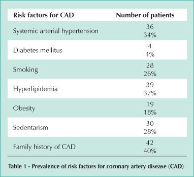
Table 1 shows the prevalence of the risk factors studied for coronary artery disease.
Correlation between carotid artery IMT and right subclavian artery IMT - Taking into consideration a > 0.8 mm value for the right subclavian artery IMT, 41 carotid arteries were IMT-free, whereas 30 (73%) right subclavian arteries showed IMT > 0.8 mm. Out of the 65 carotid arteries with early thickening, 59 (91%) subclavian arteries showed IMT > 0.8 mm.
The average IMT value obtained in the carotid artery (Chart 1) was 0.87 mm (0.5 mm minimum, 1.3 mm maximum, 0.9 mm median, and standard deviation 0.23). The mean IMT value obtained in the right subclavian artery (Chart 2) was 1.7 mm (0.4 mm minimum, 2.8 mm maximum, 1.1 mm median, and 0.46 standard deviation). The analysis of reproducibility of the method indicated a 10.2% error between the [two] investigators.
The correlation coefficient between the carotid artery IMT and the right subclavian artery IMT was 0.31%, with a 95% confidence interval ranging from 0.12 to 0.47.
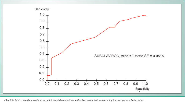
Subclavian artery IMT as an indicator of early thickening - A 0.8 mm carotid thickening was established as the standard cut-off value for the diagnosis of early IMT. Therefore, carotid IMT values under 0.8 mm were considered normal, whereas values over 0.8 mm were considered as early thickening. According to the ROC curve (Chart 3), 0.7 mm was the cut-off value for the IMT at the origin of the right subclavian artery that best characterizes the thickening.
The 0.7 mm cut-off value at the origin of the right subclavian artery, when compared to the 0.8 mm IMT reference value in the carotid arteries, showed sensitivity, specificity, PPV, NPV, and accuracy values of 91%, 27%, 66%, 65%, and 66%, respectively. Table 2 displays sensitivity levels for other IMT cut-off values in the right subclavian artery.
Discussion
The identification of artery wall changes in asymptomatic subjects indicates the need for a more strict control of cardiovascular risk factors, seeking to prevent future coronary events.
Population and hospital-based studies used non-invasive techniques to evaluate early changes in the structure and function of the artery wall, such as the measurement of the intima-media complex, investigation of endothelial dysfunction, and coronary artery calcification25,27,28.
Carotid artery IMT measurement is a safe, low-cost, and easily reproducible method suitable for identifying those patients with subclinical atherosclerotic disease and higher risks for coronary artery disease7,25,29,30.
Studies determined 0.8 mm as the reference value for early thickening of the intima-media complex associated with an increase in cardiovascular risks4,18,31-33. Groot et al conducted a study with 315 patients with familial hypercholesterolemia compared to 118 controls, and showed that an intima-media thickening of up to 0.8 mm would be considered normal. The familial hypercholesterolemia patients reached a 0.8 mm IMT value at the age of 40 years, whereas control group patients reached such a value only at 76 years of age, when their cardiovascular risk would be greater due to age4.
Asymptomatic individuals with a low to intermediate pre-test probability of coronary artery disease, and a carotid IMT value over 1 mm, are at a higher risk of developing coronary events in the future8,25,29,30,34. Multicentric studies showed that patients with an IMT greater than 1 mm have a higher risk of acute myocardial infarction within four years7,29,30. Since the objective of our study was to determine an earlier marker of cardiovascular risk, we used a 0.8 mm cut-off value.
In this study, the correlation coefficient between the carotid artery IMT and the right subclavian artery IMT was 0.31, with a 95% confidence interval ranging from 0.12 to 0.47. The fact that zero is not included in this interval indicates the significance of this correlation. However, considering the limits of the 95% confidence interval, it is clear that despite the low value of the lower limit, the upper limit indicates a good correlation between the carotid artery IMT and the right subclavian artery IMT.
In this study, considering a 91% sensitivity value, a 0.7 mm IMT was determined for the right subclavian artery (lower than the 0.8 mm reference value used for the carotid artery). This suggests that the measurement of the thickening at the origin of the subclavian artery can be an earlier marker of cardiovascular risk.
The IMT, measured by B-mode ultrasound image, consists of the distance between two echogenic lines corresponding to the lumen-intima and media-adventitia interfaces of the artery wall. Since even high-resolution vascular ultrasonography is incapable of distinguishing the intima layer from the media layer of the artery wall, the intima-media complex measurement is routinely used. An increase in the thickness of the intima-media complex may be due to thickening of the media layer or the intima layer. It is known that the atherosclerotic disease affects primarily the intima layer of the artery wall. Carotid and subclavian arteries are elastic arteries, consisting mainly of the intima layer and a very small muscle component. By contrast, in peripheral arteries such as the femoral artery, medial muscle layer prevails. Therefore, carotid and subclavian IMT represents mainly thickening of the intima layer, which is associated with the presence of atherosclerotic disease3,26,35,36.A carotid artery IMT value over 1.3 mm is considered an atherosclerotic plaque37.
One possible explanation for the earlier intima-media thickening in the right subclavian artery would be the presence of greater vessel angulation at its origin as compared to the carotid artery bifurcation. Higher speeds in the inner curvature border, as observed at the origin of the right subclavian artery, are responsible for the increase in endothelial surface stress and shear forces at the site38. The artery wall stress and the resulting greater shear force would contribute to the development of the intima-media thickening and a posterior atherosclerotic plaque at the site. Back in 1963, Texon proposed that lower inner wall pressures would favor atheroma deposition in the curvatures39. However, there are no studies in medical literature associating right subclavian artery IMT and cardiovascular risk factors.
This study observed that out of the 41 patients whose carotid arteries were IMT-free, thirty (73%) had right subclavian artery IMT values > 0.8mm. This finding shows that, even when the carotid artery is normal, the subclavian artery may show thickening of the intima-media complex. Consequently, the presence of IMT may be detected earlier in the subclavian artery rather than in the carotid artery, enabling an earlier prescription of aggressive management of cardiovascular risk factors in order to prevent future coronary events.
The authors conclude that there is an association between carotid artery IMT and the IMT at the origin of the right subclavian artery. With a 0.7 mm cut-off value, it is possible to detect IMT earlier in the subclavian artery than in the carotid arteries. The IMT at the origin of the right subclavian artery may be considered an earlier marker for the evaluation of cardiovascular risks.
Potential Conflict of Interest
No potential conflict of interest relevant to this article was reported.
References
Received on 06/02/05; revised manuscript received August 20, 2005; accepted on 10/17/05
- 1. Badimon JJ, Fuster V, Chesebro JH, Badimon L. Coronary atherosclerosis: a multifactorial disease. Circulation 1993; 87 (suppl): II3-II16.
- 2. Wittes J, Lakatos J, Probstfield J. Surrogate endpoints in clinical trials. Stat Med 1989; 8: 415-25.
- 3. Grobbee DE, Bots ML. Carotid artery intima-media thickness as an indicator of generalized atheroclerosis. J Int Med 1994; 236: 567-73.
- 4. Groot E, Hovingh GK, Wiegman A, et al. Measurement of arterial wal thickness as a surrogate marker for atherosclerosis. Circulation 2004; 109 (suppl III): III-33III-38.
- 5. Van Bortel LM. What does intima-media thickness tell us? J Hypertens 2005; 23: 37-9.
- 6. Bots ML, Grobbee DE, Hofman A, Witteman JCM. Common carotid intima-media thickness and risk of acute myocardial infaction. Stroke 2005; 36: 762-7.
- 7. O'Leary DH, Polak JF, Kronmal RA, Manolio TA, Burke GL, Wolfson SK Jr. Carotid-artery intima and media thickness as a risk factor for myocardial infarction and stroke in older adults. Cardiovascular Health Study Collaborative Research Group. N Engl J Med 1999; 340: 14-22.
- 8. Simon A, Gariepy J, Chironi G, Mengnien JL, Levenson J. Intima-media thickness: a new tool for diagnosis and treatment of cardiovascular risk. J Hypertens 2002; 20: 159-69.
- 9. Crouse JR, Tang R, Espeland MA, Terry JG, Morgan T, Mercuri M. Association of extra-cranial carotid atheroclerosis progression with coronary status and risk factors in patients with and without coronary artery disease. Circulation 2002; 106: 2061-6.
- 10. Luedemann J, Schminke U, Berger K, et al. Association between behavior-dependent cardiovascular risk factors and asyntomatic carotid atheroclerosis in a general population. Stroke 2002; 33: 2929-35.
- 11. Stein JH, Douglas PS, Srinivasan SR, et al. Distribution and cross-sectional age-related increases of carotid artery intima-media thickness in young adults. The Bogalusa Heart Study. Stroke 2004; 35: 2782-7.
- 12. Jerrard-Dunne P, Markus HS, Steckel Da, Buehler A, von Kegler S, Stizer M. Early carotid atherosclerosis and family history of vascular disease. Specific effects on arterial sites have implications for genetic studies. Arterioscler Thromb Vasc Biol 2003; 23: 302-6.
- 13. Weber F. Risk factors for subclinical carotid atherosclerosis in healthy men. Neurology 2002; 59: 524-28.
- 14. Tropeano AI, Boutouyrie P, Katsahian S, Laloux B, Laurent S. Glucose level is a major determinant of carotid intima-media thickness in patients with hypertension and hyperglycemia. J Hipertens 2004; 22: 2153-60.
- 15. Liu ML, Yilitalo K, Salonen R, Salonen JT, Taskinen MR. Circulating oxidized low-density lipoprotein and its association with carotid intima-media thickness in asyntomatic members of familial combined hiperlipidemia families. Arterioscler Thromb Vasc Biol 2004; 24: 1492-7.
- 16. Wang TJ, Nam BH, D'Agostinho RB, et al. Carotid intima-media thickness is associated with premature parental coronary heart disease. The Framinghan Heart Study. Circulation 2003; 108: 572-6.
- 17. Vaudo G, Schillaci G, Evangelista F, Pasqualini L, Verdecchia P, Mannarino E. Arterial wall thickening at different sites and its association with left ventricular hypertrophy in newly diagnosed essential hypertension. Am J Hypertens 2000; 13: 324-31.
- 18. Cuspidi C, Ambrosioni E, Mancia G, Pessina AC, Trimarco B, Znachetti A. Role o echocardiography and carotid ultrasonography in stratifying risk in patients with essential hypertension: the Assesment of Prognostic Risk Observational Survey. J Hypertens 2002; 20: 1307-14.
- 19. Zakopoulos NA, Tsivgoulis G, Barlas G, et al. Time rate of bood pressure variation is associated with increased common carotid artery intima-media thickness. Hypertension 2005; 45: 505-12.
- 20. Guidelines committee. 2003 European Society of Hypertension European Society of Cardiology guidelines for manegement of arterial hypertension. J Hypertens 2003; 21: 1011-53.
- 21. Held C, Hjemdahl P, Eriksson SV, Björkander I, Forslund L, Rehnquivist N. Prognostic implications of intima-media thickness and plaques in the carotid and femoral arteries in patiens with stable angina. Eur Heart J 2001; 22: 62-72.
- 22. Lelakis JP, Papamichael CM, Cimponeriu AT, et al. Atherosclerotic changes of extra-coronary arteries are associated with the extent of coronary atheroclerosis. Am J Cardiol 2000; 85: 949-52.
- 23. Mackinnon AD, Jerrard-Dunne P, Tizer M, Buehler A, von Kegler S, Markus HS. Rates and determinants of site-specific progression of carotid artery intima-media thickness. The carotid atheroclerosis progression study. Stroke 2004; 35: 2150-4.
- 24. Pignoli P, Tremoli E, Poli A, Oreste P, Paoletti R. Intimal plus medial thickness of the arterial wall: a directed mesurement with ultrasound imaging. Circulation 1986; 74: 1399-406.
- 25. Bots ML, Dijk JM, Oren A, Grobbee DE. Carotid intima-media thickness, arterial wall stiffness and risk of cardiovascular disease: current evidence. J Hipertens 2002; 2317-25.
- 26. Sinha AK, Eigenbrodt M, Mehta JL. Does carotid intima media thickness indicate coronary atheroclerosis? Curr Opin Cardiol 2002; 17: 526-30.
- 27. Simon A, Megnien JL, Levenson J. Coronary risk estimation and treatment of hypercholesterolemia. Circulation 1997; 96: 2449-52.
- 28. Hollander M, Hak AE, Koudstaal PJ, et al. Comparison between measures of atheroclerosis and risk of stroke. Stroke 2003; 34: 2367-73.
- 29. Chambless LE, Heiss G, Folson AR, Szklo M, Sharrett AR, Cleg LX. Association of coronary heart disease incidence with carotid arterial wall thickness and major risk factors: The Atherosclerosis Risk in Communities (ARIC) Study. Am J Epidemiol 1997; 146: 483-94.
- 30. Bots ML, Hoes AW, Koudstaal PJ, Hofman A, Grobbee DE. Common carotid intima-media thickness and risk of stroke and miocardial infaction: the Rotterdan Study. Circulation 1997; 96:1432-7.
- 31. Jadkav UM, Kaddam NN. Carotid intima-media thickness as an independent predictor of coronary artery disease. Indian Heart J 2001; 53 (4): 458-62.
- 32. Rohani M, Jogestrand T, Ekberg M, et al. Interrelation between the extent of atherosclerosis in the thoracic aorta, carotid intima-media thickness and the extent of coronary artery disease. Atherosclerosis 2005; 179 (2): 311-6.
- 33. Hodis HN, Mack WJ, Selzer RH, Liu C, Azen SP. The role of carotid arterial intima-media thickness in predicting clinical coronary events. Ann Intern Med 1998; 128: 262-9.
- 34. Aminbakhsh A, Mancini GB. Carotid intima-media thickness mesurements: waht defines an abnormality? A systematic review. Clin Invest Med 1999; 4: 149-57.
- 35. Slonen JT, Salonen R. Ultrasound B-mode imaging in observational studies of atherosclerotic progression. Circulation 1997; 87 (3 Suppl): II56-65.
- 36. Montenegro MRG. Estrutura da parede vascular. In Maffei FHA, Lastória S, Yoshida WB, Rollo HA. (ed.). Doenças Vasculares Periféricas. 3 ed. Rio de Janeiro: Medsi; 2002, v.1, 179-91.
- 37. Zanchetti A, Bond MJ, Henning M, et al. On behalf of the ELSA investigators. Risk factors associated with alterations in carotid intima-media thickness in hypertension: baseline data from the European Lacidipine Studty on Atherosclerosis. J Hyperten 1998; 16: 949-61.
- 38. Lopes OU. Estudo das doenças vasculares periféricas a partir da dinâmica dos fluidos. In: Maffei FHA, Lastória S, Yoshida WB, Rollo HA. (ed.). Doenças vasculares periféricas. 3 ed., Rio de Janeiro: Medsi; 2002, v.1, 217-31.
- 39. Texon M. The role of vascular dynamics in the development of atherosclerosis. In Sandler M, Bourne GH (ed.). Atherosclerosis and Its Origiti. New York: Academic Press, 1963: 167-95.
Publication Dates
-
Publication in this collection
06 Mar 2007 -
Date of issue
Nov 2006
History
-
Accepted
17 Oct 2005 -
Reviewed
20 Aug 2005 -
Received
02 June 2005