Abstracts
OBJECTIVE: To assess the long-term results of percutaneous balloon valvuloplasty at a single institution. METHODS: This study comprised 189 patients with pulmonary valve stenosis undergoing percutaneous balloon valvuloplasty from 1984 to 1996, whose mean age was 7.97±9.25 years. The procedure was classified as successful when the RV-PA gradient was reduced to levels < 36 mmHg; restenosis was indicated by RV-PA gradients > 36 mmHg after an effective procedure. RESULTS: After the procedure, the peak-to-peak transvalvular gradient decreased from 70.12±30.06 to 25.11 ±20.23 mmHg (P<0.001). Immediate success was obtained in 148 (78.72%) patients. A later reduction in the gradient to values < 36 mmHg was obtained in 24 other patients previously categorized as unsuccessful. Therefore, percutaneous balloon valvuloplasty was considered effective in 172 (91.01%) patients. Effectiveness increased to 93.53% (159/170) in the cases of typical morphology. Follow-up ranged from 4.39±3 years to 13.01 years. Restenosis was observed in 24 (13.95%) patients. Pulmonary regurgitation was detected in 95.1% of the patients, being more intense than mild in 29.5% of the patients. The probability of maintaining an appropriate result, at any time point, with no restenosis was 92.29% in 2 years, 87.38% in 5 years, 82.46% in 8 years, and 64.48% in 10 years. CONCLUSION: Percutaneous balloon valvuloplasty was effective and safe for the treatment of pulmonary valve stenosis with excellent short- and long-term results.
valvuloplasty; pulmonary valve stenosis; results; echocardiography
OBJETIVO: Determinar os resultados em longo prazo da valvoplastia percutânea por balão em uma instituição isolada. MÉTODOS: Estudados 189 pacientes com estenose valvar pulmonar submetidos a valvoplastia percutânea por balão, entre 1984-1996, com idade média de 7,97±9,25 anos, classificando-se como bem sucedida, a redução do gradiente VD-AP em níveis < 36mmHg e reestenose gradientes > 36mmHg após procedimento eficaz. RESULTADOS: Após o término do procedimento, o gradiente pico a pico transvalvar reduziu-se de 70,12±30,06 para 25,11±20,23 mmHg (p<0,001). Obtiveram sucesso imediato 148 (78,72%) pacientes. Houve redução posterior do gradiente para valores < 36mmHg em outros 24 pacientes categorizados sem sucesso no grupo. Assim a valvoplastia percutânea por balão foi considerada efetiva em 172 (91,01%) pacientes. A efetividade aumentou para 93,53% (159/170) nos casos de morfologia típica. O tempo de seguimento foi de 4,39±3anos até o período máximo de 13,01 anos. Observou-se reestenose em 24 (13,95%). A presença de regurgitação pulmonar foi detectada em 95,1% dos pacientes, sendo que em 29,5% com grau maior do que leve. A probabilidade de se manter um resultado adequado, até qualquer ponto no tempo, sem a ocorrência de reestenose, foi de 92,29% em 2 anos, de 87,38 % em 5 anos, de 82,46% em 8 anos e de 64,48% em 10 anos. CONCLUSÃO: A valvoplastia percutânea por balão foi efetiva e segura no tratamento da estenose valvar pulmonar com excelentes resultados imediatos e a longo prazo.
valvoplastia; estenose da valva pulmonar; resultados; ecocardiografia
ORIGINAL ARTICLE ARTIGO ORIGINAL
Short- and long- term results of percutaneous balloon valvuloplasty in pulmonary valve stenosis
Domingos M. Hatem; Iran Castro; José Carlos Haertel; Raul I. Rossi; Paulo Zielinsky; Flávio C. Leboute; Nara Pomar; Maristela Winckler; Rogério N. Kersten; Carlos Roberto Cardoso; Carlos A.M. Gottschall
Instituto de Cardiologia RS/Fundação Universitária de Cardiologia
Correspondence Correspondence to Domingos Hatem Unidade de Pesquisa do IC/FUC Av. Princesa Isabel, 370 Cep 90620-001 Porto Alegre, RS, Brazil E-mail: pesquisa@cardnet.tche.br
ABSTRACT
OBJECTIVE: To assess the long-term results of percutaneous balloon valvuloplasty at a single institution.
METHODS: This study comprised 189 patients with pulmonary valve stenosis undergoing percutaneous balloon valvuloplasty from 1984 to 1996, whose mean age was 7.97±9.25 years. The procedure was classified as successful when the RV-PA gradient was reduced to levels
< 36 mmHg; restenosis was indicated by RV-PA gradients > 36 mmHg after an effective procedure.
RESULTS: After the procedure, the peak-to-peak transvalvular gradient decreased from 70.12±30.06 to 25.11 ±20.23 mmHg (P<0.001). Immediate success was obtained in 148 (78.72%) patients. A later reduction in the gradient to values < 36 mmHg was obtained in 24 other patients previously categorized as unsuccessful. Therefore, percutaneous balloon valvuloplasty was considered effective in 172 (91.01%) patients. Effectiveness increased to 93.53% (159/170) in the cases of typical morphology. Follow-up ranged from 4.39±3 years to 13.01 years. Restenosis was observed in 24 (13.95%) patients. Pulmonary regurgitation was detected in 95.1% of the patients, being more intense than mild in 29.5% of the patients. The probability of maintaining an appropriate result, at any time point, with no restenosis was 92.29% in 2 years, 87.38% in 5 years, 82.46% in 8 years, and 64.48% in 10 years.
CONCLUSION: Percutaneous balloon valvuloplasty was effective and safe for the treatment of pulmonary valve stenosis with excellent short- and long-term results.
Key words: valvuloplasty, pulmonary valve stenosis, results, echocardiography
Pulmonary balloon valvuloplasty, initiated in 1982, was 1 of the first therapeutic procedures to use catheters for the treatment of congenital heart diseases 1,2. It was first performed in 1982 with the static technique, which is still currently used by Kan et al 1. From then on, the percutaneous pulmonary valvuloplasty technique underwent few changes; in regard to the material used, however, great progress has been made. The improvement in the technology of balloon-catheter confection allowed the production of lower-profile balloons, therefore, reducing the complications of the procedure, notably the vascular ones, enabling the performance of the procedure in neonates, in addition to improving the chances of passing across the stenotic orifice 3,4. Balloons whose diameters exceeded the ring measurement by 20% to 40% on angiography were shown to be safe and more effective than those used in the initial procedures, which adopted the diameter of the pulmonary ring as the upper limit. This increased the success rate with lower residual gradients and no increase in the number of complications 5,6.
Initially, most hemodynamic data of patients with pulmonary valve stenosis were obtained from cardiac catheterization. Currently, the use of Doppler echocardiography allows the reliable and serial assessment of the stenotic lesion by determining the gradient through the pulmonary valve in any age group, which enables one to follow the natural history of the disease 7-9.
The immediate results and the short- and medium-term follow-up of pulmonary valve dilation have already been well documented in the literature. Reports on long-term follow-up, however, are still scarce 10-19.
Methods
A noncontrolled cohort study of incidence was carried out in patients undergoing percutaneous pulmonary valvuloplasty as a treatment for pulmonary valve stenosis.
The outcome axis is dynamic, because the major clinical outcomes considered were immediate success, efficacy, and the intermediate and late results of balloon pulmonary valvuloplasty, using the measurement of the maximum instantaneous gradient on Doppler echocardiography and the peak-to-peak gradient obtained through cardiac catheterization. In regard to the temporal axis, the study may be considered mixed, because part of the data were collected in a historical (retrospective) way and part in a contemporary (prospective) way.
From September 1984 to October 1996, 252 consecutive patients diagnosed with pulmonary valve stenosis were referred to the hemodynamics department of the Instituto de Cardiologia of Rio Grande do Sul/Fundação Universitária de Cardiologia to undergo balloon pulmonary valvuloplasty.
The following patients were excluded from the study: those requiring balloon valvuloplasty as a palliative treatment for cyanosis, and those with pulmonary valve stenosis and large defects in the interventricular septum. Patients with interatrial septal defects or patent oval foramen were included in the study. The criteria for indicating percutaneous pulmonary valvuloplasty were a peak-to-peak pulmonary transvalvular systolic gradient obtained during any previous hemodynamic study or during the therapeutic procedure, or a maximum instantaneous gradient obtained on Doppler echocardiography, or both, greater than 50 mmHg with a normal cardiac index, independent of the symptoms, in addition to the evidence of right ventricular hypertrophy on the electrocardiogram with symptoms of heart failure, angina, or syncope. Two hundred and thirty-eight patients underwent percutaneous balloon valve dilation, 45 dropped out during follow-up, and 4 had only 1 echocardiogram performed within the 30 days following the procedure. The population actually studied comprised 189 patients, all of whom underwent at least 1 Doppler echocardiographic study 30 days after the procedure until 12/31/97, corresponding to 79.41% of the patients eligible for the analysis of the final follow-up.
Ninety-six (50.79%) patients were females. The mean age of the patients followed up, at the time of valvuloplasty, was 7.97±9.25 years, ranging from 0.1 to 48.88 (median of 5.04) years. No patient was a neonate, 30 (15.88%) were 1 to 12 months old, 114 (60.32%) were 1 to 10 years old, 24 (12.7%) were 10 to 20 years old, and 21 (11.1%) were older than 20 years.
The criterion used for characterizing the immediate success of the procedure was the reduction in the residual peak-to-peak systolic gradient between the right ventricle and the pulmonary artery, measured after valve dilation, to values < 36 mmHg. The efficacy of pulmonary valvuloplasty comprised the cases in which immediate success occurred or those in which, despite lack of immediate success, a later reduction was observed in the maximum instantaneous residual gradient to values < 36 mmHg in subsequent Doppler echocardiographic studies. In assessing the results of late follow-up, the same values of the gradients already used to define success or unsuccess immediately after valvuloplasty were used. The occurrence of restenosis was defined as a new elevation in the transvalvular gradient to levels > 36 mmHg after effective valvuloplasty. The patients were also analyzed in regard to the morphology of the stenotic valve, which was classified as typical, dysplastic, or complex, in the case of previous surgical valvotomy (postoperative restenosis).
The technique of valvuloplasty used was similar to that reported by Kan et al 1, in which the procedure was performed, depending on age, with the patient under general anesthesia. Special attention was given to the withdrawal tracings between the pulmonary artery and the right ventricle, as well as to the measurement of the systolic pressure in that ventricle, because the final indication of the procedure depended on these measurements. Ventriculography was performed in the left profile and in the 30o right anterior oblique projection with cranial angulation to exclude the presence of subvalvular obstruction, and to allow an appropriate measurement of the pulmonary ring, which directly influenced the choice of the diameter of the balloon. The anatomy and the diameter of the balloon to be used were defined as being up to 40% greater than the systolic diameter between the points of insertion of the pulmonary leaflets in the arterial wall. The technique with a single balloon was used in 157 patients and that with 2 balloons in 32 patients.
The Doppler-echocardiographic studies consisted initially of the sequential morphologic assessment 20.
The degree of severity of the obstruction determined by pulmonary valve stenosis was assessed and quantified with continuous Doppler. The maximum instantaneous gradient was calculated through the maximum velocity obtained with the modified Bernoulli equation. In case of associated pulmonary subvalvular obstruction, a mosaic of colors indicated the presence of flow turbulence below the plane of the pulmonary valve and also a characteristic pattern of the spectral curve on Doppler with an increase from the beginning to the end of systole. The presence and intensity of pulmonary regurgitation were also assessed through color flow mapping, as reported by Cooper et al 21, considering the extension of the regurgitating jet and its width at the origin.
In regard to the gradients, the groups were compared at baseline with the Student t test. The chi-square test was used for the categorical variables. The transvalvular gradients before and after valvuloplasty were compared with the t test for paired samples. An actuarial survival curve was built with the aid of SPSS 8 statistical software. The continuous variables were expressed as means ± standard deviation. The characterization of the result of pulmonary valvuloplasty was transformed into a dichotomous variable (success x unsuccess) using the value of 35 mmHg as the cut point.
Results
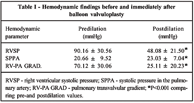
Immediately after the procedure (tab. I), the patients had a reduction in the right ventricular systolic pressure from 90.16±30.56 to 48.08±21.50 mmHg (P<0.001). The pulmonary systolic pressure had a slight elevation from 20.66±9.52 to 23.03±7.04 mmHg (P<0.001). The peak-to-peak pulmonary transvalvular gradient had a reduction from 70.12±30.06 to 25.11±20.23 mmHg (P<0.001) (tab. I).
Of the 189 patients undergoing balloon valvuloplasty, 1 could not undergo blood pressure and transvalvular gradient measurements after balloon inflation due to the occurrence of complications during the procedure, which was then interrupted. A Doppler echocardiographic study carried out after percutaneous dilation revealed the presence of an elevated residual gradient.
The 188 patients undergoing the hemodynamic measurements after the procedure were considered for analyzing the immediate results. The gradient measured represents the total residual gradient between the right ventricle and the pulmonary trunk, and the contributions of the valvular and infundibular components were not routinely individualized, which was the reason why this factor was not analyzed.
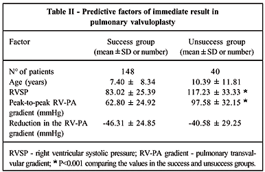
One hundred forty-eight (78.72%) patients had a peak-to-peak pulmonary transvalvular gradient < 36 mmHg, representing the group that obtained immediate success with percutaneous dilation of the pulmonary valve. On the other hand, 40 (21.28%) patients had a peak-to-peak residual gradient > 36 mmHg, characterizing an unsuccessful procedure in regard to alleviating pulmonary valve stenosis. Among the patients with typical pulmonary valve stenosis (n=170), 137 (80.59%) had immediate success after the procedure; on the other hand, among those with pulmonary valve stenosis with a dysplastic valve and complex morphology consequent to the previous surgery (n=18), the rate of success was 61.11% (P<0.05).
Analyzing separately the patients with stenosis in a dysplastic pulmonary valve (n=9) and those with a complex valvular morphology (n=9), success was obtained in 66.67% and 55.56% (NS), respectively.
The patients who did not obtain immediate success after valvuloplasty had more severe pulmonary valve stenosis, characterized by a greater peak-to-peak pulmonary transvalvular gradient (P<0.001) and a more elevated right ventricular systolic pressure (P<0.001). No difference regarding age and the degree of reduction in the peak-to-peak transvalvular gradient was observed between the 2 groups (tab. II).
Regarding the reduction in the peak-to-peak systolic gradient obtained with valvular dilation, no significant difference was observed between the groups of patients with immediate success or unsuccess (tab. II). Among the former, a reduction of 46.31±24.85 mmHg was observed, and, among the latter, a reduction of 40.58±29.25 mmHg (NS) was observed. In both groups, the reduction in the gradient ranged from -1 to -142 mmHg, an elevation of 15 mmHg being observed in 1 patient consequent to infundibular reaction.
Acute complications occurred in 8 (4.23%) patients. Dissection of the inferior vena cava occurred in 1 case, but no retroperitoneal bleeding or hematoma was observed. Two patients had convulsions during the procedure, more precisely during the inflation of the balloon catheter. Nontransient supraventricular arrhythmia (atrial fibrillation) occurred in 2 patients. One patient had deep venous thrombosis in the right lower limb after the procedure. Rupture of the tricuspid subvalvular apparatus was observed in 1 patient, determining a significant tricuspid regurgitation. The procedure was interrupted in 1 patient due to the occurrence of cardiac arrest that required resuscitation maneuvers.
For analyzing the efficacy of the valvuloplasty, the 189 patients were considered. The efficacy of valvuloplasty was assessed over an intermediate follow-up period comprising the time interval between the performance of valvuloplasty and the first Doppler echocardiography performed.
The efficacy of balloon pulmonary valvuloplasty encompassed those patients who obtained immediate success in the procedure and those who maintained a peak-to-peak residual gradient immediately after valvuloplasty > 36 mmHg and who had a spontaneous reduction to levels < 36 mmHg in the first Doppler echocardiography during follow-up.
The first Doppler echocardiography was performed 2.15±2.52 years after valvuloplasty, and a reduction in the RV-PA gradients to values below 36 mmHg was observed in 24 patients in the group with no success immediately after percutaneous balloon valvuloplasty. Of those, 22 had typical morphology, and 2 had complex morphology, but none of the latter had a dysplastic pulmonary valve.
Valvuloplasty was considered effective in 172 (91.01%) patients, 148 of whom had a reduction in the pulmonary transvalvular gradient right after the procedure, and another 24 had it during follow-up (fig. 1). One hundred and seventy (89.95%) patients had pulmonary valve stenosis with the typical dome-like morphology, an appropriate result being obtained in 159, corresponding to a success rate of 93.53%. Of the 24 patients evolving with a reduction in the transvalvular gradient, 22 had typical morphology, while 2 had complex morphology (fig. 1).
The mean follow-up duration was 4.39±3 years, until a maximum of 13.01 years. The mean maximum instantaneous pulmonary transvalvular gradient was 26.12 ±19.08 mmHg, ranging from 5 to 100 mmHg (fig. 2).
One hundred forty-nine (78.84%) patients had a maximum instantaneous gradient < 36 mmHg, characterizing the maintenance of the success of pulmonary valvuloplasty, and the patients remained free from restenosis during the follow-up period. Forty (21.16%) patients had a maximum instantaneous gradient > 36 mmHg, 16 (8.47%) of whom maintained the noneffective result (unsuccess) of valvuloplasty and 24 (12.7%) had a new elevation in the transvalvular gradient during follow-up, characterizing the occurrence of pulmonary valve restenosis (fig. 1).
Valves with typical or dysplastic stenoses, or stenoses with complex morphology were found in, respectively, 170, 9, and 10 patients. The rate of success in the group with typical stenosis remained 81.18%; for the others, 55.56% and 60% were the rates for the patients with dysplastic and complex morphology, respectively (P<005).
A new elevation in the pulmonary transvalvular gradient to levels > 36 mmHg after relief of the pulmonary valve stenosis through dilation with the balloon characterized the occurrence of restenosis. Of the 172 patients of the cohort whose procedures were successful (effective percutaneous balloon valvuloplasty), with gradients < 36 mmHg immediately after valvular dilation or later due to a reduction in infundibular hypertrophy, 24 (13.95%) had pulmonary valvular restenosis, and only 2 had a dysplastic pulmonary valve (fig. 1). The detection of valvular restenosis occurred 3.56±3.50 years after percutaneous dilation, and, in 29.2% of the cases, it occurred after 5 years of follow-up. In the group of patients with restenosis, the mean age was 7.69±7.89 years, the peak-to-peak gradient before valvuloplasty was 69.63±40.19 mmHg, and that immediately after valvuloplasty was 26.88±13.07 mmHg. No significant difference was observed between the patients who developed restenosis and those who remained free from restenosis in regard to age or the gradient before valvuloplasty and the residual gradient after valvuloplasty.
Sixty-one sequential patients, corresponding to 32.3% of the cohort, underwent the last Doppler echocardiography in a single device (HP Sonos 2500). They were carefully assessed by 2 observers and by a third in case of disagreement in regard to the degree of pulmonary regurgitation. Some degree of regurgitation was observed in 95.1% of the patients (58/61), and a more intense than mild degree was observed in 29.5% (18/61).
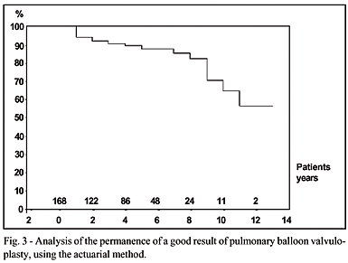
The probability of persistence of a good result in pulmonary valvuloplasty, with no restenosis, was 92.29% in 2 years, 87.38 % in 5 years, 82.46% in 8 years, and 64.48% in 10 years (fig. 3).
Discussion
The percutaneous dilation of the pulmonary valve with a balloon is currently considered the therapeutic modality of choice for the treatment of pulmonary valve stenosis in any age group and any valvular morphology 17. It is an effective and safe technique, with low indices of mortality and of incidence of important complications when used in a period other than the neonatal 12,17. The method was developed to replace surgical valvotomy, reported by Brock in 1948, with a mortality rate around 1.5 to 2% 22.
The study of the follow-up of the patients of this cohort was based on the maximum instantaneous gradient obtained on Doppler echocardiography and comparable to the gradient obtained on cardiac catheterization 23,24.
This high degree of correlation and concordance is reduced when the measurements obtained with the 2 methods are assessed unsimultaneously. This difference is usually small, < 10 mmHg. Some authors suggest that the greater gradient should be used, even if obtained in an unsedated patient, reflecting a more actual physiological condition 25.
This explains the findings in some patients in this cohort, who, at the time of valvuloplasty, had peak-to-peak gradients significantly lower than those obtained on Doppler echocardiography before valvuloplasty, even in those already known as having previously documented significant pulmonary valve stenosis.
Immediate success documented in 78.72% (148/188) of the patients, comprising all valve morphologies is consistent with those observed by other authors, which ranged from 54% to 87.1% 10,19,26. The patients who did not obtain immediate success with percutaneous dilation had a significantly greater pulmonary transvalvular gradient and right ventricular systolic pressure than those who obtained immediate success. No significant difference between the 2 groups in regard to age, sex, and degree of reduction in the peak-to-peak pulmonary transvalvular gradient was observed.
Ray et al 19, studying 139 patients, also observed that those who obtained incomplete relief of the obstruction right after valvuloplasty had a more significant degree of pulmonary valve stenosis characterized by greater RV-PA gradients and right ventricular systolic pressure. In this same study, the mean age of the patients whose procedures were unsuccessful was higher.
In the present study, the reduction in the transvalvular gradient obtained was similar in both groups, 46.31±24.85 mmHg and 40.85±29.25 mmHg (NS), respectively, in those who obtained immediate success and in those who did not.
Mendelsohn et al 27 also found an almost identical reduction in the gradients between the 2 groups, with values around 40 mmHg, suggesting that this decrease in the gradient was finite. The success of valvuloplasty related to the initial gradient value, which could have a limited reduction. The authors suggested an earlier intervention, indicating balloon valvuloplasty in patients with lower degrees of pulmonary valve stenosis, with gradient values of lower magnitude, such as 40 mmHg. The consequence of this earlier intervention would be a lower residual systolic gradient, with a consequent decrease in the occurrence of symptoms and of right ventricular hypertrophy in the long run, and a higher rate of success.
The results of the patients with dysplastic valve and residual stenosis after surgical valvotomy (61.11%) were less effective when compared with those of the group of patients with typical pulmonary valve stenosis (80.59%) P<0.05.
Ballerini et al 28 continue to perform percutaneous balloon valvuloplasty in dysplastic valves and in those with complex morphology, although with less effective results in this group of patients, aiming at avoiding or delaying the need for surgery.
The efficacy of pulmonary balloon valvuloplasty has been underestimated when only the immediate results are considered, because a progressive reduction in the transvalvular gradient is frequently documented as being similar to that observed after surgical valvotomy 3, which occurs due to a regression in the infundibular obstruction, considered a dynamic process that regresses after a variable period of time. Therefore, the apparent poor immediate result observed in some cases, mainly in the more severe pulmonary valve stenoses, may temporarily mask the results of an effective valvuloplasty, which later may be confirmed with the reduction in infundibular hypertrophy 12,29,30. In the cases of residual infundibular stenosis, complete regression may take up to 2 years to occur, and no relation with severity or the patient's age may exist 30,31.
Of the 40 patients in our case series who did not obtain immediate success, 24 (60%) evolved with a reduction in both the maximum instantaneous and the peak-to-peak residual pulmonary transvalvular gradients to levels < 36 mmHg without undergoing any additional intervention. This evolution characterized the percutaneous valve dilation as effective, despite the unsatisfactory pressure results, immediately after the procedure.
Mahnert et al 11, studying 52 patients who had undergone percutaneous balloon valvuloplasty, obtained a reduction in the mean pulmonary transvalvular gradient from 79.9±37.3 mmHg to 37.2±29.6 mmHg (P<0.001) immediately after the procedure, a residual gradient > 36 mmHg persisting in 19 patients. During a period shorter than 2 years, defined by the authors as an intermediate follow-up, the gradient assessed on catheterization or Doppler echocardiography dropped to values < 36 mmHg in 10 out of 19 (52.63%) patients, without any other additional intervention. Several reports exist in the literature on the immediate results of percutaneous balloon valvuloplasty and their short- and medium-term follow-up. Studies on the effectiveness of valvuloplasty in the long run, ie, a follow-up longer than 2 years, are scarce. In our patients assessed on Doppler echocardiography for up to 13 years, a mean of 4.39±3 years after pulmonary valvuloplasty, persistent success with permanence of a maximum instantaneous gradient < 36 mmHg was observed in 78.84% (149/189) of the patients.
Data about the follow-up of the patients in the largest case series published can be found in the VACA study 18, comprising 533 patients from 22 institutions and with a follow-up of up to 8.7 years, 77% of whom maintained gradients < 36 mmHg, a percentage similar to that observed at our institution (78.84%).
Ray et al 19 reported a case series with 139 patients, 79 undergoing catheterization 13±8.7 months after the procedure, and 81% of whom showed a peak-to-peak systolic gradient < 36 mmHg.
Rao et al 14 reported the following results of a long-term follow-up (3 to 10 years) in 80 patients undergoing percutaneous balloon valvuloplasty in 2 university-affiliated institutions: maintenance of the appropriate result in 88% of the patients in 5 years, and in 84% in 10 years.
The occurrence of complications related to pulmonary valvuloplasty is considered small. The number and severity of the complications are greater when the procedure is performed in the neonatal period. Therefore, although the technique is relatively safe, it is worth stressing that potential complications may occur and that the correct technique should be used, as should the balloon with appropriate diameter and length 17.
The presence of patent oval foramen protects against arterial hypotension. Shuck et al 32 observed that the occurrence of arterial hypotension is minimal in individuals with patent oval foramen at the time of balloon inflation, probably due to maintenance of the left ventricular filling and right atrial decompression through the oval foramen. The use of the double balloon technique 33, bifoil or trifoil balloons that allow the passage of some flow from the right ventricle around the balloon, and the use of short periods of inflation (5 s or less) reduce the occurrence of systemic hypotension.
In the follow-up studies after percutaneous pulmonary valvuloplasty, the initial impression was that the incidence of residual pulmonary valve regurgitation was small. Subsequent larger studies showed that that was a frequent finding, usually of small magnitude after successful percutaneous dilation 34. The incidence in the most recently published series ranged from 74% to 100% 10,14,18,19,29. The occurrence of residual pulmonary regurgitation after the procedure may be explained by the fact that the mechanism of valvular opening through the use of balloon catheters consists of commissural separation, rupture, or even avulsion of the leaflets 35.
In our group of patients, pulmonary insufficiency was present in 95.1% of the cases, usually being mild; however, 26.2% had a moderate reflux, and 3.3% had a severe reflux. Ray et al 19 detected pulmonary insufficiency on Doppler echocardiography in 86% of the 139 patients in their case series, but only 3 had a reflux greater than mild.
In the results of the VACA study published by McCrindle 18, residual pulmonary regurgitation was detected in 74% of the patients, being classified as trivial in 22%, mild in 45%, and moderate in 7%. No case of severe pulmonary insufficiency was detected, and the following factors were identified as favoring the occurrence of moderate pulmonary insufficiency: the ratio between the balloon and the pulmonary ring greater than 1.4, and complex valvular morphology due to a previous surgical valvotomy or the presence of valvular dysplasia.
Rao et al 14, in a case series with 85 patients from 2 university-affiliated centers, assessed the incidence of pulmonary regurgitation and performed its semiquantification using color Doppler in 4 periods of time: before valvuloplasty, 1 day after, within the period of 2 years, and, finally, in the period from 3 to 10 years. The quantification was performed using the ratio between the width of the jet in its origin and the diameter of the pulmonary ring in the short axis obtained in the parasternal window, considering that the values < 10%, 11-25%, 26-50%, and > 50% indicated, respectively, degrees of regurgitation from I to IV. The incidence of pulmonary regurgitation had a gradual, but significant, increase, being greater in the late follow-up, when present in 70 of the 80 (87.5%) patients assessed. No patient had degree IV of pulmonary insufficiency or required surgical intervention because of this reason.
The frequency of restenosis, in several case series, has ranged from 4.8% to 21% 10,14,16,35, being related to the use of an inappropriate-sized balloon and the presence of valvular dysplasia 35. In our case series, 13.95% (24/172) of the patients evolved with elevation in the gradients at varied time intervals after an effective valvuloplasty. The time of progression of the residual gradient to levels > 36 mmHg was 3.56±3.5 years, and in only 29.2% of the cases they were detected after 5 years of follow-up.
In the series by Rao et al 14, 11% of the patients had restenosis, all cases occurring in the first 2 years of follow-up. The criterion used for characterizing restenosis was an elevation in the pulmonary transvalvular gradient to levels > 50 mmHg.
In the VACA study 18, of the patients with adequate immediate results, only 12% had inadequate late results, maintaining residual gradients > 36 mmHg or requiring a new percutaneous or surgical pulmonary valvuloplasty.
Jarrar et al 16, in a study of invasive and noninvasive follow-up in children, adolescents, and adults undergoing percutaneous balloon valvuloplasty, reported restenosis in 3 of 62 patients (4.8%). The low rates of restenosis and persistence of stenosis found in the study were attributed to the use of high balloon/annulus ratios (BARs), such as 1.4±0.38 and > 1.5 in 5 adults. Although most authors recommend that the BAR should not exceed the value of 1.5, others have suggested its use above that value mainly in adults, when the residual gradient immediately after the procedure is > 35 mmHg 16.
Rao et al 36 investigated the causes of restenosis after balloon valvuloplasty and identified the following factors as predisposing to its occurrence: the use of a balloon/pulmonary ring ratio < 1.2, and gradient measured immediately after valvuloplasty > 30 mmHg.
In our cohort, the probability of the maintenance of a good result of pulmonary valvuloplasty, with no restenosis, was 94.39% in 1 year, 92.29% in 2 years, 87.38% in 5 years, 82.46% in 8 years, 64.48% in 10 years, and 56.42% in 13 years. The estimates after 10 years of follow-up were based on a smaller number of patients, and, therefore, the results observed after that period are less reliable. Rao et al 14 reported the results of a 3-to-10-year follow-up in 80 patients aged 7±6.4 years, who underwent pulmonary balloon valvuloplasty. The permanence of the success of percutaneous dilation in 1, 2, 5, and 10 years was, respectively, 94%, 89%, 88%, and 84%. In the series by Quereshi 37, with 92 patients, 76% maintained an adequate result with no need for reintervention in 5 years, and 67% in 10 years.
Our study allows the following conclusions: percutaneous balloon dilation of the pulmonary valve is effective to relieve pulmonary valve stenosis documented by the reduction in the mean gradients; pulmonary balloon valvuloplasty is an effective method and its beneficial effects remain in the medium and long runs; the maintenance of a good result was 92.29% in 2 years, 87.38% in 5 years, 82.46% in 8 years, and 64.48% in 10 years; the complications of the procedure were small and not frequent; nonsignificant residual gradients persisted in most patients, restenosis occurring in 13.95% of the cases; the prevalence of residual pulmonary insufficiency was elevated, with approximately 1/3 of the patients with regurgitation intensity greater than mild.
References
1. Kan JS, White Jr RI, Mitchell SE, Anderson JH, Gardner TJ. Percutaneous balloon valvuloplasty: A new method for trating congenital pulmonary-valve stenosis. N Engl J Med 1982;307:540-2.
2. Lázaro Castillo JL, Munayer Calderón J, Aldana Pérez T et al. Pulmonary valvuloplasty. Long term results at the Centro Medico la Raza. Arch Inst Cardiol Mex 1999;69:338-43
3. Chen CR, Cheng TO, Huang T et al. Percutaneous balloon valvuloplasty for pulmonic stenosis in adolescents and adults. N Engl J Med 1996;335:21-5.
4. Rao PS. Long term follow up results after balloon dilatation of pulmonic stenosis, aortic stenosis, and coarctation of the aorta: A review. Prog Cardiovasc Dis 1999;42:59-74.
5. Zabal C, Lince R, Buendia A, Attié F, Rios MA. Interventional cardiology in congenital heart disease. Arch Inst Cardiol Mex 1999;69:63-8.
6. Melgares R, Prieto JA, Azpitarte J. Succes determining factors in percutaneous transluminal balloon valvuloplasty of pulmonary valve stenosis. Eur Heart J 1991;12:15-23.
7. Gielen H, Daniëls O, van Lier H. Natural history of congenital pulmonary valvar stenosis: an echo and Doppler cardiographic study. Cardiol Young 1999;9: 129-35.
8. Rowland DG, Hammill WW, Allen HD, Gutgesell UP. Natural course of isolated pulmonary valve stenosis in infants and children utilizing Doppler echocardiography. Am J Cardiol 1997;79:344-9.
9. Vermilion RP, Snider R, Bengur AR, Meliones JN. Long-term assesment of right ventricular diastolic filing in patients with pulmonic valve stenosis successfully treated in childhood. Am J Cardiol 1991;68:648-52
10. Hernáez-Cobeño MA, Bermúdez-Cañete R, Herraiz I, Fernández-Pineda L, Quero-Jiménez C, Díaz-García P. Percutaneous balloon pulmonary valvuloplasty: the medium-term results in a series of 100 consecutive pediatric patients. An Esp Pediatr 1998; 49:264-72.
11. Mahnert B, Paul TH, Luhmer I, Kallfelz HC. Medium- to long-term results after percutaneous balloon pulmonary valvuloplasty in childhood. Z Kardiol 1996;85:482-8.
12. Sadr-Ameli MA, Firoozi I, Emran MTS, Hashemi MJ. Late results of balloon pulmonary valvuloplasty in adults. J Am Coll Cardiol 1995; 25(Supll.A):167.
13. Santoro G, Formigari R, Pasquini L, Zorzi A, Ballerini L. Vavuloplastica polmonare in età pediatrica:resultati immediati e follow-up a lungo termine. G Ital Cardiol 1995; 25:139-47.
14. Rao PS, Galal O, Patnana M, Wilson AD. Results of three to 10 year follow up of balloon dilatation of the pulmonary valve. Heart 1998;80:591-5.
15. TeupeCHJ, Burger W, Schräder R, Zeiher AM. Late (Five to Nine Years) Follow- Up after Balloon Dilation of Valvular Pulmonary Stenosis in Adults. Am J Cardiol 1997;80:240-2.
16. Jarrar M, Betbout F, Ben Farhat M, et al. Long-term invasive and noninvasive results of percutaneous balloon pulmonary valvuloplasty in children, adolescents, and adults. Am Heart J 1999;138:950-4.
17. Stanger P, Cassidy SC, Girod DA, Kan JS, Lababidi Z, Shapiro SR. Balloon pulmonary valvuloplasty:Results of the valvuloplastyand angioplasty of congenital anomalies registry. Am J Cardiol 1990;65:775-83.
18. McCrindle BW. Independent predictors of long-term results after balloon pulmonary valvuloplasty. Circulation 1994;89:1751-9.
19. Ray DG, Subramanyan R, Titus T, et al. Balloon pulmonary valvuloplasty: factors determining short- and long-term results. Int J Cardiol 1993;40:17-25.
20. Zielinsky P, Haertel JC, Lucchese FA. Abordagem seqüencial das cardiopatias congênitas:um enfoque ecocardiografico bidimensional. Arq Bras Cardiol 1985;45:129-44.
21. Cooper JW, Nanda NC, Philipot EF. Evaluation of valvular regurgitation by color Doppler. J Am Soc Echocardiogr 1989;2:56-66.
22. Kirklin JN, Barratt-Boyes BG. Pulmonary stenosis and intact ventricular septum. In: Kirklin JN, Barratt-Boyes BG. Cardiac Surgery. New York: Churchill Livingstone, 1992:1013-34.
23. Johnson GL, Kwan OL, Handshoe S, Noonan JA, DeMaria AN. Accuracy of combined two-dimensional echocardiography and continuous wave Doppler recordings in the estimation of pressure gradient in right ventricular outlet obstruction. J Am Coll Cardiol 1984;3:1013-8.
24. Lima CO, Sahn D, Valdes-Cruz LM, et al. Noninvasive prediction of transvalvular pressure gradient in patients with pulmonary stenosis by quantitative two-dimensional echocardiographic doppler studies. Circulation 1983;67:866-71.
25. Lim MK, Houston AB, Doig WB, Lilley S, Murtagh EP. Variability of the Doppler gradient in pulmonary valve stenosis before and after balloon dilatation. Br Heart J 1989;62:212-6.
26. Beghetti M, Oberhänsli I, Friedli B. Short and long term results of pulmonary balloon valvuloplasty in children. Schweiz Med Wochenschr 1998;128:491-6.
27. Mendelsohn AM, Bannerjee A, Meyer RA, Schwartz DC. Predictors of successful pulmonary balloon valvuloplasty: 10 year experience. Cathet Cardiovasc Diagn 1996;39:236-43.
28. Ballerini L, Mullins CE, Cifarelli A, et al. Percutaneous balloon valvuloplasty of pulmonary valve stenosis, dysplasia, and residual stenosis after surgical valvotomy for pulmonary atresia with intact ventricular septum: Long term results. Cathet Cardiovasc Diagn 1990;19:165-9.
29. Masura J, Burch M, Deanfield JE, Sullivan ID. Five-year follow-up after balloon pulmonary valvuloplasty. J Am Coll Cardiol 1993;21:132-6.
30. Fontes VF, Esteves CA, Sousa JEMK, Silva MGD, Bemborn MCB. Regression of infundibular hypertrophy after pulmonary valvuloplasty for pulmonic stenosis. Am J Cardiol 1988;62:977-9.
31. Fontes VF, Sousa JEMR, Esteves CA, Silva MVD, Cano MN, Maldonado G. Pulmonary valvuloplasty-experience of 100 cases. Int J Cardiol 1988;21:335-42
32. Shuck JW, McCormick DJ, Cohen IS, Oetgen WJ, Brinker JA. Percutaneous balloon valvuloplasty of the pulmonary valve:role of right to left shunting through a patent foramen ovale. J Am Coll Cardiol 1984;4:132-5.
33. Mullins CE, Nihill MR, Vick GW III, et al. Double balloon technique for dilation of valvular or vessel stenosis in congenital and acquired heart disease. J Am Coll Cardiol 1987;10:107-14.
34. Rao PS, Fawzy ME, Solymar L, et al. Long-term results of balloon pulmonary valvuloplasty of valvar pulmonic stenosis. Am Heart J 1988;115:1291-6.
35. Walls JT, Lababidi Z, Curtis JJ. Morphologic effects of percutaneous balloon pulmonary valvuloplasty. South Med J 1987;80:475-8.
36. Rao PS, Thapar MK, Kutayli F. Causes of reestenosis after balloon valvuloplasty for valvular pulmonary stenosis. Am J Cardiol 1988;62:979-82.
37. Qureshi SA. Practical interventional paediatric cardiology. In: Gresch ED, Ramsdale DR. Practical Interventional Cardiology. St. Louis: Mosby, 1997:343-69.
Received: 12/30/02
Accept: 6/5/03
- 1. Kan JS, White Jr RI, Mitchell SE, Anderson JH, Gardner TJ. Percutaneous balloon valvuloplasty: A new method for trating congenital pulmonary-valve stenosis. N Engl J Med 1982;307:540-2.
- 2. Lázaro Castillo JL, Munayer Calderón J, Aldana Pérez T et al. Pulmonary valvuloplasty. Long term results at the Centro Medico la Raza. Arch Inst Cardiol Mex 1999;69:338-43
- 3. Chen CR, Cheng TO, Huang T et al. Percutaneous balloon valvuloplasty for pulmonic stenosis in adolescents and adults. N Engl J Med 1996;335:21-5.
- 4. Rao PS. Long term follow up results after balloon dilatation of pulmonic stenosis, aortic stenosis, and coarctation of the aorta: A review. Prog Cardiovasc Dis 1999;42:59-74.
- 5. Zabal C, Lince R, Buendia A, Attié F, Rios MA. Interventional cardiology in congenital heart disease. Arch Inst Cardiol Mex 1999;69:63-8.
- 6. Melgares R, Prieto JA, Azpitarte J. Succes determining factors in percutaneous transluminal balloon valvuloplasty of pulmonary valve stenosis. Eur Heart J 1991;12:15-23.
- 7. Gielen H, Daniëls O, van Lier H. Natural history of congenital pulmonary valvar stenosis: an echo and Doppler cardiographic study. Cardiol Young 1999;9: 129-35.
- 8. Rowland DG, Hammill WW, Allen HD, Gutgesell UP. Natural course of isolated pulmonary valve stenosis in infants and children utilizing Doppler echocardiography. Am J Cardiol 1997;79:344-9.
- 9. Vermilion RP, Snider R, Bengur AR, Meliones JN. Long-term assesment of right ventricular diastolic filing in patients with pulmonic valve stenosis successfully treated in childhood. Am J Cardiol 1991;68:648-52
- 10. Hernáez-Cobeño MA, Bermúdez-Cañete R, Herraiz I, Fernández-Pineda L, Quero-Jiménez C, Díaz-García P. Percutaneous balloon pulmonary valvuloplasty: the medium-term results in a series of 100 consecutive pediatric patients. An Esp Pediatr 1998; 49:264-72.
- 11. Mahnert B, Paul TH, Luhmer I, Kallfelz HC. Medium- to long-term results after percutaneous balloon pulmonary valvuloplasty in childhood. Z Kardiol 1996;85:482-8.
- 12. Sadr-Ameli MA, Firoozi I, Emran MTS, Hashemi MJ. Late results of balloon pulmonary valvuloplasty in adults. J Am Coll Cardiol 1995; 25(Supll.A):167.
- 13. Santoro G, Formigari R, Pasquini L, Zorzi A, Ballerini L. Vavuloplastica polmonare in età pediatrica:resultati immediati e follow-up a lungo termine. G Ital Cardiol 1995; 25:139-47.
- 14. Rao PS, Galal O, Patnana M, Wilson AD. Results of three to 10 year follow up of balloon dilatation of the pulmonary valve. Heart 1998;80:591-5.
- 15. TeupeCHJ, Burger W, Schräder R, Zeiher AM. Late (Five to Nine Years) Follow- Up after Balloon Dilation of Valvular Pulmonary Stenosis in Adults. Am J Cardiol 1997;80:240-2.
- 16. Jarrar M, Betbout F, Ben Farhat M, et al. Long-term invasive and noninvasive results of percutaneous balloon pulmonary valvuloplasty in children, adolescents, and adults. Am Heart J 1999;138:950-4.
- 17. Stanger P, Cassidy SC, Girod DA, Kan JS, Lababidi Z, Shapiro SR. Balloon pulmonary valvuloplasty:Results of the valvuloplastyand angioplasty of congenital anomalies registry. Am J Cardiol 1990;65:775-83.
- 18. McCrindle BW. Independent predictors of long-term results after balloon pulmonary valvuloplasty. Circulation 1994;89:1751-9.
- 19. Ray DG, Subramanyan R, Titus T, et al. Balloon pulmonary valvuloplasty: factors determining short- and long-term results. Int J Cardiol 1993;40:17-25.
- 20. Zielinsky P, Haertel JC, Lucchese FA. Abordagem seqüencial das cardiopatias congênitas:um enfoque ecocardiografico bidimensional. Arq Bras Cardiol 1985;45:129-44.
- 21. Cooper JW, Nanda NC, Philipot EF. Evaluation of valvular regurgitation by color Doppler. J Am Soc Echocardiogr 1989;2:56-66.
- 22. Kirklin JN, Barratt-Boyes BG. Pulmonary stenosis and intact ventricular septum. In: Kirklin JN, Barratt-Boyes BG. Cardiac Surgery. New York: Churchill Livingstone, 1992:1013-34.
- 23. Johnson GL, Kwan OL, Handshoe S, Noonan JA, DeMaria AN. Accuracy of combined two-dimensional echocardiography and continuous wave Doppler recordings in the estimation of pressure gradient in right ventricular outlet obstruction. J Am Coll Cardiol 1984;3:1013-8.
- 24. Lima CO, Sahn D, Valdes-Cruz LM, et al. Noninvasive prediction of transvalvular pressure gradient in patients with pulmonary stenosis by quantitative two-dimensional echocardiographic doppler studies. Circulation 1983;67:866-71.
- 25. Lim MK, Houston AB, Doig WB, Lilley S, Murtagh EP. Variability of the Doppler gradient in pulmonary valve stenosis before and after balloon dilatation. Br Heart J 1989;62:212-6.
- 26. Beghetti M, Oberhänsli I, Friedli B. Short and long term results of pulmonary balloon valvuloplasty in children. Schweiz Med Wochenschr 1998;128:491-6.
- 27. Mendelsohn AM, Bannerjee A, Meyer RA, Schwartz DC. Predictors of successful pulmonary balloon valvuloplasty: 10 year experience. Cathet Cardiovasc Diagn 1996;39:236-43.
- 28. Ballerini L, Mullins CE, Cifarelli A, et al. Percutaneous balloon valvuloplasty of pulmonary valve stenosis, dysplasia, and residual stenosis after surgical valvotomy for pulmonary atresia with intact ventricular septum: Long term results. Cathet Cardiovasc Diagn 1990;19:165-9.
- 29. Masura J, Burch M, Deanfield JE, Sullivan ID. Five-year follow-up after balloon pulmonary valvuloplasty. J Am Coll Cardiol 1993;21:132-6.
- 30. Fontes VF, Esteves CA, Sousa JEMK, Silva MGD, Bemborn MCB. Regression of infundibular hypertrophy after pulmonary valvuloplasty for pulmonic stenosis. Am J Cardiol 1988;62:977-9.
- 31. Fontes VF, Sousa JEMR, Esteves CA, Silva MVD, Cano MN, Maldonado G. Pulmonary valvuloplasty-experience of 100 cases. Int J Cardiol 1988;21:335-42
- 32. Shuck JW, McCormick DJ, Cohen IS, Oetgen WJ, Brinker JA. Percutaneous balloon valvuloplasty of the pulmonary valve:role of right to left shunting through a patent foramen ovale. J Am Coll Cardiol 1984;4:132-5.
- 33. Mullins CE, Nihill MR, Vick GW III, et al. Double balloon technique for dilation of valvular or vessel stenosis in congenital and acquired heart disease. J Am Coll Cardiol 1987;10:107-14.
- 34. Rao PS, Fawzy ME, Solymar L, et al. Long-term results of balloon pulmonary valvuloplasty of valvar pulmonic stenosis. Am Heart J 1988;115:1291-6.
- 35. Walls JT, Lababidi Z, Curtis JJ. Morphologic effects of percutaneous balloon pulmonary valvuloplasty. South Med J 1987;80:475-8.
- 36. Rao PS, Thapar MK, Kutayli F. Causes of reestenosis after balloon valvuloplasty for valvular pulmonary stenosis. Am J Cardiol 1988;62:979-82.
- 37. Qureshi SA. Practical interventional paediatric cardiology. In: Gresch ED, Ramsdale DR. Practical Interventional Cardiology. St. Louis: Mosby, 1997:343-69.
 Correspondence to
Publication Dates
-
Publication in this collection
05 Apr 2004 -
Date of issue
Mar 2004
History
-
Received
30 Dec 2002 -
Accepted
05 June 2003