 The use of mesenchymal stem cells adhered to the suture filament in the closure of rat aponeurosis
ABSTRACT
Background: Abdominal wall hernia is a common disease, with an incidence of around 20%. Recent studies have shown the benefits of using stem cells, especially mesenchymal ones, to improve tissue healing.
Aims: Evaluate the use of mesenchymal stem cells derived from adipocytes adhered to a suture filament to enhance tensile strength and collagen formation in aponeurosis.
Methods: Human stem cells derived from adipocytes were adhered to a suture filament. Thirty-seven rats of the species Sprague Dawley were divided into three groups: Group 1 was the control group, Group 2 used only a regular suture filament to close abdominal aponeurosis, and Group 3 used a suture filament with stem cells. These animals were evaluated seven, 14, and 56 days after the intervention.
Results: Rupture occurred at the semilunar line and the midline. All animals from Groups 2 and 3, submitted to incision and closure, evaluated at D7 and D14, showed a rupture in the midline. However, all animals evaluated at D56 (all groups) ruptured at the semilunar line. Furthermore, tensile strength was significantly lower at D7 in Groups 2 and 3 compared to Group 1 (p<0,001). On D14, Groups 2 and 3 showed a similar increase in tensile strength, but still inferior to the one observed in Group 1 (p<0,05). On D56, all groups reached similar values (p=0,074, p>0.05). Collagen histologic analysis showed that animals from Group 3 had the highest values in all time points, and Group 2 had higher values than Group 1 in all time points (p>0,05). In graphical analysis, Groups 2 and 3, on D7, had an increase in collagen, but on D14 showed a decrease, with a similar level on D56 (p>0,05).
Conclusions: This study do not support the use of mesenchymal stem cells to improve the healing of a midline abdominal incision in healthy subjects. However, an option for future studies is to employ this filament, combined with matrices for reconstructive purposes, in areas requiring extensive repair, such as large hernias where the aponeurosis is insufficient for defect correction.
Keywords:
Incisional hernia; Aponeurosis; Mesenchymal stem cells; Tensile strength; Collagen
ARTICLE HIGHLIGHTS
Abdominal wall hernia is a globally prevalent disease, with its subtype, incisional hernia, occurring in 10 to 20% of patients after any surgical incision involving the abdominal wall.
Stem cells have been studied for cellular therapy, as drug targets and as sources for generating different tissues for drug testing.
Specifically, adipose-derived mesenchymal stem cells have shown excellent cellular proliferation and differentiation capacity, making them excellent candidates for therapeutic use.
Use of a suture filament with adhered mesenchymal stem cells in the healing of an abdominal incision is an option in the future.
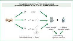
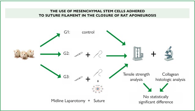
CENTRAL MESSAGE This research aimed to use mesenchymal stem cells derived from adipocytes, attached to a suture thread, to increase tensile strength and collagen formation in the abdominal aponeurosis of rats. Human stem cells derived from adipocytes were attached to a polyglactin 910 suture thread. Thirty-seven Sprague Dawley rats were used, divided into three groups. We concluded that the use of stem cells did not provide any benefit in improving tensile strength or the amount of collagen formed at the site. Future research could be conducted to evaluate whether there would be any benefit in situations of fragility, such as malnutrition, cancer, immunosuppression, or medication use.
PERSPECTIVES The use of stem cells did not show any benefit in improving tensile strength or the amount of collagen formed at the site. However, it is possible that further studies could evaluate the use of this filament, combined with matrices for reconstructive purposes, in areas requiring extensive repairs, such as large hernias where the aponeurosis is insufficient to correct the defect. Furthermore, research could be conducted to assess whether there might be any benefit in situations of fragility, such as malnutrition, cancer, immunosuppression, or medication use.
RESUMO
Racional: Hérnia da parede abdominal é uma condição comum, com incidência de aproximadamente 20%. Estudos recentes demonstraram benefícios do uso de células-tronco, especialmente as mesenquimais, para potencializar a cicatrização tecidual.
Objetivos: Avaliar o uso de células-tronco mesenquimais derivadas de adipócitos, aderidas a um fio de sutura, para aumento da força tênsil e formação de colágeno na aponeurose.
Métodos: Células-tronco humanas derivadas de adipócitos foram aderidas a um fio de sutura. Trinta e sete ratos da espécie Sprague Dawley foram divididos em três grupos: Grupo 1: controle; Grupo 2: sutura convencional no fechamento da aponeurose abdominal; Grupo 3: sutura com células-tronco. Os animais foram avaliados aos 7, 14 e 56 dias após a intervenção.
Resultados: As rupturas ocorreram na linha semilunar e na linha média. Todos os animais dos Grupos 2 e 3, avaliados em D7 e D14, apresentaram ruptura na linha média; em D56, todos os grupos apresentaram ruptura na linha semilunar. A força tênsil foi significativamente menor em D7 nos Grupos 2 e 3 em comparação ao Grupo 1 (p<0,001). Em D14, houve aumento progressivo da força tênsil nos Grupos 2 e 3, mas ainda inferior ao Grupo 1 (p<0,05). Em D56, todos os grupos apresentaram valores semelhantes (p=0,074, p>0.05). Na análise histológica, o Grupo 3 apresentou maiores valores de colágeno em todos os períodos, enquanto o Grupo 2 superou o Grupo 1, embora sem significância estatística (p>0,05). A análise gráfica demonstrou aumento de colágeno em D7 nos Grupos 2 e 3, redução em D14 e estabilização em D56 (p>0,05).
Conclusões: Este estudo não sustenta o uso de células-tronco mesenquimais para aprimorar a cicatrização de incisões abdominais em sujeitos saudáveis. No entanto, é possível que mais estudos avaliem o uso desse filamento, combinado com matrizes para fins reconstrutivos, em áreas que requerem reparos extensos, como grandes hérnias onde a aponeurose é insuficiente para a correção do defeito.
Palavras-chave:
Hérnia Incisional; Aponeurose; Célula-Tronco Mesenquimais; Resistência à Tração; Colágeno
INTRODUCTION
Praxagoras of Kos, a physician from ancient Greece, was the first to consider hernia a surgical pathology, with the earliest historical reports of abdominal wall hernia dating back to the 14th century BC31. Abdominal wall hernia is a globally prevalent disease, with its subtype, incisional hernia, occurring in 10 to 20% of patients after any surgical incision involving the abdominal wall35. Risk factors associated with the occurrence of incisional hernias include obesity (body mass index — BMI>25 kg/m), abdominal aortic aneurysm, and congenital disorders of connective tissue1,14,15,18,20,38.
The pathophysiology of incisional hernia is based on a multifactorial healing defect; the main factors involved are collagen deposition and the healing environment8. The role of collagen in wound healing, particularly in the formation of hernias, became apparent after it was shown that individuals with connective tissue disorders such as Ehlers-Danlos syndrome and Marfan syndrome exhibited a higher incidence of inguinal hernias than the general population22,23. Collagen is the extracellular matrix’s main protein responsible for its tensile strength4,9,37,43.
Stem cells are characterized by their ability to differentiate into various lineages and their high capacity for proliferation and self-regeneration2, and can also give rise to another identical and/or differentiated cell30,36.
Stem cells have been studied for cellular therapy as drug targets, and as sources for generating different tissues for drug testing. A specific type of adult stem cell, mesenchymal stem cells (MSCs), can differentiate into various lineages, exhibit immunomodulatory properties, and demonstrate regenerative potential3,10. The mechanisms of action of MSCs are known to involve paracrine effects through biomolecules, immunomodulatory effects, proliferative capacity, and potential differentiation into other cell lineages5,11,17. Preclinical studies with these cells have shown they possess an excellent capacity for tissue healing, reduce the inflammatory response, and improve many aspects of healing in various animal models10.
Specifically, adipose-derived mesenchymal stem cells have shown excellent cellular proliferation and differentiation capacity, low immunogenicity, and immunomodulatory capacity, making them excellent candidates for therapeutic use7,19,21,28,33,44,46,47. Clinical studies in various fields have been conducted with these cells, such as plastic surgery, orthopedics, immunology, cardiology, oral and maxillofacial surgery, dermatology, connective tissue diseases, neurology, nutrition, and metabolic diseases16,32,34. A recent study showed that using a suture filament with MSCs adhered to it effectively closed enterocutaneous fistulas41.
This study evaluated the role of a suture filament with adhered mesenchymal stem cells in the healing of an abdominal incision in rats.
METHODS
This study was approved by the Animal Ethics Committee (number 5482-1/2020) and Human Research Ethics Committee (number 4.119.745) of the Universidade Estadual de Campinas (UNICAMP).
The MSCs used in the study were obtained from human adipose tissue (AT) of patients undergoing aesthetic liposuction. The lipoaspirated tissue was washed with phosphatebuffered saline (PBS), and, to isolate the MSCs from AT, enzymatic digestion was performed using collagenase type 1A (Gibco Invitrogen Corporation, Grand Island — New York — USA). After the enzymatic action of collagenase, lysis of red blood cells, and successive washes with PBS, the isolated cells were cultured in Dulbecco’s Modified Eagle Medium (DMEM) low glucose (Gibco Invitrogen) with 10% fetal bovine serum (VitroCell Embriolife, Campinas, São Paulo, Brazil). The cells were incubated at 37 °C in a humid atmosphere containing 5% CO₂. Cells expanded up to the fourth passage of culture were confirmed to be MSCs by adherence to plastic, absence of Cluster of Differentiation 34 (CD34) and CD45 expression, and positive expression of CD73, CD90, and CD105, characterized by flow cytometry using specific monoclonal antibodies. The genetic stability of these cells was assessed through karyotyping and telomerase enzyme activity (TRAPeze® Telomerase Detection Kit, S7700 — Chemicon International, USA).
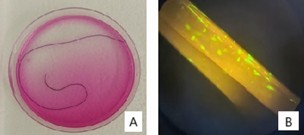
After the mesenchymal stem cells were prepared, they were adhered to the suture filament. The filament used was polyglactin 910, 15 cm long, with a thickness of 3-0 (Johnson and Johnson, reference J316, Vicryl™). One million MSCs were adhered to each suture filament using a glue made with thrombin and calcium gluconate in a 3:1 ratio, and they were kept in DMEM low glucose culture medium (Gibco Invitrogen) with 10% fetal bovine serum (VitroCell Embriolife, Campinas, São Paulo, Brazil). The filaments were incubated at 37 °C in a humid atmosphere containing 5% CO₂, and were observed every 24 hours. After 72 hours of culture, the filaments were used to treat the animals. To confirm the attachment of the mesenchymal stem cells to the suture filament and their viability after the fixation process with glue, a Live and Dead experiment was conducted (Invitrogen™ LIVE/DEAD™ Viability/Cytotoxicity Assay Kit — Green/Deep Red). Figure 1 shows the filament with the stem cells after preparation, immediately before animal use, and demonstrates the Live and Dead assay under fluorescent light.
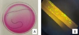
Suture Filament with Stem Cells. A: Vicryl™ filament with stem cells adhered in the culture medium. B: Live and Dead assay under fluorescent microscopy. Demonstration of living mesenchymal stem cells adhered to the suture filament.
G*Power 3.1 software, from Heinrich Heine Universität — Dussledorf, was used to calculate the sample size. Taking an elevation of 50% on tension force as clinically significant and using a significance level alpha=0.05 and a power of 0.8, it was calculated that, at each of the time points, each group should have at least four animals. As there were three time points to be studied and accounting for a 10–15% mortality of the animals, five animals were assigned to each group at each time point. As the control group would be using non-operated animals, and in the name of the 3Rs (replacement, reduction, and refinement) of animal use in experimental surgery, it was supposed that only one group of three animals would suffice to serve as a control for the whole experiment.
The experiment was performed with 37 male Sprague Dawley rats, aged eight weeks, weighing approximately 300 grams. All animals were housed in appropriately sized plastic boxes, with a 12-hour light/dark cycle, temperature 20±2°C, and ambient humidity, with free access to water and standard commercial food. All animals used in the study were obtained from the Multidisciplinary Center for Biological Research at UNICAMP, following approval of the study by the Animal Ethics Committee (number 54821/2020) of the UNICAMP.
Group 1 (control): 3 rats;
Group 2 (Vicryl™): 17 rats;
Group 3 (Vicryl™ with adhered mesenchymal stem cells): 17 rats.
The day (D) of the intervention was considered D0, with evaluations conducted on days D7, D14, and D56 following the surgery (D0).
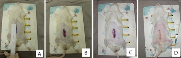
In Group 1, no intervention was performed; the animals were kept under the same conditions as the others. In Groups 2 and 3, the following surgical procedure was carried out: all animals (17+17) underwent general anesthesia, followed by a midline abdominal skin incision of approximately 4 cm. After the skin was opened, a 4 cm incision was made in the midline of the aponeurosis, with the peritoneum opened simultaneously, exposing the abdominal cavity. Subsequently, the aponeurosis was sutured using the following technique: a continuous suture encompassing the peritoneum and aponeurosis in a single layer, maintaining a suture-to-incision length ratio of 4:1, and using closely spaced stitches.
In Group 2, standard Vicryl™ 3-0 suture filament (Johnson & Johnson Medical Brazil, São Paulo, Brazil, reference J316), was used without modifications. In Group 3, the same Vicryl™ 3-0 filament, reference J316) was used, but with mesenchymal stem cells adhered to it, as described previously. The skin was then closed using Mononylon™ 4-0 filament (reference 14502, Johnson & Johnson Medical Brazil, São Paulo, Brazil), with a continuous suture in both groups.
Euthanasia was performed on days D7, D14, and D56 by deepening general anesthesia, using thiopental, at a dose of 25 mg intravenously, in accordance to the Animal Ethics Committee of the UNICAMP. After euthanasia, the Mononylon™ filament was removed from the skin, which was opened to expose the aponeurosis. A visual inspection detected any signs of secretion, infection, local reaction, and aponeurosis integrity. A segment of the aponeurosis, measuring 6 cm in width and 4 cm in length around the previous incision, was resected using a mold to standardize specimen collection. The segment was divided into cranial (1 cm) and caudal (3 cm) portions. The caudal segment was placed in saline solution and immediately sent for tensile strength analysis, while the cranial segment was placed in 10% buffered formalin for histological analysis. Figure 2 illustrates the procedures performed on the animals.
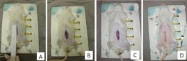
Sequence of Procedures Performed on Animals. A: Anesthetized rat; midline incision marking using a ruler; measurement of 4 cm. B: Midline incision performed with complete opening to the peritoneal cavity. C: Aponeurosis closure with "Vicryl" filament. D: Final aspect after aponeurosis and skin closure.
Group 1: three animals, with one animal at each time point (D7, D14 and D56);
Groups 2 and 3: six animals per group at D7; six animals per group at D14; and five animals per group at D56.
The variables analyzed included the tensile strength of the aponeurosis and collagen quantification using Masson’s Trichrome and Picrosirius Red staining.
Tensile strength tests were performed after removing the suture filament. A universal testing machine (Texture Analyzer, Model TA500) with a 500 N load cell (Lloyd Instruments) was used, operating at a constant traction speed of 0.6 mm/s. Atraumatic Collin-type gripping clamps were attached to the specimen’s extremities, with the clamp tips positioned laterally to the semilunar line.
For histological analysis, the aponeuroses from all animals were collected and fixed in 10% buffered formalin for 12 hours. After fixation, the tissues were washed in 70% ethanol and dehydrated in an ascending alcohol series. The fragments were cleared with xylene for two hours and embedded in plastic polymers (Paraplast Plus, St. Louis, MO, USA). The samples were sectioned using a Slee CUT 5062 RM 2165 rotary microtome (Slee Mainz, Mainz, Germany) at 5-micrometer thickness, stained with Picrosirius Red and Masson’s Trichrome, and photographed with a Leica DM2500 photomicroscope (Leica, Munich, Germany) equipped with a DFC295 camera (Leica, Munich, Germany).
All samples were photographed as follows: one image at 40x magnification covering the entire midline of the aponeurosis, and three sequential images at 100x magnification, scanning the midline from edge to edge. Collagen quantification was performed by counting 80 points per 100x magnification field in the three images of the midline of each sample stained with Picrosirius Red, with an area of 15,000 pixels per frame, using the grid mode of ImageJ software.
Statistical analyses were conducted using the JASP software, version 0.18.3 (University of Amsterdam, Netherlands). Statistical parameters included the median, mean, standard deviation, and maximum and minimum values. Analysis of variance (ANOVA) was used to analyze these parameters at different time points for continuous variables. A significance level of 5% and a statistical power of 80% were considered for determining statistical significance. In all analyses, measurements were categorized by days (D7, D14, and D56). At each time point, the three groups were compared using ANOVA. If p<0.05, a posthoc Tukey test was performed to identify which groups presented significant differences.
RESULTS
Tensile strength analysis
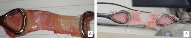
Regarding the tensile strength analysis of the aponeurosis fragments, the tissue ruptured at the semilunar line and the midline (Figure 3).
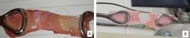
Specimen detail after rupture. A: Detail of the specimen after rupture at the semilunar line. The red circle marks the midline (no rupture), while the black circle marks the semilunar line (ruptured). B: Detail of the specimen after rupture at the midline.
In Group 1, the aponeurosis of all animals ruptured at the semilunar line. In the animals subjected to intervention (Groups 2 and 3), the rupture occurred at the midline (incision site) on days D7 and D14. Finally, on D56, all animals exhibited ruptures at the semilunar line.
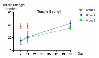
Graphical analysis of tensile strength revealed that, on D7, the tensile strength in Groups 2 and 3 was significantly lower than in Group 1 (p<0.001). A progressive and similar increase in tensile strength was observed in Groups 2 and 3 on D14. However, tensile strength remained significantly lower than in Group 1 (p<0.05). By D56, all groups exhibited very similar values, with no statistically significant differences (p=0.074). Notably, on D7 and D14, there were no significant differences between Groups 2 and 3 (p>0.05) (Figure 4).
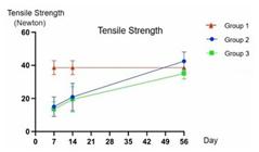
Representation of tensile strength over time: days 7, 14, and 56, comparing the groups. Tensile strength in Newtons (GraphPad Prism v10.2). D7: p<0.001; D14: p<0.05; D56: p=0.074.
Table 1 presents a descriptive analysis of tensile strength, divided by group and time point. The study compared all groups simultaneously, and, when statistical significance was detected, pairwise comparisons were performed to identify which groups exhibited significant differences. Groups 2 and 3 did not show a statistically significant difference on D7 and D14 when compared to each other (p>0.05). However, both showed a significant difference compared to Group 1 at the same time points (p<0.05). Nevertheless, on D56, there was no statistically significant difference among the groups (p>0.05) (Table 1).
Histological analysis
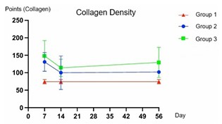
The graphical analysis indicated that Groups 2 and 3 experienced an increase in collagen on D7, followed by a decline on D14, and then stabilized at similar levels on D56. Despite these trends, no significant differences were observed among the groups at any time (p>0.05, Figure 5).
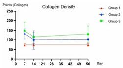
Representation of collagen density in the specimens over time: days 7, 14, and 56, comparing the groups. Points indicate the number of collagen-positive areas in the sample (GraphPad Prism v10.2). D7, D14 and D56: p>0.05.
Based on the count of collagen-positive points, the histological analysis of specimens demonstrated that Group 3 had the highest mean collagen content at all time points (Table 2). Group 2 also showed higher collagen content than Group 1 at all time points (Table 2). However, these differences were not statistically significant, as shown in Figure 5 and Table 2 (p>0.05).
Collagen density: descriptive analysis, separated by group and period. No statistically significant difference was observed among the groups (p>0.05).
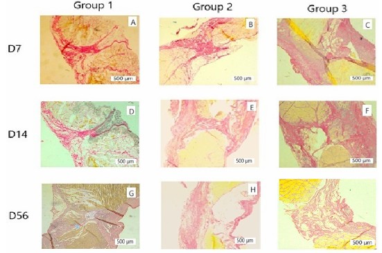
Below are representative histological images of the midline of specimens from all groups at different time points (Figure 6).
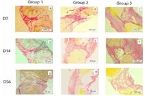
Representative photomicrographs of histological analyses of tissue samples stained with Picrosirius Red, organized by groups and analyzed periods. A, B, C: representative of D7, corresponding to groups 1, 2, and 3, respectively; D, E, F: representative of D14, corresponding to groups 1, 2, and 3, respectively; G, H, I: representative of D56, corresponding to groups 1, 2, and 3, respectively. Scale bar: 500 μm.
DISCUSSION
This study evaluated the potential effects of using mesenchymal stem cells derived from adipocytes to enhance tensile strength and collagen formation in rat aponeurosis. However, these parameters showed no improvement when stem cells were added to healthy rats.
Considering that adipose-derived mesenchymal stem cells have the ability to migrate to the injured site, we chose to use the suture coated with stem cells as their carrier because we had previously used this method, had experience with it, and had obtained satisfactory results.
The lack of difference observed between Groups 2 and 3 could raise questions as to whether the stem cells were indeed present on the suture and, if so, whether they were viable. To address this concern, we performed a Live and Dead assay, which confirmed the presence and viability of the stem cells on the suture.
One hypothesis for the lack of improvement is that wound healing is already optimized under normal conditions. Stem cells might be more beneficial in improving the healing of compromised tissues, such as those exposed to radiotherapy or in diabetic individuals6.
Most related research involves scaffolds or matrices populated with stem cells, but none have utilized suture filaments with adhered stem cells, as in the present study.
A study in Sprague-Dawley rats compared the use of sponges with and without bone marrow-derived stem cells. No differences in tensile strength between groups were found four weeks post-implantation42. Similarly, another study using collagen vehicles to deliver bone marrow-derived stem cells to rabbit aponeurosis did not report significant differences in tensile strength after eight weeks of implantation26.
The present results confirm previous findings by different authors. Mestak et al. used extracellular matrices with or without adipose tissue-derived mesenchymal stem cells in Wistar rats. After three months, their findings were mixed — some showed improved tensile strength, while others did not. The authors concluded that the use of stem cells was not advantageous29.
Conversely, Heffner et al. reported a statistically significant improvement in tensile strength after four and eight weeks when combining mesenchymal stem cells, platelet-rich plasma, and bovine collagen matrices in rats. This suggests a synergistic effect of the multiple components13.
Hansen et al.12 evaluated different meshes for pelvic floor reconstruction and observed an effect of the mesh itself on tensile strength. However, after mesh resorption at 24 weeks, there was no difference in tensile strength between groups with and without mesenchymal stem cells.
A study by van Steenberghe et al.39 used adipose- and bone marrow-derived mesenchymal stem cells adhered to a collagen matrix made from human aponeurosis implanted into rat abdominal walls. After 30 days, no difference in tensile strength was observed between groups with or without stem cells. However, the adipose-derived stem cell group significantly increased tissue vascularization.
A comprehensive review of mesenchymal stem cell use, particularly adipose-derived stem cells, in healing tendon injuries across species (rabbits and horses) reported improved tissue organization but inconsistent results in acute injuries and when combined with platelet-rich plasma40.
Another review analyzed stem cells attached to various types of mesh. Studies using biological and synthetic meshes found no improvement in tensile strength with stem cells, although vascularization did improve. The authors hypothesized that the inherent benefits of meshes could provide sufficient results27.
Finally, the two preceding studies that utilized this suture filament with adhered mesenchymal stem cells demonstrated its healing potential. The first study evaluated the efficacy of this suture filament in treating enterocutaneous fistulas. In contrast, the second combined the suture filament with a matrix containing mesenchymal stem cells for tracheal fistula repair. Both studies showed that this stem cell-enriched filament provided a statistically significant benefit in the healing process25,41. However, the main difference between these earlier and the present study lies in the tissues evaluated. The earlier studies primarily focused on epithelial tissues, such as the epidermis and respiratory epithelium, whereas this study evaluated connective tissue, specifically the aponeurosis.
Mestak et al. supported the hypothesis that stem cells exert their effects primarily on epithelial tissues. They demonstrated that rats treated with adipose-derived mesenchymal stem cells for abdominal wall hernia repair showed improved healing in the peritoneum but not in the aponeurosis29. Another study reported enhanced peritoneal formation with mesenchymal stem cell application45. Furthermore, Liu et al. found that adipose-derived mesenchymal stem cells improved epithelialization rates and collagen formation in cutaneous wounds in rats24.
Our study’s limitations include its exclusive focus on aponeurosis, without evaluating the peritoneum. Additionally, the histological analysis was limited to quantitative collagen assessment, without a qualitative evaluation of collagen types or other markers such as vascular endothelial growth factor (VEGF) and metalloproteinases, which are known to be influenced by stem cells.
Future studies could investigate the types of collagen, distinguishing between types I and III, as well as analyzing inflammatory markers and angiogenesis markers such as VEGF. Another possibility is the use of this mesenchymal stem cellenriched filament in immunocompromised animals, such as those subjected to chemotherapy, radiotherapy, or immunosuppressive medications. Moreover, this filament could be combined with matrices for reconstructive purposes in areas requiring extensive repair, such as large hernias where the aponeurosis is insufficient for defect correction.
CONCLUSIONS
The present study’s data do not show that the use of mesenchymal stem cells improves the healing of a midline abdominal incision in otherwise healthy subjects. However, an option for future studies is to employ this filament, combined with matrices for reconstructive purposes, in areas requiring extensive repair, such as large hernias where the aponeurosis is insufficient for defect correction.
DATA AVAILABILITY
The Informations regarding the investigation, methodology and data analysis of the article are archived under the responsibility of the authors.
HOW TO CITE THIS ARTICLE
Kalil J, Bustorff-Silva JM, Calderoni DR, Leme KC, Camargo GCA, Fávaro WJ, et al. ABCD Arq Bras Cir Dig. 2026;39:e1931. https://doi.org/10.1590/0102-67202026000002e1931.
REFERENCES
-
1 Antoniou GA, Georgiadis GS, Antoniou SA, Granderath FA, Giannoukas AD, Lazarides MK. Abdominal aortic aneurysm and abdominal wall hernia as manifestations of a connective tissue disorder. J Vasc Surg. 2011;54(4):1175-81. https://doi.org/10.1016/j.jvs.2011.02.065
» https://doi.org/10.1016/j.jvs.2011.02.065 -
2 Biondo-Simões ML, Morais CG, Tocchio AF, Miranda RA, Moura PA, Colla K, et al. Characteristics of the fibroplasia and collagen expression in the abdominal wall after implant of the polypropylene mesh and polypropylene/polyglecaprone mesh in rats. Acta Cir Bras. 2016;31(5):294-9. https://doi.org/10.1590/S0102-865020160050000001
» https://doi.org/10.1590/S0102-865020160050000001 -
3 Caplan AI, Dennis JE. Mesenchymal stem cells as trophic mediators. J Cell Biochem 2006;98(5):1076-84. https://doi.org/10.1002/jcb.20886
» https://doi.org/10.1002/jcb.20886 -
4 Casanova AB, Trindade EN, Trindade MR. Collagen in the transversalis fascia of patients with indirect inguinal hernia: a case-control study. Am J Surg. 2009;198(1):1-5. https://doi.org/10.1016/j.amjsurg.2008.07.021
» https://doi.org/10.1016/j.amjsurg.2008.07.021 -
5 Chamberlain G, Fox J, Ashton B, Middleton J. Concise review: mesenchymal stem cells: their phenotype, differentiation capacity, immunological features, and potential for homing. Stem Cells. 2007;25(11):2739-49. https://doi.org/10.1634/stemcells.2007-0197
» https://doi.org/10.1634/stemcells.2007-0197 -
6 Chen Y, Zhang Q, Wu Y, Branch-Brooks CD, Butler CE. Short-term influences of radiation on musculofascial healing in a laparotomy rat model. Sci Rep. 2019;9(1):11896. https://doi.org/10.1038/s41598-019-48201-5
» https://doi.org/10.1038/s41598-019-48201-5 -
7 Cui L, Yin S, Liu W, Li N, Zhang W, Cao Y. Expanded adipose-derived stem cells suppress mixed lymphocyte reaction by secretion of prostaglandin E2. Tissue Eng 2007;13(6):1185e95. https://doi.org/10.1089/ten.2006.0315
» https://doi.org/10.1089/ten.2006.0315 -
8 DuBay DA, Choi W, Urbanchek MG, Wang X, Adamson B, Dennis RG, et al. Incisional herniation induces decreased abdominal wall compliance via oblique muscle atrophy and fibrosis. Ann Surg. 2007;245(1):140-6. https://doi.org/10.1097/01.sla.0000251267.11012.85
» https://doi.org/10.1097/01.sla.0000251267.11012.85 -
9 Ferrarin DD, Malafaia O, Czeczko NG, Kubrusly LF, Sigwalt MF, Sousa EL, et al. Use of decellularized human amniotic membrane in intestinal anastomosis: a study in rats treated with 5-fluorouracil. Arq Bras Cir Dig. 2024;37:e1843. https://doi.org/10.1590/0102-6720202400049e1843
» https://doi.org/10.1590/0102-6720202400049e1843 -
10 Galipeau J, Sensébé L. Mesenchymal stromal cells: clinical challenges and therapeutic opportunities. Cell Stem Cell. 2018;22(6):824-33. https://doi.org/10.1016/j.stem.2018.05.004
» https://doi.org/10.1016/j.stem.2018.05.004 -
11 Gnecchi M, He H, Noiseux N, Liang OD, Zhang L, Morello F, et al. Evidence supporting paracrine hypothesis for Aktmodified mesenchymal stem cell-mediated cardiac protection and functional improvement. FASEB J. 2006;20(6):661-9. https://doi.org/10.1096/fj.05-5211com
» https://doi.org/10.1096/fj.05-5211com -
12 Hansen SG, Taskin MB, Chen M, Wogensen L, Vinge Nygaard J, Axelsen SM. Electrospun nanofiber mesh with fibroblast growth factor and stem cells for pelvic floor repair. J Biomed Mater Res B Appl Biomater. 2020;108(1):48-55. https://doi.org/10.1002/jbm.b.34364
» https://doi.org/10.1002/jbm.b.34364 -
13 Heffner JJ, Holmes JW, Ferrari JP, Krontiris-Litowitz J, Marie H, Fagan DL, et al. Bone marrow-derived mesenchymal stromal cells and platelet-rich plasma on a collagen matrix to improve fascial healing. Hernia. 2012;16(6):677-87. https://doi.org/10.1007/s10029-012-0941-2
» https://doi.org/10.1007/s10029-012-0941-2 -
14 Henriksen NA, Helgstrand F, Vogt KC, Jorgensen LN, Bisgaard T; Danish Hernia Database; Danish Vascular Registry. Risk factors for incisional hernia repair after aortic reconstructive surgery in a nationwide study. J Vasc Surg. 2013;57(6):1524-1530.E3. https://doi.org/10.1016/j.jvs.2012.11.119
» https://doi.org/10.1016/j.jvs.2012.11.119 -
15 Höer J, Lawong G, Klinge U, Schumpelick V. Factors influencing the development of incisional hernia. A retrospective study of 2,983 laparotomy patients over a period of 10 years. Chirurg. 2002;73(5):474-80. https://doi.org/10.1007/s00104-002-0425-5
» https://doi.org/10.1007/s00104-002-0425-5 -
16 Hur JW, Cho T-H, Park D-H, Lee J-B, Park J-Y, Chung Y-G. Intrathecal transplantation of autologous adipose-derived mesenchymal stem cells for treating spinal cord injury: a human trial. J Spinal Cord Med. 2016;39(6):655-64. https://doi.org/10.1179/2045772315Y.0000000048
» https://doi.org/10.1179/2045772315Y.0000000048 -
17 Jackson WM, Nesti LJ, Tuan RS. Concise review: clinical translation of wound healing therapies based on mesenchymal stem cells. Stem Cells Transl Med. 2012;1(1):44-50. https://doi.org/10.5966/sctm.2011-0024
» https://doi.org/10.5966/sctm.2011-0024 -
18 Judge DP, Dietz HC. Marfan’s syndrome. Lancet. 2005;366(9501):1965-76. https://doi.org/10.1016/S0140-6736(05)67789-6
» https://doi.org/10.1016/S0140-6736(05)67789-6 -
19 Kronsteiner B, Wolbank S, Peterbauer A, Hackl C, Redl H, van Griensven M, et al. Human mesenchymal stem cells from adipose tissue and amnion influence T-cells depending on stimulation method and presence of other immune cells. Stem Cell Dev. 2011;20(12):2115-26. https://doi.org/10.1089/scd.2011.0031
» https://doi.org/10.1089/scd.2011.0031 -
20 Kroese LF, Mommers EH, Robbens C, Bouvy ND, Lange JF, Berrevoet F. Complications and recurrence rates of patients with Ehlers-Danlos syndrome undergoing ventral hernioplasty: a case series. Hernia. 2018;22(4):611-6. https://doi.org/10.1007/s10029-018-1739-7
» https://doi.org/10.1007/s10029-018-1739-7 -
21 Lee JM, Jung J, Lee H-J, Jeong SJ, Cho KJ, Hwang S-G, et al. Comparison of immunomodulatory effects of placenta mesenchymal stem cells with bone marrow and adipose mesenchymal stem cells. Int Immunopharm 2012;13(2):219-24. https://doi.org/10.1016/j.intimp.2012.03.024
» https://doi.org/10.1016/j.intimp.2012.03.024 -
22 Lehnert B, Wadouh F. High coincidence of inguinal hernias and abdominal aortic aneurysms. Ann Vasc Surg. 1992;6(2):134-7. https://doi.org/10.1007/BF02042733
» https://doi.org/10.1007/BF02042733 -
23 Liem MS, van der Graaf Y, Beemer FA, van Vroonhoven TJ. Increased risk for inguinal hernia in patients with EhlersDanlos syndrome. Surgery. 1997;122(1):114-5. https://doi.org/10.1016/s0039-6060(97)90273-7
» https://doi.org/10.1016/s0039-6060(97)90273-7 -
24 Liu Y, Liu Y, Wu M, Zou R, Mao S, Cong P, et al. Adiposederived mesenchymal stem cell-loaded -chitin nanofiber hydrogel promote wound healing in rats. J Mater Sci Mater Med. 2022;33(2):12. https://doi.org/10.1007/s10856-021-06630-7
» https://doi.org/10.1007/s10856-021-06630-7 -
25 Manzini BM, Dávila JL, Volpe BB, Duarte ADSS, Côrtez MTF, Duek EAR, et al. Poly (L-Lactic Acid) cell-laden scaffolds applied on swine model of tracheal fistula. J Surg Res. 2022;277:319-34. https://doi.org/10.1016/j.jss.2022.03.024
» https://doi.org/10.1016/j.jss.2022.03.024 -
26 Marie H, Zhang Y, Heffner J, Dorion HA, Fagan DL. Biomechanical and elastographic analysis of mesenchymal stromal cell treated tissue following surgery. J Biomech Eng. 2010;132(7):074503. https://doi.org/10.1115/1.4001382
» https://doi.org/10.1115/1.4001382 -
27 Marinaro F, Sánchez-Margallo FM, Álvarez V, López E, Tarazona R, Brun MV, et al. Meshes in a mess: Mesenchymal stem cell-based therapies for soft tissue reinforcement. Acta Biomater. 2019;85:60-74. https://doi.org/10.1016/j.actbio.2018.11.042
» https://doi.org/10.1016/j.actbio.2018.11.042 -
28 McIntosh K, Zvonic S, Garrett S, Mitchell JB, Floyd ZE, Hammill L, et al. The immunogenicity of human adipose-derived cells: temporal changes in vitro. Stem Cells. 2006;24(5):1246-53. https://doi.org/10.1634/stemcells.2005-0235
» https://doi.org/10.1634/stemcells.2005-0235 -
29 Mestak O, Matouskova E, Spurkova Z, Benkova K, Vesely P, Mestak J, et al. Mesenchymal stem cells seeded on crosslinked and noncross-linked acellular porcine dermal scaffolds for long-term full-thickness hernia repair in a small animal model. Artif Organs. 2014;38(7):572-9. https://doi.org/10.1111/aor.12224
» https://doi.org/10.1111/aor.12224 -
30 Morrison SJ, Kimble J. Asymmetric and symmetric stem-cell divisions in development and cancer. Nature. 2006;441:1068-74. https://doi.org/10.1038/nature04956
» https://doi.org/10.1038/nature04956 -
31 Papavramidou NS, Christopoulou-Aletras H. Treatment of "hernia" in the writings of Celsus (first century AD). World J Surg. 2005;29(10):1343-7. https://doi.org/10.1007/s00268-005-7808-y
» https://doi.org/10.1007/s00268-005-7808-y -
32 Pers Y-M, Rackwitz L, Ferreira R, Pullig O, Delfour C, Barry F, et al. Adipose mesenchymal stromal cell-based therapy for severe osteoarthritis of the knee: A phase I dose-escalation trial. Stem Cells Transl Med. 2016;5(7):847-56. https://doi.org/10.5966/sctm.2015-0245
» https://doi.org/10.5966/sctm.2015-0245 -
33 Puissant B, Barreau C, Bourin P, Clavel C, Corre J, Bousquet C, et al. Immunomodulatory effect of human adipose tissue-derived adult stem cells: comparison with bone marrow mesenchymal stem cells. Br J Haematol 2005;129(1):118-29. https://doi.org/10.1111/j.1365-2141.2005.05409.x
» https://doi.org/10.1111/j.1365-2141.2005.05409.x -
34 Qayyum AA, Mathiasen AB, Mygind ND, Kühl JT, Jørgensen E, Helqvist S, et al. Adipose-derived stromal cells for treatment of patients with chronic ischemic heart disease (MyStromalCell trial): a randomized placebocontrolled study. Stem Cell Int. 2017;2017. https://doi.org/10.1155/2017/5237063
» https://doi.org/10.1155/2017/5237063 -
35 Schreinemacher MH, Emans PJ, Gijbels MJ, Greve JW, Beets GL, Bouvy ND. Degradation of mesh coatings and intraperitoneal adhesion formation in an experimental model. Br J Surg. 2009;96(3):305-13. https://doi.org/10.1002/bjs.6446
» https://doi.org/10.1002/bjs.6446 -
36 Tajbakhsh S, Rocheteau P, Le Roux I. Asymmetric cell divisions and asymmetric cell fates. Annu Rev Cell Dev Biol. 2009;25:671-99. https://doi.org/10.1146/annurev.cellbio.24.110707.175415
» https://doi.org/10.1146/annurev.cellbio.24.110707.175415 -
37 Tustumi F, Darce GFB, Lobo Filho MM, Abdalla RZ, Costa TN. Stapled fascial closure vs. Continuous hand-sewn suture: experimental study of the abdominal wall on porcine model and human cadaver. Arq Bras Cir Dig. 2024;37:e1800. https://doi.org/10.1590/0102-672020240007e1800
» https://doi.org/10.1590/0102-672020240007e1800 -
38 Udén A, Lindhagen T. Inguinal hernia in patients with congenital dislocation of the hip. A sign of general connective tissue disorder. Acta Orthop Scand. 1988;59(6):667-8. https://doi.org/10.3109/17453678809149421
» https://doi.org/10.3109/17453678809149421 -
39 van Steenberghe M, Schubert T, Guiot Y, Goebbels RM, Gianello P. Improvement of mesh recolonization in abdominal wall reconstruction with adipose vs. bone marrow mesenchymal stem cells in a rodent model. J Pediatr Surg. 2017;52(8):1355-62. https://doi.org/10.1016/j.jpedsurg.2016.11.041
» https://doi.org/10.1016/j.jpedsurg.2016.11.041 -
40 Veronesi F, Salamanna F, Tschon M, Maglio M, Aldini NN, Fini M. Mesenchymal stem cells for tendon healing: what is on the horizon? J Tissue Eng Regen Med. 2017;11(11):3202-19. https://doi.org/10.1002/term.2209
» https://doi.org/10.1002/term.2209 -
41 Volpe BB, Ada SSD, Ribeiro TB, Stocchero I, Kharmandayan P, Saad STO, et al. Mesenchymal stromal cells from adipose tissue attached to suture material enhance the closure of enterocutaneous fistulas in a rat model. Cytotherapy. 2014;16(12):1709-19. https://doi.org/10.1016/j.jcyt.2014.08.004
» https://doi.org/10.1016/j.jcyt.2014.08.004 -
42 Wang D, Ji Z-L, Wang J-M, Tan Y-Y. Bone morphogenetic protein-12 inducing tenogenic differentiation of mesenchymal stem cells enhances healing of linea alba incision. Exp Ther Med. 2018;16(6):5067-72. https://doi.org/10.3892/etm.2018.6861
» https://doi.org/10.3892/etm.2018.6861 - 43 White B, Osier C, Gletsu N, Jeansonne L, Baghai M, Sherman M, et al. Abnormal primary tissue collagen composition in the skin of recurrent incisional hernia patients. Am Surg. 2007;73(12):1254-8.
-
44 Yoo KH, Jang IK, Lee MW, Kim HE, Yang MS, Eom Y, et al. Comparison of immunomodulatory properties of mesenchymal stem cells derived from adult human tissues. Cell Immunol. 2009;259(2):150-6. https://doi.org/10.1016/j.cellimm.2009.06.010
» https://doi.org/10.1016/j.cellimm.2009.06.010 -
45 Zhao Y, Zhang Z, Wang J, Yin P, Zhou J, Zhen M, et al. Abdominal hernia repair with a decellularized dermal scaffold seeded with autologous bone marrow-derived mesenchymal stem cells. Artif Organs. 2012;36(3):247-55. https://doi.org/10.1111/j.1525-1594.2011.01343.x
» https://doi.org/10.1111/j.1525-1594.2011.01343.x -
46 Zuk PA, Zhu M, Ashjian P, Ugarte DA, Huang JI, Mizuno H, et al. Human adipose tissue is a source of multipotent stem cells. Mol Biol Cell. 2002;13(12):4279-95. https://doi.org/10.1091/mbc.e02-02-0105
» https://doi.org/10.1091/mbc.e02-02-0105 -
47 Zuk PA, Zhu M, Mizuno H, Huang J, Futrell JW, Katz AJ, et al. Multilineage cells from human adipose tissue: implications for cell-based therapies. Tissue Eng. 2001;7(2):211-28. https://doi.org/10.1089/107632701300062859
» https://doi.org/10.1089/107632701300062859
Edited by
-
Editor:
Nelson Adami Andreollo https://orcid.org/0000-0001-7452-1165