 Spitz nevus in a patient with oculocutaneous albinism: dermoscopic and histopathologic correlation
Dear Editor,
In oculocutaneous albinism (OCA), an autosomal recessive disorder characterized by total or partial absence of melanin production, melanocytic lesions pose a diagnostic chal-lenge due to their atypical dermoscopic presentation.1 The present report describes a patient with OCA followed by dig-ital mapping, in whom a Spitz nevus (SN) was identified and confirmed by histopathologic examination.
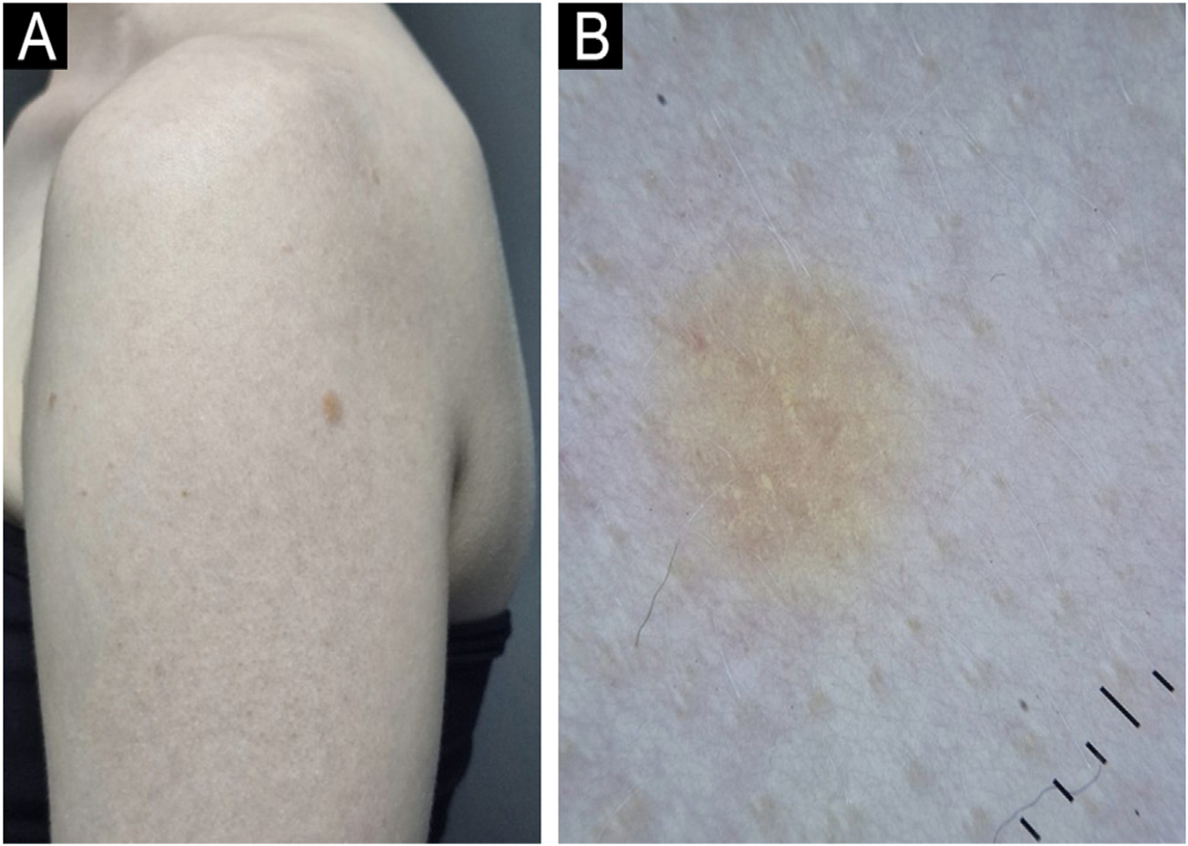
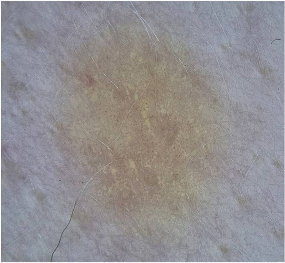
A 25-year-old female patient with OCA, with no family history of albinism or other skin diseases, underwent digital dermoscopic follow-up at a tertiary dermatology center due to multiple atypical nevi. During the dermoscopic follow-up, a 0.4 cm light brown papule was identified on the left upper limb. Dermoscopy revealed globular structures with a yel-lowish appearance and uniform distribution throughout the lesion (Fig. 1) and fine punctate vessels, distributed globally and symmetrically (Fig. 2). Given the atypical appearance of the lesion, excision was chosen. The lesion area had been examined approximately one year prior without any lesion being detected.

(A) Patient with oculocutaneous albinism presenting with a 0.4 cm non-pigmented nevus on the left upper limb. (B) Dermoscopy with polarized light reveals yellowish structures in the central region.
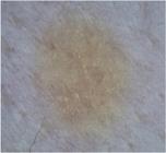
Higher dermoscopic magnification with polarized light showing millimeter-sized punctate vessels, distributed globally and symmetrically throughout the lesion.
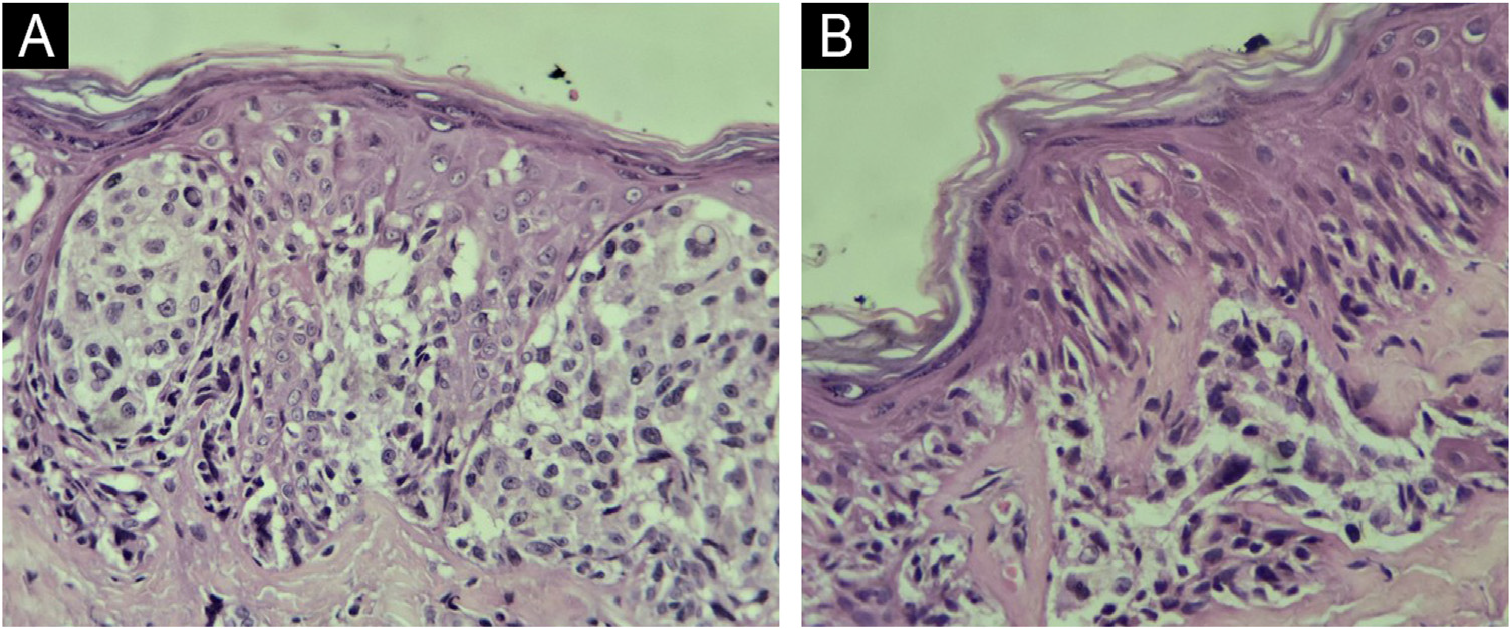
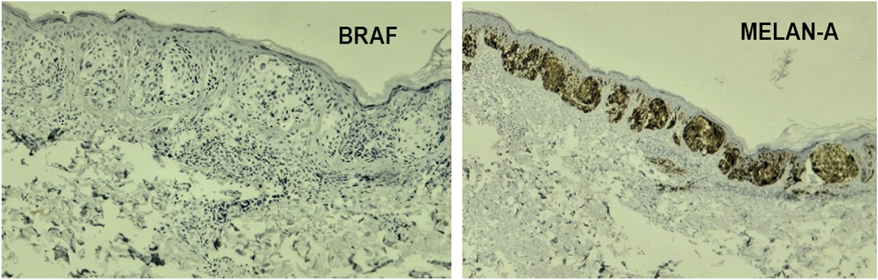
The evaluation of the histological sections revealed a well-defined, compound melanocytic proliferation, formed by large and varied nests of epithelioid and fusiform melanocytes. The dermal component was restricted to the superficial reticular dermis. Some cells showed ample cyto-plasm, sometimes granular, with discrete cytological atypia. Junctional nests showed separation gaps in relation to the adjacent epidermis, in addition to eosinophilic globular structures compatible with Kamino bodies. Subtle acan-thosis was observed, without mitotic activity or pagetoid dissemination of melanocytes. There was no melanocytic pigmentation (Fig. 3). Immunohistochemistry showed pre-served p16 expression, negative BRAF test, and positivity for Melan-A (Fig. 4). The test was reviewed by two experi-enced dermatopathologists, who confirmed the diagnosis of Spitz nevus.
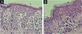
Patient’s histopathologic examination. (A) Large nests of epithelioid and spindle cells with ample cytoplasm. Slight acanthosis can be observed. (Hematoxylin & eosin, ×100). (B) Irregular nests of epithelioid and spindle cells, presence of Kamino bodies (Hematoxylin & eosin, ×100).
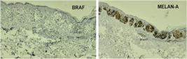
Immunohistochemistry examination of the patient: left showing negative BRAF staining in cells of interest (BRAF ×10); right showing positive Melan A staining in cells of interest (Melan A ×10).
Spitz nevus is a melanocytic neoplasm consisting of epithelioid and/or spindle cells. Clinically, it presents as a solitary, well-defined papule or nodule, usually <1 cm, with rapid initial growth and predominance on the limbs of young adults. The classic dermoscopic presentation involves a punctate vascular pattern in about 50% of cases, with reg-ularly distributed monomorphic vessels on a homogeneous pink background. Other, less common patterns include retic-ular or homogeneous depigmentation.2
Histologically, it is characterized by symmetry, well-defined delimitation, presence of epithelioid/fusiform melanocytes, progressive dermal maturation, mild cytologi-cal atypia, Kamino bodies, occasional mitoses, and discrete lymphocytic inflammatory infiltrate.3,4 The preserved p16 expression observed in the patient reinforces the lesion’s benignity, since its loss is associated with malignant Spit-zoid neoplasms. Melan-A, in turn, confirmed the melanocytic nature.5
From a dermoscopic point of view, the yellowish globules observed may correspond to the melanocytic nests identified histologically, whose absence of pigment in the context of OCA results in a yellowish hue instead of the homogeneous pink background coloration usually observed.
Regarding molecular biology, activating mutations in BRAF and NRAS are known to be rare in Spitz nevi, although age may influence their occurrence. Fusions involving BRAF are described in about 5% of epithelioid lesions, and a small percentage may evolve into melanoma.6,7 The neg-ative result for BRAF in this case is consistent with the expected profile for Spitz nevi, which usually show gene fusions that are not detectable by this method.8
Despite the extensive description of Spitz nevi in the lit-erature, there is still a scarcity of data on their dermoscopic presentation in patients with oculocutaneous albinism, which reinforces the relevance of this report. In particular, the observation of yellowish structures under polarized light is highlighted, which, in hypopigmented lesions, may corre-spond to clusters of melanocytic cells, a finding of potential diagnostic value. During the literature review, the authors did not find a dermoscopic description of Spitz nevus in patients with oculocutaneous albinism.
Research data availability
Does not apply.
References
- 1 Bakos RM, Argenziano G, Zalaudek I, Masiero NC, Zoratto G, Cartell A, et al. Dermatoscopy of pigmented melanocytic nevi in patients with oculocutaneous albinism. J Am Acad Dermatol. 2009;60:487-9.
- 2 Casso EM, Grin-Jorgensen C, Grant-Kels JM. Spitz nevi. J Am Acad Dermatol. 1992;27:901-13.
- 3 Chatzopoulos K, Syrnioti A, Linos K. Spitz melanocytic tumors: a fascinating 75-year journey. Genes (Basel). 2024;15:195.
- 4 Gerami P, Chen A, Sharma N, Patel P, Hagstrom M, Kancherla P, et al. BRAF mutated and morphologically Spitzoid Tumors, a subgroup of Melanocytic Neoplasms difficult to distinguish from true Spitz Neoplasms. Am J Surg Pathol. 2024;48:538-45.
- 5 Garrido-Ruiz MC, Requena L, Ortiz P, Pérez-Gómez B, Alonso SR, Rodríguez Peralto JL. The immunohistochemical profile of Spitz nevi and conventional (non-Spitzoid) melanomas: a baseline study. Mod Pathol. 2010;23:1215-24.
- 6 McAfee JL, Scarborough R, Jia XS, Azzato EM, Astbury C, Ronen S, et al. Combined utility of p16 and BRAF V600E in the evaluation of spitzoid tumors: superiority to PRAME and correlation with FISH. J Cutan Pathol. 2023;50:155-68.
- 7 Ferrara G, Gianotti R, Cavicchini S, Salviato T, Zalaudek I, Argen-ziano G. Spitz tumor, and spitzoid melanoma: a comprehensive clinicopathologic overview. Dermatol Clin. 2013;31:589-98, viii.
- 8 Sainz-Gaspar L, Sánchez-Bernal J, Noguera-Morel L, Hernández-Martín A, Colmenero I, Torrelo A. Spitz Nevus and other spitzoid tumors in children - part 1: clinical, histopathologic, and immunohistochemical features. Actas Dermosifiliogr (Engl Ed). 2020;111:7-19. English, Spanish.
Edited by
-
Editor
Sílvio Alencar Marques.