
 Chronic poisoning by Pteridium spp. - case report
ABSTRACT
Pteridium spp. is one of the most common genera of toxic plants in the national territory, and its consumption results in acute consequences with high intake over a short period or chronic manifestations when ingestion occurs in small portions over a long period. This work is a report on chronic intoxication by Pteridium spp. in a cow approximately four years old. The animal presented with persistent hematuria as the main clinical manifestation, which was associated with progressive weight loss, decreased voluntary dry matter intake, and apathy. A tumor in the urinary bladder was identified via videoscopy and was confirmed to be a hemangioma responsible for persistent hematuria. Additionally, squamous cell carcinoma (SCC) and papilloma were identified in the upper digestive tract.
Keywords:
squamous cell carcinoma; hemangioma; hematuria; bracken fern
RESUMO
Pteridium spp. é um dos gêneros de plantas tóxicas mais comuns no território nacional, e seu consumo resulta em consequências agudas com alta ingestão em curto período ou manifestações crônicas quando a ingestão ocorre em pequenas porções por um longo período. Este trabalho é um relato de intoxicação crônica por Pteridium spp. em uma vaca de aproximadamente quatro anos de idade. O animal apresentou hematúria persistente como a principal manifestação clínica, associada à perda de peso progressiva, à diminuição da ingestão voluntária de matéria seca e à apatia. Um tumor na bexiga urinária foi identificado via videoscopia e confirmado como sendo um hemangioma responsável pela hematúria persistente. Além disso, carcinoma de células escamosas (CCE) e papiloma foram identificados no trato digestivo superior.
Palavras-chave:
carcinoma de células escamosas; hemangioma; hematúria; samambaia
INTRODUCTION
Cattle farming is one of the most important economic activities in Brazil, with the largest commercial cattle herd in the world, the second largest consumer, and the second largest exporter of beef in the global ranking. The productivity of the national herd is negatively affected by health problems related to consumption and poison caused by the ingestion of toxic plants, leading to significant economic losses. The main factors responsible for these intoxications are lack of adequate pasture and food scarcity (Barbosa et al., 2007).
Ferns of the genus Pteridium spp. are among the most significant toxic plants in cattle production, as they are considered invasive, difficult to control, and are present in almost the entire Brazilian territory. The plant contains several active compounds, including ptaquiloside, which is described as the most toxicologically important compound for ruminants and humans. It is a carcinogenic compound with chronic progression, and its toxicity varies according to the ingested dose and the duration of consumption (Ribeiro, 2018).
The objective of this study was to report the case of a cow treated at the Ruminant Medical Clinic of the Veterinary Hospital at the Federal University of Minas Gerais (UFMG), presenting clinical signs and necropsy findings consistent with chronic intoxication by Pteridium spp.
CASE REPORT
A 4-year-old crossbred cow weighing approximately 400 kg was treated at the Ruminant Medical Clinic of the Veterinary Hospital at the Federal University of Minas Gerais (HV-UFMG). The animal belonged to a property in the municipality of Brumadinho, MG and remained in a paddock with access to ferns of the genus Pteridium spp.
The main complaints were progressive weight loss, dysuria, reddish-colored urine, and clots. According to the owner, the animal began to show moderately reddish urine approximately 18 months ago.
On clinical examination, low body condition score (BCS 2.5), brittle hair, and 6% dehydration were observed. Additionally, the cow showed apathy, kyphosis posture, dysuria, and submandibular edema, with reddish-colored urine and a large quantity of clots (Figure 1 A and B).
Cardiac auscultation revealed tachycardia (112 BPM) and arrhythmia. On respiratory evaluation, coarse crackles were observed in the trachea and pulmonary areas. To confirm the presence of red blood cells in the urine and to analyze other components, a sample was collected for urinalysis. In addition, blood was collected for complete blood count and biochemical analysis to assess the severity and progression of the clinical condition.
Videoscopy of the lower urinary tract was performed, allowing visualization of many clots in the urethra and bladder, as well as the presence of a pedunculated brownish mass on the bladder mucosa near the trigone region.

A: 4-year-old cow with a history of progressive weight loss and dysuria, showing a kyphosis posture upon clinical inspection. B: Reddish urine from the attended cow undergoing sedimentation, characterizing hematuria.
A complete blood count revealed severe anemia (hematocrit 11%), characterized as normocytic, hypochromic, and non-regenerative (Table 1). In the biochemical analysis, the cow presented hypoalbuminemia (19.01 mg/dL), hypoproteinemia (4.20 g/dL), and liver enzymes below the reference values for the species (Table 1).
Owing to the severe anemia presented by the animal, a transfusion of five liters of fresh whole blood was performed. One day after the transfusion, there was an improvement in the respiratory pattern, a decrease in heart rate, and an increase in appetite. A second complete blood count was performed 48 hours after the blood transfusion, showing a moderate increase in packed cell volume to 15% (Table 1). In the second biochemical analysis, urea and plasma protein levels remained below the reference values; however, an increase in liver enzymes was observed (Table 1). On the third day after blood transfusion, due to continuous hematuria, the animal's overall clinical condition worsened. Given the overall clinical picture and unfavorable prognosis, euthanasia and necropsy were performed.
During macroscopic examination, pale ocular, oral, and vulvar mucous membranes were observed.
Urinalysis revealed many red blood cells in the urine, confirming hematuria, along with a high concentration of proteins and glycosuria (Table 2).
The subcutaneous tissue, capsule/serosa of the internal organs, mesentery, and omentum are slightly icteric. In the initial portion of the esophagus, there were dozens of exophytic projections ranging from 0.2 to 0.6 cm in diameter, whitish in color, with an irregular surface forming finger-like projections, suggestive of multifocal papilloma (Fig. 2 A).
The liver was diffusely yellowish and friable, with slightly rounded edges and a mildly prominent lobular pattern, suggestive of moderate diffuse steatosis. In the outer cortex of the kidneys, multiple areas ranging from 0.4 to 2.0 cm in diameter were observed, yellowish-white, smooth, and wedge-shaped upon sectioning, extending towards the medullary region, suggestive of renal infarction (Fig. 2 B). In the urinary bladder, there were dozens of multifocal, dark red nodular areas, shiny, irregular, and raised, ranging from 0.2cm to 2.5cm in diameter.
Erythrogram showing non-regenerative, normocytic, and hypochromic anemia, associated with the leukogram of the attended cow

Macroscopic evaluation of the necropsy of the cow reported in the case. A: Papilloma in the mid-region of the cervical esophagus (indicated by the arrow). B: Outer cortex of the kidneys with multifocal yellowish-white, smooth, and wedge-shaped areas upon sectioning (indicated by the arrow), extending towards the medullary region. C: Bladder mucosa with dozens of multifocal, dark red, shiny, irregular, and raised nodular areas, ranging from 0.2 cm to 2.5 cm in diameter (black arrow). D: Visualization of a multilobulated and pedunculated nodule (hemangioma). Source: Veterinary Pathology, HV-UFMG.
Microscopic evaluation revealed that the urinary bladder had benign, expansive, well-demarcated, and non-encapsulated neoplastic proliferation. The neoplasm was composed of homogeneous vascular structures, with intraluminal blood accumulation lined by a single layer of spindle-shaped cells with an elongated cytoplasm and indistinct borders. The nuclei were elongated and centrally located with condensed chromatin and no visible nucleoli. In the epithelium, there were multifocal to coalescent areas of thickening, with intense cytoplasmic vacuolation, karyomegaly, and occasionally eosinophilic intracytoplasmic bodies, known as Melamed-Wolinska bodies (epithelial dysplasia) (Fig. 3 B). Based on these characteristics, the neoplasm was diagnosed as hemangioma.
In the kidney, swelling of the tubular epithelium was observed in multifocal areas with increased cytoplasmic eosinophilia, pyknosis, and karyorrhexis (coagulative necrosis). In some areas, the process also affected the glomeruli. In the interstitium adjacent to necrotic areas, a moderate infiltrate was observed, consisting of neutrophils along with some lymphocytes, plasma cells, and macrophages.
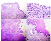
Histopathology of the cow reported in the case. A: Bladder (40X magnification), benign mesenchymal neoplastic proliferation composed of homogeneous vascular structures (hemangioma) (arrow). B: Bladder (100X magnification), epithelium with areas of thickening, intense cytoplasmic vacuolation, karyomegaly, and occasionally eosinophilic intracytoplasmic bodies (Melamed-Wolinska bodies) (epithelial dysplasia) (arrow). C: Esophagus (40X magnification) with well-defined, expansive, exophytic epithelial neoplastic proliferation composed of cells from the squamous layer, with squamous differentiation from the epithelium and the formation of finger-like projections (papilloma) (arrow). D: Esophagus (40X magnification) and some moderately dilated mucous glands with squamous metaplasia (arrow). Moderately hyperplastic epithelium. Source: Veterinary Pathology, HV-UFMG.
In the esophagus, there was a well-defined, expansive, and exophytic epithelial neoplastic proliferation composed of cells from the squamous layer, with squamous differentiation from the epithelium and the formation of finger-like projections (Fig. 3 C). The cells exhibited an eosinophilic cytoplasm with indistinct borders. The nuclei were large, oval to rounded, and had fine stippled chromatin. There was also slight swelling of the adjacent keratinocytes with cytoplasmic vacuolation, characterizing a papilloma (Fig. 3 B).
DISCUSSION
The cow presented clinical alterations, complementary test results, necropsy findings, and histopathological examination results consistent with chronic poisoning by Pteridium spp., including persistent hematuria, bladder hemangiomas, and neoplasms of the upper digestive tract (Evans, 1964; Singh et al., 1973; Yamane et al., 1975; Souto et al., 2006a, 2006b). Souto et al. (2006a) reported the occurrence of chronic poisoning by P. aquilinum in three cattle that presented with hematuria and anemia. These authors diagnosed hemangiomas in the bladder of these animals, and papillomas were found in the upper digestive tract of two animals.
Pteridium spp. is commonly known in Brazil as "samambaia," "samambaia do campo," or "pluma" (Ribeiro, 2018). There are many risk factors that predispose animals to natural intoxication by Pteridium spp., but the lack of available or offered food has been pointed out as the main cause, even though it is not palatable. Additionally, some animals develop a habit of consuming the plant even when food is available (Riet-Côrrea et al., 2007).
One of the main clinical signs was persistent hematuria, which resulted in regeneratively ineffective anemia. Bovine enzootic hematuria is a chronic form of poisoning by Pteridium spp. that occurs in animals that consume less than 10 g/kg per day over a long period. Clinical signs begin after months or even years of the plant's ingestion by cattle (Tokarnia et al., 2000). In these cases, aplastic anemia may occur, leading to leukopenia, thrombocytopenia, hemostatic disorders, increased bleeding time, and elevated fibrinogen levels (Evans, 1964; Yamane et al., 1975).
The serum creatinine levels were within the reference values for the species. However, a significant increase in the urine protein/creatinine ratio (9.27) was observed, along with +4 glycosuria in the urine dipstick test. During necropsy, multifocal yellowish-white smooth areas were noted in the outer cortex of the kidneys, which extended in a wedge shape into the medullary region upon sectioning, thus characterizing areas of infarction. Renal microscopy revealed multifocal coagulative necrosis, and a moderate infiltrate composed of neutrophils along with some lymphocytes, plasma cells, and macrophages adjacent to the necrotic area, indicating glomerular and renal tubular impairment. Although renal lesions were confirmed by histopathological examination, they represented, on average, only 25% of the renal involvement.
Serum creatinine levels indicated preserved renal function; however, the animal exhibited significant protein loss in urine and glycosuria due to vascular compromise in the area affected by the vesical hemangioma. These laboratory and histopathological findings, when considered together, demonstrate that the lesions responsible for proteinuria were of post-renal origin. Additionally, the decrease in plasma proteins, particularly albumin, leads to a decrease in oncotic pressure in the blood vessels, explaining the observed submandibular edema (Singh et al., 1973).
The esophagus showed well-defined, expansive, and exophytic epithelial neoplastic proliferations composed of squamous layer cells, with squamous differentiation from the epithelium and formation of finger-like projections, suggestive of papilloma. Species of Pteridium spp. contain several carcinogenic, mutagenic, and immunosuppressive principles, including ptaquiloside norsesquiterpene and quercetin (Campo 1997). The association of these carcinogenic substances present in ferns with infection by bovine papillomavirus is strongly correlated with the etiology of bovine enzootic hematuria (Campo et al. 1992; Campo 2002; Wosiacki et al. 2005).
CONCLUSION
The clinical presentation, lesions, and neoplasms in the urinary bladder and esophagus found in the cow in this study are common in chronic poisoning by Pteridium spp., particularly in the species P. arachnoideum.
REFERENCES
- BARBOSA, R.R.; RIBEIRO FILHO, M.R.; SILVA, I.P.; SOTO-BLANCO, B. Plantas tóxicas de interesse pecuário: importância e formas de estudo. Acta Vet. Bras., v.1, p.1-7, 2007.
- CAMPO, M.S. Animal models of papillomavirus pathogenesis. Virus Res., v.89, p.249-261, 2002.
- CAMPO, M.S. Review bovine papillomavirus and cancer. Vet. J., v.154, p.175-188, 1997.
- CAMPO, M.S.; JARRETT, W.F.; BARRON, R. et al. Association between bovine papillomavirus type 2 and bracken ferns and bladder cancer in cattle. Cancer Res., v.52, p.6898-6904, 1992.
- EVANS, W.C. Bracken poisoning of farm animals. Vet. Rec., v.76, p.365-372, 1964.
- RIBEIRO, D.S.F. Validação de método e determinação de ptaquilosídeo e pterosina B por cromatografia líquida de alta eficiência em samambaias e no leite de vacas. 2018. 81f. Tese (Doutorado em Medicina e Cirurgia Veterinária) - Escola de Veterinária, Universidade Federal de Minas Gerais, MG.
- RIET-CORREA, F.; SCHILD, A.L.; LEMOS, R.A.A.; BORGES, J.R.J. Doenças de ruminantes e equídeos. São Paulo, 2007, p.173-176.
- SINGH, A.K.; JOSHI, H.C.; RAY, S.N. Studies on bovine haematuria. 1. Haematological and biochemical observations on the blood of cattle suffering from haematuria. Indian J. Anim. Sci., v.43, p.296-299, 1973.
- SOUTO, M.A.A.; KOMMERS G.D.; BARROS C.S.L. et al. Neoplasias do trato alimentar de bovinos associadas ao consume espontâneo de samambaia (Pteridium aquilinum). Pesqui. Vet. Bras., v.26, p.112-122, 2006a.
- SOUTO, M.A.A.; KOMMERS G.D.; BARROS C.S.L. et al. Neoplasmas da bexiga associados à hematúria enzoótica bovina. Ciênc. Rural, v.36, p.1647-1650, 2006b.
- TOKARNIA, C.H.; DOBEREINER, J.; PEIXOTO, P.V. et al. Plantas tóxicas do Brasil. Rio Janeiro: Helianthus, 2000. p.142-185.
- WOSIACKI, S.R.; BARREIRO, M.A.C.; ALFIERI, A.F.; ALFIERI, A.A. Semi-nested PCR for detection and typing of bovine Papillomavirus type 2 in urinary bladder and whole blood from cattle with enzootic haematuria. J. Virol. Methods, v.126, p.215-219, 2005.
- YAMANE, O.; HAYASHI, T.; SAKU, S. Studies on blood coagulation disorders in domestic animals. Thrombelastograms of normal cattle and cattle affected with bracken poisoning. Jpn. J. Vet. Sci., v.37, p.577-583, 1975.


