
ABSTRACT
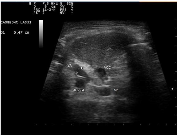
Portosystemic shunt (PSS) is an anomalous vascular connection between the portal venous system and the systemic circulation. These deviations connect the main portal vein (PV) or some portal branches to the vena cava (VC) or, less commonly, to the azygos vein (AV). The purpose of this case report was to describe the diagnosis of PSS in a dog classified as porto-azygos. This diagnosis is considered uncommon compared to other portosystemic shunts using ultrasonography and portography. The subject was a male dog, Yorkshire, 8 months old, presented neurological signs characterized by head press, ataxia, tremors and episodes of temporary blindness and deafness. Ultrasonographic examination revealed a dilated and curved anomalous vessel with approximately 0.6cm of diameter and turbulent flow seen through pulsed and color Doppler, and segmental dilation of the azygos vein. The portography revealed enhancement by iodinated contrast in the jejunal vein, the portal vein and an anomalous vessel flowing towards the azygos vein in the craniodorsal region of the abdomen. The PSS was surgically corrected with an ameroid constrictor. Ultrasonography and portography were effective at detecting and characterizing the portoazygos shunt despite some limitations.
Keywords:
diagnostic imaging; canine; portosystemic deviatio
 211x172mm (96 x 96DPI).
 211x172mm (96 x 96DPI).
 254x203mm (96 x 96DPI).