
 Tracheal collapse in a pony breed horse - case report
ABSTRACT
This case report aims to present the clinical, diagnostic, and therapeutic aspects of a case of tracheal collapse in a Pony breed horse with a history of progressive respiratory noise. The animal exhibited noisy exhalation, along with tracheal and pulmonary stridor. Radiographic and endoscopic examinations confirmed grade 4 tracheal collapse, associated with intermittent dorsal displacement of the soft palate. A tracheostomy was performed as a therapeutic measure, but the respiratory condition worsened. Given the poor prognosis, euthanasia was chosen, and the diagnosis of severe tracheal collapse was confirmed through necropsy. Although rare in horses, tracheal collapse should be considered in the differential diagnosis when signs such as dyspnea and respiratory noise are present. Radiography and endoscopy are essential tools for diagnosis; however, due to the severity of the clinical condition, surgical intervention was not possible.
Keywords:
dyspnea; respiratory noise; radiography; endoscopy
RESUMO
O presente relato tem como objetivo descrever os aspectos clínicos, diagnósticos e terapêuticos de um caso de colapso traqueal em um equino da raça Pônei, com histórico de ruídos respiratórios progressivos. O animal apresentava expiração ruidosa, além de estridor traqueal e pulmonar. Exames radiográficos e endoscópicos revelaram colapso traqueal grau 4, associado a deslocamento dorsal intermitente do palato mole. Foi realizada traqueostomia como medida terapêutica, porém houve agravamento do quadro respiratório. Devido ao prognóstico desfavorável, optou-se pela eutanásia, sendo o diagnóstico de colapso traqueal severo confirmado por meio de necropsia. Embora raro em equinos, o colapso traqueal deve ser incluído no diagnóstico diferencial quando há sinais como dispneia e ruídos respiratórios. Radiografia e endoscopia são métodos fundamentais para confirmação do diagnóstico. Em virtude da gravidade do quadro clínico, a realização de intervenção cirúrgica não foi possível.
Palavras-chave:
dispneia; ruídos respiratórios; radiografia; endoscopia
INTRODUCTION
The term tracheal collapse refers to a narrowing of the tracheal lumen, characterized by dorsoventral flattening of the tracheal rings, which may affect the cervical and/or thoracic portions of the trachea. This condition can lead to partial or complete airway obstruction (Auer et al., 2019).
Tracheal collapse in horses is considered uncommon; however, similar to dogs, it occurs more frequently in miniature animals, typically Ponies and American Miniature Horses. The actual incidence of this condition is likely higher than what is reported in the literature, as diagnosis is usually only made once the animal presents with respiratory distress, indicating an advanced stage of the disease (Every et al., 2020).
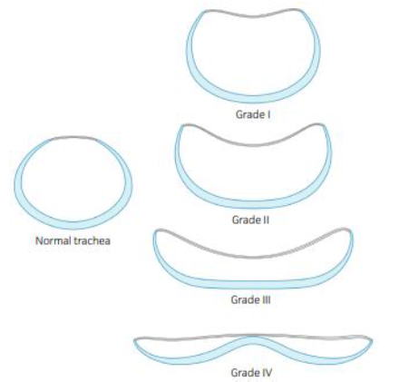
Tracheal collapse may be associated with congenital abnormalities, degenerative changes, deformities of the cartilaginous rings, paratracheal abscesses, trauma, or even increased respiratory effort in cases of severe pulmonary infections. The main cause of tracheal collapse described in adult American Miniature Horses is chondromalacia of the tracheal rings-a degenerative process that gradually leads to rupture of the cartilaginous rings (Aleman et al., 2008; Auer et al., 2019; Peyrecave-Capo et al., 2023). Clinical signs vary depending on the severity and location of the collapse and are classified into four grades (Fig. 1), following the Csystem originally established in dogs. Grade 1 corresponds to approximately a 25% reduction in airway diameter with a dorsally dependent tracheal membrane; Grade 2 involves a 25-50% reduction in tracheal lumen diameter with slight widening of the tracheal rings and muscle; Grade 3 reflects a 50-75% reduction in lumen diameter with moderate flattening of the tracheal rings; and Grade 4 indicates a 75-100% reduction in tracheal lumen diameter with severe flattening of the tracheal rings and dorsal inversion of the ventral tracheal margin (Every et al., 2020).
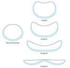
Affected animals may present with chronic cough, dyspnea, tachypnea, dysphagia, and commonly exhibit respiratory noises (wheezing and stertor). Clinical signs may be exacerbated by excitement, stress, heat, and physical exercise (Rosa and Martinez, 2022).
Radiography and endoscopy are commonly used diagnostic methods for investigating tracheal collapse. In laterolateral radiographic projections, dorsoventral flattening of the tracheal lumen is commonly observed (Barakzai, 2013). Endoscopy, in turn, reveals the abnormal shape of the tracheal lumen (Auer et al., 2019).
Treatment can be conservative or surgical, depending on the degree of tracheal collapse. The conservative approach is indicated for patients with up to 50% reduction of the tracheal lumen and consists of the administration of bronchodilators and mucolytic agents. Additionally, procedures such as tracheostomy and oxygen therapy may be performed (Gonçalves et al., 2004; Barakzai, 2013). For patients with more than 50% reduction, surgical intervention is recommended. Depending on the location and extent of the lesion, placement of extraluminal ring prostheses or intraluminal stents is considered, aiming to increase the airway diameter and restore the tracheal lumen (Barakzai, 2013; Couëtil et al., 2004; Couëtil and Hawkins, 2013; Every et al., 2020).
The objective of this study is to report a case of tracheal collapse, addressing the clinical, diagnostic, and therapeutic aspects.
ETHICAL ASPECTS
The present study refers to a clinical case report, with no experimental nature; therefore, submission to the Animal Use Ethics Committee (CEUA) was not required. All procedures were part of routine veterinary care and were conducted in accordance with animal welfare principles. The animal’s legal guardian authorized the procedures and the use of clinical data and images for academic and scientific purposes.
CASE REPORT
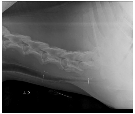
An 8-year-old Pony breed equine, weighing 118kg, was admitted to Veterinary Clinic X with a history of intense abnormal respiratory noises for approximately six months. According to the anamnesis, the owner reported that initially the noises were mild and intermittent, observed only during physical exertion or episodes of excitement. About three months prior, the noises intensified and began to occur more frequently, even when the animal was at rest. During the general physical examination, the animal was standing, alert, with a body condition score of 3, mucous membranes normocolored, capillary refill time of 2 seconds, elevated rectal temperature of 39.0°C, heart rate of 48 beats per minute (bpm), and respiratory rate of 13 breaths per minute (bpm). Specific examination revealed forced inspiration and expiration, with noisy expiration, absence of nasal or paranasal sinus discharge, presence of tracheal stridor, and pulmonary stridor on the left side. Blood was collected via jugular venipuncture, and the complete blood count showed leukocytosis (16.7x10³/µL) due to neutrophilia (14.195x10³/µL), indicating an acute infectious process. Initially, recurrent laryngeal neuropathy and dorsal displacement of the soft palate were suspected. For diagnostic purposes, lateral radiographic projections of the cervical and thoracic regions were performed first. The images allowed visualization of a reduced lumen between the distal third of the cervical trachea and the proximal thoracic trachea, with the thoracic portion exhibiting greater lumen reduction (Fig. 2).
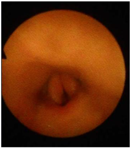
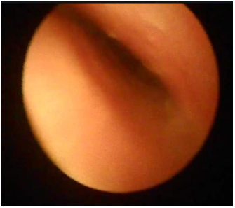
Additionally, the animal underwent airway endoscopy, which revealed intermittent dorsal displacement of the soft palate (Fig. 3) and irregularity of the tracheal lumen in the dorsoventral direction (Fig. 4).
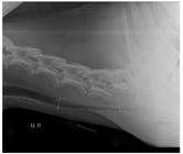
Radiographic image of the trachea in laterolateral projection, showing lumen reduction between the distal third of the cervical trachea and the proximal thoracic trachea.
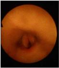
Intermittent dorsal displacement of the soft palate. Note that the epiglottis is not visualized in this image.
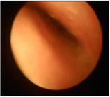
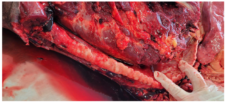
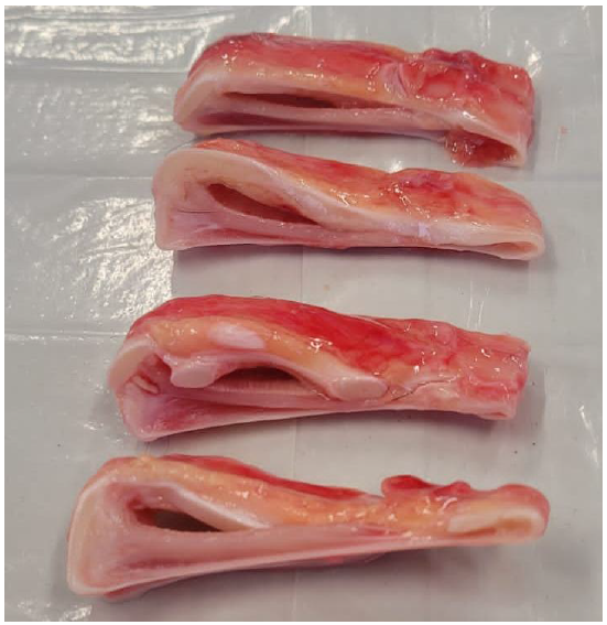
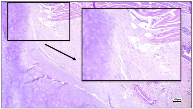
Based on the findings from the physical examination, radiography, and endoscopy, a diagnosis of grade 4 tracheal collapse was established, along with intermittent dorsal displacement of the soft palate, explaining the respiratory distress presented by the animal. Initially, treatment was initiated for pneumonia, as suggested by fever, pulmonary auscultation, and leukocytosis on the blood count. The following protocol was instituted: Dipyrone (25 mg/kg) intravenously, once daily for 1 day; Penicillin (40,000 IU/kg) intramuscularly, twice daily for 5 days. The patient was kept in a well-ventilated environment, free from stimuli that could exacerbate respiratory distress. After recovery from pneumonia, a tracheostomy was performed to relieve respiratory difficulty in preparation for a possible staphylectomy procedure. The patient was sedated using detomidine (10 µg/kg) and butorphanol (0.1 mg/kg), and the tracheostomy was carried out in the cervical region, where the tracheal rings were palpably intact. The tracheostomy provided temporary relief until the endotracheal tube was removed, and the effects of the sedative wore off. After this period, the patient developed severe respiratory distress, requiring immediate re-administration of sedatives, reinsertion of the endotracheal tube, and continuation of oxygen therapy. Due to the poor prognosis, immediate euthanasia was elected. Necropsy confirmed the severity of the tracheal collapse, with dorsoventral flattening most evident in the distal portion of the cervical trachea and throughout the thoracic trachea, with near-complete collapse in the terminal portion of the thoracic trachea (Figures 5 and 6). Histopathological analysis of the tracheal fragment revealed a submucosa composed of preserved loose connective and adipose tissue, as well as hyaline cartilage, smooth muscle, and adventitia without significant structural alterations in the examined sample (Fig. 7), despite the previously described macroscopic changes.
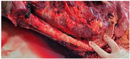
Post-mortem macroscopic image of the trachea showing severe tracheal collapse. Note that in the distal thoracic segment of the trachea, the collapse is more pronounced, compromising nearly 100% of the tracheal lumen.
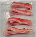
Transverse sections of the tracheal segment showing dorsoventral flattening of the tracheal rings.
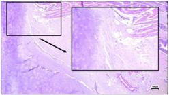
Histological section shows fragments of trachea with preserved loose connective and adipose tissue in the submucosa. Hyaline cartilage, smooth muscle, and adventitia are also preserved.
DISCUSSION
Concomitant conditions are present in several cases of tracheal collapse, including aspiration pneumonia, sinusitis, dental problems, and dorsal displacement of the soft palate (Aleman et al., 2008). In the case reported, the animal presented intermittent dorsal displacement of the soft palate along with pneumonia.
In many cases of tracheal collapse, both the cervical and thoracic trachea are affected, leading to characteristic inspiratory sounds and increased abdominal effort during expiration (Auer et al., 2019). In this case, the animal showed involvement of both the cervical and thoracic portions of the trachea; however, the abnormal sounds occurred during expiration, with increased effort observed during both inspiration and expiration.
Tachycardia and tachypnea are common clinical signs at rest in animals with tracheal collapse, due to respiratory inefficiency caused by the condition (Couëtil & Hawkins, 2013). In the present case, the patient exhibited both inspiratory and expiratory effort; however, tachypnea was not observed at rest, according to the physiological reference values proposed by Feitosa (2020).
Microscopically, tracheal collapse is often characterized by multifocal areas of hypocellularity in the hyaline cartilage, replaced by fibrous and/or fibrocartilaginous connective tissue, associated with reduced basophilic staining of the ground substance, consistent with degenerative changes (Peyrecave-Capo et al., 2023). In the present case, endoscopy not only demonstrated the severity of the collapse but also enabled the detection of intermittent displacement of the soft palate, highlighting its importance for accurate diagnosis.
Although the use of extraluminal prostheses and intraluminal stents has been reported in ponies and miniature horses, the outcomes remain inconsistent and are frequently associated with significant complications, such as persistent coughing, intraluminal granulation tissue formation, and recurrent airway obstruction (Aleman et al., 2008; Mellish, 2025). In the present case, surgical intervention with extraluminal implants or intraluminal stents was not initially considered, as the first attempt consisted of a tracheostomy, aiming to alleviate respiratory distress and enable a potential staphylectomy. However, at the end of the procedure, following removal of the endotracheal tube and reduction of the sedative effect, the patient developed severe respiratory distress, requiring reinfusion of sedatives, reinsertion of the endotracheal tube, and maintenance under oxygen therapy. Given the unfavorable prognosis, immediate euthanasia was elected. In severe cases of tracheal collapse, euthanasia may become necessary due to the lack of response to clinical management and the limitations of available surgical options (Aleman et al., 2008; Mellish, 2025).
In a post-mortem examination, Peyrecave-Capo et al. (2023) described in horses multifocal areas of hypocellularity of the hyaline cartilage, replaced by fibrous and/or fibrocartilaginous connective tissue, associated with reduced basophilic staining of the ground substance, consistent with degenerative changes. In the present case, microscopic evaluation of the tracheal fragment revealed preserved tissue (Fig. 7), despite the evident macroscopic deformity of the rings observed during the post-mortem examination (Fig. 6). Although changes compatible with tracheal collapse were identified macroscopically, no significant histological alterations were observed in the analyzed sample. A similar situation has been reported in miniature horses, in which collapse could be confirmed clinically and macroscopically, but not always accompanied by microscopic changes, with chondromalacia being described only in some animals (Aleman et al., 2008). This discrepancy may be related to the functional and dynamic nature of the condition, in which narrowing occurs mainly during respiration, without evident structural degeneration, or to the possibility of extrinsic compression without direct involvement of the tracheal tissue (Mellish, 2025). Furthermore, it should be considered that the absence of histological findings may reflect early or focal lesions not represented in the examined fragments.
CONCLUSION
Tracheal collapse is a rarely diagnosed condition in the equine species, yet it is a relevant respiratory disorder. Clinical signs such as inspiratory and/or expiratory dyspnea accompanied by abnormal respiratory sounds should be considered as differential diagnoses for tracheal collapse. Plain radiography in the laterolateral projection proved to be an important tool for patient diagnosis and prognosis. However, endoscopy allows for a better assessment of the severity of the collapse, especially since reference values for tracheal lumen measurement in ponies are not available in the literature. Due to the severity of the tracheal collapse presented by the animal, surgical intervention was not feasible.
REFERENCES
- ALEMAN, M.; JORGE NIETO, E.; BENAK, J.; JOHNSON, L.R. et al. Tracheal collapse in American Miniature Horses: 13 cases (1985-2007). J. Am. Vet. Med. Assoc., v.233, p.1302-1306, 2008.
- AUER, J.A.; STICK, A.; PRANGET, T. et al. 2019. Equine surgery. 5.ed. [Oxford]: Elsevier, 2019. p.802-803.
- BARAKZAI, S. Tracheal collapse in a Shetland pony: a case study. Livestock, v.18, p.90-92, 2013.
- COUËTIL L.L.; GALLATIN, L. L.; BLEVINS, W.; KHADRA, I. Treatment of tracheal collapse with an intraluminal stent in a miniature horse. J. Am. Vet. Med. Assoc., v.255, p.1727-1732, 2004.
- COUËTIL, L.L.; HAWKINS, J.F. Respiratory diseases of the horse: a problem-oriented approach to diagnosis & management. Manson Publishing, London, 2013. 238p.
- EVERY, L.J.; HOSTNIK, E.T.; HOSTNIK, L.D. et al. Radiographic tracheal lumen to vertebral ratios in the normal American miniature horse. Equine Vet. J., v.52, p.428-434, 2020.
- FEITOSA, F.L.F. Semiologia veterinária: a arte do diagnóstico. 4.ed. Rio de Janeiro: Roca, 2020. 841p.
- GONÇALVES, R.C., SILVA, D.P.G., BORGES, A.S., et al. Colapso traqueal em minipônei. Semin. Ciênc. Agr., v.25, p.59-62, 2004.
- MELLISH, M. Tracheal collapse in equines: diagnosis, management, and surgical challenges. Equine Vet. Educ., v.37, p.467-470, 2025.
- PEYRECAVE-CAPO, X.; MUNOZ-DIAZ, J.; FERRER, G.R. et al. Acute tracheal trauma and chronic tracheal collapse in an asthmatic mature pony. Equine Vet. Educ., v.35, p.241-247, 2023.
- ROSA, M.J.; MARTINEZ, L.M.R. Left rotation and dorsoventral collapse of the trachea on an adult Dartmoor cross-gelding: Imaging diagnosis. Equine Vet. Educ., v.34, p.473-477, 2022.
The research data are available within the article itself.

 Source:
  Source: Veterinary Clinic X, 2023.
 Source: Veterinary Clinic X, 2023.
 Source: Veterinary Clinic X, 2023.
 Source: Veterinary Clinic X, 2023.
 Source: Veterinary Clinic X, 2023.
 Source: Image provided by Laboratory X, 2024.