ABSTRACT
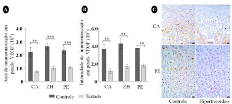
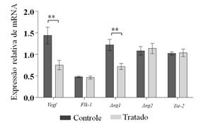
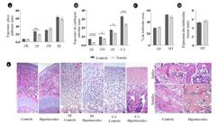
The effects of excess of maternal thyroxine associated with postnatal hyperthyroidism at bone growth and proliferative and angiogenic profile of cartilage were studied. Sixteen adult Wistar rats were divided into treated and control groups. The offspring of the treated group received L-thyroxine from weaning to 40 days-old. At weaning, plasma assay of free T4 was measurement on female rats. In the offspring, the following assessments were performed: measurement of total T3 and free T4, histomorphometry analysis of the thyroid, measurement of body weight and length and width of the femur. In femoral growth cartilage, immunostaining of CDC-47, gene or protein expression of VEGF, Flk-1, Ang1, Ang2 and Tie2 were evaluated. Data were analyzed using Student’s t-test. Free T4 was significantly higher in treated rats and total T3 and free T4 were significantly higher in offspring. The width of the femur was significantly lower in treated animals. There was lower immunoreactivity of CDC-47, VEGF and lower expression of gene transcripts for VEGF and Ang1. We concluded that the excess maternal thyroxine associated with postnatal hyperthyroidism reduces the width of the femoral shaft, the cell proliferation and gene and protein expression of VEGF and gene expression of Ang1 on the growth cartilage in rats.
Keywords:
rat; thyroxine; congenital hyperthyroidism; growth cartilage; angiogenesis
 Thumbnail
 Thumbnail
 Thumbnail
 Thumbnail
 Thumbnail