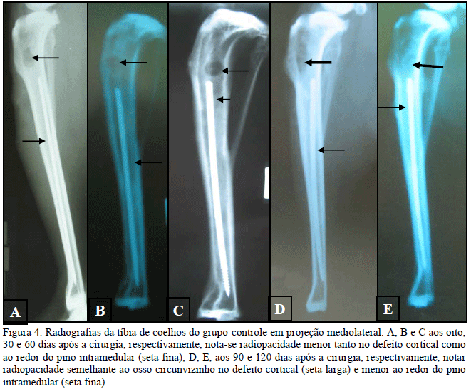
This study evaluated a sinthetic hydroxyapatite-lignin composite for osseoingration between metallic implant and bone tissue. Twenty New Zealand rabbits were used. A defect was made a the proximal region of lateral surfaces of both tibias. An intramedullary pin was inserted in the medullary cavity of the left tibia after filling the cavity with 1,000mg of the composite and covering the thread part of the pin with the composite. The same procedure was made in control tibias without the composite. Clinical evaluation was based on inflammatory reaction, dehiscence, lameness, pain, and tibial circumference. Radiographs were made immediately after surgery and then on days eight, 30, 60, 90, and 120. Most of the animals presented normal clinical progression. Radiographies of treated group showed decrease in the defect and medullary cavity radiopacity to the same pattern of the surrounding bone, while the contrary happened in control group; but, in this group, the medullary cavity remained radioluscent at the last observation date. It can be concluded that the hydroxyapatite-lignin composite did not show clinical signs of rejection and that radiopacity of the tissue in the bone defect and surrounding the pin was similar to bone radiopacity, suggesting that the material promoted osseointegration.
rabbit; osseointegration; composite; implant; hydroxyapatite; lignin