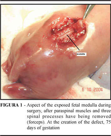
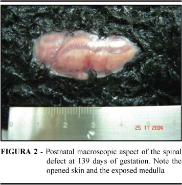
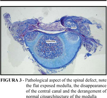
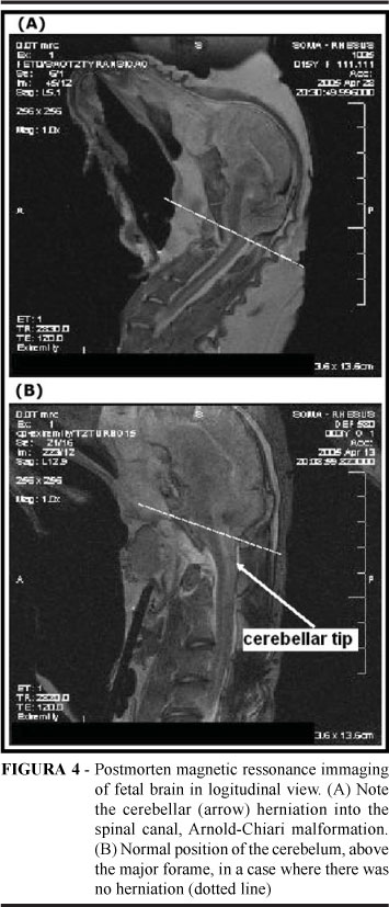
PURPOSE: To produce a myelomeningocele-like human defect in the ovine fetus and validate this experimental model in our population. METHODS: A prospective study on 12 pregnant sheep of a crossed Hampshire/Down breed where a spinal defect was surgically created between Day 75 and Day 77 after conception. The technique consisted of a hysterotomy with exposure of fetal hind limbs and tail up to the mid spine. Fetal skin, paravertebral muscles, and 4 posterior spinal arches were excised, exposing the spinal cord. Duramater was opened and the medulla was incised until the medullar canal. Animals were euthanized at 139 days of gestation for fetal evaluation. The central nervous system was submitted to post-mortem magnetic resonance imaging (MRI) and the spine was submitted to pathological examination. RESULTS: The defect was created in 13 fetuses and 5 survived. Mean gestational age at necropsy was 121.6 days (varying from 93 to 145 days). Macroscopically, the defect was present in 4 cases. Microscopy revealed a flattened medulla with disappearance of the medullar canal and disruption of normal medullar architecture with neuronal apoptosis and/or fusion of the piamater and duramater. The MRI showed herniation of the cerebellum into the cervical canal and syringomyelia. CONCLUSIONS: The surgically produced defect mimics the defect found in the human fetus, including the Arnold-Chiari malformation. Post-mortem MRI was used for the first time in our study and proved an excellent alternative for demonstrating the cerebellar herniation. We standardized the technique for creating the defect in our population.
Models animal; Meningomyelocele; Spinal dysraphism; Fetus; Sheep