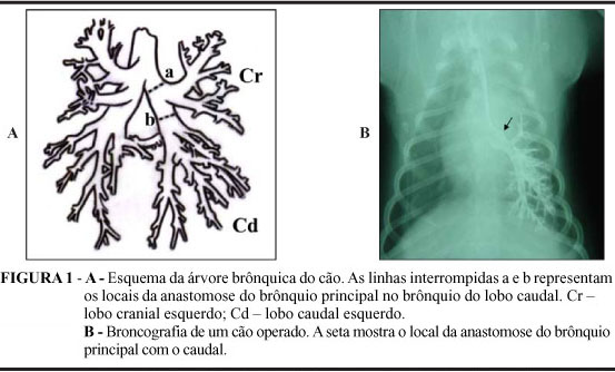
PURPOSE: To study the bronchial and vascular anastomosis in the autologus lobar lung implant and show if exist complications in transplants occuping only half of pleural cavity. METHODS: We studied 15 male from 8 to 14kg. After pneumonectomy in the left chest wall we replaced the caudal lobe. In the late post operative we made bronchography and perfusion's scintigraphy and analysed the lungs after sacrified the animal. RESULTS: Four dogs died, one by early dehiscence of bronchial anastomosis, one by infection, and two by lung infarct after occlusion of lung vein in place of anastomosis. The relative perfusion of right and left lung were 72,7% and 27,3% respectively. The bronchography didn't show stenosis nor other alterations in bronchial anastomosis. After opened the thorax we saw that caudal lobe there was filled all pleural cavity without shift of the mediastinum. CONCLUSIONS: The study showed that the end to end suture without protection don't carry the complications to the anastomosis. Didn't occour complications, in the fact the replaced lobes occupated only half pleural cavity. The most important complication was pulmonary infarct and total dehiscence of the chest wall. The bronchography was efficient to study bronchial anastomosis. Scintigraphy was useful to quantify the relative pulmonary mass functioning in vivo.
Pneumonectomy; Autologus transplantation; Lung; transplantation; Dogs