Bioactive Compounds from Dioscorea bulbifera L. Obtained by Combined Ultrasound-Assisted and Solid-Liquid Extraction
Abstract
The aim was to extract bioactive compounds from the husk and pulp of Dioscorea bulbifera L. by the combination of ultrasound and solid-liquid extraction techniques, as well as to evaluate the antioxidant potential. The extraction at 80 °C for 60 minutes showed the highest content of total phenolic compounds (TPC), 715.53 ± 8.00 mg EAG 100 g-1, for the husk, while the extraction at 70°C for 45 minutes showed 235.50 ± 25.30 mg EAG 100 g-1, for the pulp. The husk and pulp extracts showed flavonoid content of 363.63 ± 8.92 and 102.44 ± 1.51 mg EC 100 g-1, respectively. The antioxidant potential was evaluated according to the FRAP assay (125.09 ± 8.52 and 32.76 ± 0.65 µM ferrous sulfate g-1), the removal of H2O2 (29% and 41%), the ABTS radical assay (66.88 ± 0.93 and 14.93 ± 0.31 µM Trolox g-1) and the β-carotene/linoleic acid system, (84% and 47%) for husk and pulp, respectively. The bioaccessibility was obtained around 25% (pulp) and 16% (husk) accessible for absorption in the intestine. The combination of ultrasound and solid-liquid extraction methods proved to be effective in extracting bioactive compounds, with possible applications in the food industry.
Keywords:
“Cara-moela”; Unconventional Food Plants; Simulated gastrointestinal digestibility; Antioxidant capacity.
HIGHLIGHTS
Dioscorea bulbifera L. showed bioactive compounds in the husk and pulp.
The extraction methods were optimized by mathematical modeling.
Unconventional food plant (Dioscorea bulbifera L.) showed antioxidant potential.
The husk and pulp showed bioaccessibility by the simulated gastrointestinal digestibility assay.
INTRODUCTION
Dioscorea bulbifera L. is a tuber belonging to the Dioscoreaceae family, of the genus Dioscorea, native to Africa, but is a tuber widely cultivated and consumed in the tropics and subtropics [1]. It is considered a type of air yam and popularly known as “cara-moela”, air yam and butterfly yam. Its cultivation and use are not widespread due to lack of knowledge of its nutritional and application potential. This tuber belongs to the group of Unconventional Food Plants (UFPs). The UFPs can be described as food species that have one or more edible parts such as roots, tubers, bulbs, rhizomes, leaves, fruits, but without daily use, but with potential to supply the food chain. UFPs can be consumed both fresh and processed. For this reason, interest in UFPs has been growing recently, as these plants can be sources of a wide variety of nutrients, such as proteins, carbohydrates, minerals, vitamins, dietary fiber, and phenolic compounds, but they are still little explored food sources [2,3].
Scientific studies on Dioscorea bulbifera L. are still scarce, but what is in the literature has shown that this tuber has potential for introduction in human food. The tuber has a low lipid content, between 0.85 and 1.23 g/100 g, and a high carbohydrate content, around 71.25 g/100 g [4,5], in addition to having bioactive compounds directly related to different biological activities, such as anticancer activity [6], anti-inflammatory activity and antioxidant [4,7,8]. The bioactive compounds are secondary metabolites via the shikimate or acetate pathway and consist of an aromatic ring and a benzene ring with one or more hydroxyl groups. Depending on the number of phenolic rings and the structure of the interconnected rings, phenolic compounds can be classified into two groups: compounds of a flavonoid nature and compounds of a non-flavonoid nature [9,10]. In addition to being responsible for the color of food (such as yellow, orange, red and blue pigments), for the taste and flavor (such as vanillin and eugenol) of food. Bioactive compounds have a specific metabolic and physiological action in the human body, modulating antioxidant defense and inflammatory and mutagenic processes [11].
The functionality of bioactive compounds in foods is related to their availability after reaching the gastrointestinal tract [12]. To be absorbed by enterocytes, bioactive compounds must first be released from the food matrix and solubilized by gastrointestinal fluids during digestion. However, various physicochemical conditions within the gastrointestinal tract can influence their bioaccessibility, either enhancing or impairing the solubility and stability of these compounds [13,14]. Moreover, polyphenols often undergo substantial chemical modifications and/or transformations as they pass through the digestive system, which significantly affects their absorption profiles. While some metabolites are readily absorbed in the small intestine, others reach the colon, where they may undergo further transformation by the gut microbiota or be excreted without being absorbed. Consequently, a comprehensive understanding of the dynamic interaction among digestion, absorption, and metabolism is essential for accurately assessing the health-promoting potential of bioactive compounds [15,16].
Thus, in vitro, and in vivo digestibility or bioaccessibility or bioavailability studies are extremely relevant to provide a depth knowledge regarding interactions between nutrients and its food components [15]. Digestibility refers to the proportion of a nutrient that is absorbed by the individual and is typically determined by calculating the difference between the amount of the nutrient ingested and the amount excreted in the feces. In contrast, bioaccessibility denotes the fraction of a nutrient or bioactive compound that is released from the food matrix during digestion and is available for absorption across the intestinal epithelium. Bioavailability, on the other hand, encompasses the rate and extent to which an active compound is absorbed and reaches its target site within the body to exert its physiological effect [16,17].
Furthermore, to better understand the behavior of bioactive compounds in a food matrix, it is important to consider how to obtain them. The extraction of bioactive compounds can be categorized into conventional and non-conventional techniques [18]. The conventional extraction would cause the degradation of thermo-sensitive compounds resulting from long processing time and high extraction temperature. All techniques of solid liquid extraction (SLE) techniques take advantage of the higher solubility that some compounds have in different solvents but are often very time-consuming and can require large quantities of toxic and non-biodegradable organic solvents, as well as using a large amount of energy in the form of heat [19].
On the other hand, ultrasound-assisted extraction, which is considered as the non-conventional technology requires lesser solvent, shorter extraction time and produces higher extraction yield, in addition to minimizing the degradation of thermo-sensitive compounds [20,21]. After treatment with ultrasound, the cell walls are disrupted, as the physical forces created during acoustic cavitation cause disruption in plant tissue, increasing the diffusion rate and enhancing the mass transfer of cell content, which helps in the release of bioactive compounds [22,23]. This assists in compound bioaccessibility as compounds are easily released from plant matrix in gastrointestinal and thus being available for intestinal absorption [18,21]. For this purpose, the extraction of bioactive compounds should also be studied based on the application of different processes, such as acid hydrolysis in different species of Dioscorea, hydro-distillation for extracting bioactive compounds in yam, ultrasound in various plant crops, solid-liquid extraction with application of different solvents in Dioscorea bulbifera L. To optimize the various extraction processes and minimize possible degradation of bioactive compounds, a combination of extraction processes can be applied. The aim was to extract bioactive compounds from the husk and pulp of Dioscorea bulbifera L. by the combination of ultrasound and solid-liquid extraction techniques, as well as to evaluate the antioxidant potential.
MATERIAL AND METHODS
Samples and preparation
The samples used were from Dioscorea bulbifera L., obtained in Mandaguari/PR/Brazil. The samples were washed and sanitized with sodium hypochlorite (Anhembi, Brazil) solution (100 ppm) for 15 minutes and the husk and pulp fractions were separated. For enzymatic inactivation, the fractions were submitted to citric acid (Synth, Brazil) solution (2.4%) at 80°C for 5 minutes [5]. Subsequently, the samples were dried in an oven (LUCADEMA 82/150, Brazil) at 40°C until constant mass. The dried samples were ground, stored in low-density polyethylene (LDPE) packages, vacuum sealed (GSVAC/GS420, Brazil), protected from light and under refrigeration (4°C) until the extractions were carried out.
Extraction of bioactive compounds
The extractions of bioactive compounds from the husk and pulp from Dioscorea bulbifera L. were carried out according to the Box-Behnken 32 experimental design, considering the variables temperature and time, as shown in Table 1.
Factors and levels of the Box-Behnken 32 experimental design for extraction of bioactive compounds from husk and pulp from Dioscorea bulbifera L.
Assays (E) were carried out separately for husk and pulp from Dioscorea bulbifera L. in the proportion of 1:50 (m/v) in deionized water. The samples were submitted to an ultrasonic bath (SolidSteel/SSBu, Brazil) 40 kHz for 10 minutes at room temperature and subsequently submitted to a stirring Dubnoff orbital metabolic bath (Matoli/170M013, Brazil) at temperatures and times according to the experimental design (Table 1). At the end of the extraction times, the samples were centrifuged (Solab/SL-700, Brazil) at 6000 rpm for 20 minutes, filtered and the contents of total phenolic compounds, response variable, were evaluated in the extracts. Subsequently, the extracts were stored in amber flasks under refrigeration at 4°C until the time of the other analyses.
Colorimetric analysis for quantification of bioactive compounds
Total phenolic compounds
The total phenolic compounds content was determined in the extracts by mixing 60 µL of extracts with the addition of 3,000 µL of deionized water and 300 µL of Folin-Ciocalteau reagent (Dinâmica, Brazil), waiting for 3 minutes and adding 900 µL of sodium carbonate (15%) and 1740 µL of deionized water. Gallic acid - EGA (Sigma-Aldrich, USA) was used as a standard, and the results were expressed in gallic acid equivalents (mg EGA) 100 g-1, calculated by fitting the standard curve of gallic acid at concentrations from 25 at 650 mg L-1 in a spectrophotometer (Drawell/DU-8800RS, Brazil) at 765 nm [24].
Total flavonoid content
The determination of the total flavonoid content was based on the mixture of 500 µL of extracts with 2.5 mL of deionized water, 150 µL of sodium nitrite (Dinâmica, Brazil) (5%), after 6 minutes 300 µL of aluminum chloride hexahydrate (marca) were added. (10%), and after resting for 5 minutes, 1 mL of 1 M NaOH (Dinâmica, Brazil) was added. The analysis took place in a spectrophotometer (KASVI/K37-UVVIS, Brazil) at 510 nm. Catechin (Sigma-Aldrich, USA) was used, as a standard, in concentrations from 24.5 to 350 mg L-1 and the results were expressed in milligrams of catechin equivalent (mg CE) 100 g-1 [25].
Content of total carotenoids
The total carotenoid contents were estimated in the extracts at absorbances of 646.8, 663.2 and 470 nm in a spectrophotometer (KASVI/K37-UVVIS, Brazil). The results were obtained based on Eq.1, 2 and 3, and expressed in μg 100 g-1 [26].
Antioxidant capacity assays
ABTS radical scavenging activity
The evaluation of antioxidant activity was performed by the ABTS radical scavenging method with modifications [27]. Aliquots of 90 µL of the samples (husk and pulp extract) were transferred to test tubes with 9,000 µL of the ABTS•+ radical (Sigma-Aldrich, USA), previously prepared, after 6 minutes of the reaction, the samples were analyzed at 734 nm in a UV-Visible spectrophotometer (Drawell/DU-8800RS, Brazil). The ethyl alcohol solvent (Dinâmica, Brazil) was used as a blank and the calibration curve was performed with the Trolox standard (6-Hydroxy-2,5,7,8-tetramethylchroman-2-carboxylic acid) (Sigma-Aldrich, USA) at a concentration of 100 to 2000 µM and the results were expressed in µM trolox g-1.
Auto-oxidation by the β-carotene/linoleic acid system
Antioxidant activity was evaluated by the β-carotene/linoleic acid system [28]. The β-carotene/linoleic acid emulsion (Sigma-Aldrich, USA) was prepared by mixing 10 mg of β-carotene (Sigma-Aldrich, USA) with 10 mL of chloroform (Dinâmica, Brazil), then 2 mL of this solution was added to 100 mg of linoleic acid (Sigma-Aldrich, USA) and 400 mg of Tween 20 (Dinâmica, Brazil). The chloroform was completely evaporated on a rotary evaporator (MARCONI/442835, Brazil). Pre-oxygenated deionized water was added for 30 minutes until absorbance remained between 0.6 and 0.7 nm. A 3.5 mL aliquot of the β-carotene/linoleic acid emulsion was mixed with 350 µL of extract at concentrations of 40 mg mL-1. The solutions were incubated in a water bath (Nova instruments/NI 1215, Brazil) at 50°C. For control, Trolox standard (0.2 mg mL-1) was used. The initial absorbance was determined immediately after adding the samples to the system for the determination of time zero. The emulsion oxidation was determined by spectrophotometry (Drawell/DU-8800RS, Brazil) at 470 nm after 120 minutes of incubation. The antioxidant capacity was calculated in terms of percentage of oxidation inhibition, according to Eq. 4.
Where: AOA is the percentage of oxidation inhibition, DRC is the control degradation rate = ln (a/b)/120, DRS is the sample degradation rate = ln (a/b)/120, a is the initial absorbance at time 0 and b is the absorbance at 120 minutes.
Removal of hydrogen peroxide (H2O2)
The removal activity was determined by mixing 0.5 mL of H2O2 (Dinâmica, Brazil) (0.1 mmol L-1) with 0.5 mL of extract, 0.05 mL of ammonium molybdate (Nuclear, Brazil) (3% w/v), 5 mL of sulfuric acid (Synth, Brazil) (2 mol L-1), 3.5 ml of potassium iodide (Nox, Brazil) (1.8 mol L-1). For the pulp, concentrations of 40, 20 and 10 mg mL-1 were used, while for the husk were 40 and 20 mg mL-1. The titration of negative (glucose, - Vetec, Brazil) and positive (ascorbic acid - Synth, Brazil) controls at the same concentrations of the extracts was performed with sodium thiosulfate (Dinâmica, Brazil) (5 mmol L-1). Removal capacity was calculated according to Eq. 5.
Where: V0= volume used in the titration of the control and V1= volume used in the titration of the samples.
Iron Reduction Method (FRAP)
The determination of the iron reduction activity was carried out in test tubes, to which 150 µL of extract were added at concentrations of 2.5 to 40 mg mL-1, 2850 µL of FRAP reagent and kept in the dark in a water bath at 37°C for 30 minutes. The samples were evaluated in a spectrophotometer (Drawell/DU-8800RS, Brazil) at 593 nm. The ferrous sulfate (Dinâmica, Brazil) was used as standard in the concentrations of 100 - 2000 μM. Results were expressed as μM ferrous sulfate g-1 [29].
Simulated gastrointestinal digestibility
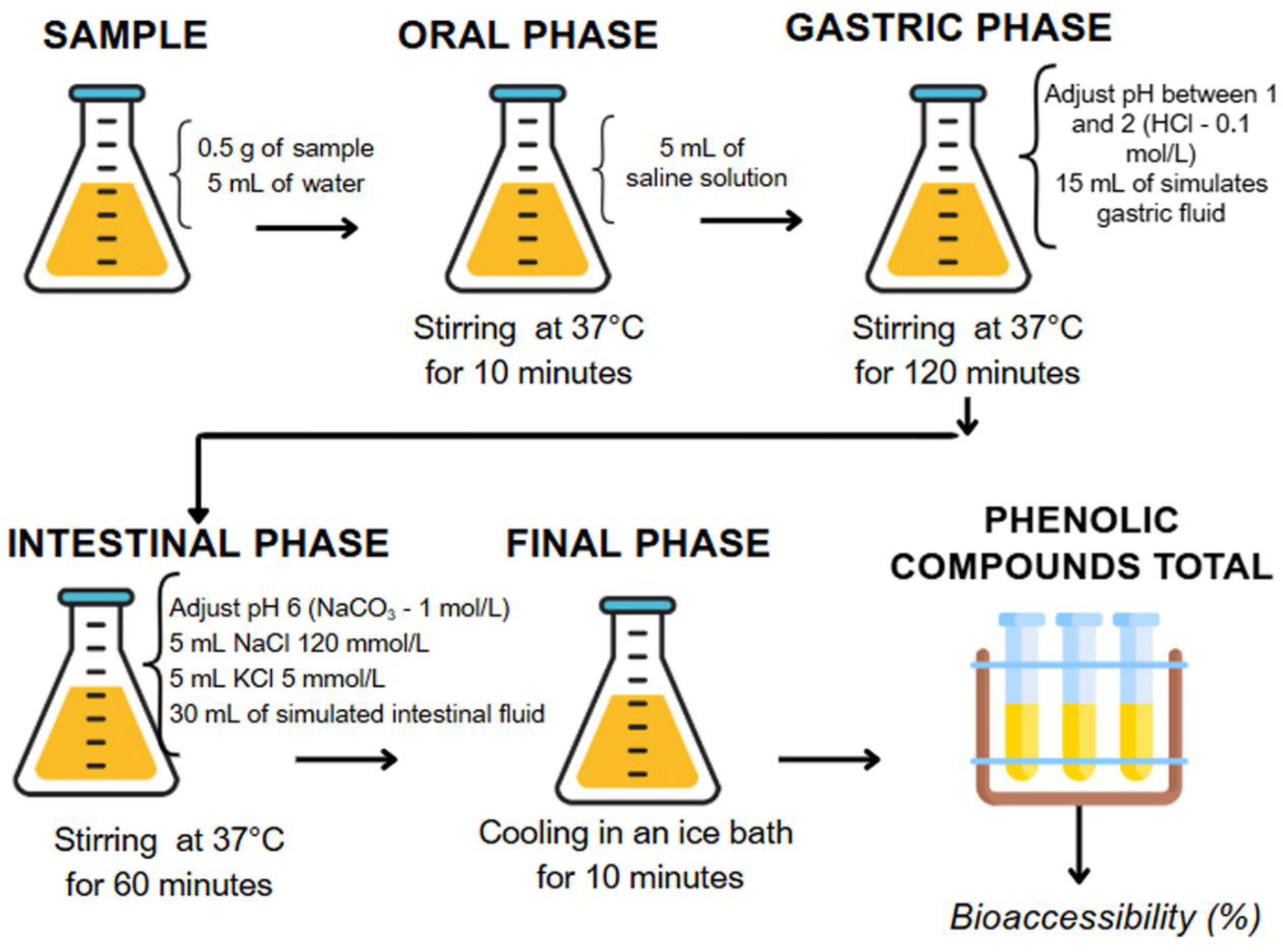
The bioaccessibility was performed by simulating the digestibility by in vitro model [30,31]. Initially, 0.5 g of sample were mixed with 5 mL of deionized water and submitted to digestion simulation in the oral, gastric, and intestinal phases. For the oral phase, 5 mL of saline solution (simulated saliva) was added, and they were kept at 37°C for 10 minutes. To start the gastric phase, the pH was adjusted between 1 and 2, and then 15 mL of simulated gastric fluid was added and kept at 37°C for 120 minutes. In the intestinal phase, the pH was adjusted to 6 and then 5 mL of 120 mmol L-1 NaCl, 5 mL of 5 mmol L-1 KCl and 30 mL of simulated intestinal fluid were added, kept at 37°C for 60 minutes. As can be seen in Figure 1. All steps were performed at 37°C in a Dubnoff orbital metabolic bath (MATOLI/170M013, Brazil) under agitation. To stop the digestion process, the samples were placed in an ice bath for 10 minutes. All reagents used in this analysis were supplied by Dinâmica (Brazil).
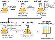
Representation of the phases of simulated gastrointestinal digestion from husk and pulp from Dioscorea bulbifera L.
In each phase, aliquots were taken to determine total phenolic compounds (TPC). Bioaccessibility was calculated using Eq. 6.
Where: D = TPC (mg GAE 100g-1) content after intestinal digestion; I = TPC (mg GAE 100g-1) content before digestion.
Statistical analyses
All analyzes were performed in triplicates and results were expressed as mean ± standard deviation. Data were analyzed by analysis of variance (ANOVA) and significant differences between means were analyzed by the Tukey test, p ≤ 0.05, using Statistica 7.0 (USA) and Minitab (USA) software’s.
RESULTS AND DISCUSSION
Analysis of bioactive compounds
Bioactive compounds from the husk and pulp of Dioscorea bulbifera L. were extracted and quantified based on the levels of total phenolic compounds, expressed in Table 2, according to the Box-Behnken design. It was found that the husk extracts presented contents between 545.30 and 715.53 mg GAE 100 g-1, considerably higher than those obtained for the pulp, which ranged from 166.95 to 235.50 mg GAE 100 g-1.
Content of total phenolic compounds for each husk and pulp extract of Dioscorea bulbifera L.
In addition to differences in region, climate, cultivation method, growth conditions, maturation stage, storage conditions that can interfere with the chemical composition of the husk and pulp, the content of total phenolic compounds are also influenced by the extraction method and parameters such as temperature, time and, especially, the solvent used [32,33].
Solid-liquid extraction is a process that involves the transfer of solute from a solid matrix to a solvent. It is an operation often used to extract important food components [32]. The efficiency of this extraction method varies with the size of the solid particles, due to the contact surface of the solute with the solvent, with the temperature, increasing permeability of cell walls and membranes with the application of heat, with the choice of solvent, influencing the solubility of the compound by the interaction with the solute, contact time between solvent and solid particles, among other factors such as surface tension, viscosity and agitation, which help in the mass transfer mechanism.
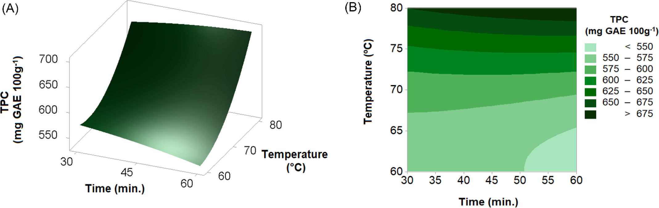
In this study, the solid-liquid extraction was preceded by ultrasound (UAE) which helped to break the cell walls, releasing the intracellular compounds into the solvent. In this case, only water was used, as in addition to helping mass transfer, it is not an aggressive solvent, generating less environmental impact and facilitating future applications [33]. In Figure 2 are (A) the results of the response surface methodology and (B) the contour curves. It can be observed that the bark extract submitted to a higher temperature (80°C) and a longer time (60 min.), resulted in the highest CFT content, around 715 mg EAG 100 g-1.
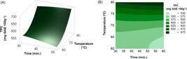
Response surface and contour curves for analysis of total phenolic compounds from extracts from the husk of Dioscorea bulbifera L.
The results may be associated with the presence of fibers, since there are compounds of phenolic origin that are structurally bonded to the fibers and, with the action of temperature, become more malleable, being released directly into the extraction solvent [32]. Knowing that the husk of Dioscorea bulbifera L. has around 4.90 g 100 g-1 of fiber, significant levels of phenolic compounds were expected when subjected to higher temperatures [5], as occurred in the E9 assay. From the data obtained for extracting the husk, a complete second-order quadratic mathematical model was obtained to determine the TPC content (dependent variable) of the husk extracts as a function of temperature and time (independent variables) which can be expressed by Equation 7.
Where T = temperature (°C); t = time (min.)
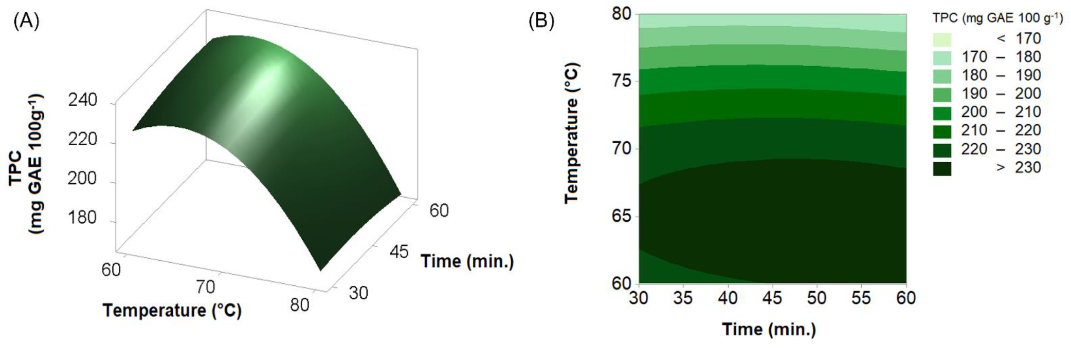
By maximizing the mathematical model, 699.31 ± 14.30 mg EAG 100 g-1 were obtained for the content of total phenolic compounds in the condition of 80°C and 60 minutes, as extraction parameters for the husk extract. The time factor was not significant while the temperature factor had an influence on the variations of the total phenolic compound’s contents of the husk extracts of Dioscorea bulbifera L. In Figure 3 are the results of the TPC content of the pulp extract, being (A) the results of the response surface methodology and (B) the contour curves. Extractions at a temperature of 70°C and 45 minutes resulted in the highest TPC content, around 235 mg EAG 100 g-1.
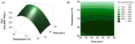
Response surface and contour curves for analysis of total phenolic compounds from extracts from the pulp of Dioscorea bulbifera L.
In the pulp extract, it can be observed that the increase in temperature resulted in lower levels of TPC. This effect results from the heat treatment applied, which may lead to the degradation of bioactive compounds and interfere with the hydrolysis of bonds between phenolic compounds and associated proteins and/or carbohydrates, thereby potentially decreasing or increasing the total phenolic content [34,35].
From the data obtained for pulp extraction, a second order quadratic mathematical model was obtained to determine the TPC content (dependent variable) of extracts from the pulp of Dioscorea bulbifera L. as a function of temperature (independent variable), the variable time did not have a significant influence, being disregarded from the model, expressed by Eq. 8.
Where T = temperature (°C)
The maximization of the mathematical model allowed to obtain, at the optimal point, 235.98 ± 6.00 mg GAE 100 g-1 for the content of total phenolic compounds in the condition of 64.4 °C, for temperature, and 50.3 minutes of time extraction method for the pulp extract of Dioscorea bulbífera L. The time and temperature factors showed different behavior, and the time was not significant while the temperature factor had an influence on the variations of the phenolic compound’s contents of the pulp extracts, as can be seen in the main effects graph. There are interactions between phenolic compounds and the porous structure of carbohydrates. In this sense, as the pulp of Dioscorea bulbifera L. contains about 75.70g 100 g-1 of carbohydrates, the results of the contents of total phenolic compounds for the pulp extracts may indicate such associations [5]. Therefore, the differences between the results for the pulp and husk extracts are justified due to the structural differences of these fractions.
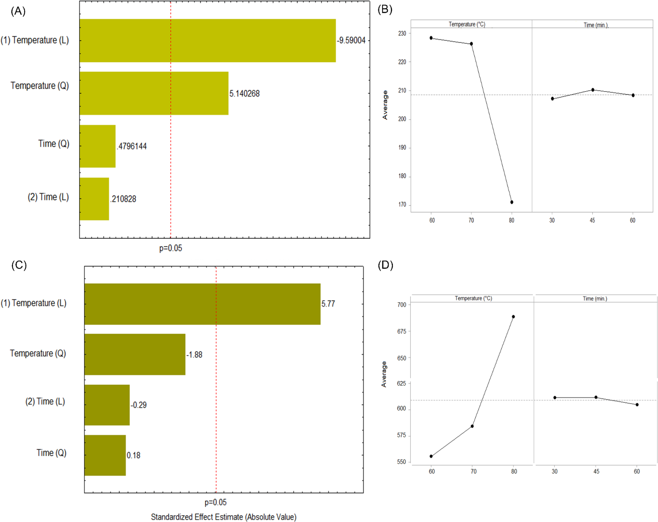
Based on the analysis of the Pareto charts and main effects plots, it is evident that temperature was, indeed, the most influential factor on the response variable for both the pulp and husk of Dioscorea bulbifera L. (Figure 4). In the pulp, the linear effect of temperature was highly significant (p < 0.05), with a negative trend, indicating that increasing the temperature significantly reduced the response values Figure 4A and Figure 4B. In the husk, temperature also showed a significant effect, but with a positive trend, resulting in higher response values at elevated temperatures, Figure 4C and Figure 4D. In both samples, processing time did not have a statistically significant effect, indicating that it had a lower impact under the evaluated conditions.
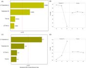
Pareto diagrams (A and C) and effects plots (B and D) for the pulp and husk of Dioscorea bulbifera L., respectively
In addition, statistical analysis (ANOVA) was performed for both the pulp and husk of Dioscorea bulbifera L., which confirmed that temperature was the most influential factor affecting the response variable, with clear statistical significance (Table 3).
Analysis of variance (ANOVA) results for the Box-Behnken 32 experimental design applied to the husk and pulp of Dioscorea bulbifera L.
It can be observed that, for both the pulp and the husk of Dioscorea bulbifera L., the overall model was highly significant, with p < 0.0001 for the mean/intercept. Regarding the pulp, the ANOVA results showed that only the linear effect of temperature (p = 0.0045) was statistically significant, indicating that increasing temperature had a relevant impact on the response variable. The quadratic effects and those related to processing time, on the other hand, were not significant (p > 0.05), suggesting that time did not substantially influence the response under the evaluated conditions. As for the husk, both the linear (p = 0.0007) and quadratic (p = 0.0068) effects of temperature were significant, indicating a nonlinear relationship between temperature and the response, with a possible optimal point. Conversely, the linear and quadratic effects of processing time were not statistically significant in either sample (p > 0.65), demonstrating that time had little to no influence on the response variable under the tested conditions, reinforcing that temperature is also the main determining factor for the husk.
The analysis of the bioactive potential also took place in relation to the levels of total flavonoids and total carotenoids, as shown in Table 4, but only with the extracts that presented the best results according to the experimental design, that is, for the husk at 80°C for 60 minutes (E9) and for the pulp at 70°C for 45 minutes (E5).
Content of total flavonoids and total carotenoids for husk and pulp of Dioscorea bulbifera L.
The husk and pulp fractions showed considerable levels of total flavonoids, with the skin having the highest result, 363.63 mg CE 100 g-1, when compared to the pulp. Flavonoids represent the predominant class of active compounds, accounting for approximately 39.6% of the total bioactive constituents. These metabolites are biosynthesized by plants predominantly via the shikimic acid and mevalonate pathways. Notably, flavonoids exert potent antioxidant activity by neutralizing reactive oxygen and nitrogen species (ROS and RNS), thereby contributing to cellular protection and playing a significant role in the prevention of oxidative stress-related diseases [36,37].
In relation to total carotenoids, the available content in the extracts was estimated, and the husk extract of Dioscorea bulbifera L. had the highest content, about 2.13 µg 100 g-1, when compared to the pulp extract. Carotenoids are fat-soluble pigments with a polyene chemical structure consisting of conjugated double bonds, enabling these compounds to absorb excess energy from other molecules and eliminate reactive oxygen species (ROS) and free radicals. Thus, through the results of the analysis of total phenolic compounds, total flavonoids and total carotenoids, there is an indication of bioactivity of the husk and pulp fractions of Dioscorea bulbifera L.
Antioxidant profile of bioactive compounds
The antioxidant capacity can be identified from different mechanisms of antioxidant action; thus, it is important that its evaluation is done by more than one methodology. Accordingly, the evaluation was conducted using both hydrogen atom transfer (HAT) and single electron transfer (SET) mechanisms. The HAT mechanism was assessed through the β-carotene/linoleic acid auto-oxidation system, while the SET mechanism was evaluated using the FRAP and hydrogen peroxide (H₂O₂) scavenging assays. Additionally, the ABTS assay was employed, as it encompasses both HAT and SET mechanisms simultaneously [38,39].
In the present study, the hydrogen peroxide removal, ABTS, FRAP and β-carotene/linoleic acid assays were used to estimate the antioxidant capacity of the husk and pulp extracts of Dioscorea bulbífera L. (Table 5). It is important to highlight that the ABTS assay enables the assessment of antioxidant capacity through the scavenging of the 2,2'-azinobis (3-ethylbenzothiazoline-6-sulfonic acid) free radical, a stable species generated via chemical or enzymatic reactions. This method is suitable for estimating the antioxidant activity of both hydrophilic compounds, such as phenolics and lipophilic compounds, such as carotenoids [40]. In the β-carotene/linoleic acid autoxidation system, antioxidant activity is evaluated based on the inhibition of peroxyl radicals (LOO•) generated by the reaction of linoleic acid with oxygen. These radicals normally interact with β-carotene, leading to its discoloration. However, if antioxidants are present in the sample, they react with the peroxyl radicals, thereby delaying the decrease in β-carotene absorbance [41].
The FRAP method evaluates the antioxidant activity by reducing the ferric-tripyridyltriazine complex (Fe3+-TPZ) to the ferrous complex (Fe2+-TPZ), a complex that has an intense blue-violet color, being considered an assay with high reproducibility and high correlation with compounds phenolics. Based on the results obtained from the antioxidant capacity analyses, it can be inferred that both the husk and pulp fractions of Dioscorea bulbifera L. exhibit a significant antioxidant profile, primarily associated with the presence of compounds such as carotenoids and total phenolic compounds.
Study of simulated gastrointestinal digestibility
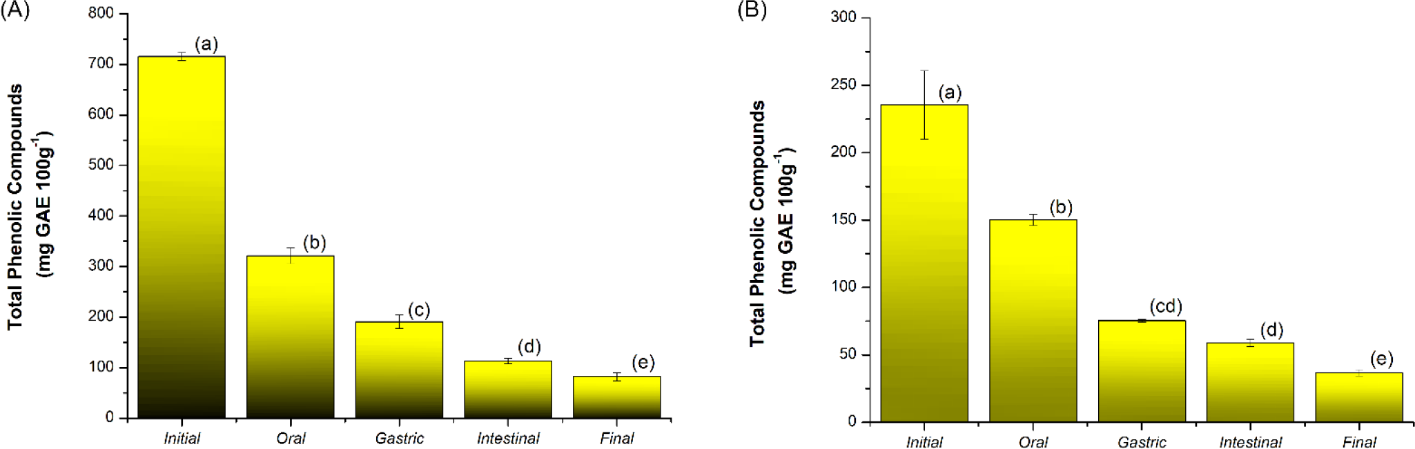
Simulating gastrointestinal digestion provides insights into the behavior and bioaccessibility of bioactive compounds within a food matrix during the digestive phases, offering valuable information for both researchers and the food industry [42]. Thus, the contents of total phenolic compounds (TPC) were evaluated for the husk and pulp fraction of Dioscorea bulbifera L. for each phase of the simulated digestion, as shown in Figure 5. From the simulated gastrointestinal digestion process, it can be seen that the TPC content decreased considerably when comparing the results at the beginning of the digestion process, during the process until the final phase for both the husk (Figure 5A) and the pulp (Figure 5B) of Dioscorea bulbifera L. TPC levels ranged from 715 to 82 mg EAG 100 g-1, and in all phases there was a statistical difference (p ≤ 0.05) between the values of these bioactive compounds.
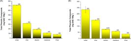
Total phenolic compounds of the fraction of the husk (A) and pulp (B) of Dioscorea bulbifera L. at each phase of simulated gastrointestinal digestibility.
The TPC contents of the pulp fraction of Dioscorea bulbifera L. showed similar behavior to that of the husk fraction, with a decrease in values from 235 to 37 mg EAG 100g-1, however, between the gastric and intestinal phases there was no significant difference by the Tukey test (p ≤ 0.05). It can be observed that in the husk and pulp fractions there was a greater loss of oral digestion in relation to the other phases, however, there was no difference in the profile of the percentage of bioavailability.
In the husk and pulp fractions, there was a greater loss in oral digestion, with no differences in the profile of the percentage of bioavailability, showing a decrease at each stage of the simulated digestion. A factor that may be directly related to the nature of the phenolic compounds, about their interactions with simulated fluids, mainly in relation to their instability when subjected to pH variations.
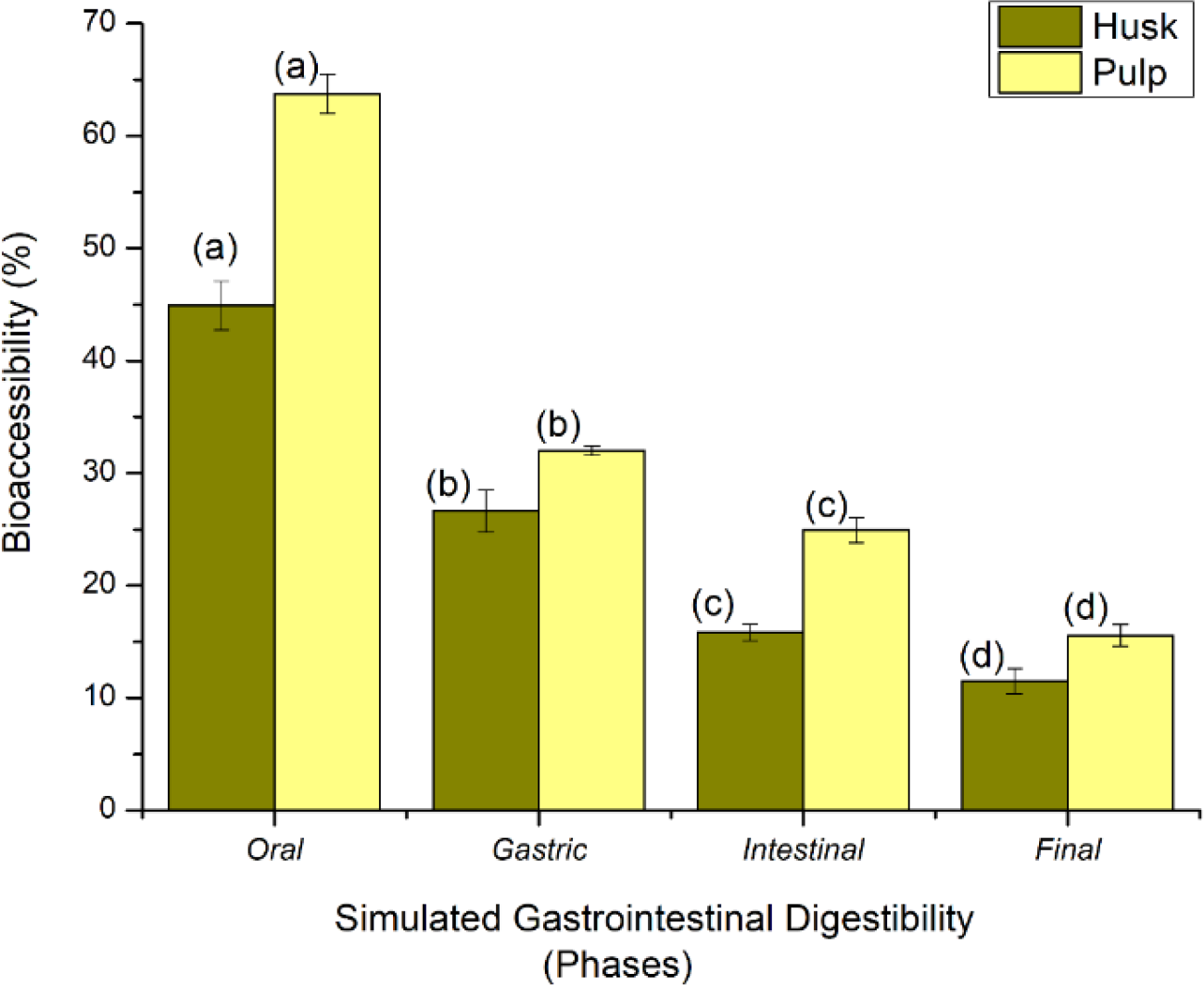
Regarding bioaccessibility, it is known that it is a parameter related to the ability of a compound extracted from the food matrix to be absorbed by intestinal cells after the gastrointestinal digestion process. In this sense, from the TPC results, it can be observed that approximately 16% of the total phenolic compounds in the husk and 25% of the pulp were considered bioaccessible to be absorbed in the intestine, and that despite the husk having a higher TPC content, the pulp fraction had a higher bioaccessibility (Figure 6).
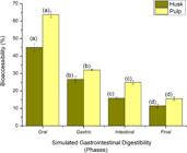
Bioaccessibility of total phenolic compounds in husk and pulp fractions for each stage of simulated gastrointestinal digestion.
The results of the husk and pulp fraction may have been influenced by the interaction of phenolic compounds with the structure of the food matrix. In this sense, the higher the fiber content, the lower the TPC content, as the fibrous structure makes it difficult to release phenolic compounds that are associated with it. Furthermore, it is possible that the phenolic compounds present in the samples interacted with other constituents, such as proteins, polysaccharides, or even components of the simulated digestive system, potentially affecting their stability [43]. Certain phenolic compounds found in human foods may exhibit reduced stability under high pH conditions due to their specific structural characteristics. Investigating these properties can aid in predicting the stability of individual phenolic compounds across varying pH environments. Additionally, some phenolic compounds may undergo structural transformations under acidic conditions, which can hinder their detection by the Folin-Ciocalteu assay [44].
Studies indicate that the stability of phenolic compounds is strongly dependent on their chemical structure. Phenolic acids tend to be resistant to digestive processes, whereas flavonols are primarily degraded during digestion. Throughout the digestive process, total phenolic compounds (TPC) may be degraded, released from the food matrix, or bound to other compounds or constituents present in the digestive fluids [45]. Moreover, merely 5 to 10% of the total phenolic compounds present in a food matrix are able to reach the small intestine [46], thus the TPC content present in both Dioscorea bulbifera L. fractions showed significant bioaccessibility for be absorbed in the intestine.
CONCLUSION
In this context, it is concluded that the combination of the ultrasound-assisted technique with the solid-liquid extraction method, provided the extraction of bioactive compounds from Dioscorea bulbifera L. The fractions of husk and pulp showed bioactive potential, according to the antioxidant activity methods (hydrogen peroxide, ABTS, FRAP and β-carotene/linoleic acid removal) as well as demonstrated bioaccessibility through simulated gastrointestinal digestion. Therefore, Dioscorea bulbifera L. husk and pulp fractions may be potential sources for isolation and purification of bioactive compounds, as well as applications in the food industry.
-
Funding:
This research received no external funding.
Acknowledgments:
The authors gratefully acknowledge the support of CAPES.
Data availability statement:
Research data are available in the body of the manuscript.
REFERENCES
- 1 Godfrey EO, Esther II, Faith O. Proximate Composition, Levels of Some Essential Mineral Elements and Anti-Nutritional Components of Some Yam Species Found in Minna, Niger State. Biol Med Nat Prod Chem. 2022; 12:9-16.
- 2 Milião GL, Oliveira APH, Soares LS, Arruda TR, Vieira ÉNR, Leite Junior BRC. Unconventional Food Plants: Nutritional Aspects and Perspectives for Industrial Applications. Future Foods. 2022; 5:100124.
- 3 Setyawan N, Maninang JS, Suzuki S, Fuji Y. Variation in the Physical and Functional Properties of Yam (Dioscorea Spp.) Flour Produced by Different Processing Techniques. Foods. 2021; 10(6):10061341.
- 4 Uchenna CJ, Morenike DF, Chioma OL. Activation of Cotyledon as Nutrients and Antioxidant Enhancer in Oryza Sativa, Dioscorea Bulbifera and Phaseolus Vulgaris for Food Formulation. Sci Dev. 2022; 3(1):31-45.
- 5 Carneiro GR, Crema NM, Züge LCB, Ribeiro LF. Physical and Chemical Evaluation of Husk and Pulp Flours of Dioscorea Bulbifera L. with the Possibility of Application in Bakery. Braz J Dev. 2020;6(12):96201-11.
- 6 Yadav SA, Hasan S, Gnanaselvan S, Baskaran S, Danaraj J. Biological Activities and Nanoparticle Synthesis of Dioscorea Bulbifera and Its Mechanistic Action - An Extensive Review. Pharm Nanotechnol. 2024;12(5):379-90.
- 7 Park SY, Truong VL, Jeon SG, Choe SY, Rarison RHG, Yoon BH, et al. Anti-Inflammatory and Prebiotic Potential of Ethanol Extracts and Mucilage Polysaccharides from Korean Yams (Dioscorea Polystachya and Dioscorea Bulbifera). Foods. 2025;14(2):173.
- 8 Chaniad P, Tewtrakul S, Sudsai T, Langyanai S, Kaewdana K. Anti-Inflammatory, Wound Healing and Antioxidant Potential of Compounds from Dioscorea Bulbifera L. Bulbils. PLoS One. 2020;15(12):e0243632.
- 9 Yang Z, Amrit BK, Zhao W, Shi L, Wu H, Barrow C, et al. Bioaccessibility and Bioavailability Changes of Phenolic Compounds in Pumpkins (Cucurbita Moschata): A Review. Food Biosci. 2022;47:101753.
- 10 Melini V, Melini F, Acquistucci, R. Phenolic Compounds and Bioaccessibility Thereof in Functional Pasta. Antioxidants. 2020;9(4):343.
- 11 Silva MM, Reboredo FH, Lidon FC. Food Colour Additives: A Synoptical Overview on Their Chemical Properties, Applications in Food Products and Health Side Effects. Foods. 2022;11(3):379.
- 12 Miskinis RAS, Nascimento LÁ, Colussi R. Bioactive Compounds from Acerola Pomace: A Review. Food Chem.2023;404:134613.
- 13 Dima C, Assadpour E, Nechifor A, Dima S, Li Y, Jafari SM. Oral Bioavailability of Bioactive Compounds; Modulating Factors, in Vitro Analysis Methods, and Enhancing Strategies. Crit Rev Food Sci Nutr. 2024;64(24):8501-39.
- 14 Rodrigues DB, Marques MC, Hacke A, Loubet Filho PS, Cazarin CBB, Mariutti LRB. Trust Your Gut: Bioavailability and Bioaccessibility of Dietary Compounds. Curr Res Food Sci. 2022;5:228-33.
- 15 Núñez-Gómez V, González-Barrio R, Periago MJ. Interaction between Dietary Fibre and Bioactive Compounds in Plant By-Products: Impact on Bioaccessibility and Bioavailability. Antioxidants. 2023;12(4):976.
- 16 Oracz J, Nebesny E, Zyzelewicz D, Budryn G, Luzak B. Bioavailability and Metabolism of Selected Cocoa Bioactive Compounds: A Comprehensive Review. Crit Rev Food Sci Nutr. 2020;60(12):1947-85.
- 17 Demarco M, Moraes JO, Matos ÂP, Derner RB, Neves FF, Tribuzi G. Digestibility, Bioaccessibility and Bioactivity of Compounds from Algae. Trends Food Sci Technol. 2022;121:114-28.
- 18 Yusoff IM, Mat Taher Z, Rahmat Z, Chua LS. A Review of Ultrasound-Assisted Extraction for Plant Bioactive Compounds: Phenolics, Flavonoids, Thymols, Saponins and Proteins. Food Res Int. 2022;157:111268.
- 19 Didion YP, Tjalsma TG, Su Z, Malankowska M, Pinelo M. What is next? The Greener Future of Solid Liquid Extraction of Biobased Compounds: Novel Techniques and Solvents Overpower Traditional Ones. Sep Purif Technol. 2023;320:124147.
- 20 Fu X, Wang D, Belwal T, Xie J, Xu Y, Li L, et al. Natural Deep Eutectic Solvent Enhanced Pulse-Ultrasonication Assisted Extraction as a Multi-Stability Protective and Efficient Green Strategy to Extract Anthocyanin from Blueberry Pomace. LWT - Food Sci Technol. 2021;144:111220.
- 21 Siddiqui H, Sultan Z, Yousuf O, Malik M, Younis K. A Review of the Health Benefits, Functional Properties, and Ultrasound-Assisted Dietary Fiber Extraction. Bioactive Carbohydr Diet Fibre. 2023;30:100356.
- 22 Wang J, Xie B, Sun Z. Quality Parameters and Bioactive Compound Bioaccessibility Changes in Probiotics Fermented Mango Juice Using Ultraviolet-Assisted Ultrasonic Pre-Treatment during Cold Storage. LWT - Food Sci Technol. 2021;137:110438.
- 23 Hadidi M, Ibarz A, Pouramin S. Optimization of Extraction and Deamidation of Edible Protein from Evening Primrose (Oenothera Biennis L.) Oil Processing by-Products and Its Effect on Structural and Techno-Functional Properties. Food Chem. 2021;334:127613.
- 24 Singleton VL, Rossi JA. Colorimetry of Total Phenolics with Phosphomolybdic-Phosphotungstic Acid Reagents. Am J Enol Vitic. 1965;16:144-58.
- 25 Meyers KJ, Watkins CB, Pritts MP, Liu RH. Antioxidant and Antiproliferative Activities of Strawberries. J Agric Food Chem. 2003;51:6887-92.
- 26 Lichtenthaler HK. [34] Chlorophylls and Carotenoids: Pigments of Photosynthetic Biomembranes. In: 1987; pp. 350-382.
- 27 Re R, Pellegrini N, Proteggente A, Pannala A, Yang M, Rice-Evans C. Antioxidant Activity Applying an Improved ABTS Radical Cation Decolorization Assay. Free Radic Biol Med. 1999;26(9-10):1231-7.
- 28 Miller HE. A Simplified Method for the Evaluation of Antioxidants. J Am Oil Chem Soc. 1971;48:91.
- 29 Thaipong K, Boonprakob U, Crosby K, Cisneros-Zevallos L, Hawkins BD. Comparison of ABTS, DPPH, FRAP, and ORAC Assays for Estimating Antioxidant Activity from Guava Fruit Extracts. J Food Compos Anal. 2006;19(6-7):669-75.
- 30 Dantas AM, Mafaldo IM, Oliveira PML, Lima MS, Magnani M, Borges GSC. Bioaccessibility of Phenolic Compounds in Native and Exotic Frozen Pulps Explored in Brazil Using a Digestion Model Coupled with a Simulated Intestinal Barrier. Food Chem. 2019;274:202-14.
- 31 Bornhorst GM, Singh RP. Kinetics of in Vitro Bread Bolus Digestion with Varying Oral and Gastric Digestion Parameters. Food Biophys. 2013;8:50-9.
- 32 Tzia C, Liadakis G. Extraction Optimization in Food Engineering. CRC Press, 2003; ISBN 9780824741082.
- 33 Damodaran S, Parkin KL, Fennema OR. Fennema’s Food Chemistry; 4th ed.; CRC Press, 2007; ISBN 978-0849392726.
- 34 Xie S, Lei Y, Chen H, Liu S, Lin X, Guo Z, et al. Physicochemical Properties and Structural Study of Heat Treatment-Modified Chinese Yam (Dioscorea Opposita Thunb.) Starch-Ferulic Acid Complexes. Foods. 2025;14(10):1761.
- 35 Franková H, Musilová J, Árvay J, Šnirc M, Jančo I, Lidiková J, et al. Changes in Antioxidant Properties and Phenolics in Sweet Potatoes (Ipomoea Batatas L.) Due to Heat Treatments. Molecules. 2022;27(6):1884.
- 36 Zahra M, Abrahamse H, George BP. Flavonoids: Antioxidant Powerhouses and Their Role in Nanomedicine. Antioxidants. 2024;13(8):922.
- 37 Martins T, Fonseca BM, Rebelo I. Antioxidant Effects of Chalcones during the Inflammatory Response: An Overall Review. Curr Med Chem. 2021;28(37):7658-713.
- 38 Gulcin İ. Antioxidants: A Comprehensive Review. Arch Toxicol. 2025;99(5):1893-997.
- 39 HDT, M. Advancing In Vitro Antioxidant Activity Assessment: A Comprehensive Methodological Review and Improved Approaches for DPPH, FRAP and H2O2 Assays. J Nat Ayurvedic Med. 2023;7(4):000431.
- 40 Sadeer NB, Montesano D, Albrizio S, Zengin G, Mahomoodally MF. The Versatility of Antioxidant Assays in Food Science and Safety-Chemistry, Applications, Strengths, and Limitations. Antioxidants. 2020;9(8):709.
- 41 Abeyrathne EDNS, Nam K, Ahn DU. Analytical Methods for Lipid Oxidation and Antioxidant Capacity in Food Systems. Antioxidants. 2021;10(10):1587.
- 42 Gonçalves SM, Freitas-Silva O, Mariutti LRB, Teodoro AJ. A Review of in Vitro Methods to Evaluate the Bioaccessibility of Phenolic Compounds in Tropical Fruits. Crit Rev Food Sci Nutr. 2024;64(6):1780-90.
- 43 Tomé-Sánchez I, Martín-Diana AB, Peñas E, Frias J, Rico D, Jiménez-Pulido I, et al. Bioprocessed Wheat Ingredients: Characterization, Bioaccessibility of Phenolic Compounds, and Bioactivity During in Vitro Digestion. Front Plant Sci. 2021;12:790898.
- 44 Pasquet PL, Julien-David D, Zhao M, Villain-Gambier M, Trébouet D. Stability and Preservation of Phenolic Compounds and Related Antioxidant Capacity from Agro-Food Matrix: Effect of PH and Atmosphere. Food Biosci. 2024,57:103586.
- 45 Li CX, Wang FR, Zhang B, Deng ZY, Li HY. Stability and Antioxidant Activity of Phenolic Compounds during in Vitro Digestion. J Food Sci. 2023;88(2):696-716.
- 46 Dima C, Assadpour E, Dima S, Jafari SM. Bioavailability and Bioaccessibility of Food Bioactive Compounds; Overview and Assessment by in Vitro Methods. Compr Rev Food Sci Food Saf. 2020;19(6):2862-84.
-
Editor-in-Chief:
Bill Jorge Costa
-
Associate Editor:
Bill Jorge Costa