Design, Construction and Instrumentation of Photobioreactor for Microalgae
Abstract
The cultivation of microalgae has been a biotechnological technique with many advances throughout history; however, depending on the growth conditions, the production of carbohydrates, proteins, lipids, vitamins, polyphenols and terpenes (metabolites primary and secondary) has been favored or affected. This work consists of designing and building a photobioreactor with internal blue LED light to monitor the pH, temperature, carbon dioxide (CO2) concentration and light intensity in real time to increase the current production yields of biomass and metabolites with great potential for biotechnological applications. With the construction of the photobioreactor, the growth kinetics of the microalgae C. vulgaris were determined by applying the Taguchi experimental statistical methodology (mixotrophic medium) to evaluate its antioxidant activity. Like independent variables: substrate concentration (sodium nitrate, NaNO3) and light exposure time (photoperiod). The dependent variables were as follows: cell growth, biomass, substrate consumption (UV‒vis, 301 λ), and metabolites with antioxidant activity (ABTS•+ and DPPH• radical methods). In conclusion, the construction of a photobioreactor for microalgae cultivation allows greater control of growth factors such as CO2, pH, temperature and dissolved O2 and allows the microalgae growth medium to be isolated from the outside and thus avoid contamination by microbial agents from the environment. Similarly, the best growth condition for C. vulgaris with antioxidant activity is a concentration of 3.6 mM NaNO3 and a low photoperiod time of 8 hours of light and 16 hours of darkness, generating an equivalent response of Trolox 16 µM/mL for ABTS•+ and equivalents of Trolox 6 µM/mL for DPPH•.
Keywords:
Yields; metabolites; real time; scaling; Taguchi; optimization.
HIGHLIGHTS
Construction of a photobioreactor that allows establishing optimal conditions for microalgae growth.
Mixotrophic growth with internal blue-LED light source at photobioreactor level.
Effect of sodium nitrate concentration as substrate and photoperiod time on cell growth of C. vulgaris.
Evaluation of antioxidant activity in C. vulgaris microalgae.
INTRODUCTION
Microalgae can be autotrophic or heterotrophic. The first group is where organisms use inorganic compounds as a carbon source, which in turn can be photoautotrophs; that is, they use light as energy to obtain equally energetic compounds that serve them for their development, or chemoautotrophs, that is, through the oxidation of inorganic compounds, they synthesize energetic molecules [1]. Among the main molecules that are synthesized through any type of metabolism, starch and lipids are considered primary metabolites, which are those that are formed for the main functions of the organism. Another type of primary metabolite that is synthesized in the same way is proteins, which are obtained via the metabolism of nitrated compounds such as ammonium [2]. Therefore, the chemical composition of microalgae is directly influenced by the carbon source used as a substrate for the synthesis of primary metabolites [3,4]. These metabolites have important applications, such as in biomass production, pigments, biodiesel, and biogas and the extraction of useful compounds in cosmetic products [5].
The growth of different genera and species of microalgae, as well as the yield of metabolites that are useful for different economic activities, depends on various factors, such as the type and intensity of light, temperature, pH, available amount of O2 and CO2, nutrients, etc. [6]. In this way, there is a need for means and equipment that promote the growth and production of microalgae. Furthermore, these methods maximize the extraction of any metabolite with potential application at the industrial level. In the following work, the design, instrumentation and construction of a photobioreactor are presented with adjustable biosensors for the volume of the growth medium of the microalgae. These sensors are designed for monitoring the dissolved oxygen content, pH and temperature, with real-time monitoring through a data acquisition card connected to a computer where with the help of a graphic program, the REAL-TIME signal of each of the parameters can be recorded. In addition, the photobioreactor differs from the other sensors because the type of LED light arrangement is in the range of blue colors (wavelengths close to 400 to 450 nm) and because it also has a light intensity sensor external to the photobioreactor, with which it will be possible to know the trajectory, especially the amount of light that passes through the culture medium, which is essential for high-yield photosynthesis [7].
MATERIAL AND METHODS
Photobioreactor
The first was the design of the photobioreactor through SolidWorks 2019 software, with which the stainless-steel metal components and the different finishes that are required in them were designed, such as the input and output holes for sensors and the collection of biological samples. In addition, the plastic components of the photobioreactor, such as the dissolved oxygen (O2), pH, temperature and carbon dioxide (CO2) microbubble diffuser sensors, were digitized; with these sensors, the disposition of the components for data acquisition for the growth of microalgae remains under an arrangement that allows monitoring of the growth conditions in real time and is favorable. Finally, through the same software, an LED light source was designed in the blue color range, with which the best effect was sought for the development of microalgae photosynthesis, which was optimized [8].
For the photobioreactor instrumentation, an embedded system data acquisition card was used so that, through the graphic interface program, the voltage in the sensors was detected in real time for pH, temperature, dissolved oxygen (O2) and LED light sources, which were converted into measurable parameters through the construction of calibration curves, and the growth kinetics were carried out.
The calibration curves were built from solutions of known concentrations of the analytes to be measured; that is, for the temperature sensor, distilled water was cooled and heated in a temperature range of 0 to 94 °C in triplicate, and by means of linear regressions, the equation of the straight line with which the measurable value, or the answer, in the problem solutions was obtained. In the case of the calibration curve of the dissolved oxygen (O2) sensor, distilled water was sterilized to eliminate the oxygen dissolved in water [9]; later, it was subjected to agitation until saturation, which could be determined when there were no variations in voltage in the sensor. The corresponding linear regression was performed, and the corresponding equation was established to calculate the concentration in % of dissolved oxygen in mg/L [9].
Finally, for the pH sensor, a calibration curve was constructed with a Buffers scale in the pH range of 1 to 11.5, the corresponding equation was obtained, and it was validated with commercial problem samples of the Golden-Bell® brand. In the case of the light source, the measurement of the Luxes was carried out in a range of 0 to 1120 by varying them through the graphic interface program interface, and with this, it was possible to program the adjustment in real time of the light intensity by entering the dataset by the user.
To construct the calibration curve of the blue LED light source, a light-insulating plastic cylinder 17 cm in diameter and 45 cm in height was constructed. The tube has a perforation on one side of an approximate size of 6 cm wide by 25 cm high where the lux meter was placed to measure the light source on the part: upper front, lower front, side top right, bottom right, top left, bottom left, and top and bottom back. By opening the cylinder, it was only possible to place the lux detection sensor with the MASTECH® Digital Light Meter instrument (MS6612), and each observation was taken in triplicate at different points for greater precision.
Specifically, the accessory devices of the photobioreactor include a thermocouple with J-type connections, a pH electrode from the HANNA® instrument brand of technology input, a sensor for dissolved oxygen from the DFROBOT® brand and an LED light source that is emitted under a blue wavelength and is described in detail in the design and instrumentation section, each of which was calibrated and connected to a circuitry designed under the PROTEUS® 8.0 program.
Microbial kinetics
Growth kinetics were carried out at 10 days under a Taguchi L4 (22) experimental design where the independent variable was the concentration of NaNO3, which was 3.4 mM or 16.4 mM, as well as the photoperiod, which was 8 hours of light and 16 hours of darkness (8:16). The second photoperiod was 16 hours of light and 8 hours of darkness (16:8). CO2 injections were performed every 24 h for 20 s at a flow of 5 mg/mL such that there was mixotrophic growth and the medium was not acidified, since if this occurred, it can cause the cessation of cell growth and even microbial death. For the design of the experiments, the response variables used were as follows: cell growth by the counting method in a Neubauer chamber, biomass by dry weight, substrate consumption by light absorption of nitrate ions in the growth media, pH, oxygen dissolved in the medium, and antioxidant activity through the stability of the radicals of ABTS•+ and DPPH•.
Growth cell: To analyze the formation of new cells during kinetics, a low microscopic count was performed with a bright-field Neubauer camera on the No. 40 objective, where it was not necessary to stain the cells with a dye such as methylene blue since it is possible to distinguish viable cells from those with damaged cell walls owing to the intense green color that they present [10].
Biomass: This parameter was determined via the dry weight technique, where with the help of aluminum trays at constant weight (initial weight), 1 mL of sample was heated in an electric oven for 24 hours at a temperature of 60 °C. After that, the tray was weighed again (final weight) with the sample, and on the basis of the difference in weight (final weight - initial weight), the weight of the cells taken in the sample per millilitre of medium was calculated [11].
Substrate consumption: To analyze the amount of organic substrate that the microalga consumes and that was transformed into primary and secondary metabolites, a calibration curve of known concentrations of NaNO3 ranging from 0 to 50 mM was constructed, and through absorbance readings via spectrophotometry at a wavelength of 301 nm, it was possible to quantify the amount of nitrate ions dissolved in the reaction medium, which could be used to establish how much nitrate is available in the medium and that it was translated to a substrate not consumed by the microalgae.
pH: pH readings were carried out at time zero as a reference. For this purpose, a HANNA® Instrument brand potentiometer was necessary, for which a sample volume of 5 mL was required to be contained in 25 mL Falcon tubes.
Dissolved oxygen concentration: Through the ROOT® Oxygen sensor, the dissolved oxygen concentration was monitored in real time and was determined to be less than 30% in the medium. This sensor was calibrated for saturation in the medium depending on its agitation [12].
Antioxidant activity: To quantify the products formed, the ABTS•+ and DPPH• techniques were carried out, which indirectly measure the concentration of polyphenolic compounds (mostly) with said activity. To do this, cell rupture was carried out with glass beads by washing with deionized water under a centrifuge at 4000 rpm for 5 minutes. This operation was carried out three times to eliminate the salts dissolved in the medium. The volume of deionized water used in washing was 1 mL. Once the cells were washed, they were contacted with glass beads suspended in 1 mL of deionized water in a centrifuge at 4000 rpm to subsequently collect the supernatant and evaluate the stability of the synthetic radicals ABTS•+ and DPPH•. For the ABTS•+ method, an ABTS•+ radical solution was prepared at a concentration of 7 mM, an aqueous solution of ammonium persulfate at a concentration of 2.5 mM, and a stock of Trolox at a concentration of 80 μM/mL. A calibration curve was subsequently generated for Trolox equivalents (μM/mL) at a wavelength of 734 nm. Once the calibration curve was constructed, the percentage of inhibition of the ABTS•+ radical was calculated when it was in contact with the Trolox solution, and the concentration in Trolox equivalents was also calculated. To determine the activity of the sample, a volume of 10 μL of sample was diluted with 990 μL of adjusted ABTS•+ solution, and the corresponding readings and calculations were performed to express the results in equivalents of Trolox g/L [13]. For the DPPH• method, a radical solution of DPPH• was prepared at a concentration of 0.046 g/L, and a stock of Trolox was prepared at 80 μM/mL. A calibration curve was subsequently generated for Trolox equivalents (μM/mL) at a wavelength of 515 nm. Once the calibration curve was constructed, the percentage of inhibition of the DPPH• radical was calculated when it was in contact with the Trolox solution, and the concentration in equivalents of Trolox was also calculated. To determine the activity of the sample, 100 μL of sample was diluted with 3900 μL of adjusted DPPH• solution. The corresponding readings and calculations were performed to express the results in Trolox equivalents (μM/mL) [14].
RESULTS
The results obtained are presented below, addressing various areas that involve the design and construction of the photobioreactor for microalgae growth, as well as tests that validate the functionality of the equipment in such a way that one can be certain of its application at a biotechnological level.
Mechanical design
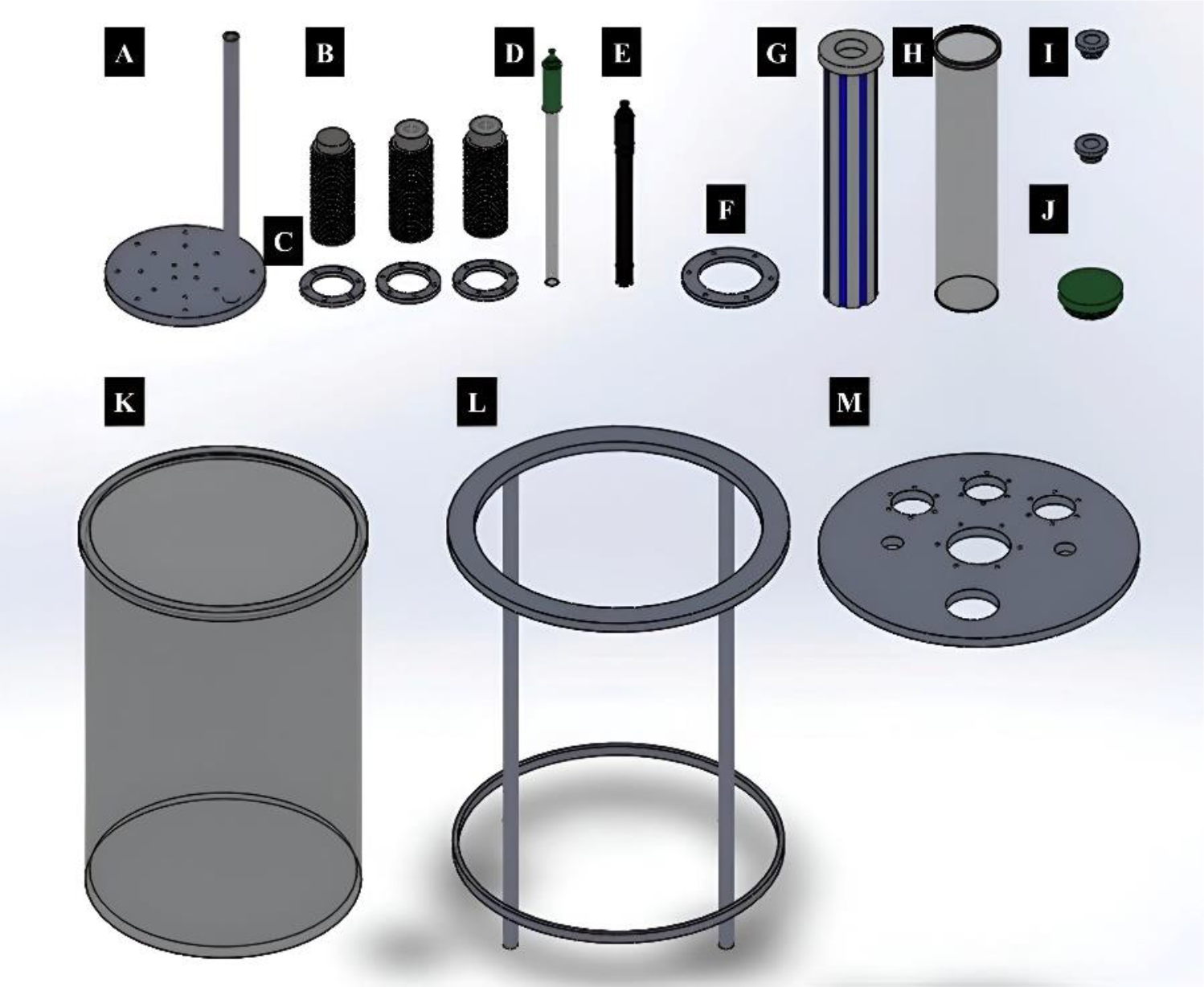
The proposed photobioreactor consists of two stainless steel covers, which are referred to as "cover A" and "cover B" or stainless-steel covers. Cover A is welded to a stainless-steel support that has four rods that provide support and stability to the photobioreactor. Cover A will receive a Pyrex® glass container or "growth medium container", which will contain the microalgae with all the nutrients and other abiotic factors that favor their growth. Once the container is placed, lid cover B is placed above the glass vessel, which has a plurality of sensors that measure the pH, temperature, and oxygen, as well as a CO2 diffuser, to collect samples and, in its center, the LED light source. The sensors are connected, as described above, to the embedded system data acquisition card, which allows real-time monitoring of electrical signals in the culture medium, which makes it different from others previously developed. Figure 1 shows the components that make up the photobioreactor.
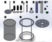
Growth photobioreactor of microalgae with real-time monitoring of pH, temperature and dissolved oxygen (O2), as well as light intensity control. A) Diffuser. B) Holder for pH, temperature and dissolved oxygen sensors. C) Sensors covering (B). D) pH sensor. E) Dissolved oxygen sensor. F) Blue LED light source cover. G) Blue LED light source. H) Internal vessel for the light source. I) PLA plugs for sample collection and CO2 injection. J) PLA cap for future accessories. K) External vessel for microalgae growth medium. L) Support for a photobioreactor with 4 posts. M) Stainless steel cover.
Cover M is intended to cellar the microalgae culture medium, as well as to place all the accessories (sensors for dissolved oxygen, temperature, pH, a sampling hose, a carbon dioxide inlet hose that connects with a diffuser and, finally, the LED light source). In the center of cover B, the blue LED light source is placed, which provides the light energy necessary to carry out photosynthesis and where the type of photoperiod can be varied in addition to the intensity.
Through the graphic interface program, it is possible to modulate the intensity of light in such a way that it can be adapted to the type of microalgae that is intended to grow, since not all varieties of these photosynthetic organisms react in the same way to exposure to light, which does not imitate its use to a single type of microalgae.
Finally, the LED light array in Figure 1 is inserted into a Pyrex® glass tube called the "inner tube", which protects it from damage due to immersion of the light source in the culture medium, which would cause a short circuit. In turn, this internal tube is placed inside an "external vessel" with a capacity of 10 L of the same material (Pyrex® glass), which will contain the growth medium for the microalgae and a diffuser for CO2 microbubbles, the microalgae and sensors for the detection of biochemical signals. Once the photobioreactor was designed, the acquisition of the different sensors, stainless steel materials, Pyrex® glass and the embedded system data acquisition card, the factory and the instrumentation started.
Instrumentation
To detect the varying responses in the photobioreactor and translate them, it was necessary to acquire the embedded system card, which, through voltage measurements by pH sensors, temperature, dissolved oxygen and light intensity, can be used to monitor conditions in real time.
The embedded system card is an analog input (AI), analog output (AO), digital entry and output. This card connects to the feed computer through USB and wireless connections (WiFi). A and B account for connectors of the expansion port (MXP). Already within the graphic program, the signals are distinguished with the names of the connectors CONNECTORA/DIIO1 and CONNECTORB/DIIO1. Analog connectors or inputs can work in a range of 0 to 10 volts, which connects the necessary sensors for the photobioreactor.
Once the card was comforted and connected to a computer through the graphic interface program, the voltage detected with multiple sensors could be shown.
A) Thermocouple
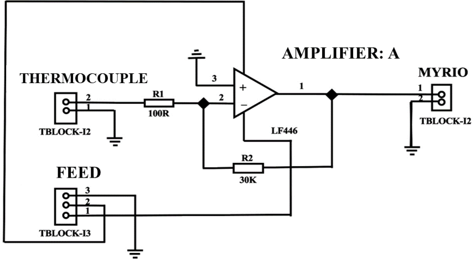
The thermocouple used has CEIV® brand connections with a 3/6” bulb diameter and a 1 m cable extension; its coating is fiberglass, and the cable exterior coating is a metal mesh that can resist temperatures up to 5000 °C. For the thermocouple connection, it was necessary to design and build the circuit to detect the variation in voltage on the embedded system card, for which the following design for the construction of the electronic plate in Figure 2 was used.
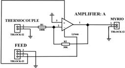
Circuit designed for thermocouple connection and the embedded system data acquisition card in the photobioreactor for microalgae growth.
The thermocouple is fed by the source of power to 24 volts, and from this sensor, the other sensors used in the photobioreactor are connected in series.
B) Dissolved oxygen electrode (O2)
The sensor used for the detection of dissolved oxygen in aqueous medium was a dissolved oxygen sensor SEN0237-A compatible with Arduino®, which contains 3 connections: GND, VCC and an analog output; its detection range is 0 to 20 mg/L. The connections are of type BNC and operate 5 volts. Through this sensor, it is possible to obtain measurements in mg/L and %. On the other hand, this sensor already has its factory electronic card, so the circuitry design is not necessary, so only the connection was made directly to the CONNECTORB/DIIO1 port for its calibration in the graphic program and, subsequently, its validation.
C) pH electrode
This electrode is from the HANNA® Instruments brand and was calibrated with commercial tampons from the SIGMA® brand for pH values of 4 and 7. It works at 12 volts and has an analog input BNC compatible with the Embedded System Card data acquisition. For the instrumentation, this sensor was directly connected to port CONNECTORB/DIIO1 of the card as well as a source of power to 24 volts, which feeds all the photobioreactor sensors; once connected, the voltage variations were recorded through the graphic interface program, and with them, a calibration curve that will later be described was convenient.
D) Light intensity
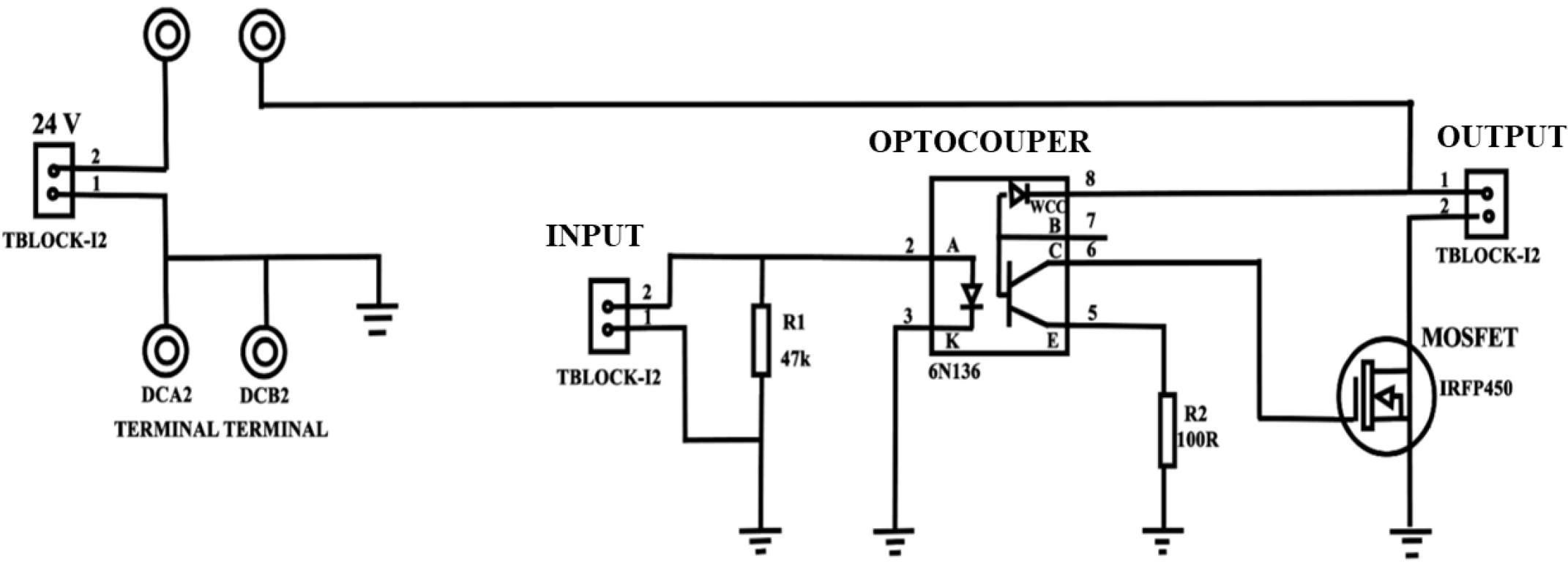
For the LED light source of the photobioreactor, it was necessary to design and build an electronic board with which the variation in light intensity (Luxes) was carried out through the graphic program, so the corresponding diagram is presented below in Figure 3.
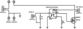
Circuit designed to connect the LED light source and the embedded system data acquisition card in the photobioreactor to control the light intensity measured in Luxes during microalgae growth.
The electrical card of the LED light source was directly connected to port G of Figure 8, and in this way, the variation could be carried out by the user through the graphic program.
Sensor calibration
A) Temperature
The first sensor to be calibrated was the thermocouple (temperature sensor), for which a standard distilled water solution was obtained; with the help of ice baths and heating on a magnetic grid, temperature variations were made. The temperature variations ranged from 0 to 90 °C, and the upper limit was because above this temperature, the water began to boil. The increases were gradual at ±2 °C.
B) Dissolved oxygen (O2)
For the calibration of the dissolved oxygen sensor, distilled water samples are used, where the water naturally reaches a saturation of 20% oxygen in the medium [15], and when subjected to high temperatures without evaporation, the dissolved oxygen is eliminated, so the concentration is equal to 0%. This percentage refers to the oxygen available in the water when it is dissolved and does not imply the atoms that make up the water molecule [16]. On the other hand, as the water undergoes an agitation process, oxygen is incorporated into the water until it reaches saturation, that is, 100%. To determine what the oxygen saturation would be, the samples were subjected to stirring under an electric grill with a magnetic stirrer, with which the change in voltage was reported as the stirring time passed and stopped until the electrode did not present significant changes, with which the assumption was made that oxygen saturation had already been reached in the medium. The time intervals for taking the voltage sample as distilled water were stirred every minute.
C) pH
The pH sensor, in which a series of dilutions was built through standard SIGMA® brand buffer solutions where the pH was varied in increments of 0.5 starting from a buffer solution of 1 to a higher range of 11.5. The reason for not working with a scale from 1 to 14 for the pH calibration curve was because of the experimentation in the growth of microalgae, and specifically for a biological model of C. vulgaris, the optimum pH for growth is 10, so it is not necessary to prepare alkaline buffers above 12, since they are not necessary and imply an additional expense for the operating process because Golden-Bell® brand buffers were used in the curve. Below is the reference that was added to the work where the optimal pH of the microalgae is indicated. The different buffers to which they should be prepared were made using monobasic phosphate buffer and ethylamine.
D) Light intensity
The intensity of the light at which the microalgae growth kinetics are going to work is determined by the user, and the measurement of the Luxes emitted by the blue LED light source was carried out through a Digital Light Meter of the MASTECH® (MS6612) brand, for which measurements were taken at four points of the light source: front, right side, left side and rear. Measurements were taken at high and low points of each of the LED light strips, and the measurements were made at duty cycle variations of ±0.05 working in a range of 0 to 1 duty cycles. The calibration was carried out offline because to isolate the light source from any external emission, a plastic cylinder was built with which different measurements were made as mentioned in the materials and methods section; however, once the reactor was operated online, the intensity must not vary unless, through the software of the equipment, the working conditions are indicated.
The value of r2 in the Pearson correlation was 0.998, which allows the graphic program to be programmed so that the user can set the work Luxes and the program to make adjustments to the corresponding work cycles. In this way, the photobioreactor was instrumented, checking the signals detected in the graphic interface program and having a record of the signals measured at an interval of 1 s for subsequent experimental treatment.
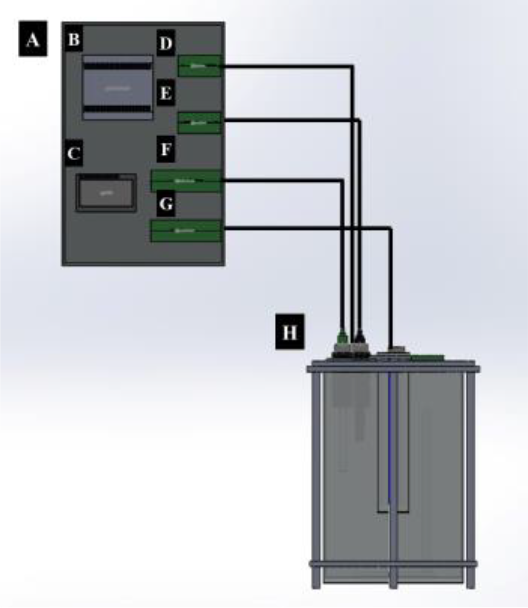
As mentioned, the signals detected by various sensors have been validated, and it is possible to start with exploratory kinetics for microalgae. The appearance of the photobioreactor is shown in Figure 4.
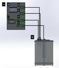
Final appearance of the photobioreactor. A) Cabinet with electronic components. B) A 24-volt power source. C) Data acquisition card. D) Electronic arrangement of the temperature sensor. E) Electronic arrangement of the dissolved oxygen sensor. F) Electronic arrangement of the pH sensor. G) Electronic arrangement for light intensity regulation. H) Photobioreactor mounted with real-time monitoring sensors and a blue LED light source.
Microbial kinetic growth
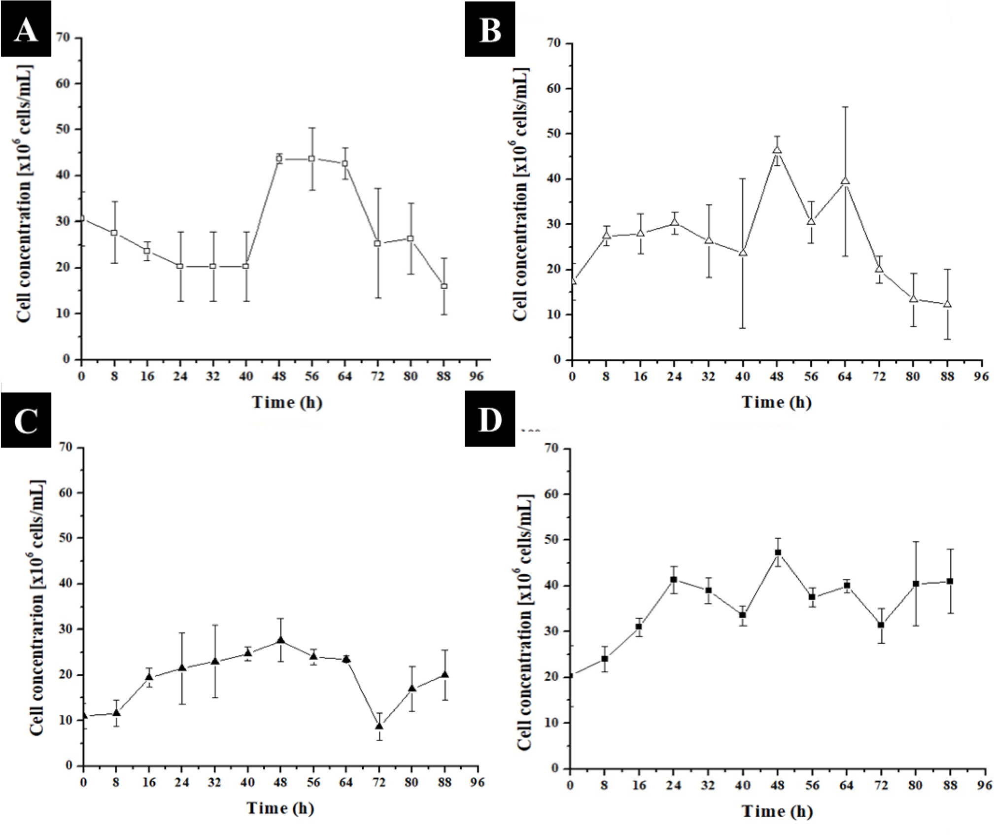
As indicated, for the analysis of the microbial growth of C. vulgaris at the photobioreactor level, a Taguchi design was carried out and implemented where the arrangement was of the L4(22) type with two factors with 4 experimental runs. The factors evaluated were the substrate concentration under low and high conditions. The second factor was the time or type of photoperiod, which was equal under low and high conditions. The low substrate condition, which was sodium nitrate, was 3.6 mM, and the high substrate condition was 16.4 mM. In terms of the type of photoperiod, the low-light conditions included 8 hours of light and 16 hours of darkness. The high-light photoperiod conditions included 16 hours of light and 8 hours of darkness. The behavior observed in the growth of C. vulgaris with the built photobioreactor is presented in Figure 5, in which there was greater control of the aseptic conditions since once the kinetics had begun, the microalgae, together with the growth medium, remained completely isolated from the environment.
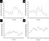
Microbial growth of C. vulgaris in mixotrophic medium after 20 s of CO2 injection. A) 3.6 mM sodium nitrate and B) 16.4 mM sodium nitrate, all previous runs were carried out under long photoperiods, that is, 16 hours of light and 8 hours of darkness, at an internal temperature of approximately 30 °C. C) 3.6 mM sodium nitrate and D) 16.4 mM sodium nitrate, all previous runs were carried out under short photoperiods, that is, 8 hours of light and 16 hours of darkness, at an internal temperature of approximately 30 °C. n=3, α=0.05.
As shown in Figure 5, short photoperiods are favorable for cell division in the microalga since up to 50x106 cells/mL were reached in the medium, beginning the exponential phase, as shown in Figure 5A and 5B, at hour 40. That is, it took longer for the microalgae to adapt and begin growing when working with a long photoperiod. On the other hand, under low substrate conditions, but under short photoperiods, it takes a short time for the microalgae to acclimatize to the growth medium to begin the development phase, beginning in both cases (Figure 5C and 5D) at hour 8 after the start of the kinetics. Similarly, under this short photoperiod, as the substrate concentration increased, the growth of the microalgae was inhibited, reaching a maximum of 50x106 cells/mL under 3.6 mM NaNO3 at 10 °C; however, when the concentration of NaNO3 was increased to 16.4 mM, a maximum of 29x106 cells/mL was reached.
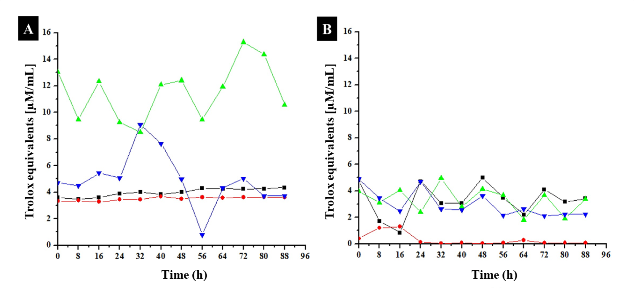
Finally, the antioxidant activity obtained is presented where it is possible to observe significant signals corresponding to secondary metabolites of polar (polyphenols) and nonpolar (terpenes) natures since activity was obtained by the two methods evaluated for the radicals ABTS•+ and DPPH•, respectively. The behavior of the treatments is presented in Figure 6.
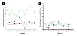
Antioxidant activity of C. vulgaris in mixotrophic medium at the photobioreactor level after 20 s of CO2 injection. A) Activity evaluated via the ABTS•+ method: ν 3.6 mM sodium nitrate and θ 16.4 mM sodium nitrate. All previous runs were carried out under long photoperiods, that is, 16 hours of light and 8 hours of darkness, at an internal temperature of approximately 30 °C. B) Activity was evaluated via the DPPH• method: π 3.6 mM sodium nitrate and λ 16.4 mM sodium nitrate. All previous runs were carried out under short photoperiods, that is, 8 hours of light and 16 hours of darkness, at an internal temperature of approximately 30 °C. n=3, α=0.05.
Figure 6 shows that under any of the conditions at the photobioreactor level, the synthesis of polar secondary metabolites with antioxidant activity is promoted; therefore, C. vulgaris contains these molecules as a possible defense mechanism. On the other hand, as shown in Figure 11, the maximum antioxidant activity is maintained within the conditions of a low substrate level and a low photoperiod, although there is also activity under a high substrate level and a high photoperiod; however, this result only shows activity under certain hours of growth, specifically within hours 24 to 48, which is different from what was observed in Figure 6A, where the activity is maintained and even increases from hour 40 to hour 72. As shown in Figure 6, when cultivated in a mixotrophic medium at the flask level, C. vulgaris retained the ability to synthesize metabolites with antioxidant activity, as these metabolites have nonpolar characteristics. The majority of the experimental conditions show signals with activity, but in the same way, it can be observed that there is a condition where the microalga is not capable of synthesizing them with great significance.
Finally, the kinetic parameters were calculated; for the C. vulgaris growth condition of 3.6 mM NaNO3 and a high photoperiod of 16 hours of light and 8 hours of darkness, the doubling time (td) was 4.32 hours. a specific growth rate (µ) of 0.0266 h-1 and a division rate (δ) of 0.2314 h-1. In terms of the calculated performance, Yx/s is 2.94, Yp_ABTS•+/s is 3.83, and Yp_DPPH•/s is 4.8. For the conditions of 16.4 mM NaNO3 and a high photoperiod of 8 hours of light and 16 hours of darkness, we have a doubling time (td) of 4.3287 hours, a specific growth rate (µ) of 0.0264 h-1 and a division speed (δ) of 0.231 h-1. The calculated performances have a value of Yx/s of 2.21, Yp_ABTS•+/s of 3.37, and Yp_DPPH•/s of 0.0048. For the conditions of 3.6 mM NaNO3 and a high photoperiod of 8 hours of light and 16 hours of darkness, a doubling time (td) of 3.76 hours, a specific growth rate (µ) of 0.0465 h-1 and a division speed (δ) of 0.2658 h-1 were used. The calculated performances are Yx/s = 2.75, Yp_ABTS•+/s = 17.79, and Yp_DPPH•/s = 2.4. Finally, for the conditions of 16.4 mM NaNO3 and a high photoperiod of 16 hours of light and 8 hours of darkness, we have a doubling time (td) of 3.85 hours, a specific growth rate (µ) of 0.0424 h-1 and a division speed (δ) of 0.2594 h-1. In terms of the calculated yields, Yx/s is 5.6, Yp_ABTS•+/s is 10.83, and Yp_DPPH•/s is 10. Therefore, the best conditions for growth are those under a low photoperiod of 8 hours of light, since this promotes cell growth, as well as an increase in yields on the basis of antioxidant activity. Additionally, the substrate concentration affects cell division and therefore growth since, from the four experimental runs in the Taguchi design, the shortest acclimatization and the beginning of microbial growth are under low substrate conditions (3.6 mM NaNO3) as well as low exposure to blue LED light (8 hours of light and 16 hours of darkness).
DISCUSSION
Within the previous analysis that has been carried out on the construction of the photobioreactor, different works can be mentioned in which no invention similar to the one proposed in this work has been found. For this purpose, searches were made in the database of works in search of patents or that have been registered as patents. The inventions are as follows:
“Lighting device for bioreactors” [17], with which a very homogeneous light distribution of high intensity in the cylindrical photobioreactor is achieved by means of SMD light-emitting diodes (surface device technology), with which the carrier is directly mounted, which is designed as a printed circuit board. The direct assembly together with the compact design of the SMD LEDs allows very dense assembly, preferably greater than 30%, preferably greater than 60%, when multilayer circuit boards are used even up to 75% coverage of the surface of the support. with LEDs. For example, warm white LEDs with a spectrum similar to that of sunlight can be used, possibly in combination with colored LEDs or LEDs that emit in the UV range. Through the combined use and computer-aided control of monochrome LEDs and those with a white spectrum similar to sunlight, different light intensities and spectra can be generated. With the external illumination device on the transparent walls of the reactor, an almost homogeneous light distribution can be achieved within the reactor (also) radially.
On the other hand, a photobioreactor with pulsating light-emitting diodes (LEDs) was developed for profitable photo fixation of carbon dioxide (CO2). Preferably, LEDs emit narrowly focused light at a wavelength of 660 nm to optimize carbon dioxide sequestration while minimizing energy costs. It is used in the photosynthesis of algae [18]. Moreover, the work entitled "Cylinder type multilayered photobioreactor" can also be mentioned, where a cylindrical multiphotobioreactor for mass cultivation of microalgae is proposed. More particularly, the present invention involves a photobioreactor for the mass cultivation of microalgae that uses a plate-shaped or circular light source consisting of a light-emitting diode (LED), an organic light-emitting diode (OLED) and a flexible light-emitting diode (LED), which are slim to minimize the bulk energy efficiency [19]. Therefore, the goal of the present invention is to provide a cylindrical photobioreactor that is thinner than a light source such as a spherical LED lamp or a fluorescent lamp, which has a high productivity per volume and can satisfy various application conditions. crop by separately attaching a setting. The cylindrical light source is made of a flexible LED (flexible light-emitting diode). It is preferable that the outer shape of the reaction chamber is cylindrical. Additionally, the cylindrical light source can be stacked one or more times in the reaction chamber. Although the invention has not been compared in its entirety with other existing works regarding the production of biomass or some other metabolites, it is certain that gathering all the conditions for optimal growth in the equipment and placing the sensors in the indicated places provide correct monitoring of the growth conditions and ensure that there are high levels of production since the working conditions are mixotrophic, which is the optimal growth condition that, in addition to the morphology of the equipment, will help in this. Prior to this work, work has been carried out at the flask level by our work team, where the growth medium is heterotrophic, and it has been observed that, with the conditions working, significant yields have been found in comparison with those currently existing, for which reason, when going from a flask-level experiment to a photobioreactor level with the construct, the response levels will increase and therefore be competitive [20].
Similarly, there is a photobioreactor for mass production of microalgae via a plater-shaped light source, which has high power efficiency, where the invention is a photobioreactor for the mass production of microalgae, in which an LED (light emitting diode), an OLED (organic light emitting diode) and a flexible LED are provided in a substrate to efficiently transmit light and enable a change in the light source [21]. Moreover, we can talk about a photobioreactor with baffles integrally provided with an internal light source for high-density cultivation of microalgae” (KR101043583B1 register), where the invention involves a photobioreactor for high-concentration cultivation of microalgae and, more particularly, a photobioreactor for improving the efficiency of stirring an internal light source made of an LED (light-emitting diode) for improving light utilization efficiency and gas and microalgae biomass. The present invention involves a photobioreactor in which a baffle is integrally formed to maximize microbial culture efficiency [21].
Finally, another work that can be referred to is the one carried out by Song and coauthors in 2019, where a high-efficiency photobioreactor for microalgae was developed under a gas exchange system for a thermal power plant and where this equipment was compared with open tanks in open air. The innovation of the designed equipment is that it has a central artificial light source that performs better in the growth of microalgae and is able to regulate and adjust the concentration of CO2 to exchange this gas with O2 to carry out mixotrophic metabolism. The artificial light source was fiber optic, generating efficient and economical equipment; however, the dimensions of the equipment were small. The intensity of the applied light was 1000 lux, and as a result, it was concluded that the exchange of CO2 and O2 gases was achieved in the growth process of the Chlorella pyrenoida microalgae, generating an expense 100 times greater than that of outdoor cultures; therefore, it can be a process that implies energy expenditure but that achieves adequate growth of the microalgae. On this basis, the design of the equipment described in this work presents characteristics that can provide adequate growth and significant positive results for the growth of microalgae since, just to name a few characteristics, the equipment also has CO2 feeding for mixotrophic growth and working under a recommended light intensity, as described in the work, since the photobioreactor designed for this work ranges from 0 to 1100 lux [21].
The arrangement and type of sensors for the design of the photobioreactor in this work are encouraging for its use at an industrial level, and there is nothing similar to it, which has allowed for the recently awarded IP in the "utility model" category.
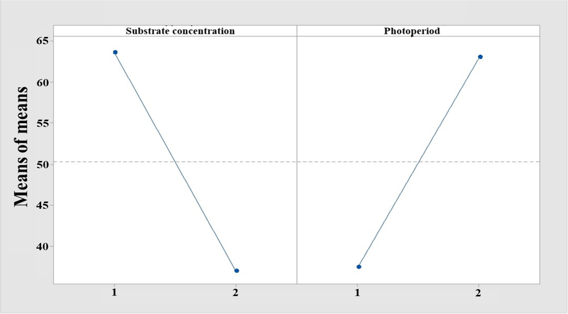
The analysis was carried out with the Taguchi design with microbial growth as a response. Figure 7 shows the main effects diagram for means whose behavior was as follows:
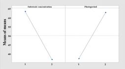
Analysis of the main effects of the sodium nitrate NaNO3 concentration and photoperiod type on cell growth. 1) NaNO3 (3.6 mM) or a short photoperiod of 8 hours of light and 16 hours of darkness. 2) 16.4 mM NaNO3 or long photoperiods of 16 hours of light and 8 hours of darkness. n=3, α=0.05.
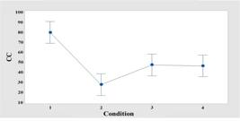
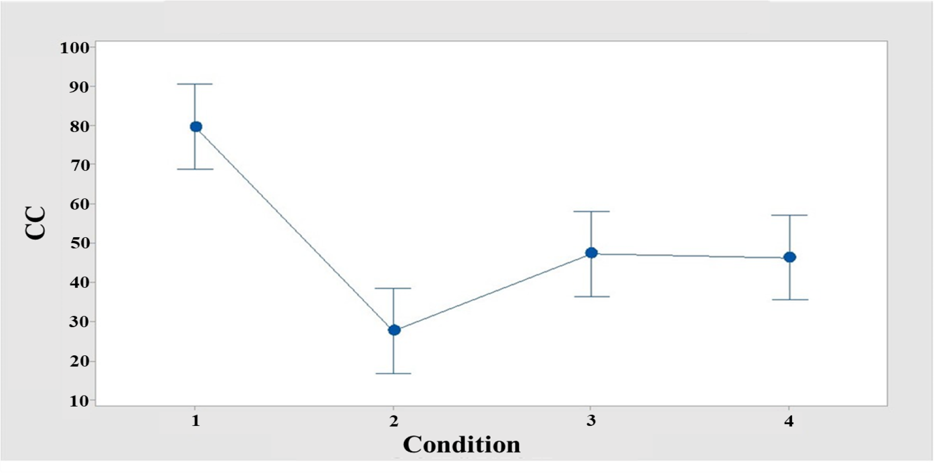
ANOVA of the cell growth (CC) of C. vulgaris with sodium nitrate NaNO3 as the substrate and CO2 injections. Mixotrophic environment. 1) 3.6 mM and 16 hours of light and 8 hours of darkness. 2) 16.4 mM and 8 hours of light and 16 hours of darkness. 3) 3.6 mM and 8 hours of light and 16 hours of darkness. 4) 16.4 mM and 16 hours of light and 8 hours of darkness. n=3, α=0.05.
As shown in Figure 7, the highest microbial growth rates occur when a low concentration of substrate is used, as growth is favored and cell division is promoted. On the other hand, long photoperiods significantly affect cell growth and are favored when the microalga is exposed to blue LED-type light for a relatively long period of time; specifically, at 16 hours of light and 8 hours of darkness, the ability of the microalga to absorb greater amounts of light begins, and the ability of the microalgae to promote its division at this wavelength is promoted.
For a final understanding of the behavior of this variable, a one-way ANOVA test was carried out to determine which experimental conditions promote greater and better cell growth, where the null hypothesis is that all the means analyzed for the maximum cell concentration at hour 48 are equal and, as an alternative hypothesis, the assumption that not all means are equal. The behavior of the observations is shown in Figure 8.
The data obtained show that under these four levels, there is a reliability value for a r2 of 88.66%, where there is an F value of 20.6 and a P value of 0.000, which indicates that at least one condition is different. and therefore, you can have experiments where you have a greater production of cells compared to the rest. This condition involves a low concentration of NaNO3 (3.6 mM) and a long photoperiod (16 hours of light and 8 hours of darkness), as shown in Figure 8, which confirms the results obtained in Figure 7 for the main effects. Similarly, the variations in the results only overlap under short photoperiod conditions when low (3.6 mM) and high (16.4 mM) concentrations of NaNO3 are evaluated, as well as when a high concentration of NaNO3 is used but a short photoperiod is used; thus, if these conditions are used, the results are very similar, and high microbial growth will not be obtained.
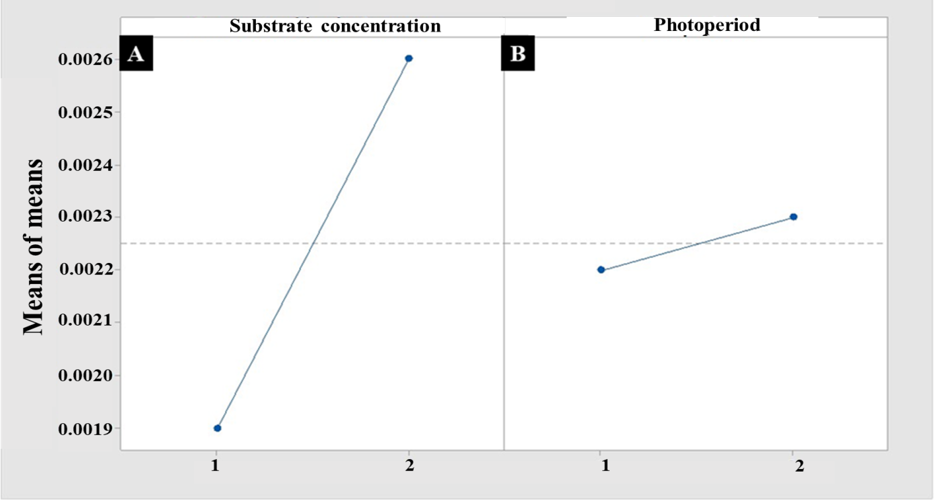
To demonstrate that the responses are affected positively or negatively by the substrate concentration, the main effects analysis was carried out for substrate consumption (NaNO3), which is presented in Figure 9.
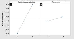
Analysis of the effects of the sodium nitrate NaNO3 concentration and photoperiod type on biomass. (A) 1=3.6 mM, 2=16.4 mM NaNO3. (B) 1= photoperiod 8 hours of light and 16 hours of darkness, 2= photoperiod 16 hours of light and 8 hours of darkness. n=3, α=0.05.
An increase in the concentration of NaNO3 results in an increase in the biomass generated. However, compared with what was obtained in Figure 10 for cell growth (CC), a greater number of cells are observed at a lower concentration of NaNO3, which means that when working with a concentration of 3.6 mM NaNO3, the microalgae, which are limited in the substrate, seek preservation, and therefore, their division is induced to generate new cells; however, this does not mean that these cells necessarily have to develop large sizes that increase in weight. On the other hand, when working under high substrate conditions (16.4 mM), division is not greatly promoted; therefore, the cells formed have abundant substrate to significantly increase their size, with this phenomenon occurring when biomass tests are performed. Like dry weight, an increase in the recorded weight of the cellular precipitates is detected, which is why this increase in biomass occurs. To corroborate this hypothesis, a post hoc test of Pearson correlations was carried out between cell growth and the biomass generated, where it was determined that there was no correlation or a directly proportional relationship between the responses. The value obtained from the correlation is 0.167, meaning that the model and the behavior can explain only 16.7% of the behavior or, in other words, only this percentage is reliable so that there is linear behavior of both variables.
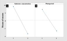
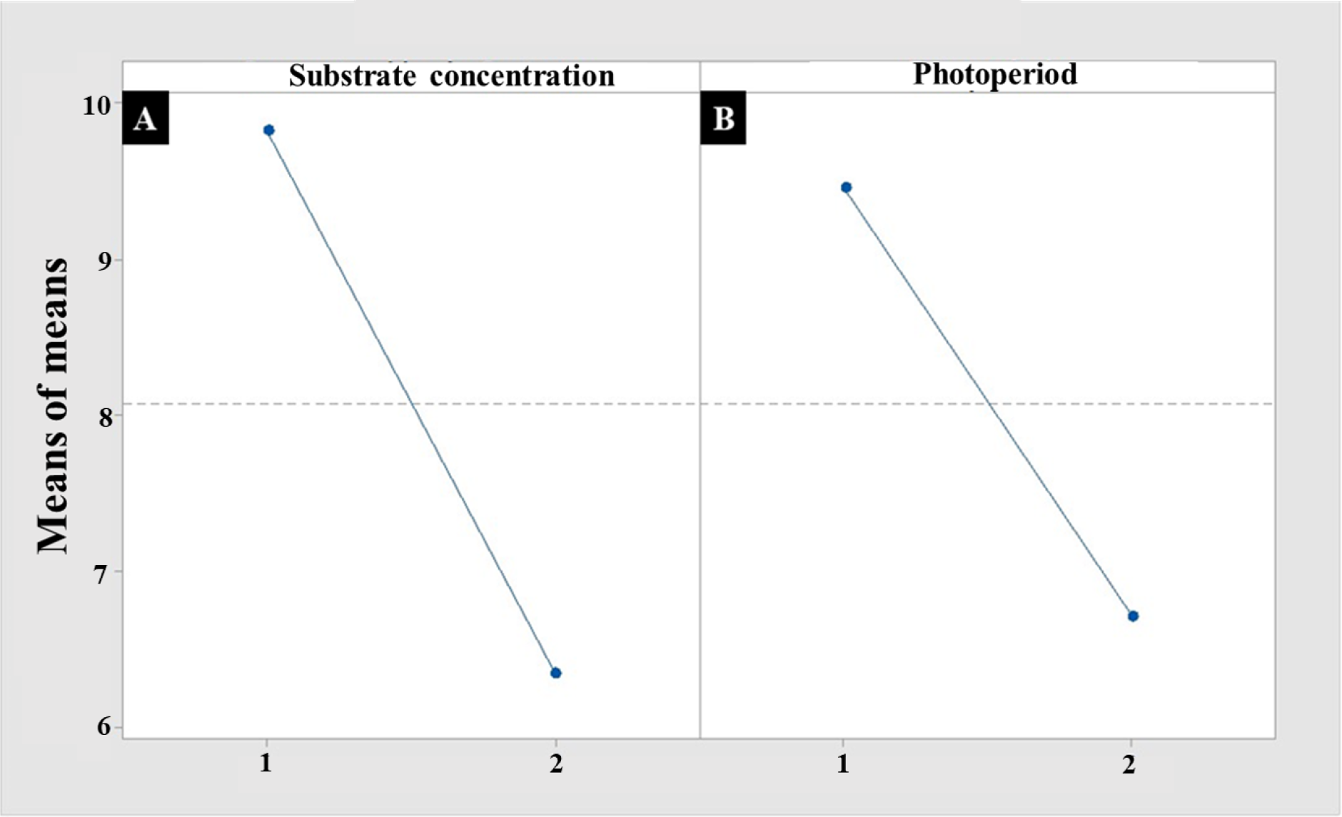
Analysis of the main effects on antioxidant activity by the ABTS•+ method with respect to the concentration of sodium nitrate NaNO3 and type of photoperiod. (A) 1=3.6 mM, 2=16.4 mM NaNO3. (B) 1= photoperiod 8 hours of light and 16 hours of darkness, 2= photoperiod 16 hours of light and 8 hours of darkness. n=3, α=0.05.
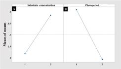
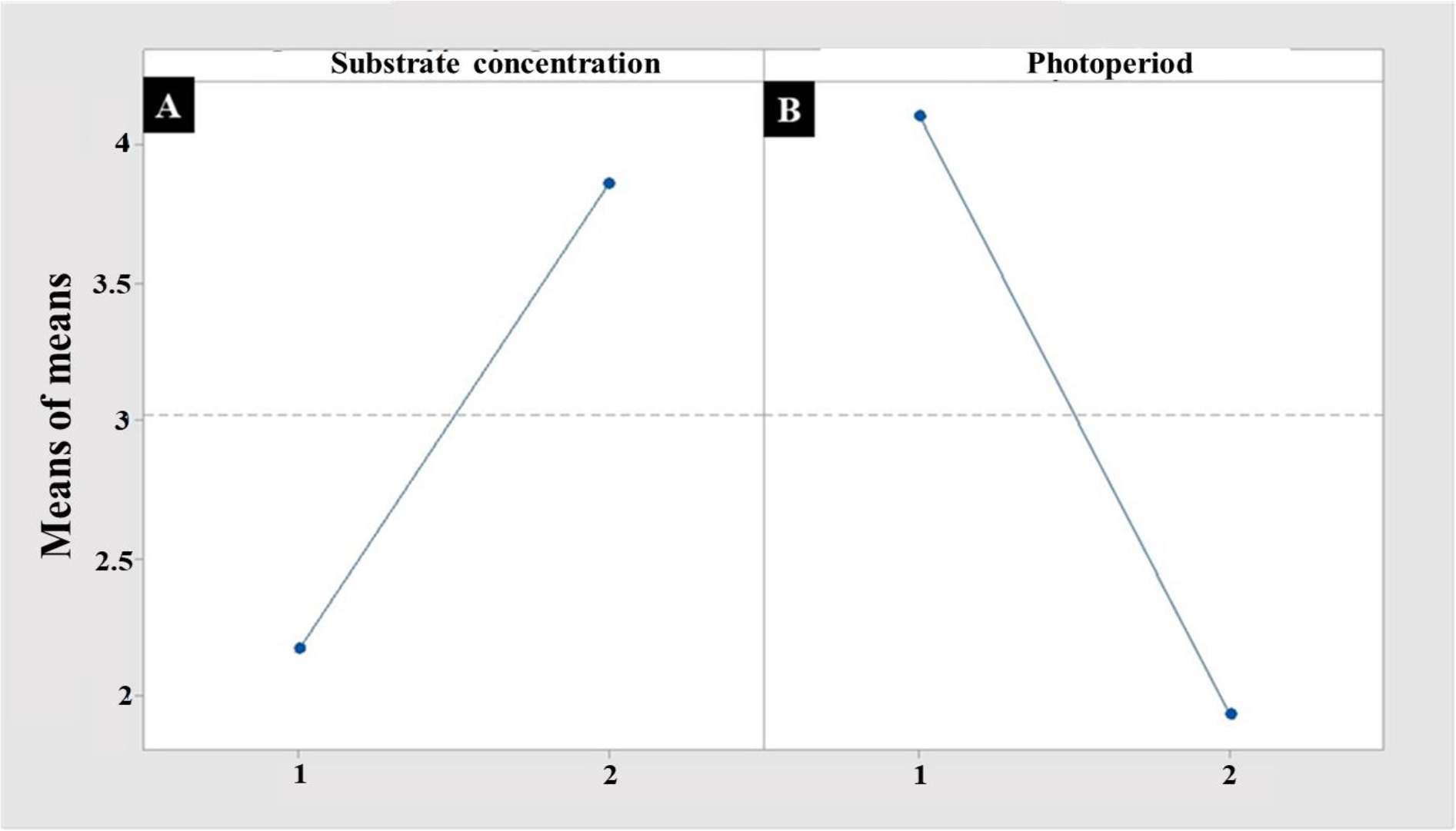
Analysis of the main effects on antioxidant activity by the DPPH• method with respect to the concentration of sodium nitrate NaNO3 and type of photoperiod. (A) 1=3.6 mM, 2=16.4 mM NaNO3. (B) 1= photoperiod 8 hours of light and 16 hours of darkness, 2= photoperiod 16 hours of light and 8 hours of darkness. n=3, α=0.05.
Within the objectives of this work, in addition to increasing the efficiency of the growth of microalgae with respect to the formation of new cells and biomass, it takes advantage of the molecules or metabolites inside since there is evidence of a wide range of groups. function that can be used for biotechnological metabolites (such as polyphenols) as well as nonpolar or fat-soluble metabolites (such as terpenes) through the stability of synthetic free radical use. One of these uses, as previously mentioned, is antioxidant activity. Therefore, after the corresponding extraction, Figure 10 shows the quantification of the polar or water-soluble
For a reinforced conclusion, the graph resulting from Taguchi's analysis is presented below in Figure 10.
Figure 10 clearly shows that the substrate concentration promotes the greatest response for antioxidant metabolites and that this, in turn, is favored when working at a concentration of 3.6 mM. In addition, the photoperiod plays an important role in promoting the same activity, unlike what was observed in Figure 10B, since the slope is more pronounced and is similar to that generated for the substrate in Figure 10A. The photoperiod that favored the antioxidant response was 8 hours of light and 16 hours of darkness.
Continuing with the conclusions of the Taguchi analysis, Figure 11 presents the main effects with respect to nonpolar metabolites.
For this situation, Figure 11 shows that the photoperiod has the greatest influence on the best response evaluated and that, within this range, short photoperiods of 8 hours of light promote synthesis. However, in this case, the substrate concentration of 3.6 mM affects the synthesis; that is, the behavior of the response is inverse to that observed in Figure 11 (B), which means that it is possible to select the type or nature of metabolites for possible application if working under low or high substrate conditions and thereby develop specific products.
CONCLUSION
It can be concluded that the photobioreactor can record biochemical signals in real time, such as pH, temperature, and oxygen concentration, during the growth of microalgae, which allows control of the growth conditions, making correct growth and establishing optimal growth conditions possible. On the other hand, with the regulation of the intensity of light, the effect on cell growth can be explored and analyzed, and the results indicate that the higher the light intensity is, the greater the growth by promoting photosynthesis or if there is a negative effect where growth is observed. decreased because of photooxidation. With respect to the growth kinetics carried out in Chlorella vulgaris via the Taguchi experimental design, the conditions promote the synthesis of metabolites with antioxidant activity for both polar molecules (polyphenols) and nonpolar molecules (terpenes), favoring synthesis and growth. The cells were cultured under a substrate concentration of NaNO3 (3.6 mM) and a low photoperiod (8 hours of light and 16 hours of darkness) with blue light. However, it was possible to observe and conclude that caution should be taken with the injection of CO2 since if it is not metabolized by the microalgae, the growth medium can become acidic since carbonic acid is formed; therefore, as there is an acidic medium, the microalgae can die or inhibit its growth since the optimal pH is 9.8, so it is suggested to carry out future experiments where the gas injection time is varied. Finally, with respect to specific antioxidant activity, the best conditions were a concentration of 3.6 mM NaNO3 and a low photoperiod time of 8 hours of light and 16 hours of darkness, generating a response of 16 µM/mL for ABTS•+ and 6 µM/mL DPPH•; thus, C. vulgaris can be used at a biotechnological level for the production of functional foods, cosmetics or medicines.
PATENTS
PATENT for photobioreactor design in the UTILITY MODEL category. Year of granting innovation, 2023. Registration: MX/u/202100055. Year 2023.
-
Funding:
Call for Support for Proyectos de Desarrollo e Innovación Tecnológica para Estudiantes 2020 del TecNM, with the project entitled “Evaluación de la fuente de sustrato sobre cinéticas de crecimiento de C. vulgaris a nivel matraz” (8959.20-P), to the project of call 2021, entitled “Efecto de la fuente de sustrato sobre cinéticas de crecimiento de C. vulgaris a nivel matraz” (7662.20-P), and to the project entitled “Diseño e instrumentación de fotobiorreactor tubular para cultivo fotoheterotrófico de C. vulgaris” (10019.21-P), as well as for the project in 2022, entitled “Determinación de terpenos y actividad antioxidante de C. vulgaris bajo crecimiento fotoheterotrófico a nivel fotobiorreactor” (13825.22-P) and the Proyect 2023 (16898.23-P).
Data availability statement:
Research data are not available.
Acknowledgments:
We thank the “Consejo Nacional de Humanidades Ciencia y Tecnología” (CONAHCYT) for PhD scholarship 0456297331 (JACR).
REFERENCES
- 1 Arias PMT, Martínez RAD, Cañizares RO. Biodiesel production from microalgae: culture parameters that affect lipid production. Acta Bio. 2013 Jan;8(1):43-68.
- 2 Aravantinou AF, Manariotis ID. Effect of operating conditions on Chlorococcum sp growth and lipid production. J. Environ. Chem. Eng. 2016 Mar;4(1):1217-1223.
- 3 Usher PK, Ross AB, Camargo-Valero MA, Tomlin AS, Gale WF. An overview of the potential environmental impacts of large-scale microalgae cultivation. Biofuels. 2014 Jul;5(3):331-49.
- 4 Andrade LM, Andrade CJ, Dias M, Nascimento C, Mendes MA. Chlorella and spirulina microalgae as sources of functional foods. Food Process Technol. 2018 Jan;6(1):45-58.
- 5 Ohse S, Derner RB, Ozorio RA, Braga MVDC, Cunha P, Lamarca CP, et al. Production of biomass and carbon, hydrogen, nitrogen and protein contents in microalgae/Producao de biomassa e teores de carbono, hidrogenio, nitrogenio e proteina em microalgas. Ciência Rural. 2009 Sep;39(6):1760-8.
- 6 Ramsundar P, Guldhe A, Singh P, Bux F. Assessment of municipal wastewaters at various stages of treatment process as potential growth media for Chlorella sorokiniana under different modes of cultivation. Bioresour. Technol. 2017 Mar;227:82-92.
- 7 Nzayisenga JC, Farge X, Groll SL, Sellstedt A. Effects of light intensity on growth and lipid production in microalgae grown in wastewater. Biotechnol. biofuels. 2020 Jan;13(4):1-8.
- 8 Blair MF, Kokabian B, Gude VG. Light and growth medium effect on Chlorella vulgaris biomass production. J. Environ. Chem. Eng. 2014 Mar;2(1):665-74.
- 9 Yuan H, Edlebeck J, Wolf M, Anderson M, Corradini M, Klein S, et al. Simulation of supercritical CO2 flow through circular and annular orifice. J. Nucl. Eng. Radiat. Sci. 2015 Mar;1(2):021003.
- 10 Ma’mun S, Wahyudi A, Raghdanesa AS. Growth rate measurements of Chlorella vulgaris in a photobioreactor via a Neubauer-improved counting chamber and a densitometer. Earth Environ. Sci. 2022 Apr;963(1):012015.
- 11 Zhu CJ, Lee YK. Determination of biomass dry weight of marine microalgae. J. Appl. Phycol. 1997 Apr;9:189-94.
- 12 Kazbar A, Cogne G, Urbain B, Marec H, Le-Gouic B, Tallec J, et al. Effect of dissolved oxygen concentration on microalgal culture in photobioreactors. Algal Res. 2019 May; 39:101432.
- 13 Kuskoski EM, Asuero AG, Troncoso AM, Mancini-Filho J, Fett R. [Application of various chemical methods to determine antioxidant activity in fruit pulp]. Ciênc. Tecnol. Aliment. 2005 Sep;25:726-732.
- 14 Guevara M, Vívenes EZ, León M, Acosta M. [Antioxidant activity of a vegetable oil enriched with the cultivated microalga Dunaliella salina (Chlorophyceae)]. AquaTechnica. 2019 Mar;1(1):40-46.
- 15 Beltrán-Álvarez R, Ramírez-Lozano JP, Sánchez-Palacios J. [Behavior of temperature and dissolved oxygen in the Picachos Dam, Sinaloa, Mexico]. Hydrobiol. 2012 Apr;22(1):94-7.
- 16 Lehr F, Posten C. (2021). Lighting device for bioreactors. Register: DE202007013406U1.
- 17 Aramco services. (2021). Carbon neutralization system (CNS) for CO2 sequstering. Registro: WO2007047805.
- 18 Inha ind partnership inst. (2021). Cylinder type multilayered photobioreactor. Register: KR100945153.
- 19 Coronado-Reyes JA, Acosta-Ramírez E, Martínez-Olguín MV, González-Hernández JC. Antioxidant Activity and Kinetic Characterization of Chlorella vulgaris Growth under Flask-Level Photoheterotrophic Growth Conditions. Molecules. 2022 Sep;27(19):6346.
- 20 Without name. (2021). Photobioreactors with baffles integrally provided with an internal light source for high-density cultivation of microalgae. Register: KR101043583B1
- 21 Song C, Liu Q, Qi Y, Chen G, Song Y, Kansha Y, et al. Absorption-microalgae hybrid CO2 capture and biotransformation strategy-A review. Int. J. Greenh. Gas. Con. 2019 Sep;88:109-117.
-
Editor-in-Chief:
Alexandre Rasi Aoki
-
Associate Editor:
Ana Cláudia Barana