Co-encapsulation of Tamoxifen and Curcumin in PLA Nanoparticles Increases the Antiproliferative Activity over B16-F10 Cells
Abstract
Tamoxifen (TAM) and curcumin (CUR) are compounds investigated for cancer treatment; however, tamoxifen is associated with side effects, especially reducing red blood cells, and curcumin has low bioavailability. Therefore, combining these compounds in an encapsulation system represents a promising therapeutic strategy. Poly-lactic acid nanoparticles (PLA-NPs) containing CUR, TAM, and their combination (CUR-TAM) were prepared via emulsification-solvent evaporation. The encapsulation efficiency was similar for both CUR and TAM, whether encapsulated individually or together, with approximately 57% for TAM and 92% for CUR. The nanoparticles exhibited spherical morphology, with an average size of approximately 200 nm. Zeta potential values were -16 mV for TAM, -26 mV for CUR, and -17 mV for CUR-TAM. In vitro release studies over 120 hours revealed 57% release for CUR and 49% for TAM in the CUR-TAM NPs. Hemolysis testing at 96 hours showed low hemolytic activity for all NPs while the antiproliferative activity against B16-F10 cells demonstrated a significant reduction in cell viability with CUR-TAM NPs (23%), compared to 89% for TAM NPs and 41% for CUR NPs. Combining CUR and TAM in nanoparticles is promising, offering potential therapeutic benefits while minimizing adverse effects.
Keywords:
Nanotechnology; Drug delivery; Hemolysis and Melanoma.
HIGHLIGHTS
Curcumin and Tamoxifen co-encapsulation is efficiently obtained.
Curcumin and Tamoxifen in PLA nanoparticles reduces the Tamoxifen hemolysis effect.
Curcumin and Tamoxifen in PLA nanoparticles is more efficient than free Tamoxifen against melanoma.
INTRODUCTION
Melanoma is a skin cancer often associated with high mortality rates around the world among cutaneous tumors [1,2]. Melanoma treatment uses several approaches such as immune checkpoint inhibitors, targeted therapies, vaccines, cytokines, sequential treatment, intravenous oncolytic virus, antivascular endothelial growth factors, targeting inhibitory molecules, and T-cell agonists [3,4].
The Tamoxifen (TAM) 2-[4-[(1Z)-1,2-diphenylbut-1-enyl] phenoxy]-N, N-dimethylethanamine, is a selective estrogen modulator [5], widely used in the treatment of breast cancer [6]. Melanoma has been reported through the cytotoxic activity of mitochondrial damage [7], suppression of protein kinase phosphorylation [8], expression of pro-apoptotic factors of Bax [9] and caspases activation [10]. However, despite being a drug with high oral bioavailability, it has long-term side effects such as an increased risk of endometrial cancer [11,12], hepatotoxicity [13] and thromboembolic events [14]. An alternative to avoid the side effects, has been the association of TAM with other molecules aiming to increase the therapeutic effect and reduce side effects [9].
Curcumin (CUR) is a polyphenol extracted from Curcuma longa that has shown cytotoxic activity in tumor cell lines, including melanoma [15]. CUR's activity in melanoma occurs through different targets, such as induction of apoptosis pathways, and inhibition of tumor proliferation, migration, and invasion interfering in growth factors protein expression [16-18]. Moreover, CUM and TAM are associated with mitochondrial damage and increased reactive oxygen species [19-21]. Researchers have investigated the synergistic effect of CUR and TAM in niosomal constructs, reveled the increase of cell death in MCF7 breast cancer cells and also, the pre-treatment with CUR followed by TAM in A375 and G361, melanoma cell lines, has shown increased cell death signaling, and reactive oxygen species (ROs) generation[10,22].
Nanotechnology has explored the delivery of drugs and compounds to overcome limitations such as low bioavailability and specificity in the target tissue [23,24]. The advantages of this co-encapsulation are the reduction of adverse effects [25], drug resistance [26], synchronized pharmacokinetics [27] increasing the therapeutic efficacy [28,29] and encourages patients to treatment [30].
Polymeric nanoparticles derived from polyesters such as Poly-lactic acid (PLA) are extensively studied due to their biopharmaceutical advantages [31]. These nanoparticles (NPs) offer gastric stability when administered via the oral route [32], enabling the incorporation of ligands [33,34]. They have been successfully used for the delivery of various therapeutic agents, such as insulin and vaccines, through different routes, including mucosal, muscular, and venous administration, demonstrating their versatility beyond the oral route [35]. However, the search for less toxic alternatives remains a key therapeutic target[36,37]. In melanoma, PLA-NP have shown the potential to increase treatment efficacy and reducing of side effects [38,39], due to their specific delivery capacity to the tumor tissue [40,41]. In vivo models presented an increase in animal survival and a delay in tumor progression [42].
Seeking understands if CUR together with TAM in NPs improve the effect of TAM and the side effects, the current study has co-encapsuled TAM and CUR into PLA nanoparticles and done its characterization. The biological assessment of CUR-TAM NPs was conducted using two approaches. The first involved evaluating the effect of CUR-TAM NPs on erythrocyte cytotoxicity, as this is one of the most severe side effects of TAM treatment. The second approach was to assess their antitumor activity in murine melanoma cells (B16F10 cells).
MATERIAL AND METHODS
Tamoxifen (≥ 99%, 563.64 Da), Curcumin (≥ 65%, 368.38 Da) 12, Poly (L-lactic acid) (PLA, 85,000 - 160,000 Da), Polyvinyl alcohol (PVA, 31KDa, 88% hydrolyzed), Iscove’s Modified Dulbecco’s medium, Polysorbate 80, Penicillin, Streptomycin, 3-(4,5-dimethyl-2-thiazolyl)- 2,5 diphenyl- 2H- tetrazolium bromide (MTT), trypan blue was purchased from Sigma-Aldrich (USA). Ethyl acetate, glucose, sodium acetate was purchased from Biotec (Brazil), dimethylsulphoxide and acetic acid were purchased from Química Moderna (Brazil), dichloromethane and isopropyl alcohol from Fmaia (Brazil), sucrose, phosphate buffered saline (PBS) from Vetec Química Fina (Brazil), ethanol and methanol from J. T Baker (USA), fetal bovine serum were purchased from Gibco (USA).
Preparation of PLA nanoparticles containing TAM, CUR, and CUR-TAM
The PLA NPs were prepared by the single-emulsion solvent evaporation method [43], with modifications. The CUR was solubilized in ethyl acetate (2.25mg/mL); TAM in dimethyl sulfoxide (25.0 mg/mL), and PLA was solubilized in dichloromethane (50mg/mL), composing the organic phase. This organic phase was added to the aqueous phase containing PVA (10 mL) 0.5% and sonicated (Ecosonics) for 4 min at 60 Hz. After the emulsion was submitted to evaporation under negative pressure, to eliminate the organic solvents. NPs were separated by centrifugation (6,720 g for 30 min) and the precipitate was washed twice with ultrapure water. The NPs were resuspended in 5% sucrose and lyophilized.
Physicochemical and morphology characterization
It was determined by mean of diameter and polydispersity index by dynamic light scattering (Brookhaven Instruments Corp. USA) with an angle of 90° at 25 °C and wavelength of 659 nm. Zeta potential analysis was performed from electrophoretic mobility under an electric field (ZS-Malvern) with a potential of ± 150 mV. To morphology characterization, NPs were suspended on copper support and negatively contrasted with 2% uranyl acetate (m/v) in aqueous solution and analyzed by transmission electron microscopy (TEM -JEOL JEM 1400), using 80 kV.
Encapsulation Efficiency (EE%)
The EE% was determined by the indirect method according to Cho and coauthors (2012) with modifications [44]. The supernatant was diluted 1:10 (v/v) in methanol, then filtered and analyzed by High-Performance Liquid Chromatography (HPLC). The mobile phase was methanol, 0.5% acetic acid and ethanol (70:15:15) v/v, with an isocratic flow rate of 0.8 mL/min and an injection volume of 60 μL. The photodiode array (PDA) detector at a wavelength of 417 nm for CUR and 240 nm for TAM. Chromatography was performed in reverse phase using a C18 column. Thus, the EE% was determined from (Equation 1).
Stability
The stability of the NPs containing CUR and TAM was determined through the diameter particle, polydispersity index and zeta potential for time intervals of 7, 30, 60 and 90 days. The NPs were stored at 25 °C, 5 °C and -15 °C.
In vitro release profile
The NPs samples were diluted in solution of PBS with 1% polysorbate 80 (pH 7.4) at concentrations of 250 µg/mL for CUR and 150 µg/mL for TAM. This dispersion of NPs was incubated under orbital shaking at 150 rpm, 37 °C. At pre-determined times (1, 3, 5, 8, 12, 24, 48, 72, 96, and 120 h) the NPs were centrifuged (7,200 g for 30 min) and the supernatant was removed and analyzed by HPLC to quantify the drug released. With the photodiode array detector at a wavelength of 417 nm for CUR and 240 nm for TAM. The precipitate containing the NPs was resuspended in a fresh PBS solution and incubated until next sampling.
Hemocompatibility Assay
The cytotoxicity against human red blood cells was performed according to [45] with modifications. The experiment was approved by the Institutional Human Ethics Committee of the Universidade Estadual do Centro-Oeste, Brazil (Registration n° 840169/2014). Heparinized blood (5 mL) obtained from healthy volunteers was centrifuged (1,200 g for 5 min) to remove plasma, and the blood was then washed with PBS buffer (10 mM, pH 7.4 and NaCl 0.85%) and centrifuged. After washing, the packed cell volume was adjusted to 2% with a PBS containing glucose (1.08 mg/mL) penicillin (0.3 mg/mL) and streptomycin (0.5 mg/mL). The red blood cells were incubated at different times (24, 48, 72 and 96 h) at 37 °C with CUR, TAM, CUR-TAM free forms, and CUR-NPs, TAM-NPs or CUR-TAM NPs, at concentrations of 200 µg/mL for CUR in ethanol and 100 µg/mL for TAM in ethanol, under constant agitation. The control analyses were conducted by incubating the red blood cells with distilled water, PBS buffer, ethanol control used to drug dilution. At the determined times, the suspension of red blood cells was centrifuged at 1,200 g for 5 min. The hemoglobin released by the rupture of red blood cells was determined in the supernatant by measuring the absorbance at 540 nm, and the percentage of hemolysis was calculated using (Equation 2).
Where:
Abs sample: absorbance of sample
Abs 100%: absorbance of control in water (100% hemolysis)
Antiproliferative activity assay
The antiproliferative tumoral activity assay was tested using B16-F10 (ATCC CRL-6475) cells line, murine melanoma cells. The cells were cultured in complete DMEM media (Sigma) enriched with 10% of fetal bovine serum and 1% of penicillin and streptomycin. Once the 80% of confluence was obtained, the cells were submitted to viability cell assay using the MTT. This method is based on the ability of cells to convert the yellow-colored MTT into an insoluble purple compound called formazan, which occurs when there is integrated mitochondrial capacity. The amount of formazan is determined spectroscopically after solubilization in organic solvent. The cells were plated at a density 1x10⁴ cells/mL in a 96-well culture plate and incubated for 24 h at 37 °C and under 5% CO2. Free CUR and CUR-NPs (40.0, 10.0 e 3.0 µg/mL), free TAM, TAM-NPs (24.0, 6.0 e 2.0 µg/mL), and free CUR-TAM and CUR-TAM NPs (40.0 CUR+ 24.0 TAM, 10.0 CUR+ 6.0 TAM and 3.0 CUR+2.0 TAM µg/mL), added on the complete cell media and then incubated for 24 and 72 h.
After incubation with the respective nonencapsulated drugs, the MTT (2 mg/mL) was add and incubated for 3 h at 37 °C. Supernatant was removed and added 50 µL of ethanol and 150 µL of PBS and isopropyl alcohol at 1:1 proportion (v/v). The absorbance was reading at 570 and 630 nm, the reading at 630 nm was performed to minimize the effect of turbidity caused by viable cells and the reading at 570 nm is related to formazan. The results were presented as the percentage of cell viability, defined by the number of living cells after receiving the treatment compared to the negative control cells (only media). The final value was found by subtracting the value obtained at 630 nm from the values at 570 nm, cell viability was calculated through the equation (Equation 3):
Statistical Analysis
Statistical comparisons of the results obtained in the determination of size, polydispersity index, stability and cytotoxicity in red blood cells and antitumoral in B16-F10 cell line, were performed using ANOVA with 95% confidence and Tukey's post-test. The differences were considered statistically significant with p<0.05. Data obtained on encapsulation efficiency and in vitro release were compared using a T-test for independent variables and a stability t-test for dependent variables. The data obtained in the in vitro release were submitted to mathematical models of zero-order, first-order, second order, and Higuchi, in addition to the Korsmeyer-Peppas model using the KinetDS software.
RESULTS
Obtaining and characterization of TAM-NPs, CUR-NPs, CUR-TAM NPs
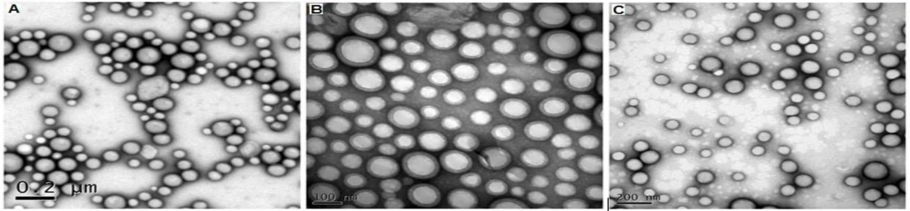
The results showed NPs homogeneity and a monomodal distribution considering that there was no statistical difference about the diameter and symmetrical distribution about 190 nm. The empty NPs showed a zeta potential of -15 mV, whereas NPs containing CUR had a zeta potential of -26 mV and CUR-TAM and TAM-NPs a zeta potential of -17 mV and -16 mV, respectively (Table 1). Regarding encapsulation efficiency (EE%), approximately 57% were obtained for TAM and 92% for CUR (Table 2). The NPs presented a spherical or oval shape, without the presence of aggregates (Figure 1).
Mean diameter and polydispersity index obtained by photon correlation spectroscopy, and zeta potential for TAM, CUR and CUR-TAM NPs (n=3).
Encapsulation efficiency for curcumin (CUR); tamoxifen (TAM); curcumin and tamoxifen (CUR-TAM) nanoparticles.Results expressed as mean ± SD (n=5).
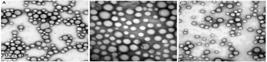
Ultrastructure of NPs obtained by transmission electron microscopy. The groups are: (A) CUR NPs, (B) TAM NPs, (C) CUR-TAM.
Stability
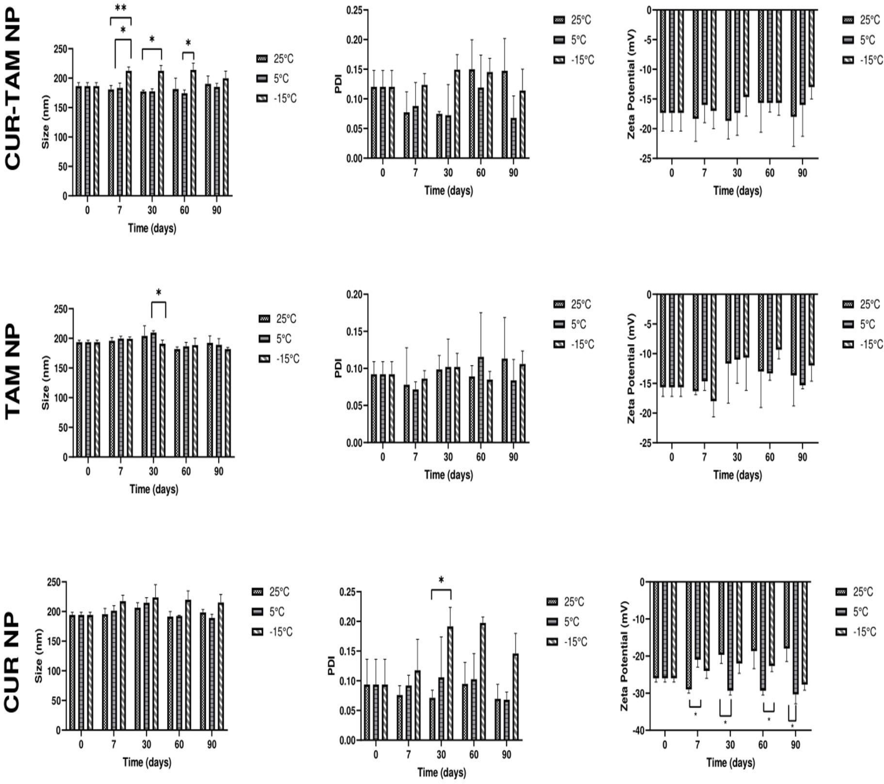
Regarding the diameter, the CUR-TAM NPs showed a variation in the average size only after 7 days at -15°C, indicating lower stability compared to TAM NPs, which exhibited a size variation only after 30 days at the same temperature. In contrast, CUR NPs did not show any variation in their average diameter.
The polydispersity index remained unchanged at all evaluated temperatures. Concerning the zeta potential, only TAM NPs showed a variation after 7 days at 25°C, while CUR and CUR-TAM NPs remained stable (Figure 2).
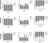
Stability parameters of nanoparticles in different temperatures during 90 days of evaluation. (*) significant difference with p<0.05 in Two-way ANOVA with 95% confidence and Tukey's post-test. In vitro release profile
Despite these variations, all types of nanoparticles analyzed remained stable in terms of size, polydispersity index, and zeta potential when stored at different temperatures for 90 days, suggesting that these storage conditions are suitable.
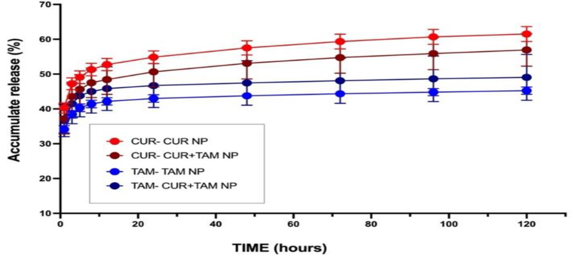
After 120 hours, the cumulative release of CUR was 61% for CUR NPs and 57% for CUR-TAM NPs, showing no significant difference between the two. For TAM the cumulative release was 45% for TAM NPs and 49% for CUR-TAM NPs, with no significant difference observed (Figure 3).
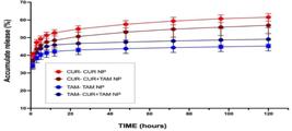
In vitro release profile of CUR and TAM in PLA NPs containing TAM, CUR and CUR-TAM, for a periodof 120 h.
To define the mathematical model that best suited the release profile, it was compared the zero-order, first and second order, Higuchi and Korsmeyers-Peppas models, aiming to obtain the model with correlation coefficient (r) closest to 1. For CUR, TAM and CUR-TAM NPs the equation that best represents the release profile was the Korsmeyer-Peppas model, in which there is closer to 1. In this, prolonged release occurs by diffusion mechanism (Table 3).
To evaluate the release mechanism of TAM and CUR from the NPs, it was used the Korsmeyer-Peppas model, that provides the release exponent (n) which express the type of releasing. The model considers that values of n ≤ 0.43 are related to a diffusion mechanism (Fickinian transport) and n ≥ 0.85 polymer erosion (non-Ficknian transport), while values of n in the range of 0.43 and 0.85 indicate anomalous transport where the diffusion mechanism is associated with erosion [46]. The n value of all NPs was below 0.43, indicating a release by diffusion (Table 4).
Evaluation of the release mechanism using the Korsmeyer-Peppas model of PLA nanoparticles containing CUR, TAM and CUR-TAM.
Analysis of cytotoxicity in red blood cells
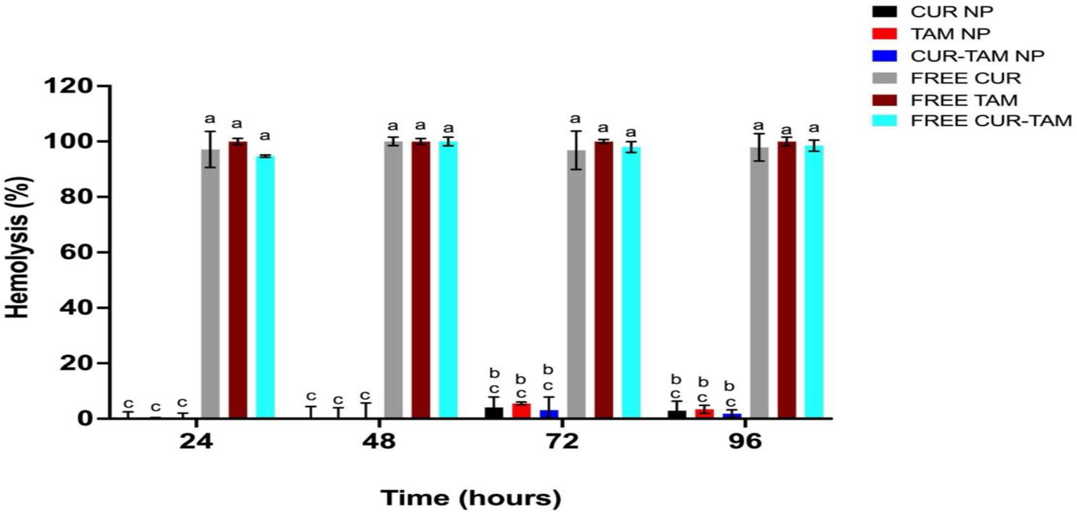
The results for free CUR and TAM showed that both compounds induce significant hemolysis. Free CUR caused nearly 100% hemolysis after 24 hours, while free TAM reached 100% hemolysis at the same time point, with this effect maintained up to 96 hours (Figure 4).
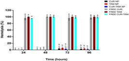
Percentage of hemolysis after treatment with NPs and free drugs, at concentrations of 200 µg/mL for CUR and 100 µg/mL for TAM, at different times. Hemolysis was determined by measuring the absorbance at 540 nm. a ,b, c, different letters represent statistical inequalities with p≤ 0.005 and equal letters indicate statistical equality.
Different from free forms, all NPs samples did not show hemolytic activity at 24 and 48 h of incubation. After 72 and 96 h, a low percentage of hemolysis was observed, with a large difference with free forms. The control with ethanol alone showed the same result as the negative control (buffer), demonstrating that the ethanol used to dilute the drugs in the free form did not interfere with the analyzes.
Analysis of cytotoxic effect in tumor cells
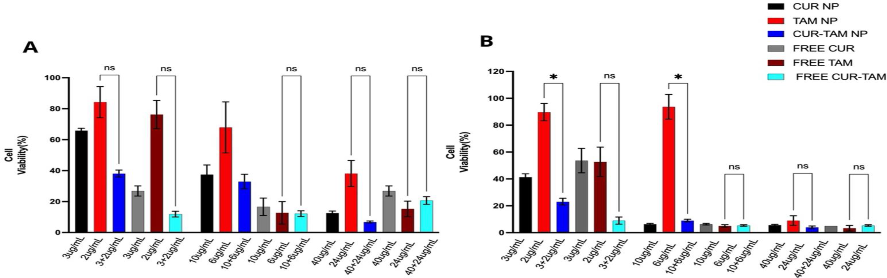
The B16F10 cells were incubated for 24 h and 72h with following drug concentration: CUR (3 μg /mL, 10 μg /mL, 40 μg /mL); TAM (2 μg /mL, 6 μg/mL, 24 μg/mL) and CUR+TAM (3 μg/mL+ 2 μg/mL, 10 μg/mL + 6 μg /mL, 40 μg /mL + 24 μg /mL) in free form and nonencapsulated. As negative control, it was used empty nanoparticle and the solvents (free evaluation), and both showed 100% cell viability.
The cytotoxic effect in melanoma cells was significantly obtained at 72h in the low concentration of CUR+TAM NP compared to TAM NP (Figure 4).
The effect was not observed when comparing high concentrations of free TAM and CUR+TAM, indicating that co-encapsulation of CUR and TAM yields better results than using TAM alone, whether encapsulated or free (Figure 5).
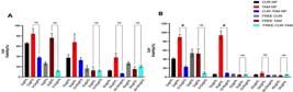
Effect of CUR, CUR-TAM and TAM (NPS), and free CUR, TAM and CUR-TAM on the viability of B16F10 cells. The graphs A and B represent 24 h and 72h, respectively, of incubation with NPs and free compounds. The ANOVA followed by Tuckey statical analysis was used (ns) non-significant, (*) p≤ 0.005.
DISCUSSION
The primary objective of the present work was to develop NPs incorporating CUR and TAM, both separately and in combination, to evaluate their release profile, toxicity, and efficacy against the proliferation of neoplastic cells (B16-F10).
Regarding NP preparation, they were prepared via single-emulsion solvent evaporation and presented satisfactory characteristics. The average size was approximately 200 nm, which is consistent with previous studies using the same compounds [47,48] and the zeta potential for CUR NPs was -26 mV, for TAM NP was -16mV and for CUR-TAM NP was -17mV, been satisfactory, considering that below -10 mV are indicative of potential instability [49,50]. Both characteristics were confirmed by microscopy, where is possible to see the predomination of size and low aggregation.
Upon comparing the encapsulation efficiencies of CUR and TAM, CUR is encapsulated more efficiently than TAM. A similar result was observed by Alhajamee and coauthors (2022) [51] in lipid nanoparticles containing CUR and TAM, which exhibited lower encapsulation efficiency compared to when each drug was encapsulated separately, aligned with our findings.
Regarding release in vitro experiments, a pattern was observed. Initially, nearly 0 up to 15 hours, there is a rapid release, possibly attributed to the adsorption of both TAM and CUR onto the surface of the nanoparticles, a characteristic often observed and a limitation of nanoparticle-based systems [52,53]. On In the other hand, after the initial period, the release is slow and consistent util 120h, showing a controlled system. This profile may be attributed to their hydrophobic characteristics, which affect their solubility in aqueous media [54,55].
Also release studies conducted in PBS (pH 7.4) under sink conditions with 10% CUR and TAM showed values of 0.319 mg/mL for TAM [56] and 0.310 mg/mL for CUR [57]. Also release studies conducted by our group in PBS (pH 7.4), presented value below of 0.43 suggesting that the prolonged release mechanism is primarily diffusion-based considering Korsmeyer-Peppas model where values n ≤ 0.43 indicate diffusion-controlled (Fickian transport) [58].
The biological analysis seeking to evaluate the NPs cytotoxic, by using red blood cells, and the antitumoral activity by using B16-F10 murine melanoma cells. TAM has been linked to hemolytic anemia in multiple studies [59-61] and presents hemolytic activity when tested in in vitro models [31,62]. The interaction of TAM with human erythrocytes causes morphological changes implicating in stomatocytes formation that affecting proteins located in the cytoplasmic portion of the erythrocyte membrane [63] and consequently, change the cell function. Additionally, TAM may disrupt membrane structural integrity, leading to alterations in membrane proteins and modifications in the structure of the cytoskeleton and/or plasma membrane proteins of red blood cells [64].
Considering the need to assess the cytotoxic effect in a systemic manner, and the important relationship between TAM and erythrocyte, the ex vivo hemolysis assays were done. The encapsulation has had a highly significant reduction compared with free form, demonstrating its compatibility with physiological process. Studies have already demonstrated that only the encapsulation of TAM in polymeric nanoparticles have advantages compared to free form [65,66]. And the association of free CUR and TAM freely [67,68]. TAM nanoparticles have been extensively studied in in vitro and in vivo [69,70] reenforcing that the nanoencapsulation brings advantages over free forms. Regarding the combining of free CUR and TAM, other researchers have also highlighted the benefits, but there is no researching showing these compounds co-encapsulated in PLA nanoparticles [71,72].
Despite our results, the co-encapsulation of CUR and TAM was formed like other studies that analyzed isolated forms. The efficiency of co-encapsulation of TAM with other drugs and its impact in cancer cytotoxicity was already demonstrated by combination of TAM with quercetin, where the authors found great impact [73,74]. Also, the co-encapsulation of CUR and TAM with deblock nanopolymer [75,76].
Conversely, the TAM PLA-NPs and CUR-PLA-NPs cause great cytotoxic effect reducing the viability in tumoral cell line (B16-F10) comparing to TAM and CUR free forms. In addition, the efficiency of co-encapsulation reduced tumoral cell viability and is significantly higher than the isolated forms. The encapsulation TAM with other compounds have also already demonstrated to be efficient on reduction of tumoral cells viability. The combination of TAM with quercetin showed great impact in tumor suppression of MCF-7 cells improving bioavailability [77,78]. Also, the co-encapsulation of CUR and TAM with deblock nanopolymer, improved the apoptosis of tamoxifen-resistant MCF-7 cancer cells and reduced the cytotoxic effect in normal cells caused by TAM in free form [79].
Furthermore, at high concentrations, the reduction in tumor viability after 72 hours was similar for both TAM nanoparticles and free TAM (Figure 4), even though the TAM NP group had about 40% drug release. This suggests that nanoparticles can reduce toxicity without compromising the antitumoral efficacy when used at appropriate doses. The main found of our results, it was the demonstration that combination of both drugs in nanoencapsulation positively impacts in TAM hemolysis, reducing it, and improve the TAM cytotoxicity effect especially after 72 hours of NPs compared to free form (Figure 4). This effect may have occurred due to the slow release of CUR and TAM from the NPs, considering the in vitro release assay where TAM was close to 50% and CUR was 60%, even after 120 h of experiment. The low concentration CUR-TAM NP is more effective in reducing the viability of B16-F10 tumor cells than CUR-TAM free form, showing that co-encapsulation is important to trigger cytotoxic effect to the tumor and consider the significant adverse effects associated with free TAM. This is supported by our hemolysis results and the literature [80,81] where the reduction in toxicity is a common benefit of sustained release systems [65,82].
Although our results are promising and provide the basis for the innovative field of drug co-encapsulation for cancer treatment, the system has a limitation regarding drug-loading capacity, in case been necessary increase TAM dose for treatment [83]. But these results are still an important breakthrough, especially considering its significant reducing of issues caused by TAM treatment to fight against tumoral cells.
CONCLUSION
The technological of the preparation of PLA NPs containing CUR-TAM is feasibility, and the physicochemical analysis the results were desirable found for nanostructured systems. In addition, the systems obtained were stable for three months in terms of size and zeta potential and presented a controlled release in vitro. The CUR-TAM NPs showed promising results decreasing the cytotoxic of TAM in blood cells and cell proliferation B16-F10 melanoma cells.
-
Funding:
The authors are grateful to the “Conselho Nacional de Desenvolvimento Científico e Tecnológico (CNPq) for financial support. This study was also supported in part by the Coordenação de Aperfeiçoamento de Pessoal de Nível Superior - Brasil (CAPES) - Finance Code 001; “Financiadora de Estudos e Projetos (FINEP-Brazil)”, and Fundação Araucária.
Acknowledgments:
The authors would like to thank Central de Microscopia - Complexo do Centro de Apoio à Pesquisa (COMCAP/Universidade Estadual de Maringá) for technical support.
Data Availability Statement:
Research data are only available upon request for corresponding author.
REFERENCES
-
1 Carr S, Smith C, Wernberg J. Epidemiology and Risk Factors of Melanoma. Surg Clin North Am 2020;100:1-12 https://doi.org/10.1016/j.suc.2019.09.005
» https://doi.org/10.1016/j.suc.2019.09.005 -
2 Ahmadi F, Karamitanha F, Ramezanpour A. Clustering trends of melanoma incidence and mortality: A worldwide assessment from 1995 to 2019. Aust J Dermatol 2022;63. https://doi.org/10.1111/ajd.13882
» https://doi.org/10.1111/ajd.13882 -
3 Guo W, Wang H, Li C. Signal pathways of melanoma and targeted therapy. Signal Transduct Target Ther 2021;6:424. https://doi.org/10.1038/s41392-021-00827-6
» https://doi.org/10.1038/s41392-021-00827-6 -
4 Villani A, Potestio L, Fabbrocini G, Troncone G, Malapelle U, Scalvenzi M. The Treatment of Advanced Melanoma: Therapeutic Update. Int J Mol Sci 2022;23:6388. https://doi.org/10.3390/ijms23126388
» https://doi.org/10.3390/ijms23126388 -
5 Stearns V. Active Tamoxifen Metabolite Plasma Concentrations After Coadministration of Tamoxifen and the Selective Serotonin Reuptake Inhibitor Paroxetine. J Natl Cancer Inst 2003;95:1758-64. https://doi.org/10.1093/jnci/djg108
» https://doi.org/10.1093/jnci/djg108 -
6 Yu Q, Xu Y, Yu E, Zheng Z. Risk of cardiovascular disease in breast cancer patients receiving aromatase inhibitors vs. tamoxifen: A systematic review and meta-analysis. J Clin Pharm Ther 2022;47:575-87. https://doi.org/10.1111/jcpt.13598
» https://doi.org/10.1111/jcpt.13598 -
7 Moreira PI, Custódio J, Moreno A, Oliveira CR, Santos MS. Tamoxifen and Estradiol Interact with the Flavin Mononucleotide Site of Complex I Leading to Mitochondrial Failure. JBC 2006;281:10143-52. https://doi.org/10.1074/jbc.M510249200
» https://doi.org/10.1074/jbc.M510249200 -
8 Matsuoka H, Tsubaki M, Yamazoe Y, Ogaki M, Satou T, Itoh T, et al. Tamoxifen inhibits tumor cell invasion and metastasis in mouse melanoma through suppression of PKC/MEK/ERK and PKC/PI3K/Akt pathways. Exp Cell Res 2009;315:2022-32. https://doi.org/10.1016/j.yexcr.2009.04.009
» https://doi.org/10.1016/j.yexcr.2009.04.009 -
9 Ghasemi M, Malek M, Javanmard ShH, Ghasemi A, Esfahani HN, Vaseghi G. Atorvastatin enhances anti-apoptotic effects of tamoxifen on melanoma cancer cells. BMJ 2019;120:752-6. https://doi.org/10.4149/BLL_2019_125
» https://doi.org/10.4149/BLL_2019_125 -
10 Chatterjee SJ, Pandey S. Chemo-resistant melanoma sensitized by tamoxifen to low dose curcumin treatment through induction of apoptosis and autophagy. Cancer Biol Ther 2011;11:216-28. https://doi.org/10.4161/cbt.11.2.13798
» https://doi.org/10.4161/cbt.11.2.13798 -
11 Emons G, Mustea A, Tempfer C. Tamoxifen and Endometrial Cancer: A Janus-Headed Drug. Cancers (Basel) 2020;12:2535. https://doi.org/10.3390/cancers12092535
» https://doi.org/10.3390/cancers12092535 -
12 Janacova L, Faktor J, Capkova L, Paralova V, Pospisilova A, Podhorec J, et al. SWATH-MS Analysis of FFPE Tissues Identifies Stathmin as a Potential Marker of Endometrial Cancer in Patients Exposed to Tamoxifen. J Proteome Res 2020;19:2617-30. https://doi.org/10.1021/acs.jproteome.0c00064
» https://doi.org/10.1021/acs.jproteome.0c00064 -
13 Elefsiniotis IS, Pantazis KD, Ilias A, Pallis L, Mariolis A, Glynou I, et al. Tamoxifen induced hepatotoxicity in breast cancer patients with pre-existing liver steatosis. Eur J Gastroenterol Hepatol 2004;16:593-8. https://doi.org/10.1097/00042737-200406000-00013
» https://doi.org/10.1097/00042737-200406000-00013 -
14 Parikh RP, Odom EB, Yu L, Colditz GA, Myckatyn TM. Complications and thromboembolic events associated with tamoxifen therapy in patients with breast cancer undergoing microvascular breast reconstruction: a systematic review and meta-analysis. Breast Cancer Res Treat 2017;163:1-10. https://doi.org/10.1007/s10549-017-4146-3
» https://doi.org/10.1007/s10549-017-4146-3 -
15 Abbasalizadeh F, Alizadeh E, Bagher Fazljou SM, Torbati M, Akbarzadeh A. Anticancer Effect of Alginate-chitosan Hydrogel Loaded with Curcumin and Chrysin on Lung and Breast Cancer Cell Lines. Curr Drug Deliv 2022;19:600-13. https://doi.org/10.2174/1567201818666210813142007
» https://doi.org/10.2174/1567201818666210813142007 -
16 Yu T, Chen C, Sun Y, Sun H, Li T, Meng J, et al. ABT-737 sensitizes curcumin-induced anti-melanoma cell activity through facilitating mPTP death pathway. Biochem Biophys Res Commun 2015;464:286-91. https://doi.org/10.1016/j.bbrc.2015.06.144
» https://doi.org/10.1016/j.bbrc.2015.06.144 -
17 Nabavi SM, Russo GL, Tedesco I, Daglia M, Orhan IE, Nabavi SF, et al. Curcumin and Melanoma: From Chemistry to Medicine. Nutr Cancer 2018;70:164-75. https://doi.org/10.1080/01635581.2018.1412485
» https://doi.org/10.1080/01635581.2018.1412485 -
18 Abd. Wahab NA, H. Lajis N, Abas F, Othman I, Naidu R. Mechanism of Anti-Cancer Activity of Curcumin on Androgen-Dependent and Androgen-Independent Prostate Cancer. Nutrients 2020;12:679. https://doi.org/10.3390/nu12030679
» https://doi.org/10.3390/nu12030679 -
19 Mortezaee K, Salehi E, Mirtavoos-mahyari H, Motevaseli E, Najafi M, Farhood B, et al. Mechanisms of apoptosis modulation by curcumin: Implications for cancer therapy. J Cell Physiol 2019;234:12537-50. https://doi.org/10.1002/jcp.28122
» https://doi.org/10.1002/jcp.28122 -
20 Kianamiri S, Dinari A, Sadeghizadeh M, Rezaei M, Daraei B, Bahsoun NE-H, et al. Mitochondria-Targeted Polyamidoamine Dendrimer-Curcumin Construct for Hepatocellular Cancer Treatment. Mol Pharm 2020;17:4483-98. https://doi.org/10.1021/acs.molpharmaceut.0c00566
» https://doi.org/10.1021/acs.molpharmaceut.0c00566 -
21 Pavón N, Buelna-Chontal M, Correa F, Yoval-Sánchez B, Belmont J, Hernández-Esquivel L, et al. Tamoxifen inhibits mitochondrial membrane damage caused by disulfiram. Biochem Cell Biol. 2017;95:556-62. https://doi.org/10.1139/bcb-2017-0027
» https://doi.org/10.1139/bcb-2017-0027 -
22 Fatemizadeh M, Tafvizi F, Shamsi F, Amiri S, Farajzadeh A, Akbarzadeh I. Apoptosis Induction, Cell Cycle Arrest and Anti-Cancer Potential of Tamoxifen-Curcumin Loaded Niosomes Against MCF-7 Cancer Cells. Iran J Pathol 2022;17:183-90. https://doi.org/10.30699/IJP.2022.124340.2356
» https://doi.org/10.30699/IJP.2022.124340.2356 -
23 Patra JK, Das G, Fraceto LF, Campos EVR, Rodriguez-Torres M del P, Acosta-Torres LS, et al. Nano based drug delivery systems: recent developments and future prospects. J Nanobiotechnology 2018;16:71. https://doi.org/10.1186/s12951-018-0392-8
» https://doi.org/10.1186/s12951-018-0392-8 -
24 Cui W, Zhao H, Wang C, Chen Y, Luo C, Zhang S, et al. Co-encapsulation of docetaxel and cyclosporin A into SNEDDS to promote oral cancer chemotherapy. Drug Deliv 2019;26:542-50. https://doi.org/10.1080/10717544.2019.1616237
» https://doi.org/10.1080/10717544.2019.1616237 -
25 Zafar S, Akhter S, Garg N, Selvapandiyan A, Kumar Jain G, Ahmad FJ. Co-encapsulation of docetaxel and thymoquinone in mPEG-DSPE-vitamin E TPGS-lipid nanocapsules for breast cancer therapy: Formulation optimization and implications on cellular and in vivo toxicity. Eur J Pharm Biopharm 2020;148:10-26. https://doi.org/10.1016/j.ejpb.2019.12.016
» https://doi.org/10.1016/j.ejpb.2019.12.016 -
26 Shafiei-Irannejad V, Samadi N, Salehi R, Yousefi B, Rahimi M, Akbarzadeh A, et al. Reversion of Multidrug Resistance by Co-Encapsulation of Doxorubicin and Metformin in Poly(lactide-co-glycolide)-d-α-tocopheryl Polyethylene Glycol 1000 Succinate Nanoparticles. Pharm Res 2018;35:119. https://doi.org/10.1007/s11095-018-2404-7
» https://doi.org/10.1007/s11095-018-2404-7 -
27 Jain AK, Thanki K, Jain S. Co-encapsulation of Tamoxifen and Quercetin in Polymeric Nanoparticles: Implications on Oral Bioavailability, Antitumor Efficacy, and Drug-Induced Toxicity. Mol Pharm 2013;10:3459-74. https://doi.org/10.1021/mp400311j
» https://doi.org/10.1021/mp400311j -
28 Feng C, Zhang H, Chen J, Wang S, Xin Y, Qu Y, et al. Ratiometric co-encapsulation and co-delivery of doxorubicin and paclitaxel by tumor-targeted lipodisks for combination therapy of breast cancer. Int J Pharm 2019;560:191-204. https://doi.org/10.1016/j.ijpharm.2019.02.009
» https://doi.org/10.1016/j.ijpharm.2019.02.009 -
29 Odeh F, Naffa R, Azzam H, Mahmoud IS, Alshaer W, Al Bawab A, et al. Co-encapsulation of thymoquinone with docetaxel enhances the encapsulation efficiency into PEGylated liposomes and the chemosensitivity of MCF7 breast cancer cells to docetaxel. Heliyon 2019;5:e02919. https://doi.org/10.1016/j.heliyon.2019.e02919
» https://doi.org/10.1016/j.heliyon.2019.e02919 -
30 Luque-Michel E, Sebastian V, Larrea A, Marquina C, Blanco-Prieto MJ. Co-encapsulation of superparamagnetic nanoparticles and doxorubicin in PLGA nanocarriers: Development, characterization and in vitro antitumor efficacy in glioma cells. Eur J Pharm Biopharm 2019;145:65-75. https://doi.org/10.1016/j.ejpb.2019.10.004
» https://doi.org/10.1016/j.ejpb.2019.10.004 -
31 Altmeyer C, Karam TK, Khalil NM, Mainardes RM. Tamoxifen-loaded poly(L-lactide) nanoparticles: Development, characterization and in vitro evaluation of cytotoxicity. Mat Sci Eng: C 2016;60:135-42. https://doi.org/10.1016/j.msec.2015.11.019
» https://doi.org/10.1016/j.msec.2015.11.019 -
32 Lima IA de, Khalil NM, Tominaga TT, Lechanteur A, Sarmento B, Mainardes RM. Mucoadhesive chitosan-coated PLGA nanoparticles for oral delivery of ferulic acid. Artif Cells Nanomed Biotechnol 2018;46:993-1002. https://doi.org/10.1080/21691401.2018.1477788
» https://doi.org/10.1080/21691401.2018.1477788 -
33 Afsharzadeh M, Hashemi M, Babaei M, Abnous K, Ramezani M. PEG-PLA nanoparticles decorated with small-molecule PSMA ligand for targeted delivery of galbanic acid and docetaxel to prostate cancer cells. J Cell Physiol 2020;235:4618-30. https://doi.org/10.1002/jcp.29339
» https://doi.org/10.1002/jcp.29339 -
34 Tang H, Chen J, Wang L, Li Q, Yang Y, Lv Z, et al. Co-delivery of epirubicin and paclitaxel using an estrone-targeted PEGylated liposomal nanoparticle for breast cancer. Int J Pharm 2020;573:118806. https://doi.org/10.1016/j.ijpharm.2019.118806
» https://doi.org/10.1016/j.ijpharm.2019.118806 -
35 Elsey J, Bubley JA, Zhu L, Rao S, Sasaki M, Pollack BP, et al. Palladium based nanoparticles for the treatment of advanced melanoma. Sci Rep 2019;9:3255. https://doi.org/10.1038/s41598-019-40258-6
» https://doi.org/10.1038/s41598-019-40258-6 -
36 Da Silveira Nogueira Lima JP, Georgieva M, Haaland B, de Lima Lopes G. A systematic review and network meta-analysis of immunotherapy and targeted therapy for advanced melanoma. Cancer Med 2017;6:1143-53. https://doi.org/10.1002/cam4.1001
» https://doi.org/10.1002/cam4.1001 -
37 Palliyage GH, Hussein N, Mimlitz M, Weeder C, Alnasser MHA, Singh S, et al. Novel Curcumin-Resveratrol Solid Nanoparticles Synergistically Inhibit Proliferation of Melanoma Cells. Pharm Res 2021;38:851-71. https://doi.org/10.1007/s11095-021-03043-7
» https://doi.org/10.1007/s11095-021-03043-7 -
38 Wang Z, Yu Y, Wang C, Li J, Pang Y. Advances in the Application of Nanomaterials to the Treatment of Melanoma. Pharmaceutics 2022;14:2090. https://doi.org/10.3390/pharmaceutics14102090
» https://doi.org/10.3390/pharmaceutics14102090 -
39 Xu M, Asghar S, Dai S, Wang Y, Feng S, Jin L, et al. Mesenchymal stem cells-curcumin loaded chitosan nanoparticles hybrid vectors for tumor-tropic therapy. Int J Biol Macromol 2019;134:1002-12. https://doi.org/10.1016/j.ijbiomac.2019.04.201
» https://doi.org/10.1016/j.ijbiomac.2019.04.201 -
40 Xiong W, Guo Z, Zeng B, Wang T, Zeng X, Cao W, et al. Dacarbazine-Loaded Targeted Polymeric Nanoparticles for Enhancing Malignant Melanoma Therapy. Front Bioeng Biotechnol 2022;10. https://doi.org/10.3389/fbioe.2022.847901
» https://doi.org/10.3389/fbioe.2022.847901 -
41 Clemente N, Miletto I, Gianotti E, Sabbatini M, Invernizzi M, Marchese L, et al. Verteporfin-Loaded Mesoporous Silica Nanoparticles’ Topical Applications Inhibit Mouse Melanoma Lymphangiogenesis and Micrometastasis In Vivo. Int J Mol Sci 2021;22:13443. https://doi.org/10.3390/ijms222413443
» https://doi.org/10.3390/ijms222413443 -
42 Drača D, Edeler D, Saoud M, Dojčinović B, Dunđerović D, Đmura G, et al. Antitumor potential of cisplatin loaded into SBA-15 mesoporous silica nanoparticles against B16F1 melanoma cells: in vitro and in vivo studies. J Inorg Biochem 2021;217:111383. https://doi.org/10.1016/j.jinorgbio.2021.111383
» https://doi.org/10.1016/j.jinorgbio.2021.111383 -
43 Antonio E, dos Reis Antunes Junior O, Marcano RGDJV, Diedrich C, da Silva Santos J, Machado CS, et al. Chitosan modified poly (lactic acid) nanoparticles increased the ursolic acid oral bioavailability. Int J Biol Macromol 2021;172:133-42. https://doi.org/10.1016/j.ijbiomac.2021.01.041
» https://doi.org/10.1016/j.ijbiomac.2021.01.041 -
44 Cho YA, Lee W, Choi JS. Effects of curcumin on the pharmacokinetics of tamoxifen and its active metabolite, 4-hydroxytamoxifen, in rats: possible role of CYP3A4 and P-glycoprotein inhibition by curcumin. Due Pharmazie, 2012;67:124-30. https://doi.org/10.1007/s12272-011-1117-9
» https://doi.org/10.1007/s12272-011-1117-9 -
45 Espada R, Valdespina S, Alfonso C, Rivas G, Ballesteros MP, Torrado JJ. Effect of aggregation state on the toxicity of different amphotericin B preparations. Int J Pharm 2008;361:64-9. https://doi.org/10.1016/j.ijpharm.2008.05.013
» https://doi.org/10.1016/j.ijpharm.2008.05.013 -
46 Siepmann J. Modeling of drug release from delivery systems based on hydroxypropyl methylcellulose (HPMC). Adv Drug Deliv Rev 2001;48:139-57. https://doi.org/10.1016/S0169-409X(01)00112-0
» https://doi.org/10.1016/S0169-409X(01)00112-0 -
47 Hu FX, Neoh KG, Kang ET. Synthesis and in vitro anti-cancer evaluation of tamoxifen-loaded magnetite/PLLA composite nanoparticles. Biomaterials 2006;27:5725-33. https://doi.org/10.1016/j.biomaterials.2006.07.014
» https://doi.org/10.1016/j.biomaterials.2006.07.014 -
48 Silva-Buzanello RA da, Ferro AC, Bona E, Cardozo-Filho L, Araújo PHH de, Leimann FV, et al. Validation of an Ultraviolet-visible (UV-Vis) technique for the quantitative determination of curcumin in poly(l-lactic acid) nanoparticles. Food Chem 2015;172:99-104. https://doi.org/10.1016/j.foodchem.2014.09.016
» https://doi.org/10.1016/j.foodchem.2014.09.016 -
49 Sarnatskaya V, Shlapa Y, Yushko L, Shton I, Solopan S, Ostrovska G, et al. Biological activity of cerium dioxide nanoparticles. J Biomed Mater Res A 2020;108:1703-12. https://doi.org/10.1002/jbm.a.36936
» https://doi.org/10.1002/jbm.a.36936 -
50 Abdelbary AA, Li X, El-Nabarawi M, Elassasy A, Jasti B. Effect of fixed aqueous layer thickness of polymeric stabilizers on zeta potential and stability of aripiprazole nanosuspensions. Pharm Dev Technol 2013;18:730-5. https://doi.org/10.3109/10837450.2012.727001
» https://doi.org/10.3109/10837450.2012.727001 -
51 Alhajamee M, Marai K, Al Abbas SMN, Homayouni Tabrizi M. Co-encapsulation of curcumin and tamoxifen in lipid-chitosan hybrid nanoparticles for cancer therapy. Mater Tech 2022;37:1183-94. https://doi.org/10.1080/10667857.2021.1926811
» https://doi.org/10.1080/10667857.2021.1926811 -
52 Rezaei A, Nasirpour A. Evaluation of Release Kinetics and Mechanisms of Curcumin and Curcumin-β-Cyclodextrin Inclusion Complex Incorporated in Electrospun Almond Gum/PVA Nanofibers in Simulated Saliva and Simulated Gastrointestinal Conditions. Bionanoscience 2019;9:438-45. https://doi.org/10.1007/s12668-019-00620-4
» https://doi.org/10.1007/s12668-019-00620-4 -
53 Moin A, Wani SUD, Osmani RA, Abu Lila AS, Khafagy E-S, Arab HH, et al. Formulation, characterization, and cellular toxicity assessment of tamoxifen-loaded silk fibroin nanoparticles in breast cancer. Drug Deliv 2021;28:1626-36. https://doi.org/10.1080/10717544.2021.1958106
» https://doi.org/10.1080/10717544.2021.1958106 -
54 Di Francesco M, Primavera R, Summa M, Pannuzzo M, Di Francesco V, Di Mascolo D, et al. Engineering shape-defined PLGA microPlates for the sustained release of anti-inflammatory molecules. JCR 2020;319:201-12. https://doi.org/10.1016/j.jconrel.2019.12.039
» https://doi.org/10.1016/j.jconrel.2019.12.039 -
55 D’Addio SM, Bukari AA, Dawoud M, Bunjes H, Rinaldi C, Prud’homme RK. Determining drug release rates of hydrophobic compounds from nanocarriers. Philos Trans A Math Phys Eng Sci 2016;374:20150128. https://doi.org/10.1098/rsta.2015.0128
» https://doi.org/10.1098/rsta.2015.0128 -
56 SreeHarsha N, Hiremath JG, Chilukuri S, Aitha RK, Al-Dhubiab BE, Venugopala KN, et al. An Approach to Enhance Dissolution Rate of Tamoxifen Citrate. Biomed Res Int 2019;2019:1-11. https://doi.org/10.1155/2019/2161348
» https://doi.org/10.1155/2019/2161348 -
57 Lu Y, Lin M, Zong J, Zong L, Zhao Z, Wang S, et al. Highly bioavailable curcumin preparation with a co-grinding and solvent-free process. Food Sci Nutr 2020;8:6415-25. https://doi.org/10.1002/fsn3.1930
» https://doi.org/10.1002/fsn3.1930 -
58 Siepmann J. Modeling of drug release from delivery systems based on hydroxypropyl methylcellulose (HPMC). Adv Drug Deliv Rev 2001;48:139-57. https://doi.org/10.1016/S0169-409X(01)00112-0
» https://doi.org/10.1016/S0169-409X(01)00112-0 -
59 Ching CK, Smith PG, Long RG. Tamoxifen-associated hepatocellular damage and agranulocytosis. The Lancet 1992;339:940. https://doi.org/10.1016/0140-6736(92)90991-B
» https://doi.org/10.1016/0140-6736(92)90991-B -
60 Montes A, Powles TJ, O’Brien MER, Ashley SE, Luckit J, Treleaven J. A toxic interaction between mitomycin C and tamoxifen causing the haemolytic uraemic syndrome. Eur J Cancer 1993;29:1854-7. https://doi.org/10.1016/0959-8049(93)90537-P
» https://doi.org/10.1016/0959-8049(93)90537-P -
61 Thangaraju M, Vijayalakshmi T, Sachdanandam P. Effect of tamoxifen on lipid peroxide and antioxidative system in postmenopausal women with breast cancer. Cancer 1994;74:78-82. https://doi.org/10.1002/1097-0142(19940701)74:1<78::aid-cncr2820740114>3.0.co;2-l
» https://doi.org/10.1002/1097-0142(19940701)74:1<78::aid-cncr2820740114>3.0.co;2-l -
62 Pedroso LS, Fávero GM, de Camargo LEA, Mainardes RM, Khalil NM. Effect of the o-methyl catechols apocynin, curcumin and vanillin on the cytotoxicity activity of tamoxifen. J Enzyme Inhib Med Chem 2013;28:734-40. https://doi.org/10.3109/14756366.2012.680064
» https://doi.org/10.3109/14756366.2012.680064 -
63 Suwalsky M, Hernández P, Villena F, Aguilar F, Sotomayor CP. Interaction of the Anticancer Drug Tamoxifen with the Human Erythrocyte Membrane and Molecular Models. Z Naturforsch C J Biosci 1998;53:182-90. https://doi.org/10.1515/znc-1998-3-407
» https://doi.org/10.1515/znc-1998-3-407 -
64 Cruz Silva MM, Madeira VMC, Almeida LM, Custódio JBA. Hemolysis of human erythrocytes induced by tamoxifen is related to disruption of membrane structure. Biochim Biophys Acta - Biomembranes 2000;1464:49-61. https://doi.org/10.1016/S0005-2736(99)00237-0
» https://doi.org/10.1016/S0005-2736(99)00237-0 -
65 Altmeyer C, Karam TK, Khalil NM, Mainardes RM. Tamoxifen-loaded poly(L-lactide) nanoparticles: Development, characterization and in vitro evaluation of cytotoxicity. Mat Sci Eng C Mater Biol Appl 2016;60:135-42. https://doi.org/10.1016/j.msec.2015.11.019
» https://doi.org/10.1016/j.msec.2015.11.019 -
66 Shenoy DB, Amiji MM. Poly(ethylene oxide)-modified poly(ɛ-caprolactone) nanoparticles for targeted delivery of tamoxifen in breast cancer. Int J Pharm 2005;293:261-70. https://doi.org/10.1016/j.ijpharm.2004.12.010
» https://doi.org/10.1016/j.ijpharm.2004.12.010 -
67 Jain AK, Swarnakar NK, Godugu C, Singh RP, Jain S. The effect of the oral administration of polymeric nanoparticles on the efficacy and toxicity of tamoxifen. Biomaterials 2011;32:503-15. https://doi.org/10.1016/j.biomaterials.2010.09.037
» https://doi.org/10.1016/j.biomaterials.2010.09.037 -
68 Kapse SV, Gaikwad RV, Samad A, Devarajan PV. Self nanoprecipitating preconcentrate of tamoxifen citrate for enhanced bioavailability. Int J Pharm 2012;429:104-12. https://doi.org/10.1016/j.ijpharm.2012.02.042
» https://doi.org/10.1016/j.ijpharm.2012.02.042 -
69 Khuroo T, Verma D, Talegaonkar S, Padhi S, Panda AK, Iqbal Z. Topotecan-tamoxifen duple PLGA polymeric nanoparticles: Investigation of in vitro, in vivo and cellular uptake potential. Int J Pharm 2014;473:384-94. https://doi.org/10.1016/j.ijpharm.2014.07.022
» https://doi.org/10.1016/j.ijpharm.2014.07.022 -
70 Khalil NM, do Nascimento TCF, Casa DM, Dalmolin LF, de Mattos AC, Hoss I, et al. Pharmacokinetics of curcumin-loaded PLGA and PLGA-PEG blend nanoparticles after oral administration in rats. Colloids Surf B Biointerfaces 2013;101:353-60. https://doi.org/10.1016/j.colsurfb.2012.06.024
» https://doi.org/10.1016/j.colsurfb.2012.06.024 -
71 Kakkar V, Muppu SK, Chopra K, Kaur IP. Curcumin loaded solid lipid nanoparticles: An efficient formulation approach for cerebral ischemic reperfusion injury in rats. Eur J Pharm Biopharm 2013;85:339-45. https://doi.org/10.1016/j.ejpb.2013.02.005
» https://doi.org/10.1016/j.ejpb.2013.02.005 - 72 Anitha A, Sreeranganathan M, Chennazhi KP, Lakshmanan V-K, Jayakumar R. In vitro combinatorial anticancer effects of 5-fluorouracil and curcumin loaded N,O-carboxymethyl chitosan nanoparticles toward colon cancer and in vivo pharmacokinetic studies. Eur J Pharm Biopharm 2014;88:238-51. c.
-
73 Ruttala HB, Ko YT. Liposomal co-delivery of curcumin and albumin/paclitaxel nanoparticle for enhanced synergistic antitumor efficacy. Colloids Surf B Biointerfaces 2015;128:419-26. https://doi.org/10.1016/j.colsurfb.2015.02.040
» https://doi.org/10.1016/j.colsurfb.2015.02.040 -
74 Chatterjee SJ, Pandey S. Chemo-resistant melanoma sensitized by tamoxifen to low dose curcumin treatment through induction of apoptosis and autophagy. Cancer Biol Ther 2011;11:216-28. https://doi.org/10.4161/cbt.11.2.13798
» https://doi.org/10.4161/cbt.11.2.13798 -
75 El-Beshbishy HA, Mohamadin AM, Nagy AA, Abdel-Naim AB. Amelioration of tamoxifen-induced liver injury in rats by grape seed extract, black seed extract and curcumin. Indian J Exp Biol 2010;48:280-8. https://doi.org/10.1016/j.jep.2013.03.065
» https://doi.org/10.1016/j.jep.2013.03.065 -
76 Jiang M, Huang O, Zhang X, Xie Z, Shen A, Liu H, et al. Curcumin Induces Cell Death and Restores Tamoxifen Sensitivity in the Antiestrogen-Resistant Breast Cancer Cell Lines MCF-7/LCC2 and MCF-7/LCC9. Molecules 2013;18:701-20. https://doi.org/10.3390/molecules18010701
» https://doi.org/10.3390/molecules18010701 -
77 Jain AK, Thanki K, Jain S. Co-encapsulation of Tamoxifen and Quercetin in Polymeric Nanoparticles: Implications on Oral Bioavailability, Antitumor Efficacy, and Drug-Induced Toxicity. Mol Pharm 2013;10:3459-74. https://doi.org/10.1021/mp400311j
» https://doi.org/10.1021/mp400311j -
78 Lockhart JN, Stevens DM, Beezer DB, Kravitz A, Harth E. Dual drug delivery of tamoxifen and quercetin: Regulated metabolism for anticancer treatment with nanosponges. JCR 2015;220:751-7. https://doi.org/10.1016/j.jconrel.2015.08.052
» https://doi.org/10.1016/j.jconrel.2015.08.052 - 79 Hajigholami S, Veisi Malekshahi Z, Bodaghabadi N, Najafi F, Shirzad H, Sadeghizadeh M. Nano Packaged Tamoxifen and Curcumin; Effective Formulation against Sensitive and Resistant MCF-7 Cells. Iran J Pharm Res 2018;17:1-10.
-
80 Zavareh HS, Pourmadadi M, Moradi A, Yazdian F, Omidi M. Chitosan/carbon quantum dot/aptamer complex as a potential anticancer drug delivery system towards the release of 5-fluorouracil. Int J Biol Macromol 2020;165:1422-30. https://doi.org/10.1016/j.ijbiomac.2020.09.166
» https://doi.org/10.1016/j.ijbiomac.2020.09.166 -
81 Cruz Silva MM, Madeira VMC, Almeida LM, Custódio JBA. Hemolysis of human erythrocytes induced by tamoxifen is related to disruption of membrane structure. BBA 2000;1464:49-61. https://doi.org/10.1016/S0005-2736(99)00237-0
» https://doi.org/10.1016/S0005-2736(99)00237-0 -
82 Yu X, Sun L, Tan L, Wang M, Ren X, Pi J, et al. Preparation and Characterization of PLGA-PEG-PLGA Nanoparticles Containing Salidroside and Tamoxifen for Breast Cancer Therapy. AAPS PharmSciTech 2020;21:85. https://doi.org/10.1208/s12249-019-1523-8
» https://doi.org/10.1208/s12249-019-1523-8 -
83 Zatorska M, Łazarski G, Maziarz U, Wilkosz N, Honda T, Yusa SI, et al. Drug-loading capacity of polylactide-based microand nanoparticles - Experimental and molecular modeling study. Int J Pharm 2020; 15;591:120031. https://doi.org/10.1016/j.ijpharm.2020.120031
» https://doi.org/10.1016/j.ijpharm.2020.120031
-
Editor-in-Chief:
Paulo Vitor Farago
-
Associate Editor:
Paulo Vitor Farago