Linalool Alleviates Oxidative Stress and Inflammatory Markers in Rats with CFA-Induced Rheumatoid Arthritis
Abstract
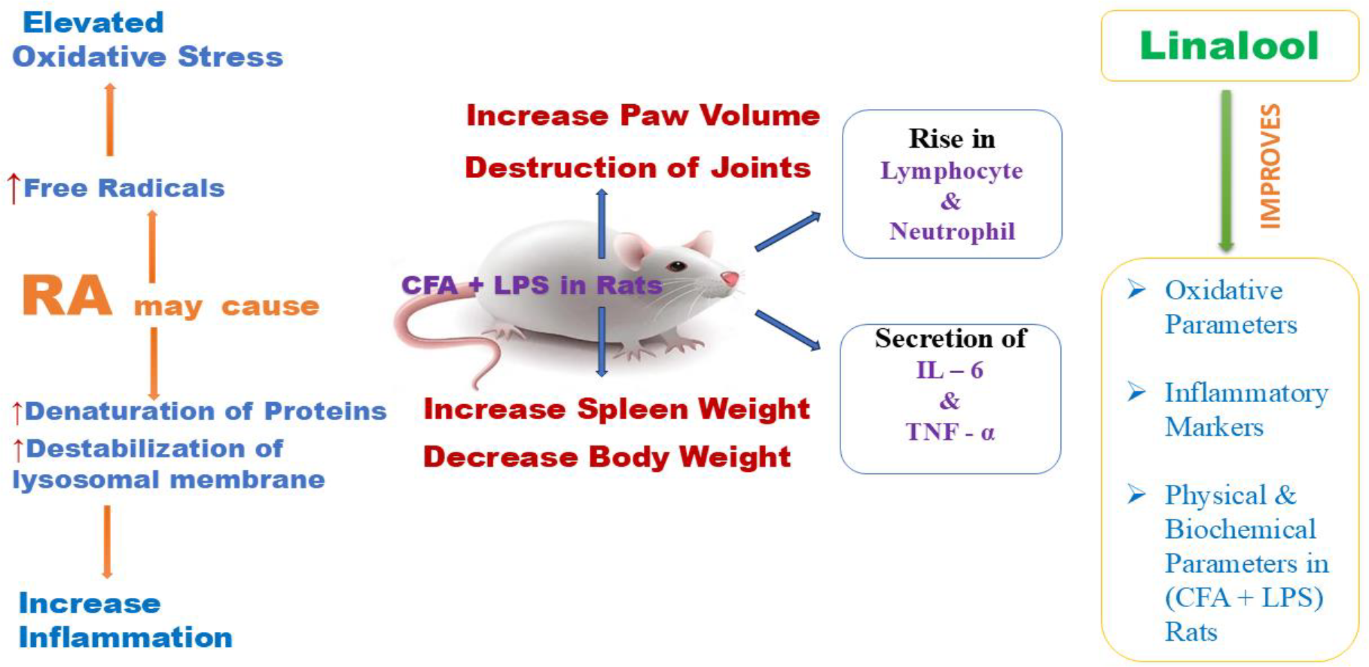
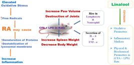
Rheumatoid Arthritis (RA) is an autoimmune and idiopathic disorder that inflames joint synovial tissue, cartilage, bone and lowers the quality of life. Many studies link RA to leukocyte-mediated inflammation and oxidative damage along with the liberation of many inflammatory cells, degradative enzymes, cytokines and chemokines. Linalool is the primary ingredient of some essential oils derived from aromatic plants containing antioxidant properties. The aim of this study was to evaluate the antirheumatic effect of Linalool through invitro, insilico and Complete Freund's adjuvant induced arthritic rat model. Antioxidant activity was measured by 2, 2-Diphenyl-1-picrylhydrazyl, Nitric oxide, Superoxide, and Hydroxyl radical scavenging assay, and anti-inflammatory activity by Human Red Blood Cell stabilization and Protein denaturation inhibition assay. Rats were stimulated with Complete Freund's adjuvant, type II collagen, and Lipopolysaccharide to develop arthritis and evaluated for biochemical and arthritic markers. Linalool exhibits concentration dependent inhibition in both antioxidant and anti-inflammatory assays at concentrations ranging from 25 to 250 µg/mL. Subsequently, both 100 mg/kg and 400 mg/kg of Linalool managed all the arthritic parameters (body weight, paw volume, splenic index, and arthritic index) effectively, including Tumor Necrosis Factor- α, Interleukin-6, Lymphocytes and Neutrophils. The insilico analysis shows a favourable protein-ligand interaction for the identified proteins. Linalool was effective in reducing inflammation, prevents joint and cartilage damage, and modulates immunological responses, suggesting it might be helpful in RA. Further research and clinical trials are needed to validate these results and explore Linalool's potential as a viable treatment option for patients suffering from this debilitating autoimmune disease.
Keywords:
Linalool; Rheumatoid Arthritis; Cytokines; Lipopolysaccharide; Complete Freund's adjuvant.
HIGHLIGHTS
CFA-induced RA rats were tested for linalool's effect on inflammatory markers.
Linalool exhibits concentration dependent inhibition ininvitro antioxidant studies.
Linalool stabilize HRBC membrane and inhibit protein denaturation.
Linalool improves inflammatory and biochemical parameters in CFA Rats.
INTRODUCTION
Rheumatoid arthritis (RA) is an idiopathic condition that involves inflammation of the synovial tissue in joints, as well as cartilage and bone. In certain cases, it may also affect areas outside of the joints [1]. It is estimated that between 0.5% and 1% of people worldwide suffer from RA, with women two to three times more likely than males to have the illness, manifested as per the epidemiological data. RA usually begins to appear between the ages of 30 and 60, however it can develop at any age [2]. The illness has a greater occurrence in developed nations, with elevated rates of occurrence reported in Northern Europe and North America in contrast to places such as Asia and Africa [3]. Recent evidence suggests that RA is influenced by both genetic and epigenetic factors, certain HLA-DRB1 alleles, in particular, as well as environmental factors such as cigarette smoke, dust exposure, and also the microbiome [4]. Due to impairment and diminished quality of life, RA has major social and economic consequences in addition to its physical health effects, making the illness a heavy burden [5]. A harmful inflammatory process occurring in the body have been observed by the recent investigations, indicated by the infiltration of synovial cells and the presence of inflammatory cells in the peripheral blood, including monocytes, polymorphonuclear neutrophils, and lymphocytes. These cells play a crucial role in causing synovial inflammation and subsequent damage to the joints [6,7]. Synovial hyperplasia is a defining hallmark of RA and is the main cause of the formation of an invasive pannus. Although the precise mechanisms that initiate RA are not fully comprehended, it can be stated that during the active phase of RA, cells originating from macrophages and fibroblasts directly cause tissue damage and sustain the complex disease process by undergoing morphological and phenotypic changes [8,9]. Moreover, Macrophages, which are the predominant proinflammatory cells in the synovium of RA, exhibit excessive activation and proliferation to secrete cytokines such as Interleukins (IL) (IL-1β and IL- 6) and Tumor necrosis factor-α (TNF-α) inducing intense inflammation by activating immune cells, stimulating fibroblasts, and polarizing T lymphocytes [10,11,12]. Based on some findings, people with RA may have higher levels of reactive oxygen species (ROS) and reactive nitrogen species (RNS) in their blood compared to normal individuals. Additionally, their endogenous enzymatic and non-enzymatic defense mechanisms may be diminishing [13]. The relationship between ROS and mitochondrial damage is strongly linked to many important pathogenic processes in RA. As a result, the adjustment of mitochondrial activity, elimination of ROS, and reduction of oxidative stress have become important areas of focus for the treatment of RA [14,15]. Modern pharmaceutical treatments, such as Disease Modifying Antirheumatic Drugs which encompass both biologic and synthetic choices, Non-Steroidal Anti-inflammatory Drugs (NSAID), and glucocorticoids, have shown encouraging results. However, the significant side effects and high cost of these therapies pose obstacles for patients seeking the most effective treatment [16,17]. Hence, it is imperative to conduct extensive research to identify and develop novel, potent medications for RA. This is necessary to address the shortcomings of current treatments, improve patient outcomes, and ultimately progress the management of this persistent ailment. Plants and components derived from plants possess bioactive properties that enable them to effectively treat inflammatory illnesses [18]. Polyphenols, terpenoids, and alkaloids function by inhibiting enzymes and modulating the immune system to reduce inflammation [19,20]. Some research indicates that the antioxidant capabilities of plant-based substances can effectively decrease inflammation by neutralizing oxidative harm [21]. Essential oils are intricate combinations of volatile molecules derived from aromatic plant components, which can be produced in many organs as secondary metabolites. The primary components of essential oils are terpenes, predominantly altered terpenes [22]. Several aromatic species' essential oils are known to contain a significant amount of the monoterpene Linalool as a volatile component. Some plants, such as mints, laurels, cinnamon, rosewood, and citrus, generate a lot of Linalool [23]. Linalool has powerful antioxidant characteristics, which assist to neutralize free radicals and protect cells from oxidative stress, according to studies [24]. This may provide protection from neurodegenerative illnesses, arthritis, cardiovascular problems, and other ailments linked to chronic oxidative stress and inflammation by lowering the production of pro-inflammatory cytokines and other inflammatory indicators [25]. The plants that produce Linalool are also used in traditional medical practices to alleviate symptoms and treat a range of acute and chronic diseases [26]. Furthermore, the therapeutic properties of Linalool, such as its ability to reduce inflammation, alleviate heightened sensitivity to pain, and provide pain relief, have been confirmed in many animal studies [27]. Linalool has undergone extensive investigation since its first identification. However, the majority of the study conducted on this compound is scattered and concealed throughout several comprehensive studies [28]. The present aim of this study is to evaluate the antirheumatic effect of Linalool in Lipopolysaccharide (LPS) accelerated, Complete Freund's adjuvant (CFA) induced arthritic rats and to identify it’s probable interaction with different proteins elevated in RA through molecular docking study.
MATERIAL AND METHODS
The chemical compound Linalool was obtained from Sigma-Aldrich, located in the United States. The bulk of the chemicals used in the invitro tests and invivo animal experiment were acquired from Sigma, and the other chemicals were purchased from reliable vendors. All the chemicals used in the experiment were of analytical grade.
Invitro Antioxidant Assay
2, 2-diphenyl-1-picrylhydrazyl Assay (DPPH)
The test used to analyze the scavenging potential of Linalool on DPPH radicals is based on the method published by Chetia P. and coauthors with some slight adjustments [29]. A reaction mixture was created by combining 0.2 mL of DPPH (100 µM in methanol) and 2.8 mL of a test solution with varying concentrations (25 µg/mL, 50 µg/mL, 150 µg/mL, 200 µg/mL, 250 µg/mL). The mixture was incubated at a temperature of 37o C for a duration of 30 minutes, after which the absorbance was determined at a wavelength of 517 nm using a spectrophotometer. Ascorbic acid was used as a reference component and prepared at an equivalent concentration to the test solution. The formula (A) expresses the proportion of inhibition of the DPPH radical, whereas the inhibitory concentration (IC50) is determined by plotting the percentage inhibition against the sample concentration.
Nitric Oxide (NO) Assay
According to Sharma, A. and coauthors, the reaction between Griess reagent and sodium nitroprusside {(10 mM in Phosphate buffer saline (PBS)} was allowed to occur to detect the presence of nitrate in a physiological solution [30]. Concisely, the reaction mixture consisting of sodium nitroprusside and Linalool in equal amounts (at various concentrations in PBS, pH 7.4) was kept at 25o C for 150 minutes. Additionally, 2 mL of the solution that had been incubated was combined with 2 mL of Griss reagent, which consists of 1% sulfanilamide in 5% phosphoric acid and 0.1% naphthylethylenediamine dihydrochloride in water. The absorbance of the resulting mixture was measured at a wavelength of 456 nm. The percentage of inhibition was calculated using the formula (A), by comparing the test result to the standard value of Ascorbic Acid.
Superoxide Anion Assay
The superoxide radical scavenging test was conducted following the method described by Chetia, P. and coauthors with some alterations [31]. To summarize, 3 mL of a test solution containing Linalool at concentrations of 25 µg/mL, 50 µg/mL, 150 µg/mL, 200 µg/mL, and 250 µg/mL were combined with 1 mL of reduced nicotinamide adenine dinucleotide at a concentration of 468 µM, and 1 mL of nitroblue tetrazolium at a concentration of 156 µM. These components were produced in a 100 mM phosphate buffer at a pH of 7.4. The reaction was initiated by adding an additional 100 µM of phenazine methosulfate, at a concentration of 60 µM. Following a 5-minute incubation at a temperature of 250 C, the absorbance of the reaction mixture was quantified at a wavelength of 560 nm. The percentage inhibition was then determined using the previously established formula (A). Vitamin C was used as a standard for comparison.
Hydroxyl Radical Scavenging Assay
The ability of Linalool to scavenge hydroxyl radicals was evaluated using the method described by Zhang, X. and coauthors with minimal adjustments [32]. Initially, an EDTA-FeCl3 solution was created by adding 0.2 mL of 1.04 mM EDTA to an equal volume of 200 µM FeCl3. In this solution, a mixture of 0.1 mL of 28 mM 2-deoxy-D-ribose, produced in 20 mM KH2PO4-KOH buffer with a pH of 7.4, 0.1 mL of 1.0 mM ascorbic acid, 0.1 mL of 1.0 mM hydrogen peroxide, and 0.1 mL of a sample with varying quantities, was combined and incubated for 1 hour at a temperature of 37o C. The absorbance was measured at a wavelength of 532 nm after adding 1 mL of a 1% solution of thiobarbituric acid and an equal volume of a 2.8% solution of trichloroacetic acid. The mixture was then incubated at a temperature of 100o C for a duration of 20 minutes, followed by cooling. The formula A stated above is used to calculate the sample's hydroxyl radical scavenging capacity, which is represented as the percentage reduction in deoxyribose degradation.
Invitro Antiinflammatory Assay
Inhibition of protein denaturation Assay
The denaturation inhibition of bovine albumin, a protein, was conducted with little modification, following the procedure used by Saleem, A. and coauthors [33]. 1 mL of the test solution (Linalool in different amounts) was added to a mixture containing 0.2 mL of 1% bovine albumin and 1.80 mL of phosphate buffered saline (PBS at pH 6.4). After being incubated in a water bath at a temperature of 37o C for a duration of 15 minutes, the solution was then heated to a temperature of 70o C for a duration of 5 minutes. Subsequently, the substance was let to reach a lower temperature while measuring the absorbance at 660 nm using a UV spectrometer. The controls used in the experiment were phosphate buffer and diclofenac, which served as the standards. The degree of inhibition of bovine albumin denaturation was quantified using the formula (B)
Inhibition of Human Red Blood Cell (HRBC) membrane disruption Assay
The HRBC membrane disruption inhibition test was conducted following the procedure outlined by Kalita, V. and coauthors [34]. Approximately 3 milliliters of blood samples obtained from healthy human volunteers who were not using NSAIDs were mixed with an equivalent volume of Alsevier's solution. After centrifuging for 20 minutes at 3000 rpm, the concentrated cells were isolated and washed three times using a 0.85% isosaline solution. Subsequently, a blood cell suspension with a volume-to-volume ratio of 10% was created in iso-saline. A mixture was prepared by combining 0.5 mL of the cell suspension with 2 mL of hypotonic saline, 1 mL of phosphate buffer (pH 7.2), and 1 mL of the test solution at various concentrations. After being incubated at a temperature of 37o C for a duration of 30 minutes and then subjected to centrifugation at a speed of 3000 rpm for 20 minutes, the liquid portion above the sediment was removed and the amount of light absorbed at a wavelength of 560 nm was measured. Diclofenac was used as a standard. The degree of inhibition of HRBC membrane disruption was assessed using the aforementioned formula (B).
FA induced RA in Rats
Ethical Clearance and Experimental animals
Following approval from the Institutional Animal Ethics Committee (Approval No: GIPS / IAC / PHD / PRO / 01 / 2022), Girijananda Chowdhury Institute of Pharmaceutical Science in Guwahati, Assam, India, a total of 44 healthy young adult nulliparous and non-pregnant Albino Wistar rats were obtained for this experimental procedure, ensuring an equal distribution of males and females. The animals exhibited a weight range of 150 to 200 grams and an age range of 8 to 10 weeks. The subjects were housed in a polypropylene cage that was well-furnished and well-ventilated. The cage followed a diurnal cycle consisting of 12 hours of sunshine and 12 hours of darkness. The ambient temperature was maintained at 22±3o C, with a relative humidity ranging from 53% to 60%. Prior to the experiment, the animals had a five-day acclimation period in the laboratory environment. During this time, they were provided with unlimited availability of pelleted food and fresh water.
Experimental design
The experimental procedure described by Brand, D.D. and coauthors was employed to produce arthritis in rats [35]. To summarize, a CII-CFA emulsion was formed by slowly adding Collagen type II (CII) to CFA, both of which were pre-cooled and had a concentration of 2 mg/mL. Day 0 involves injecting 200 µL of this mixture intradermally into the base of the tails of 38 rats consisting of 19 males and 19 females. The other six rats, who were retained as a negative control (Gp I), were not immunized. On the 11th day, animals who exhibited clinical symptoms after being exposed to CII-CFA emulsion were randomly assigned to four groups containing 6 rats each. (Gp II - Gp V). Gp II did not receive any treatment and considered as positive control, but the other three groups received the treatment of Linalool 100mg/kg, 400 mg/kg, p.o. and Methotrexate 2.5 mg/kg i.p. form day 11 to 27. On the fourteenth day, each animal in these four groups was delivered 100 µL of LPS (1 mg/mL in PBS, 7.2 pH, 0.02 M). On the fifteenth day, each animal was given a 100 μL intradermal booster dose of CII-CFA emulsion.
Evaluation of RA
The assessment and intensity of RA were determined by quantifying the size of the paws in both pairs and computing the average value using three repeated measures (n=3). The quantitative arthritis scoring in this research, as established by Shirani, K. and coauthors entails giving a numerical value ranging from 0 to 4 to various factors, depending on the extent of erythema and swelling seen [36].
Evaluated parameters
The body weight and paw volume of the rats before stimulation and at 7-day intervals up to 28 days following CII+CFA inoculation was measured. Body weight and paw volume changes were computed as a percentage. Bone density, joint space, and exudates were measured on hind paw radiographs to determine bone deterioration from 0 to 5: no change, small change, mildly moderate, moderate, somewhat severe, and severe. On day 28, animals were sacrificed and plasma was extracted from heparinized blood via retro-orbital plexus after centrifugation at 2000g×10 minutes. Plasma was stored at -20o C until biochemical analysis [37]. Every rat's spleen was removed, weighed, and the spleen-to-body weight ratio was considered to calculate the splenic index [38].
Molecular Docking
The X-ray crystal structures of 7 proteins were obtained from the RCSB Protein Data Bank (PDB) using various PDB identifiers in a .pdb format using three-dimensional (3D) coordinates, with a resolution of less than 3 Å [39]. The structure of Linalool was retrieved from PubChem (CID 6549). bearing the molecular formula C10H18O [40]. Discovery studio 2.5 prepared proteins and ligands for docking. All selected proteins had their water molecules and heteroatoms (cofactors, ions, etc.) removed. Through "Prepare Protein" tool, hydrogen atoms were introduced to the protein to fix its protonation states. The protein was purified to remove unwanted conformations, then the binding site was analyzed. The protein was made to docking requirements. After importing the ligands' 3D structures into Discovery Studio, the CharmM forcefield reduced energy. By considering an insilico pH range of 7-7.4, additional ligand conformations were produced. For enzyme protein active site docking, the lowest-energy ligand was selected. The created Ligand was then docked with the active site of the manufactured proteins using Discovery Studio 2.5's Ligand Fit process. The ligand-protein complex's binding energy was calculated using in situ ligand minimization and a non-bond list radius of 14.0 Å using the compute binding energy approach. The analysis examined binding posture and ligand orientation at the active site and ranked compounds by estimated binding energy.
Statistical Analysis
The results are presented based on the mean ± standard error of the mean (SEM). The Tukey Honestly Significant Difference (HSD) test was used after conducting a one-way analysis of variance (ANOVA) to ascertain the statistical significance (p value) in SPSS (IBM, Armonk, NY, USA). The significance thresholds were set at *p<0.05 and **p<0.01, denoting statistical significance and high statistical significance, respectively.
RESULTS
In vitro Antioxidant assay
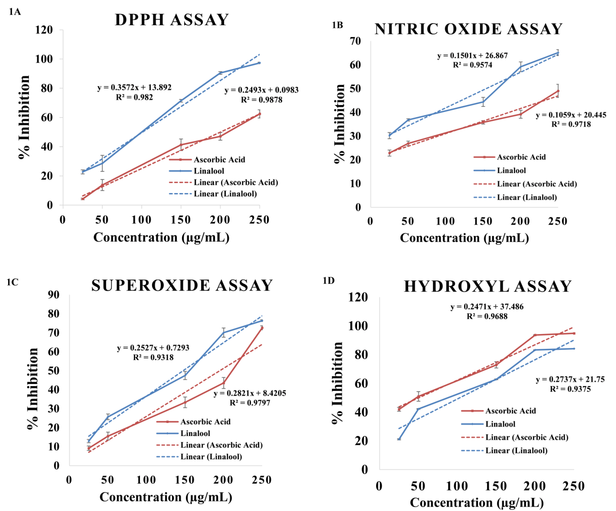
The experiment aimed to ascertain the IC50 value, which represents the concentration at which free radicals were scavenged by 50%. Notably, Linalool demonstrated a remarkable capacity to eliminate all tested reactive species in a way that depended on the concentration. The invitro evaluation of Linalool's ability to remove free radicals included a variety of tests, each designed to target certain reactive substances. The IC50 values (Mean ± SEM) obtained from three separate investigations (n = 3) were found to be at the µg/mL level. The IC50 value of Linalool, determined using the DPPH radicals scavenging test, was reported to be 101.87 ± 0.56 µg/mL (Figure 1A). In comparison, the standard had an IC50 value of 200.17 ± 2.25 µg/mL, indicating that Linalool is a more promising contender. Linalool achieved the highest inhibition rate of 97.35 ± 0.53% at a concentration of 250 µg/mL in the DPPH test, while ascorbic acid, the standard, achieved a maximum inhibition rate of 62.43 ± 2.80% at the same concentration. The IC50 values of Linalool for the NO, superoxide, and hydroxyl tests were found to be 154.12 ± 1.59 µg/mL, 147.39 ± 2.27 µg/mL, and 50.64 ± 2.65 µg/mL, respectively, in a comparable way (Figure 1B, 1C & 1D). The IC50 values for the standard were 297.08 ± 2.09 µg/mL, 194.98 ± 1.39 µg/mL, and 75.20 ± 1.50 µg/mL, respectively, when compared. Linalool has shown significant efficacy in all three of these experiments. Linalool has the greatest effectiveness, with an efficacy of 65.13±1.26 % and 76.36±0.47 % in NO and superoxide scavenging tests, respectively.
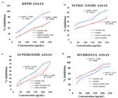
Invitro antioxidant assay of Linalool. (1A) DPPH assay; (1B) NO assay; (1C) Superoxide radical assay; (1D) Hydroxyl radical assay. Each set of data was presented as mean ± SEM (n = 3). The concentrations of 25 μg/mL, 50 μg/mL, 150 μg/mL, 200 μg/mL, and 250 μg/mL were used to evaluate both Linalool and standard, with blue representing Linalool and red representing standard.
In vitro anti-inflammatory assay
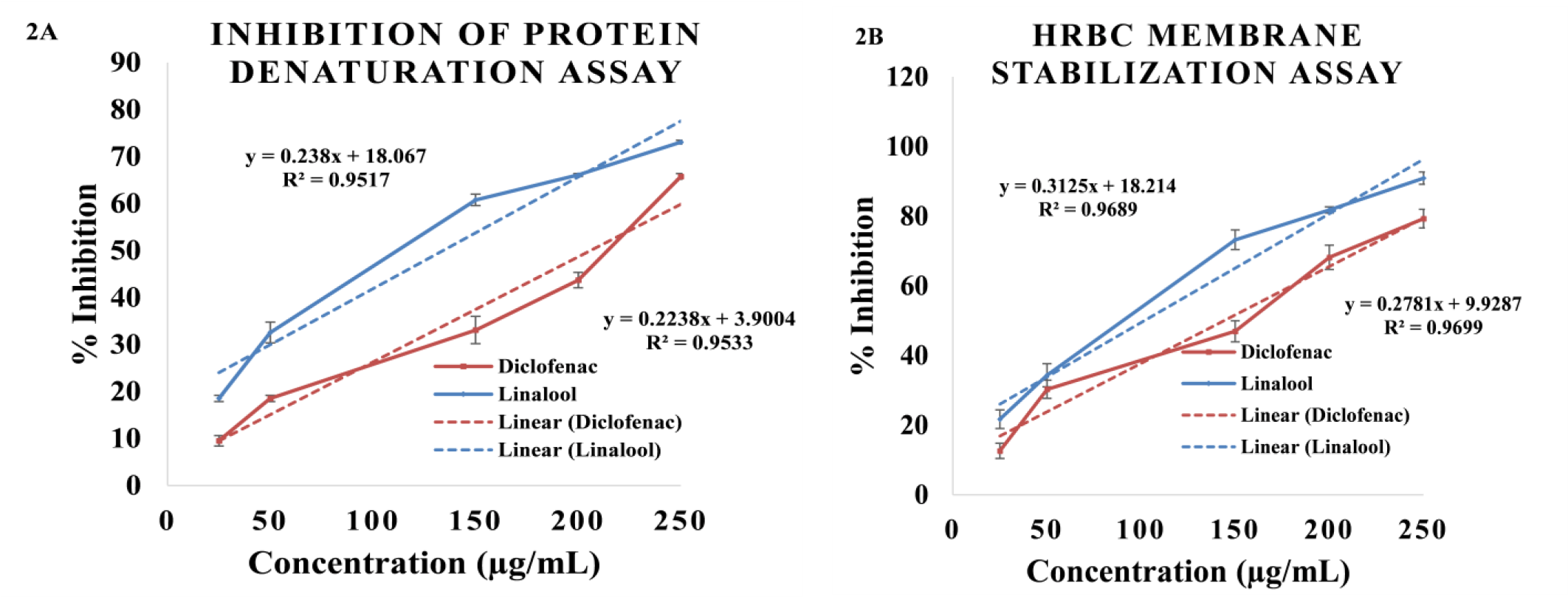
The invitro anti-inflammatory activity of Linalool was evaluated using two assays: inhibition of protein denaturation (Figure 2A) and HRBC membrane stability (Figure 2B). Linalool effectively prevented the denaturation of bovine albumin and the disruption of HRBC membranes in a concentration-dependent manner, with concentrations ranging from 25 µg/mL to 250 µg/mL. Bovine albumin was denatured by subjecting it to a temperature of 700 C for a duration of 5 minutes. The maximum inhibition of 73.05 ± 0.39 % is seen with a concentration of 250 µg/mL of Linalool. Linalool demonstrates a 50% reduction in protein denaturation at a concentration of 134.17 ± 2.06 µg/mL, whereas diclofenac displays same effect at a concentration of 205.98 ± 1.18 µg/mL. Thus, Linalool has been identified as a promising option for preventing protein denaturation. The IC50 values for Linalool in the HRBC membrane stabilization test were determined to be 101.72 ± 1.39 µg/mL, whereas the standard had a value of 144.09 ± 2.42 µg/mL. Linalool exhibited the greatest inhibition, reaching 90.91 ± 1.75%, at a dosage of 250 µg/mL.
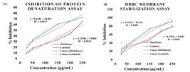
Invitro anti-inflammatory assay of Linalool. (2A) Inhibition of protein denaturation assay; (2B) HRBC membrane stabilization assay. Each set of data was presented as mean ± SEM (n = 3). The concentrations of 25 μg/mL, 50 μg/mL, 150 μg/mL, 200 μg/mL, and 250 μg/mL were used to evaluate both Linalool and standard, with blue representing Linalool and red representing standard.
Effect of Linalool in Body weight, Paw volume of CFA induced RA Rats
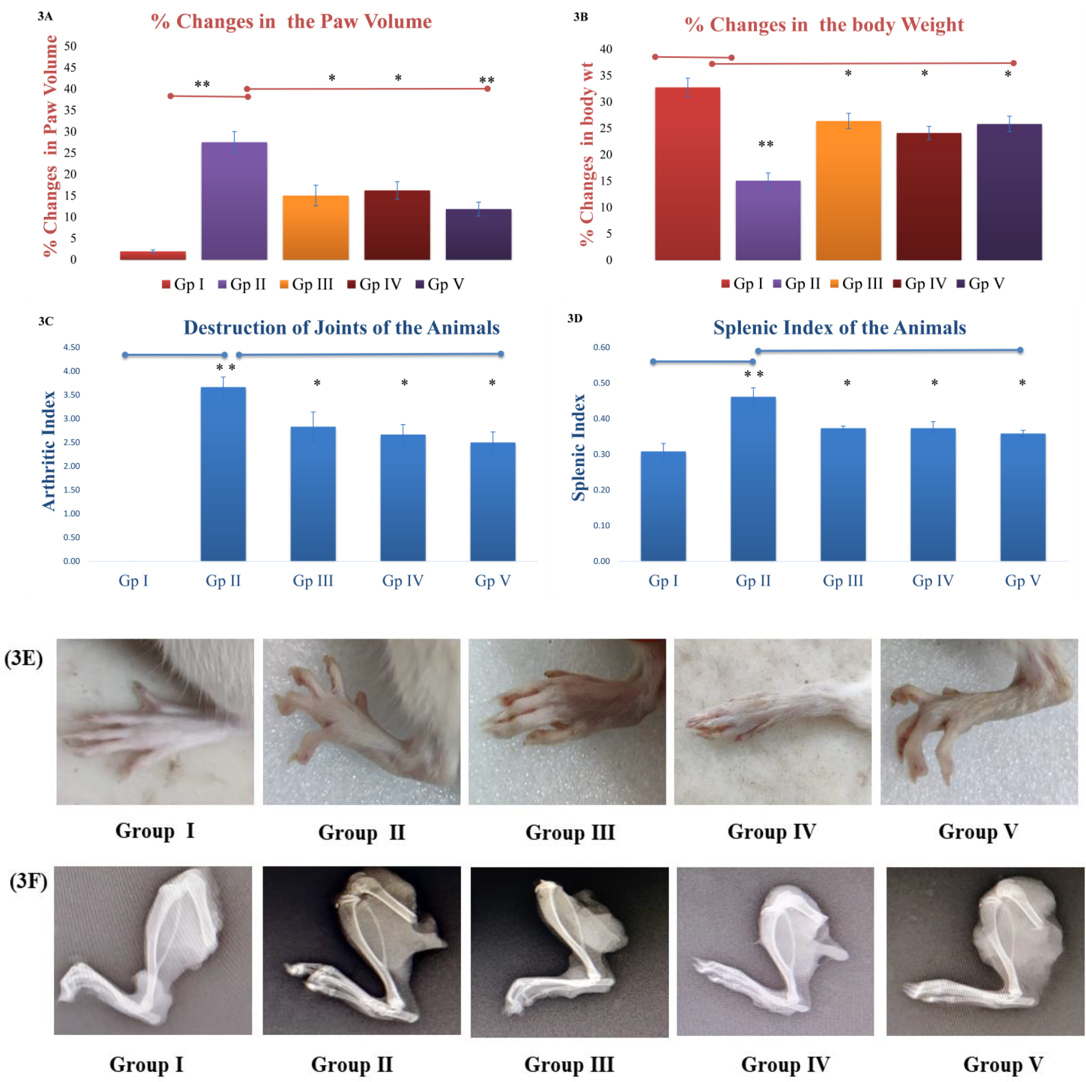
CFA rats exhibit the highest degree of similarity to human RA in terms of etiology, immunology, and genetical features [41]. Following incorporation of C II - CFA emulsion, the rats without treatment (Group II) demonstrated reduced in the body weight and elevation of paw volume indicating RA pathogenesis (Figure 3A & 3B). In Figure 3B, our findings indicate that 100 mg/kg and 400 mg/kg of Linalool, as well as methotrexate, had significant efficacy in promoting weight gain in CFA induced rats (P<0.05). This was determined by assessing the percentage change in body weight after 28 days. Similarly, both the concentration of Linalool reduced the paw swelling in arthritic induced rats (Figure 3A), but 100 mg/kg of Linalool was appeared to be significant (*P<0.05) and 400 mg/kg was found to be highly significant (**P<0.05), while measuring the % change in paw volume at the end of 27 days (Figure 3A and 3E). Linalool substantially ameliorated the delayed weight increase in rats induced by LPS-stimulated vaccination, while also reducing erythema and edema.
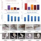
Effect of Linalool in different arthritic parameters in CFA induced RA in rats. (3A) % changes in the paw volume; (3B) % changes in the body weight; (3C) Arthritic index evaluated as per the destruction of joints; (3D) Splenic index evaluated through spleen to body weight ratio; (3E) Macroscopic images of paw volume; (3F) Radiographical image of the joints. Group I serves as the negative control, group II as the positive control, and group III, IV, and V represent animals given with Methotrexate at a dosage of 2.5 mg/kg, Linalool at dosages of 100 mg/kg and 400 mg/kg, respectively, after causing rheumatoid arthritis.
Arthritic index by Radiographic Technique (Xray)
All the rats were proceeded for radiographic analysis with a 55kVp exposure for 6.4 mAs at day 28. A prominent bony erosion and swelling of soft tissue was observed in Group II, leading to wrecking of bones and stenosis of joint spaces. Interestingly, Linalool 100 mg/kg (Gp IV) and 400 mg/kg (Gp V), was significantly effective (P<0.05) in reducing arthritic index in immunized rats manifested by slowing down cartilage destruction and synovial hyperplasia. (Figure 3C and 3F).
Splenic Index of the Animals
Following the sacrifice of the rats on day 28, the spleens were extracted and the spleen to body weight ratio was measured to calculate the splenic index [42]. As depicted in Figure 3D, the splenic index for the positive control rats (Group II) is significantly higher than the normal rats (Group I). It is fascinating to mention that Linalool at the concentration of 100 mg/kg (Gp IV) and 400 mg/kg (Gp V) is highly efficient to reduce the splenic index (P<0.05).
Biochemical Analysis
The impact of Linalool on the hematological parameters of rats with induced arthritis was also examined. CFA induced rats (Gp II) demonstrated elevation of neutrophils and lymphocytes which was found to be significantly decreased by Linalool 100 mg/kg (Gp IV) and 400 mg/kg (Gp V), P<0.05. The administration of both Methotrexate at a dosage of 2.5 mg/kg, Linalool at a dosage of 100 mg/kg, and 400 mg/kg resulted in a decline of AST, ALT, and ALP levels in the blood of rats with induced arthritis. However, this reduction was not significant. The TNF-α and IL-6 were appeared to be much higher in Arthritic rats (Group II). Interestingly, Linalool 400 mg/kg (Group V) can significantly decrease these cytokines levels (P<0.05). Surprisingly, both concentrations of Linalool, 100 mg/kg (Gp IV) and 400 µg/mL (Gp V), may significantly reduce the levels of IL-6. Furthermore, the effectiveness of Linalool is greater than that of the conventional methotrexate. The findings are shown in Table 1.
Molecular Docking Study
The protein ligand interaction of Linalool with nine proteins involved in the pathogenesis of RA was observed and the binding energy was tabulated (Table 2). Methotrexate was used as standard in this molecular docking study. The present study exhibited favorable binding energy of Linalool with proteins such as Inducible nitric oxide synthase (iNOS) and Myeloperoxidase. The highest docked score of -6.1 kcal/mol was seen for iNOS, whereas the lowest docked score of -5.1 kcal/mol was observed for IL-1beta. The standard methotrexate demonstrated the highest docking score of -11.6 kcal/mol for iNOS and lowest -7.2 kcal/mol in case of CYP450. Drugs authorized by the FDA found to have binding energies ranging from -5.63 to -6.85 kcal/mol. The interaction of amino acids with Linalool and Methotrexate through different bonds has been shown as supplementary file. Linalool is forming conventional hydrogen bonding with ARG953 of JAK3, GLY43 of IL6 and MET95, LYS94 of IL-1β respectively
Binding energy, Conventional hydrogen bond and animo acid residues in the interaction of Linalool and Methotrexate with selected proteins involved in RA.
DISCUSSION
Linalool, a monoterpene, is used for medical applications owing to its numerous pharmacological properties [43]. The study examined the potential protective properties of Linalool against RA produced by CFA in rats. This was done by analyzing many factors like the arthritic index, splenic index, paw volume, body weight, and certain biochemical markers. The antioxidant activity and in vitro anti-inflammatory effects of Linalool were also confirmed. The molecular docking studies was carried out to identify the probable interaction of Linalool with selected proteins. Multiple studies have shown the participation of many mechanisms of oxidative stress mediated by free radicals in the development of RA [44]. The free radical scavenging potential of Linalool is carried out through DPPH, NO, Superoxide and Hydroxyl radical scavenging assays within the range of 25 µg/mL to 250 µg/mL, revealing a consistent and progressive inhibitory effect in a dose dependent manner. Due to spare electron delocalization, DPPH seems stable. A quantifiable color shift from violet to yellow occurs when an antioxidant contributes an electron or hydrogen atom to the DPPH radical, reducing it to a non-radical state [45]. Linalool has the ability to transfer a hydrogen atom from its hydroxyl group (-OH) to the DPPH radical, and itself becomes a radical. However, this new radical is stabilized by the resonance structures present in the Linalool molecule [46]. An aqueous solution of sodium nitroprusside (SNP) interacts with oxygen to create nitrite ions (NO₂⁻), resulting in a measurable pink-colored azo dye using Griess reagent [47]. In this study, the Linalool was found to be competing with oxygen to react with NO radicals that produces nitrite. Increased amounts of NO and superoxide contribute to oxidative stress, leading to the impairment of cellular components. The situation is further exacerbated by generation of hydroxyl radicals from superoxide in the presence of ferrous ions [48]. Nicotinamide adenine dinucleotide reacts with phenazine methosulfate to create blue formazan following interaction with nitroblue tetrazolium to produce superoxide anions in vitro [49]. Linalool may neutralize superoxide anion by donating hydrogen or electrons from its hydroxyl group, revealing hydrogen peroxide or oxygen. In the context of inflammatory circumstances, the presence of pro-inflammatory cytokines triggers the activation of the iNOS in monocyte/macrophages, neutrophil granulocytes, and several other cell types to liberate NO implicating in the pathogenesis of tissue damage observed in inflammatory autoimmune disorders, such as RA [50,51]. Interestingly, NO and superoxide anion rapidly interact with each other to form peroxynitrite, worsening the disease condition further [52]. The observed results demonstrated a prominent capacity of Linalool to scavenge the free radical in invitro experiments. Protein denaturation in tissue is a well-documented factor that contributes to the development of arthritic and other inflammatory disorders. Loss of contact forces denature proteins in the cellular membrane, weakening it and releasing proinflammatory mediators that cause pain and inflammation in RA. Increased NO production in the inflammatory joint microenvironment may also denature RA proteins [53]. Lysosome contains similar structure to RBC that may release proteolytic enzymes when damaged. This enzyme release may damage cartilage and joints [54]. This investigation already confirmed Linalool's capacity to decrease NO and other free radicals invitro. Further, the protective effect of Linalool in the stabilization of HRBC and protein denaturation may make it more likely to be useful for RA. In rats with LPS-accelerated CFA-induced RA, Linalool was tested for antirheumatic effects. Linalool significantly improved body weight, arthritic index, splenic index, paw volume, joint and cartilage deformation in arthritic rats. Although various proinflammatory mediators affect CFA-induced arthritis in rats, IL-6 has the most influence [55]. These mediators damage tissue and articular cartilages in CFA induced rats, causing RA symptoms [38,56]. Continued injection of LPS enhances the release of inflammatory mediators, resulting in increased destruction. Linalool 400 mg/kg substantially reduced TNF-α and IL-6 levels in CFA-induced arthritic rats' blood which may be attributed to the interaction of Linalool with IL-6 with a conventional hydrogen bond at GLY43 and TNF-α, with a pi-alkyl interaction at TYR 119 residues, respectively, as seen in molecular docking studies. IL-6 governs the acute phase response, while both TNF- and IL-6 regulate inflammation throughout RA [57]. Assessing TNF-α and IL-6 levels in CFA-induced arthritic rats' serum, including intervention groups, is crucial. The rats treated with CFA also had bigger paws, lighter bodies, deteriorating joints and cartilage, and higher arthritic and splenic indices. Numerous studies believe that inflammatory regulator release, endothelial dysfunction, iNOS activation, and increased myeloperoxidase production may trigger the immune response in RA inflammation. Linalool was found to be interacted well with both iNOS and Myeloperoxidase in molecular docking study. The spleen regulates immune response by creating more immune cells and filtering dead cells, hence spleen enlargement is typical in chronic inflammatory disorders [58]. In rats stimulated with CFA, immune cell filtration removes dead red and white blood cells, raising the spleen index. Our research shows that Linalool improves paw edema and splenic index, suggesting a noteworthy impact on RA development. The Linalool-treated CFA-induced arthritic rats' blood neutrophil and lymphocyte counts, the main inflammatory cells, decreased significantly. Molecular docking is a powerful tool for finding ligands that bind to known protein structures. This method is essential for protein-based medication development. The "lock-key" mechanism involves electrostatic, hydrogen bonding, hydrophobic, and van der Waals interactions when determining ligand-receptor contact [59]. In this work, Linalool was docked with seven distinct proteins that are overexpressed in RA such as iNOS, Myeloperoxidase, JAK3, IL-6, TNF-α, CYP450, IL-1β etc. The results showed a significant interaction of Linalool with the selected proteins involved in the pathogenesis of RA. This observation was found to be consistent with the results our invitro and invivo animal studies indicating the potentiality of Linalool as a candidate to be useful in the treatment of RA. These findings suggest that Linalool may reduce inflammation and deformation of joints, bones, and cartilages in CFA-induced rats by inhibiting IL6, iNOS, major inflammatory cell production, and free radical-mediated oxidative stress. However, the study had significant limitations. This preclinical study focuses on invitro, invivo lab experiments with animal models and molecular docking. Further experimental validation is necessary to corroborate the found binding affinities and potential therapeutic implications identified by docking research. These experiments may not accurately mirror human reactions. Additionally, few animals are used for experiments. The study does not address high-dose Linalool side effects or safety. It is crucial to do additional research, which should consist of thorough preclinical investigations and carefully planned clinical trials, in order to confirm these findings and establish the safety and effectiveness of Linalool as a potential treatment for people with RA.
CONCLUSION
The study described in this article offers insight into the possible therapeutic effectiveness of Linalool in the setting of RA. This research investigated the antirheumatic properties of the substance by in vitro experiments utilizing free radical scavenging test and anti-inflammatory assay, as well as invivo experiments using rats with RA caused by CFA. The acquired findings were validated by assessing the binding score in molecular docking research using a few chosen proteins. The findings indicated that Linalool efficiently eliminates the free radicals in a way that is depending on its concentration. It also has notable in vitro anti-inflammatory effects. Linalool administration in the CFA-induced arthritic rat model results in enhancements in several disease indices, including body weight, arthritic index, splenic index, paw volume, and joint and cartilage damage. Significantly, Linalool reduced the levels of TNF-α and IL-6 in the serum of rats with CFA-induced arthritis. The results demonstrate that Linalool exhibits promise as a possible treatment for RA by reducing inflammation, protecting joints and cartilage from injury, and regulating immunological responses. Given the acknowledged constraints of this study, it is crucial to carry out more research and clinical studies to validate the results and investigate the possibility of Linalool as a feasible therapy for people afflicted with this incapacitating autoimmune condition.
-
Funding:
This research received no external funding
Acknowledgments:
The authors would like to acknowledge Girijananda Chowdhury Institute of Pharmaceutical Science, Guwahati, Assam, for providing the facilities to carry out these studies.
Data Availability and Supplementary Material:
REFERENCES
- 1 Guo Q, Wang Y, Xu D, Nossent J, Pavlos NJ, Xu J. Rheumatoid arthritis: pathological mechanisms and modern pharmacologic therapies. Bone Res. 2018;6:15.
- 2 Venetsanopoulou AI, Alamanos Y, Voulgari P V, Drosos AA. Epidemiology and Risk Factors for Rheumatoid Arthritis Development. Mediterr J Rheumatol. 2023;34(4):404-413.
- 3 Raucci F, Iqbal AJ, Saviano A, Minosi P, Piccolo M, Irace C, et al. IL-17A neutralizing antibody regulates monosodium urate crystal-induced gouty inflammation. Pharmacol Res. 2019;147:104351.
- 4 Scherer HU, Häupl T, Burmester GR. The etiology of rheumatoid arthritis. J Autoimmun. 2020;110:102400.
- 5 Alten R, Van de Laar M, de Leonardis F, Tietz N, Guerreiro M, van Vollenhoven R. Physical and Emotional Burden of Rheumatoid Arthritis: Data from RA Matters, a Web-Based Survey of Patients and Healthcare Professionals. Rheumatol Ther. 2019;6(4):587-597.
- 6 Bala A, Mondal C, Haldar PK, Khandelwal B. Oxidative stress in inflammatory cells of patient with rheumatoid arthritis: clinical efficacy of dietary antioxidants. Inflammopharmacology. 2017;25(6):595-607.
- 7 Weyand CM, Goronzy JJ. The immunology of rheumatoid arthritis. Nat Immunol. 2021;22(1):10-8.
- 8 Németh T, Nagy G, Pap T. Synovial fibroblasts as potential drug targets in rheumatoid arthritis, where do we stand and where shall we go? Ann Rheum Dis. 2022;81(8):1055-1064.
- 9 Wu J, Li Q, Jin L, Qu Y, Liang BB, Zhu XT, et al. Kirenol Inhibits the Function and Inflammation of Fibroblast-like Synoviocytes in Rheumatoid Arthritis in vitro and in vivo. Front Immunol. 2019;10.1304.
- 10 Yang X, Chang Y, Wei W. Emerging role of targeting macrophages in rheumatoid arthritis: Focus on polarization, metabolism and apoptosis. Cell Prolif. 2020;53(7):e12854.
- 11 Kurowska-Stolarska M, Alivernini S. Synovial tissue macrophages in joint homeostasis, rheumatoid arthritis and disease remission. Nat Rev Rheumatol. 2022;18(7):384-397.
- 12 Wei J, Tang Y, Qin S, Ma X, Zhong W, Yang P, et al. Laggera alata Attenuates Inflammatory Response by Regulating Macrophage Polarization in Rheumatoid Arthritis Mice. Mol Biotechnol. 2024;66(8):1934-1941.
- 13 Quiñonez-Flores CM, González-Chávez SA, Del Río Nájera D, Pacheco-Tena C. Oxidative Stress Relevance in the Pathogenesis of the Rheumatoid Arthritis: A Systematic Review. Biomed Res Int. 2016;2016:6097417.
- 14 Jing W, Liu C, Su C, Liu L, Chen P, Li X, et al. Role of reactive oxygen species and mitochondrial damage in rheumatoid arthritis and targeted drugs. Front Immunol. 2023;14:1107670.
- 15 Wang X, Fan D, Cao X, Ye Q, Wang Q, Zhang M, et al. The Role of Reactive Oxygen Species in the Rheumatoid Arthritis-Associated Synovial Microenvironment. Antioxidants (Basel, Switzerland). 2022;11(6):1153.
- 16 Radu AF, Bungau SG. Management of Rheumatoid Arthritis: An Overview. Cells. 2021;10(11):2857.
- 17 Shams S, Martinez JM, Dawson JRD, Flores J, Gabriel M, Garcia G, et al. The Therapeutic Landscape of Rheumatoid Arthritis: Current State and Future Directions. Front Pharmacol. 2021;12:680043.
- 18 Riaz M, Khalid R, Afzal M, Anjum F, Fatima H, Zia S, et al. Phytobioactive compounds as therapeutic agents for human diseases: A review. Food Sci Nutr. 2023;11(6):2500-2529.
- 19 Siddiqui T, Khan MU, Sharma V, Gupta K. Terpenoids in essential oils: Chemistry, classification, and potential impact on human health and industry. Phytomedicine Plus. 2024;4(2):100549.
- 20 Rudrapal M, Khairnar SJ, Khan J, Dukhyil A Bin, Ansari MA, Alomary MN, et al. Dietary Polyphenols and Their Role in Oxidative Stress-Induced Human Diseases: Insights Into Protective Effects, Antioxidant Potentials and Mechanism(s) of Action. Front Pharmacol. 2022;13:1-15.
- 21 Gonfa YH, Tessema FB, Bachheti A, Rai N, Tadesse MG, Nasser Singab A, et al. Anti-inflammatory activity of phytochemicals from medicinal plants and their nanoparticles: A review. Curr Res Biotechnol. 2023;6:100152.
- 22 Masyita A, Mustika Sari R, Dwi Astuti A, Yasir B, Rahma Rumata N, Emran T Bin, et al. Terpenes and terpenoids as main bioactive compounds of essential oils, their roles in human health and potential application as natural food preservatives. Food Chem X. 2022;13:100217.
- 23 Peana AT, D’Aquila PS, Panin F, Serra G, Pippia P, Moretti MDL. Anti-inflammatory activity of linalool and linalyl acetate constituents of essential oils. Phytomedicine. 2002;9(8):721-6.
- 24 Gunaseelan S, Balupillai A, Govindasamy K, Ramasamy K, Muthusamy G, Shanmugam M, et al. Linalool prevents oxidative stress activated protein kinases in single UVB-exposed human skin cells. PLoS One. 2017;12(5):e0176699.
- 25 Pereira I, Severino P, Santos AC, Silva AM, Souto EB. Linalool bioactive properties and potential applicability in drug delivery systems. Colloids Surf B Biointerfaces. 2018;171:566-578.
- 26 He J, Halitschke R, Baldwin IT, Schuman MC. Natural variation in linalool metabolites: One genetic locus, many functions? J Integr Plant Biol. 2021;63(8):1416-1421.
- 27 Kim MG, Kim SM, Min JH, Kwon OK, Park MH, Park JW, et al. Anti-inflammatory effects of linalool on ovalbumin-induced pulmonary inflammation. Int Immunopharmacol. 2019;74:105706.
- 28 Kamatou GPP, Viljoen AM. Linalool - a Review of a Biologically Active Compound of Commercial Importance. Nat Prod Commun. 2008;3(7):1183-1192
- 29 Ahmed AB, Chetia P. MAO - B Inhibitor “ Selegiline ” Attenuates Oxidative Stress and Arthritic Parameters in an Invitro Repurposive Study. J Biomed Eng. 2023;40(2):171-181.
- 30 Sharma A, Sharma L, Goyal R. GC/MS Characterization, in-vitro Antioxidant, Anti-inflammatory and Antimicrobial Activity of Essential Oils from Pinus Plant Species from Himachal Pradesh, India. J Essent Oil-Bearing Plants. 2020;23(3):522-531.
- 31 Chetia P, Bala A, Khandelwal B, Haldar PK. Comparative in vitro Free Radical Scavenging Property of β-carotene and Naringenin with Respect to Vitamin C and N-acetyl Cysteine. Pharmacologia. 2012;3:724-728.
- 32 Zhang X, Guo Y, Guo L, Jiang H, Ji Q. In Vitro Evaluation of Antioxidant and Antimicrobial Activities of Melaleuca alternifolia Essential Oil. Biomed Res Int. 2018;2018:2396109.
- 33 Saleem A, Saleem M, Furqan M. Antioxidant , anti-in fl ammatory and antiarthritic potential of Moringa oleifera Lam : An ethnomedicinal plant of Moringaceae family. South African J Bot. 2020;128:246-256.
- 34 Kalita V, Pegu P, Chetia P. Phytochemical Screening and Evaluation of Antioxidant, Anti-Microbial and Anti-Inflammatory Activity of “Curcuma Caesia.” Int J Pharm Sci Res. 2019;10(2):846.
- 35 Brand DD, Latham KA, Rosloniec EF. Collagen-induced arthritis. Nat Protoc. 2007; 2(5):1269-75.
- 36 Shirani K, Iranshahi M, Askari VR, Gholizadeh Z, Zadeh AA, Zeinali M, et al. Comparative evaluation of the protective effects of oral administration of auraptene and umbelliprenin against CFA-induced chronic inflammation with polyarthritis in rats. Biomed Pharmacother. 2021;139:111635.
- 37 Ananth DA, Rameshkumar A, Jeyadevi R, Aseervatham GSB, Sripriya J, Bose PC, et al. Amelioratory effect of flavonoids rich Pergularia daemia extract against CFA induced arthritic rats. Biomed Pharmacother. 2016;80:244-52.
- 38 Chen G, Song Y, Ma F, Ma Y. Anti-arthritic activity of D-carvone against complete Freund’s adjuvant-induced arthritis in rats through modulation of inflammatory cytokines. Korean J Physiol Pharmacol. 2020;24(6):453-62.
- 39 Sussman JL, Lin D, Jiang J, Manning NO, Prilusky J, Ritter O, et al. Protein Data Bank (PDB): database of three-dimensional structural information of biological macromolecules. Acta Crystallogr D Biol Crystallogr. 1998;54:1078-84.
- 40 Kim S, Thiessen PA, Bolton EE, Chen J, Fu G, Gindulyte A, et al. PubChem Substance and Compound databases. Nucleic Acids Res. 2016;44(D1):D1202-13.
- 41 He P, Hu Y, Huang C, Wang X, Zhang H, Zhang X, et al. N-Butanol Extract of Gastrodia elata Suppresses Inflammatory Responses in Lipopolysaccharide-Stimulated Macrophages and Complete Freund’s Adjuvant- (CFA-) Induced Arthritis Rats via Inhibition of MAPK Signaling Pathway. Evid Based Complement Alternat Med. 2020;2020:1658618.
- 42 Si M, Ma Z, Zhang J, Li X, Li R, Wang C, et al. Qingluoyin granules protect against adjuvant-induced arthritis in rats via downregulating the CXCL12/CXCR4-NF-κB signalling pathway. Pharm Biol. 2021;59(1):1441-51.
- 43 Aprotosoaie AC, Hăncianu M, Costache II, Miron A. Linalool: a review on a key odorant molecule with valuable biological properties. Flavour Fragr J. 2014;29(4):193-219.
- 44 Ferreira HB, Melo T, Paiva A, Domingues M do R. Insights in the Role of Lipids, Oxidative Stress and Inflammation in Rheumatoid Arthritis Unveiled by New Trends in Lipidomic Investigations. Antioxidants (Basel, Switzerland). 2021;10(1).45.
- 45 Sirivibulkovit K, Nouanthavong S, Sameenoi Y. Paper-based DPPH Assay for Antioxidant Activity Analysis. Anal Sci. 2018;34(7):795-800.
- 46 Julianus Sohilait H, Kainama H. Free Radical Scavenging Activity of Essential Oil of Eugenia caryophylata from Amboina Island and Derivatives of Eugenol. Open Chem. 2019;17(1):422-8.
- 47 Chelliah R, Banan-MwineDaliri E, Oh DH. Screening for antioxidant activity: Nitric oxide scavenging assay. In: Dharumadurai D, editor. Methods in actinobacteriology. Springer Protocols Handbooks. New York: Humana; 2022. p. 455-456.
- 48 Muthuvel A, Jothibas M, Manoharan C, Jayakumar SJ. Synthesis of CeO2-NPs by chemical and biological methods and their photocatalytic, antibacterial and in vitro antioxidant activity. Res Chem Intermed. 2020;46(5):2705-29.
- 49 Kumar S, Krishna Chaitanya R, Preedy VR. Assessment of Antioxidant Potential of Dietary Components. In: Preedy VR, Watson RR, editor. HIV/AIDS: Oxidative Stress and Dietary Antioxidants. Elsevier. Academic Press; 2017. p. 239-225.
- 50 Sharma JN, Al-Omran A, Parvathy SS. Role of nitric oxide in inflammatory diseases. Inflammopharmacology. 2007;15(6):252-9.
- 51 Huang JB, Chen ZR, Yang SL, Hong FF. Nitric Oxide Synthases in Rheumatoid Arthritis. Molecules. 2023; 28(11):4414.
- 52 Radi R. Oxygen radicals, nitric oxide, and peroxynitrite: Redox pathways in molecular medicine. Proc Natl Acad Sci U S A. 2018;115(23):5839-5848.
- 53 Bouhlali EDT, Hmidani A, Bourkhis B, Khouya T, Ramchoun M, Filali-Zegzouti Y, et al. Phenolic profile and anti-inflammatory activity of four Moroccan date (Phoenix dactylifera L.) seed varieties. Heliyon. 2020;6(2):e03436.
- 54 Nema NK, Maity N, Sarkar B, Mukherjee PK. Cucumis sativus fruit-potential antioxidant, anti-hyaluronidase, and anti-elastase agent. Arch Dermatol Res. 2011;303(4):247-252.
- 55 Fontes JA, Barin JG, Talor M V, Stickel N, Schaub J, Rose NR, et al. Complete Freund’s adjuvant induces experimental autoimmune myocarditis by enhancing IL-6 production during initiation of the immune response. Immunity, Inflamm Dis. 2017;5(2):163-176.
- 56 Abdel-Aziz EA, Abdel-Hameed AA. Effect of sitagliptin and methotrexate on rheumatoid arthritis induced experimentally inrats. Egypt J Basic. 2017;7(1):26-34.
- 57 Silpavathi L, Das MK, Das D. Anti-arthritic potentials of aqueous and methanolic leaf extracts of Ardisia solanacea on complete Freund’s adjuvant induced rheumatoid arthritis in rats. Adv Tradit Med. 2023;23(1):111-119.
- 58 Barrea L, Di Somma C, Muscogiuri G, Tarantino G, Tenore GC, Orio F, et al. Nutrition, inflammation and liver-spleen axis. Crit Rev Food Sci Nutr. 2018;58(18):3141-3158.
- 59 Stanzione F, Giangreco I, Cole JC. Use of molecular docking computational tools in drug discovery. Prog Med Chem. 2021;60:273-343.
-
Editor-in-Chief: Paulo Vitor Farago
-
Associate Editor: Jane Manfron Budel