In vitro Anticancer Activity of Flaxseed Oil Loaded-Polymeric Micelles in Breast Cancer Cell Line
Abstract
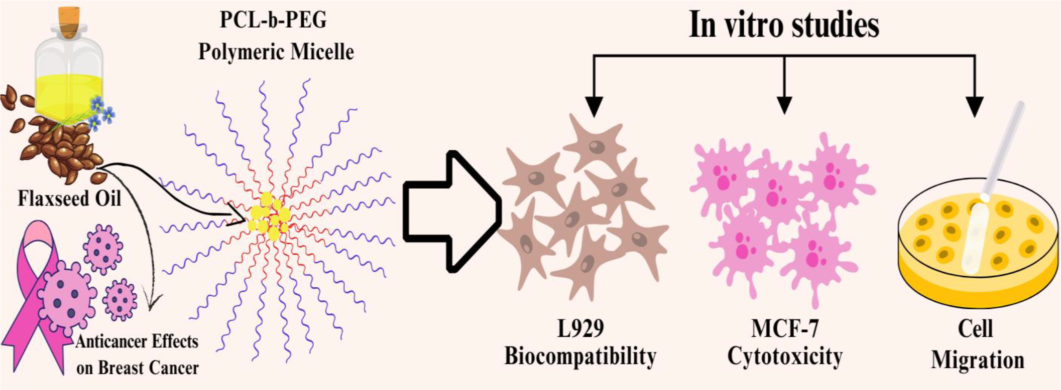
Phytochemicals have emerged as potential anticancer agents for breast cancer treatment, which remains one of the most common cancer types today. Incorporating phytochemical-based agents into polymeric nanocarriers enhances water solubility and bioavailability, making them distinct biocompatible systems. In this study, we investigated the in vitro anticancer activity of flaxseed oil-loaded PCL-b-PEG polymeric micelles for breast cancer treatment. The STEM images revealed spherical morphologies of the polymeric micelles, with sizes ranging from 95 to 260 nm depending on the amount of flaxseed oil. MTT assay was conducted on L929 and MCF-7 cells. Notably, the micelles exhibited negligible cytotoxicity towards L929 cells, while showing concentration-dependent cytotoxicity against MCF-7 cells. Additionally, the effect of the micelles on MCF-7 cell migration was evaluated, revealing a clear inhibitory effect. The findings underscore the anticancer potential of the produced polymeric micelles, thereby highlighting their potential as nanocarriers for breast cancer treatment.
Keywords:
block copolymer; breast cancer; micelle; phytochemical
HIGHLIGHTS
The micelles exhibited spherical morphology, hydrodynamic diameters ranging from 95 to 260 nm, and good colloidal stability with a low critical micelle concentration (CMC) of 0.0114 mg/mL.
Flaxseed-loaded micelles showed potent concentration-dependent cytotoxicity against MCF-7 cell line, with IC50 values of 0.08 μg/mL (24 h) and 0.096 μg/mL (48 h).
Flaxseed-loaded micelles significantly inhibited MCF-7 cell migration, reducing wound closure to 1.7% at IC50 after 48h.
The study highlights the potential of phytochemicals-based polymeric micelles as a promising and biocompatible nanocarrier system for breast cancer treatment.
INTRODUCTION
Over the past few decades, the global risk of breast cancer has increased substantially. Between 1990 and 2020, the number of new cases worldwide reached approximately 2.3 million, with around 685,000 deaths annually [1]. Current trends suggest that these numbers remain high for 2025, for example, in the United States, estimates for 2025 predict approximately 319,750 new cases in women and 42,680 deaths [2]. If these trends persist, by 2050, the annual global burden is expected to reach 3.2 million new cases and 1.1 million deaths [3], which indicates that novel and effective treatment strategies are urgently needed in this field [4] [5].
A wide variety of therapeutic strategies are applied in order to decrease the mortality rates of breast cancer, such as radiotherapy, hormone therapy, surgery and chemotherapy [6]. However, these strategies are challenging for the patients with breast cancer owing to the remarkable resistance of this cancer against such treatment methods [3]. In addition, these treatments, especially chemotherapy, cause severe side reactions by affecting not only the cancerous cells but also the healthy cells and tissues [7] ,[8]. In the last decades, phytochemicals have been extensively investigated as anticancer agents due to their fewer side effects and notable therapeutic activities. Phytochemicals are bioactive compounds found in grains, fruits, and vegetables that have anticancer, antiproliferative, antiangiogenic, and apoptotic activities [6-8]. All these properties make phytochemicals a promising tool in breast cancer treatment.
Flaxseed is the seed form of functional plant Flax (Linum usitatissimum) that has highly rich in phytochemicals such as α-linolenic acid (ALA) (major fatty acid), omega-3 fatty acids, polyunsaturated fatty acids (PUFA), phytoestrogenic lignans, fibers, antioxidants, phenolic compounds, vitamins, and minerals [9], [10]. Flaxseed exhibits a notable impact on cancer (breast, ovary, colon, prostate), diabetes mellitus, cardiovascular diseases, osteoporosis and autoimmune disorders [10]. Flaxseed oil (FO) and its extracts have been reported to have anticancer effects on breast cancer in many studies [9], [11-13].
Although natural compounds containing various phytochemicals, such as FO have been evaluated as potential candidates for the treatment of breast cancer, they have some drawbacks such as low bioavailability, rapid elimination, and low solubility. Accordingly, the encapsulation of such compounds in order to increase the bioavailability and reduce the elimination rate is a major concern in the development of effective phytochemical-based therapeutic systems [6-8], [14].
Previous studies have primarily explored flaxseed oil (FO) as a functional excipient or a carrier component in various nanocarrier systems, rather than as a therapeutic agent on its own. For instance, in nanobigel formulations combining FO with daidzein, FO played a supportive role in enhancing permeation and sustaining drug release [15]. Similarly, its use in self-microemulsifying systems increased the bioavailability of omega-3 fatty acids, demonstrating its utility for improving solubility and absorption [16]. FO has also been utilized in nanoemulsions for stabilizing drugs such as efavirenz, contributing to improved formulation stability and controlled release [17]. Furthermore, FO-based systems like Pickering emulsions and carboxymethyl chitosan-decorated proliposomes have shown advantages in terms of drug loading, digestive stability, and sustained release when compared with conventional liposomes [18], [19]. In lipid-based nanocarriers such as NLCs, FO has enhanced encapsulation efficiency and bioavailability of polyphenols like caffeic acid, underlining its excipient role in improving pharmacokinetic properties [20]. FO’s high omega-3 content has also been exploited to improve the biocompatibility and fluidity of nanocarriers, thereby aiding drug encapsulation and delivery [21]. Additionally, encapsulation approaches have focused on addressing FO’s oxidative instability, further emphasizing its role as a formulation component rather than a direct therapeutic agent [19], [22].
The delivery of the agents by encapsulating in nanoparticles, such as polymeric micelles, increases the absorption, plasma half-life and bioavailability of these drugs by increasing the stability and solubility of hydrophobic or poorly water-soluble agents [23]. In addition, serious side effects of anticancer agents can also be reduced through designing an appropriate controlled drug delivery system, which targets the desired tissue [24]. Clinical studies also indicate the efficacy of polymeric micelles as drug delivery systems due to their growing role in the cancer treatment [25]. Park and coauthors conducted a Phase III study in metastatic breast cancer patients using paclitaxel loaded polymeric micelles (Genexol-PM®). In the study, patients received either free paclitaxel or Genexol-PM® intravenously. The results demonstrated that Genexol-PM had a higher maximum tolerated dose compared to paclitaxel. Accordingly, polymeric micelles enhance the therapeutic efficacy of the encapsulated anticancer agent by allowing intravenous administration at higher tolerated concentrations [26].
Polymeric micelles are formed using amphiphilic copolymers, in which poly(ethylene glycol) (PEG) and hydrophobic linear polyesters are extensively used as hydrophilic and hydrophobic blocks [27]. PEG is a widely used polymer used in structures of polymeric micelles as it is an FDA approved, biocompatible, highly water-soluble polymer and has the ability to reduce immune reactions [28]. Polycaprolactone (PCL) is a hydrophobic polyester which is also an FDA approved polymer used in polymeric micelles as a biodegradable polymer [29], [30]. PCL-b-PEG block copolymers are synthesized to produce micelles in order to minimize the side effects of the anticancer agents by decreasing the cytotoxicity and enhancing the drug delivery to the site of action, where the hydrophobic anticancer agent can be encapsulated inside these PCL core of the micelles [31]. In addition, PCL possesses a slower degradation rate due to its high crystallinity and hydrophobicity compared to biodegradable polymers such as PLGA, PGA, PLA and PVA, among others, which allows the sustained and controlled release of anticancer agents over an extended period [32], [33]. This property makes PCL a promising nanocarrier component for the treatment of diseases such as cancer that require long-term therapy.
In this study, FO-loaded PCL-b-PEG polymeric micelles were produced and characterized as a potential polymeric drug delivery system for the breast cancer treatment, intended for intravenous administration. The anticancer activity and biocompatibility of FO-loaded polymeric micelles (FOM), which have been produced for the first time, have been investigated. Although anticancer agent loaded PCL-b-PEG or PLGA-b-PEG (polylactide-co-glycolide-b-PEG) polymeric micelles have been previously produced in many studies to increase bioavailability and loading capacity [34-36], there is no study in which FO-loaded PCL-b-PEG polymeric micelle has been produced. In contrast to the studies related to the usage of FO as a functional excipient [15-22], our work uniquely positions FO as the primary therapeutic agent. This distinction represents a novel contribution, as we aim to explore the direct biological activity and therapeutic potential of FO itself through a nanoparticle-based delivery system. By evaluating FO not only as a carrier but as a bioactive compound with intrinsic pharmacological effects, our study offers a new perspective on the therapeutic applications of this natural oil. FO was selected as the core component of the micellar system due to its rich composition of bioactive compounds, including α-linolenic acid (ALA), lignans, and phenolic acids, which are known for their antioxidants, anti-inflammatory, and anticancer properties [37], [38]. By leveraging FO’s functional benefits alongside the structural advantages of polymeric micelles, the formulation of this study offers a unique strategy that bridges nutraceutical and nanotechnology-based delivery systems. The integration of FO into polymeric micelles offers a synergistic approach to drug delivery, combining its nutritional benefits with enhanced therapeutic efficacy.
MATERIAL AND METHODS
FO was obtained from Furkan Doğal Ürünler Tic.San.Ltd.Şti. (Istanbul, Turkey). PCL-b-PEG was prepared previously according to the method of Topuzoğulları and coauthors (Mw = 25 kDa, where mPEG block is 5 kDa and PCL block is 20 kDa) [39]. Acetone, ethanol, acetic acid, thiazolyl blue tetrazolium bromide, trypan blue, penicillin-streptomycin, 3-[4,5-Dimethylthiazol-2-yl]-2,5-diphenyltetrazolium bromide (MTT) were acquired from Sigma-Aldrich. DMEM/F-12 Nutrient Mix and L-Glutamine 200 mM (100x) were obtained from Gibco. Trypsin-EDTA (Multicell) and dimethyl sulfoxide (ChemCruz) were also used. Ultrapure water was used in all aqueous solutions and obtained from Millipore water purification system.
Production of Flaxseed Oil-Loaded PCL-b-PEG Polymeric Micelles
FOMs were produced by using the solvent evaporation method. Firstly, polymer solutions were prepared by dissolving 10 mg PCL-b-PEG in 5 mL acetone. Different amounts of FO (0.1, 0.5, 1, 3, 10, 15, 20, 30, 50, 100 μL) were added into polymer solutions to investigate the effect of FO quantity on the nanocarrier system. The polymer and oil solutions were gently injected drop by drop into 20 mL ultrapure water by using a syringe while the solution was stirred at 600 rpm to prevent aggregation. The solution was stirred overnight for the evaporation of acetone. As the amount of FO increased in the polymeric micelles, the solutions gradually changed from transparent to opalescent. FO-free polymeric micelle, which will be used as a control group, were produced by using the same method. FO-free polymeric micelle was named as blank micelle (BM). FOM were named as 0.1FOM, 0.5FOM, 1FOM, 3FOM, 10FOM, 15FOM, 20FOM, 30FOM, 50FOM and 100FOM according to the amount of FO used in production of polymeric micelles. The produced polymeric micelles were lyophilized for further investigations.
Characterization
Critical micelle concentration (CMC) of the PCL-b-PEG copolymer was determined using pyrene as a fluorescent probe by measuring the fluorescence intensity ratio (I338/I333) of copolymer solutions with different concentrations using fluorescence spectroscopy (PTI QuantaMaster MD-5020 Fluorometer) [40]. Measurements were performed with an excitation wavelength of 300-360 nm, while emission wavelength was 390 nm [41]. Hydrodynamic diameter, zeta potential and polydispersity index (PDI) values of polymeric micelles were obtained with dynamic (DLS) and electrophoretic (ELS) light scattering spectroscopy using Zetasizer Nano-ZS (Malvern) analyzer. The synthesized micellar suspensions were further diluted with ultrapure water at a ratio of 1:10 (micellar suspension:water, v/v) to ensure adequate dispersion and prevent multiple scattering effects. DLS measurements were performed in triplicate, and the mean values with standard deviations were reported. Each measurement was carried out at 25°C and ultrapure water was used as a dispersant. The Fourier Transform Infrared (FTIR) spectroscopy was performed by using Shimadzu IRPrestige-21 instrument equipped with attenuated total reflectance (ATR) attachment in order to compare characteristic bands and confirm chemical structure of FO and polymeric micelles. FTIR spectra were acquired within the wavenumber range of 4000-650 cm-1. FO was analyzed in its liquid form using FTIR spectroscopy, whereas polymeric micelles were analyzed in their lyophilized form. Size and morphology of the produced polymeric micelles were examined by scanning electron microscopy (SEM) Thermo Scientific Quattro S model with scanning transmission electron microscopy (STEM) detector. The polymer solutions were dropped on carbon film coated Cu grid before SEM analysis. Images were obtained at 120,000x and 500,000x magnification. The sizes of produced polymeric micelles were determined by using ‘ImageJ’ software.
Stability Study of Micelles
After initial characterization, the stability of the micelles was evaluated under two conditions: (i) in solution and (ii) in lyophilized form.
Stability in Solution:
Freshly prepared micelles were lyophilized and subsequently reconstituted in phosphate-buffered saline (PBS). The samples were incubated at 37 °C under gentle stirring for 7 days to mimic physiological conditions. At the end of the incubation period, the hydrodynamic diameter (z-average) and polydispersity index (PDI) values of 15FOM and 50FOM were measured using DLS.
Long-Term Stability of Lyophilized Micelles:
To assess long-term storage stability, micelles were lyophilized and stored at +4 °C for 1 year. After the storage period, the samples were dissolved in PBS, and their size and PDI values were evaluated using the same DLS protocol described above.
In Vitro Studies
Cell Culture
L929 murine fibroblast cells and MCF-7 breast cancer cells were used in this study. L929 cells were cultured in DMEM (Dulbecco's Modified Eagle Medium with L-glutamine, Gibco) supplemented with 10% Fetal Bovine Serum (FBS,Gibco), 1% Penicillin-Streptomycin (Sigma). MCF-7 cells were grown in DMEM/Ham’s Nutrient Mixture F-12 (1:1) medium (Gibco) supplemented with 10% FBS (Gibco), 1% Penicillin-Streptomycin (Sigma). Both cell cultures were incubated at 37 °C in a humidified atmosphere with 5% CO2.
Cytotoxicity Assay
In this study, the cytotoxic activity of FO and FOM on L929 and MCF-7 cell lines was determined by MTT assay [42], [43]. Firstly, the cells were seeded in 96 well plates at 10,000 cells/well and incubated overnight. Subsequently, FO, 15FOM, and 50FOM, which were prepared at varying concentrations (ranging from 0.0001 to 0.1 μg/mL) by dispersing them in cell culture medium through vortexing and gentle sonication, were applied to the cells for 24 and 48 hours. After the incubation period, the medium was removed and MTT solution was added. DMSO was then added to dissolve the formazan crystals and absorbance values were read using ELISA reader (BioTek Powerwave XS2) at a wavelength of 570 nm. All measurements were in triplicate. Cell viability was measured at 24th and 48th hours post-treatment, and the results were expressed as a percentage compared to the control group. IC50 values and statistical analyses were performed using GraphPad Prism version 8.4.0. Statistical significance was determined by two-way ANOVA followed by Tukey's multiple comparison test. Differences with p < 0.001 were considered statistically significant.
Cell Migration (Wound Healing) Assay
MCF-7 cells were seeded in 24-well microplates at 50,000 cells/well and incubated for 24 hours [44]. Subsequently, the cells that reached approximately 80% confluence were scratched by a sterile micropipette tip, and FO, 15FOM and 50FOM were applied to MCF-7 cells based on the IC50 values. Cell migration was captured using a phase-contrast inverted microscope at 100x magnification and scratch distances were measured by ‘ImageJ’ software. All measurements were performed in triplicate.
RESULTS AND DISCUSSION
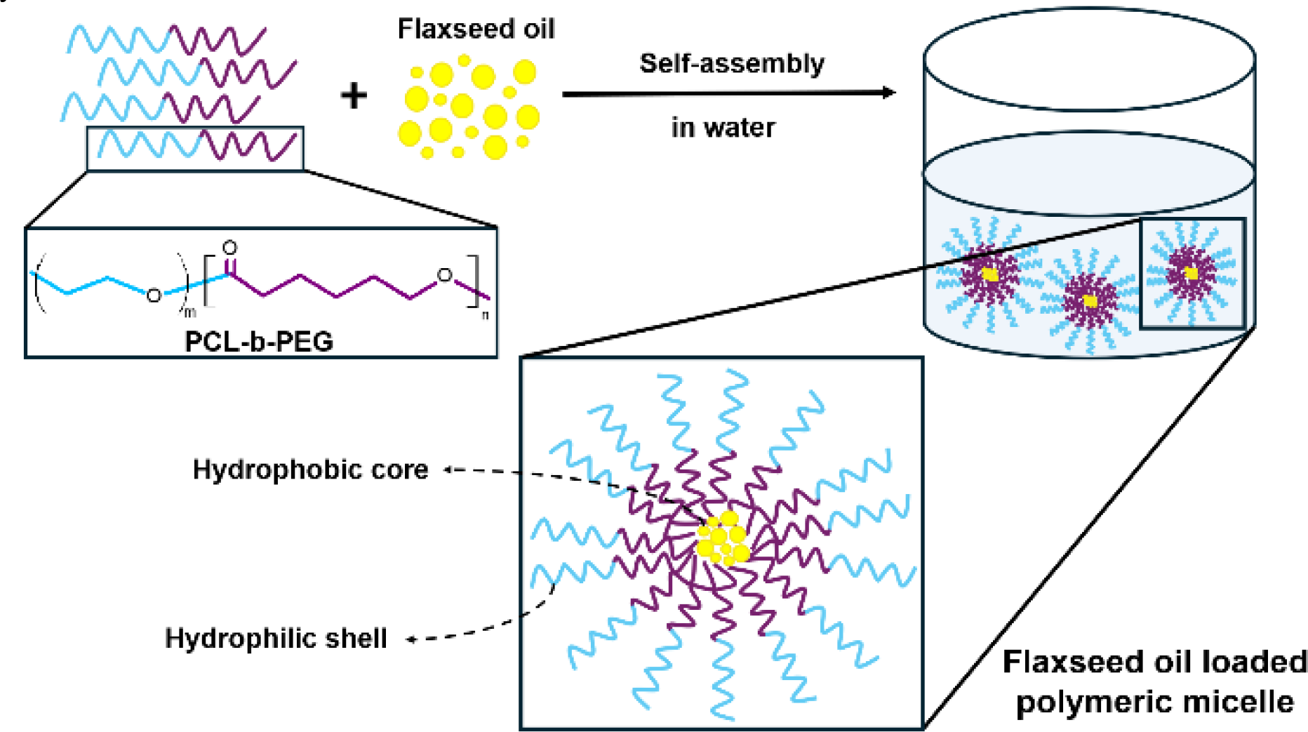
In this study, it is aimed to produce a biocompatible and water-soluble delivery system that increases the bioavailability and solubility of FO for breast cancer treatment. In line with this goal, FOMs were produced from PCL-b-PEG amphiphilic block copolymers, and the anticancer activity of these polymeric micelles against breast cancer was investigated in vitro. The enhancement of biocompatibility and solubility of the micelle is achieved by the PEG block in the copolymer, while core-forming hydrophobic block of PCL ensures the encapsulation and release of FO as anticancer agent owing to its biodegradability. Polymeric micelle formed by self-assembly of amphiphilic block copolymer in water and FO was encapsulated into micelle core due to its hydrophobicity. Hence, FO-loaded biodegradable hydrophobic core and biocompatible hydrophilic shell was obtained as shown in Figure 1. In the production of micelles, different amounts of FO were encapsulated into the micelle to evaluate the effect of the FO quantity on the micelle structure and anticancer activity.
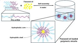
Schematic representation of the formation of FOMs, which are formed through the self-assembly of PCL-b-PEG copolymers into a hydrophobic core and a hydrophilic shell structure, and their stabilization in water, maintained through the steric stabilization provided by the PEG corona and the hydrophobic stabilization of the PCL core.
Critical Micelle Concentration of PCL-b-PEG Copolymer
Since the produced polymeric micelles are intended as potential drug delivery systems for breast cancer therapy, it is crucial that they retain their micellar structure and stability upon iv administration. The determination of the critical micelle concentration (CMC) is crucial, as iv injection leads to rapid dilution in the bloodstream, which could otherwise cause micelle disassembly and immediate release of the encapsulated therapeutic agent [45].
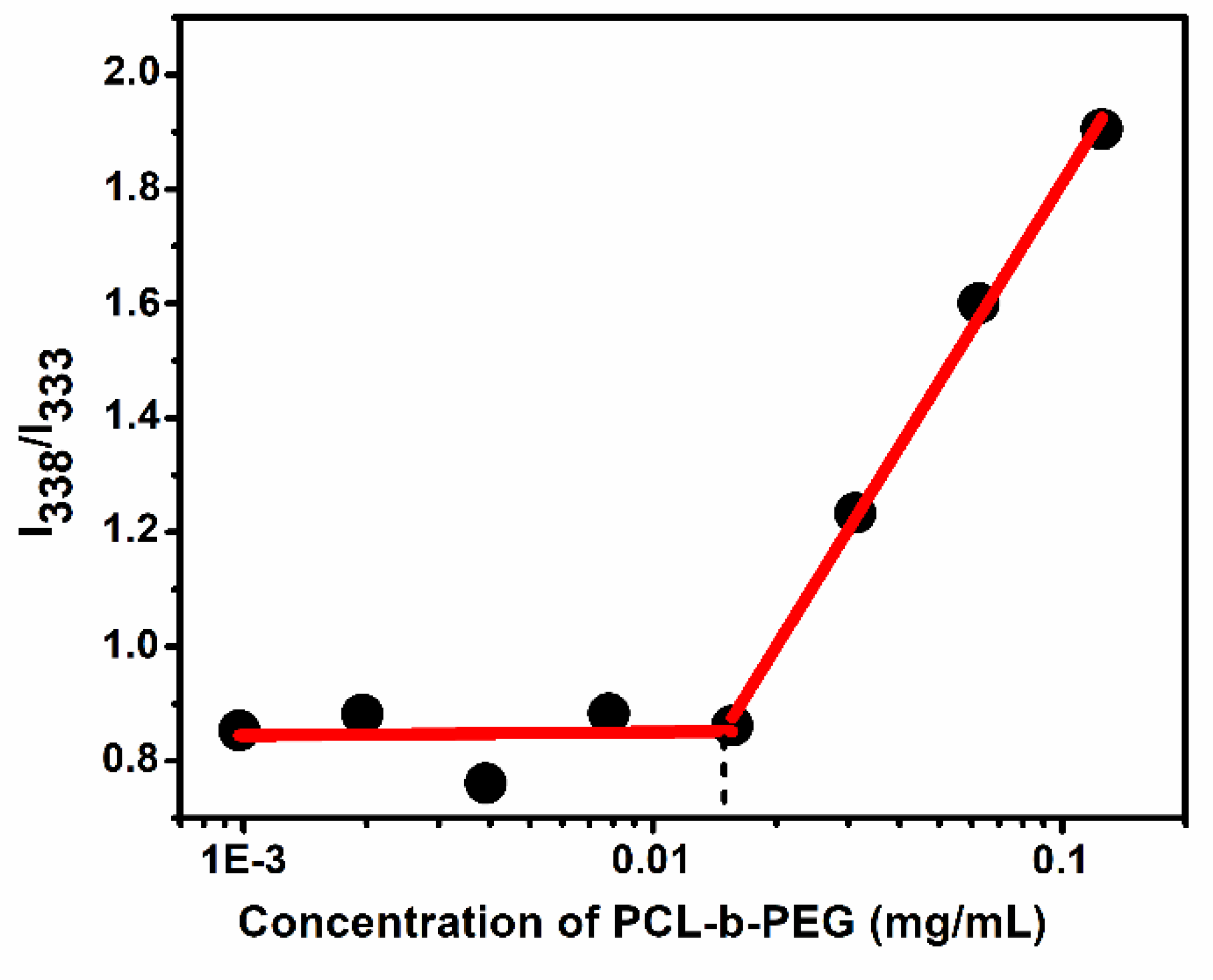
For this reason, the correlation between different concentrations of copolymers and formation of micellar structure was evaluated with a fluorescence probe. PCL-b-PEG copolymers were prepared in the concentration range from 9.8 x 10⁻⁴ to 0.125 mg/mL and a fixed concentration of pyrene was added to these copolymer solutions. The intensity ratio of I338/I333 obtained from the excitation spectrum of pyrene was investigated for different PCL-b-PEG concentrations as given in Figure 2 and the critical micelle concentration (CMC) was determined as 0.0114 mg/mL, consistent with the CMC values in the study of Lu and coauthors [46]. Lower CMC value indicates higher stability of the drug delivery system. Additionally, the large molecular weight of the hydrophobic block of PCL is one of the factors that reduce the CMC value [47].
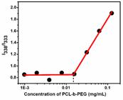
Change in fluorescence intensity ratio (I338/I333) of pyrene depending on the concentration (mg/mL) of PCL-b-PEG copolymer.
Synthesis and Characterization of Flaxseed Oil-Loaded Micelles
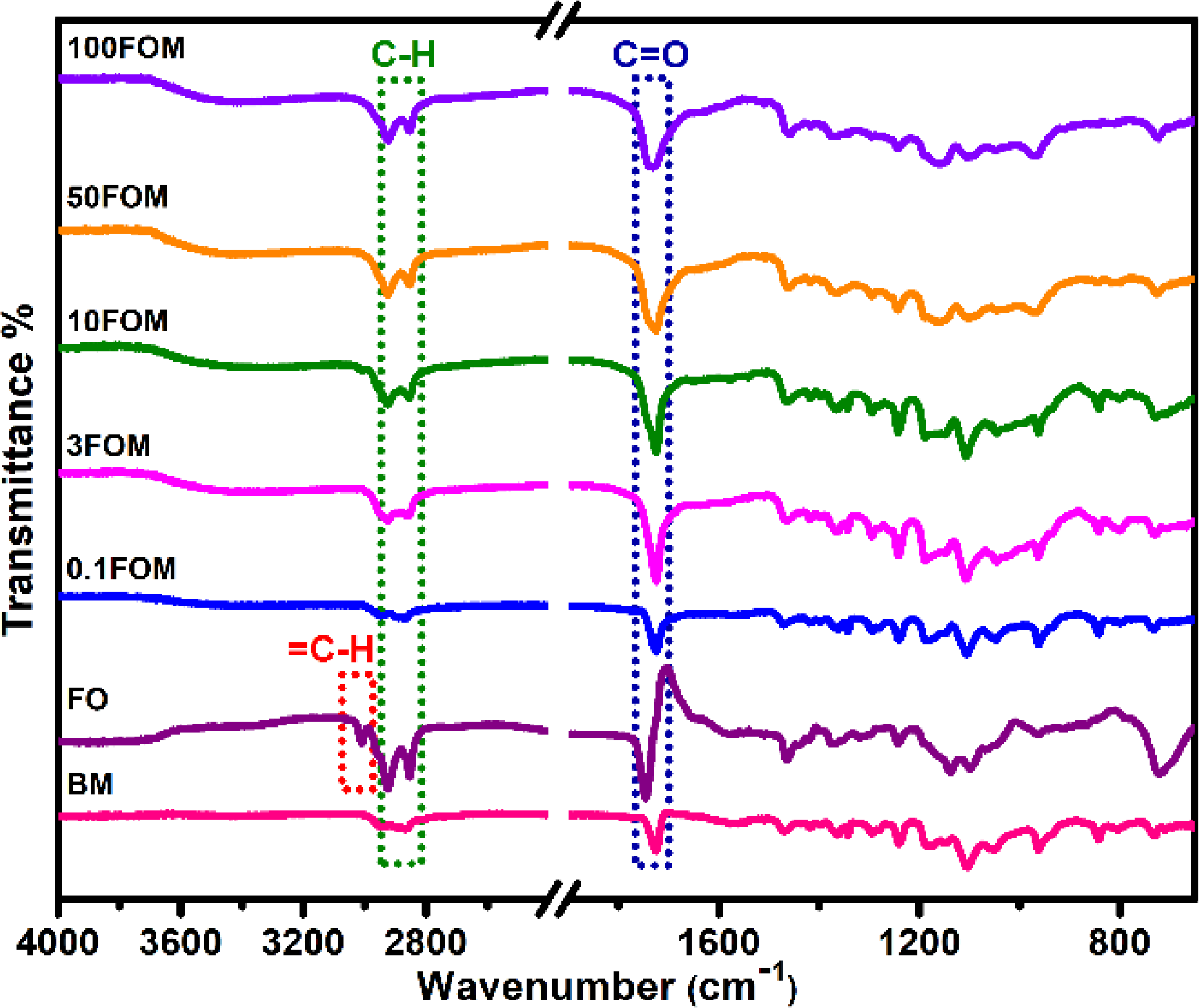
After the production of FOM, they were freeze-dried and characterized by FTIR spectroscopy to determine characteristic bands of polymeric micelles and FO, as well as to investigate the encapsulation of FO into polymeric micelles. FTIR spectra of FO, blank and FOM are shown in Figure 3.
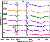
As shown in Figure 3, bands at 2960 cm-1 and 2880 cm-1 indicate C-H stretching in FO and the copolymer. In all spectra of polymeric micelles, characteristic bands at 1720 cm-1 correspond to C=O stretching belonging to the PCL block of polymeric micelles. In the spectrum of FO, there are two characteristic bands at 3120 cm-1 and 1745 cm-1 belonging to =C-H in fatty acid chains and to C=O in fatty acyl groups [48], respectively. The fact that all the micelles loaded with FO have mostly overlapped spectra, showing that they shared the same characteristics. The C=O band of the micelle at 1720 cm-1 overlaps with the C=O band of the oil at 1745 cm-1 by increasing the amount of FO, which indicates adsorption of oil molecules onto the micelle surface. The =C-H band at 3120 cm-1, another characteristic band of FO, is not observed in the spectra of FOM. Based on this, it can be said that a small amount of oil molecules is adsorbed on the surface of micelles because small amounts of C=O groups can give sharp and strong bands while it is much harder to observe the small amounts of =C-H group due to its weak band at 3120 cm-1.
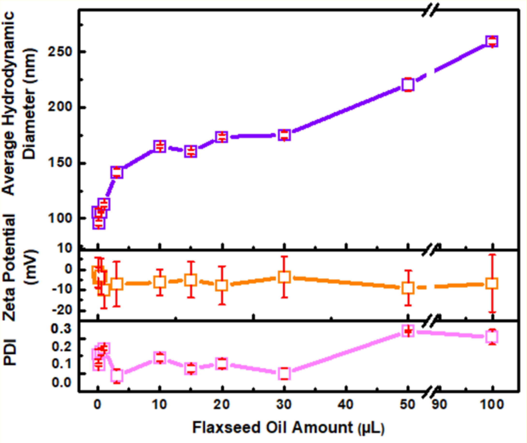
The size and surface charge of the micelles are critical parameters affecting their biological activity [49]. The hydrodynamic diameter, PDI value and zeta potential values of FOM were determined by using DLS and ELS methods. Besides, the effect of FO amount in micelles on the average size, charge and PDI values of polymeric micelles was examined. Figure 4 shows the average hydrodynamic diameter, zeta potential and PDI values of FOM according to FO amounts.
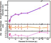
The average hydrodynamic diameters, zeta potentials and PDI values of FOMs depending on FO content.
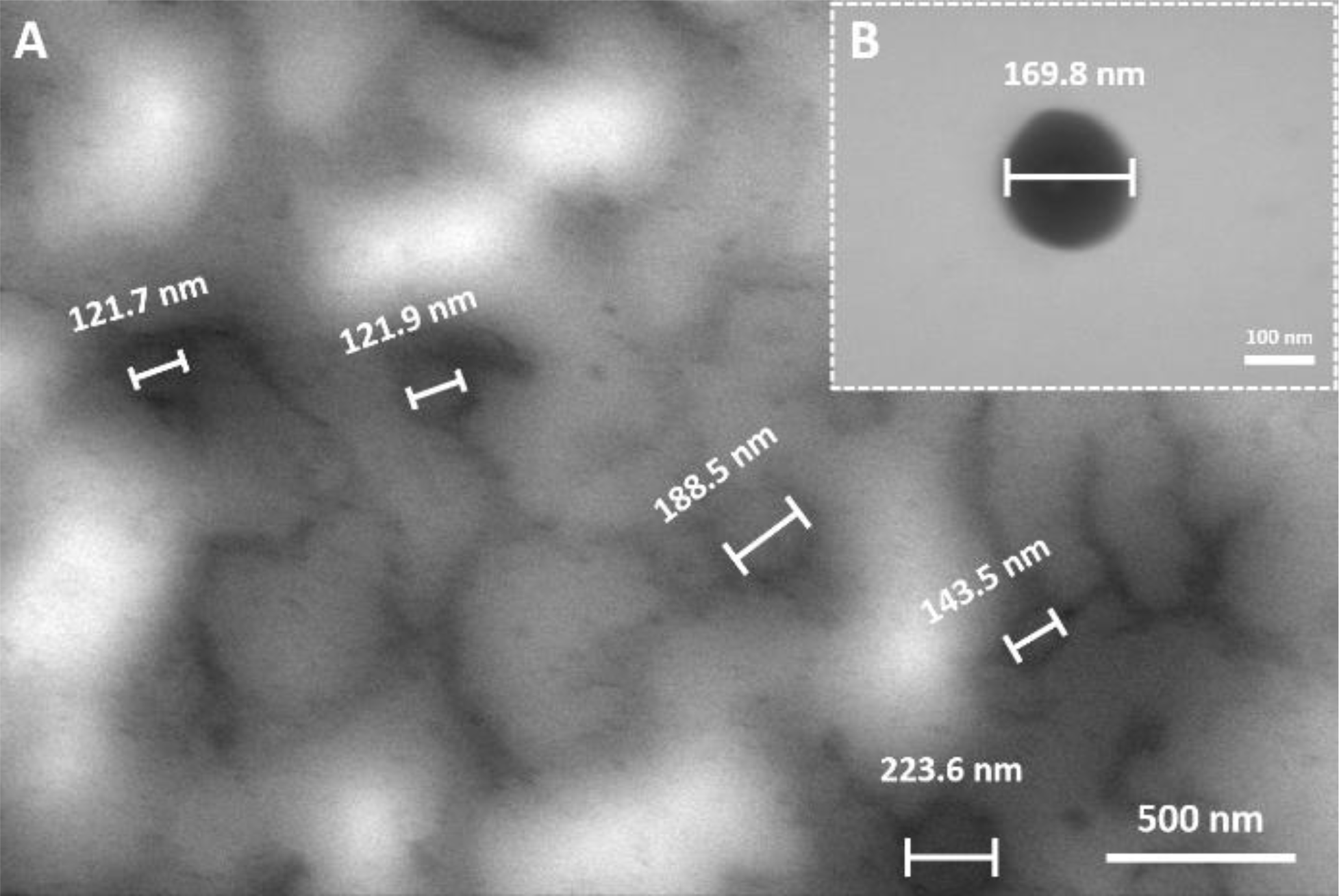
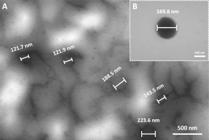
Intensity-based hydrodynamic size distributions of FOM exhibited unimodal distribution. The z-Average hydrodynamic diameters of 0.1FOM, 1FOM, 3FOM, 10FOM and 50FOM were 96, 113, 142, 165, and 221 nm, respectively. Accordingly, as the amount of FO increased, the hydrodynamic diameters of the micelles increased. In the study of Granata and coauthors, a nanocarrier system loaded with fennel essential oil (FEO) was produced, demonstrating the attainment of nano-sized structures by achieving a z-Average hydrodynamic diameter of 200 nm, similar to our study and even slightly larger than some of our micelles [50]. The PDI values of polymeric micelles ranged between 0.094 and 0.220, which reveals the narrow size distribution of the produced polymeric micelles. ELS measurements exhibited that the FOM have zeta potential values in the range of -9.98 mV and -1.34 mV. Accordingly, PCL-PEG polymeric micelles were slightly negatively charged in aqueous environment, as depicted in the literature [51]. The morphology of the produced FOM was examined by STEM. The STEM image of 15FOM (Figure 5) reveals nano-sized particles in which spherical morphologies are observed and having an average diameter of 161.5 nm. The size of 15FOM in STEM image is below its hydrodynamic diameter of 167.6 nm, in which the size difference in these two techniques arises from the hydration layer around the micelles in DLS measurements.
After the characterization of the micelles, we studied the stability of the produced micelles in solution and as lyophilized. Table 1 presents the hydrodynamic size and PDI values of 15FOM and 50FOM after 1 week in solution and after storage as a lyophilized powder for 1 year. The size of 15FOM after remaining in PBS at 37 °C did not increase while a slight decrease in size was observed for 50FOM. PDI value increased from 0.115 to 0.317 for 15FOM and decreased from 0.220 to 0.195. The PDI values after 1 week were still below or close to 0.3 which corresponds to uniform distribution of particles. As seen, the particles exhibit stability in physiological conditions for 1 week.
Z-average hydrodynamic diameter (DH) and PDI values of 15FOM and 50FOM. Day 0 measurements were obtained immediately after production. Day 7 measurements were obtained from lyophilized samples that were dissolved in PBS and incubated under gentle stirring at 37 °C for 7 days. The 1-year measurements were obtained from lyophilized samples stored for one year and subsequently dissolved in PBS.
After 1 year of storage of lyophilized micelles, the size of 15FOM was almost same while the size of 50FOM decreased from 220.9 to 162 nm. The PDI values of the samples were lower than 0.3. The decrease in the size of 50FOM can be related to the dehydration during the lyophilization process [52]. These results demonstrate that the FOMs exhibit good colloidal stability under physiological conditions over 1 week and maintain structural integrity after 1 year of storage in lyophilized form.
Certain limitations should be considered when interpreting studies involving the encapsulation and release of plant-derived oils from polymeric micellar systems. Quantification based on UV-Vis spectroscopy, while suitable for qualitative and comparative assessments, may lack the selectivity and accuracy required for precise determination of encapsulation efficiency and release behavior in complex oil-containing systems. In addition, plant-derived oils are known to be sensitive to external factors such as heat, light, and oxidative conditions, which may influence their chemical integrity during formulation and in vitro evaluation. These aspects represent general methodological limitations for oil-loaded micellar systems and highlight the need for more selective analytical techniques (e.g., HPLC or GC) and enhanced stability control in future studies.
In Vitro Cytotoxicity Assay
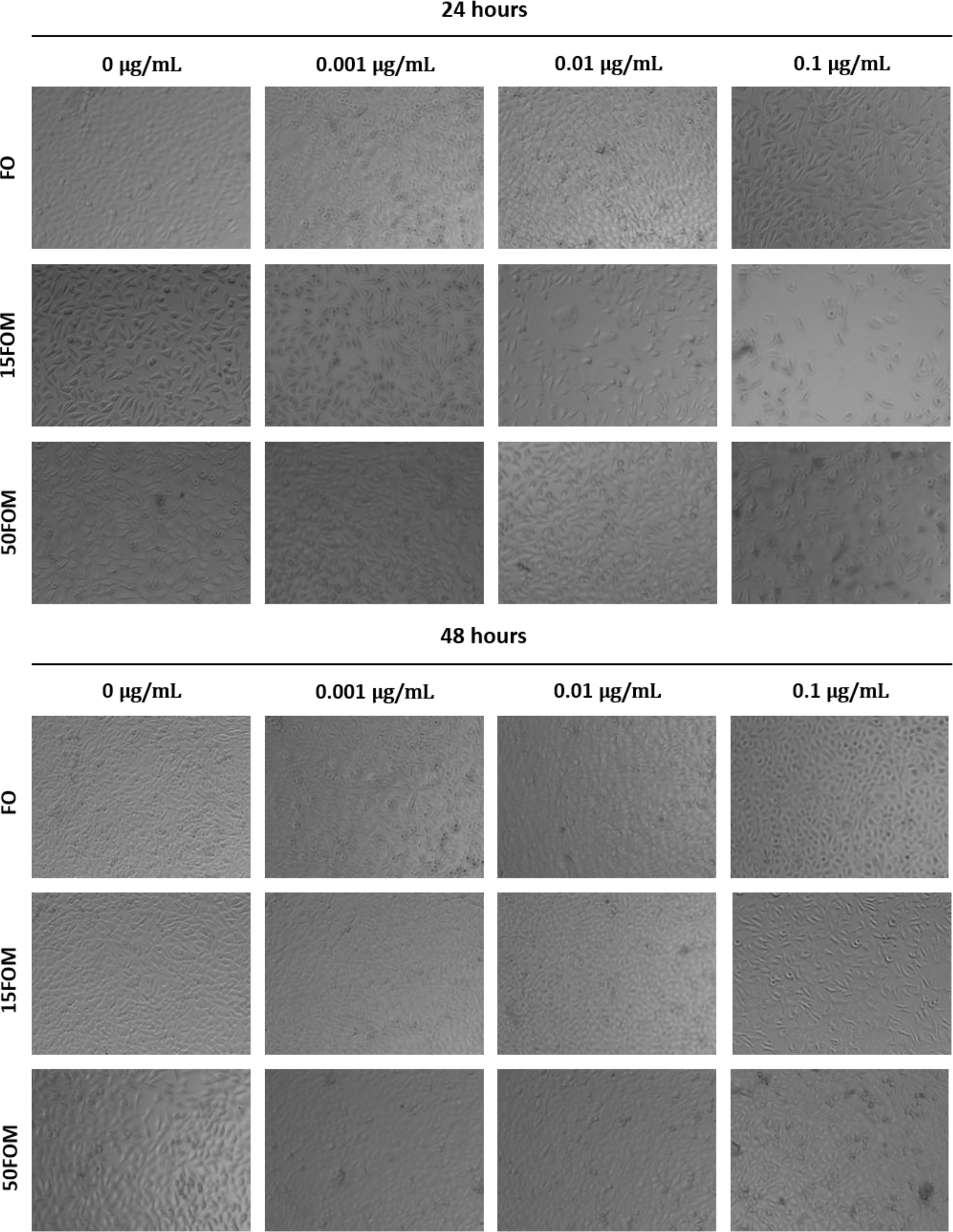
The efficacy of produced FOM in biological systems was investigated in order to examine potential use of micelles for breast cancer treatment. Firstly, the effect of the micelles on healthy cells were evaluated by the in vitro cytotoxicity studies of FO and FOM on L929 fibroblast cells using MTT analysis. As a result of MTT analysis of FO, 15FOM and 50FOM at different concentrations, cell images of L929 were obtained after 24 hours and 48 hours and the morphological changes of the cells were examined (Figure 6). In the L929 cell images obtained after 24 and 48 hours of incubation with the samples, the cells did not undergo morphological changes and no significant decrease in cell viability was observed.
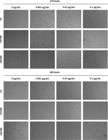
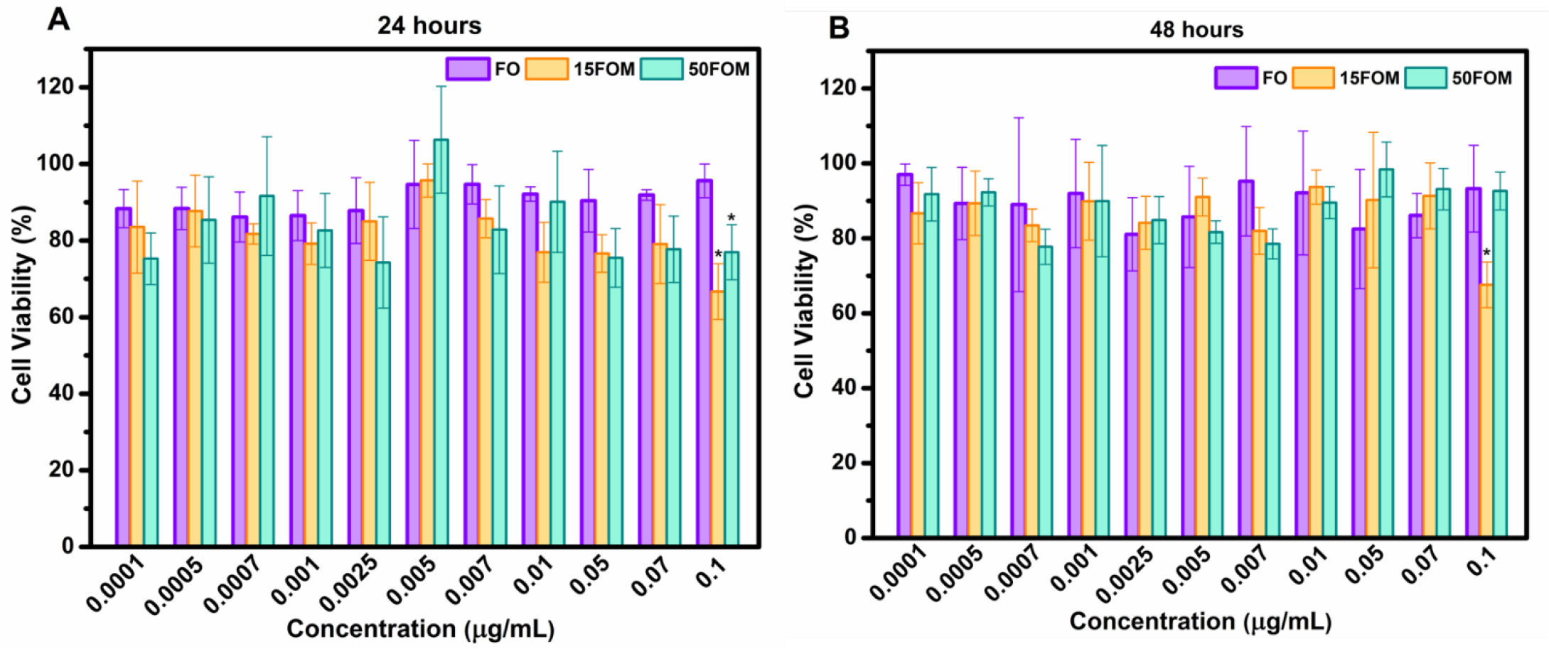
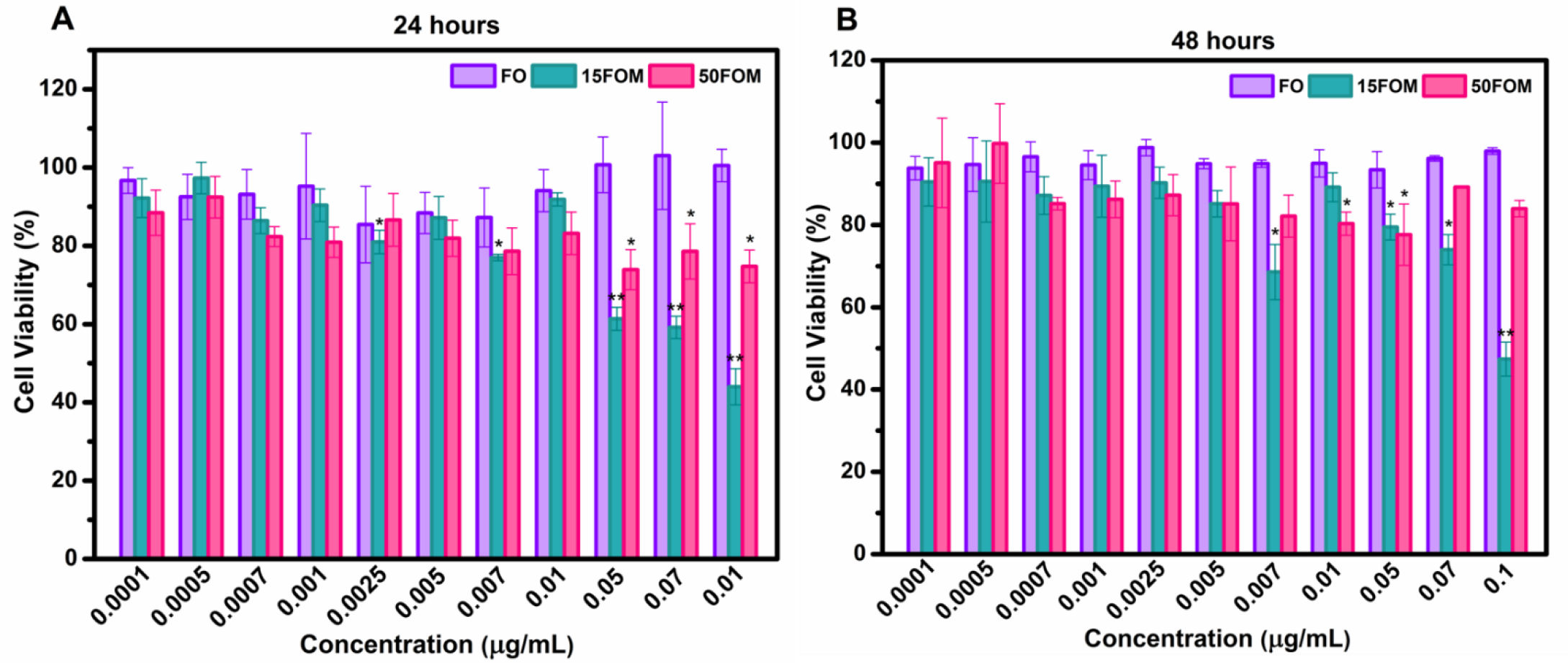
The effect of FO, 15FOM and 50FOM on viability of L929 cells is shown in Figure 7. Cell viability higher than 70% was achieved at all studied concentrations of FO, 15FOM, and 50FOM after incubation for both 24 and 48 hours. However, only the sample of 15FOM at 0.1 μg/mL caused cell viabilities lower than 70%, which are 66.69% and 67.61% for 24 and 48 hours of incubation, respectively. According to ISO 10993-5 definition, micelles did not show cytotoxic properties against fibroblast cells at the concentrations studied [53].
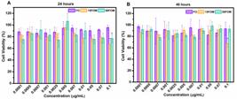
Cell viability of L929 fibroblast cells treated with FO, 15FOM, and 50FOM at different concentrations for 24 (A) and 48 (B) hours. Cell viability was assessed using the MTT assay. Data represent mean ± SD (n = 3). Statistical significance was indicated as *(p < 0.001) and ** (p < 0.001, larger mean difference) compared to the control group at the corresponding time point.
MTT assay was also performed to examine the anticancer activity of the produced FOM on MCF-7 breast cancer cells and the results are presented in Figure 8. As shown in the Figure 8, FO did not show a cytotoxic effect on MCF-7 breast cancer cells after 24 and 48 hours of incubation in the concentration range studied. It was shown in several studies that the effect of FO on MCF-7 cells was observed timeand concentration-dependently. In the study of Hu and coauthors, FO exhibited inhibition more than 50% of MCF-7 cells only after 72 hours with an IC50 value of 367.28 μg/mL [11]. Moreover, in the study of Buckner and coauthors [54], FO showed 40-60% inhibition of MCF-7 cells only after 4 days of incubation.
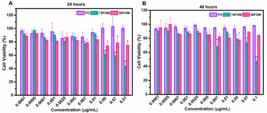
Cell viability of MCF-7 cells treated with FO, 15FOM, and 50FOM at different concentrations for 24 (A) and 48 (B) hours. Cell viability was assessed using the MTT assay. Data represent mean ± SD (n = 3). Statistical significance was indicated as *(p < 0.001) and **(p < 0.001, larger mean difference) compared to the control group at the corresponding time point.
On the other hand, FOM of 15FOM exhibited cytotoxic activity against MCF-7 cells. A decrease in cell viability was observed with increasing concentration of 15FOM. When 15FOM was at a concentration of 0.1 μg/mL, the cell viability of MCF-7 cells decreased below 50%. The IC50 value against MCF-7 cells for 15FOM was obtained as 0.08 μg/mL at 24 hours and 0.096 μg/mL at 48 hours. As seen, the produced 15FOM nanocarrier system showed the ability to inhibit the proliferation of MCF-7 cells at lower concentrations compared to the several previous studies [55-57]. It is obvious that the micellar system for delivery of FO critically increased the anticancer activity of FO both in the time and concentration aspects. Accordingly, as reported by Mason and coauthors, flaxseed oil shows anticancer activity by inhibiting the Akt and MAPK signaling pathways and altering the fatty acid profile of tumor [58]. Encapsulation of flaxseed oil within a nanocarrier is expected to further enhance these mechanisms by improving cellular uptake and thereby strengthening its anticancer activity. Moreover, the co-delivery of agents such as trastuzumab, which has demonstrated enhanced anticancer activity [59] when used in combination with flaxseed oil, by using a nanocarrier system represents a strong candidate for achieving synergistic effects in breast cancer therapy.
Additionally, 50FOM also inhibited the proliferation of MCF-7 cells compared to the FO but cell viabilities did not decrease to values lower than 70%. The reason for lower anticancer activity of 50FOM compared to 15FOM can be the larger size of the micelle which can directly affect the cancer cell uptake. In the study by Choi and coauthors, cellular uptake and cytotoxicity of particles ranging in size from 70 to 1000 nm were compared, and it was shown that even among particles with sizes between 100 and 200 nm, there was a difference in cancer cellular uptake and cytotoxicity [60].
In Vitro Cell Migration (Wound Healing) Assay
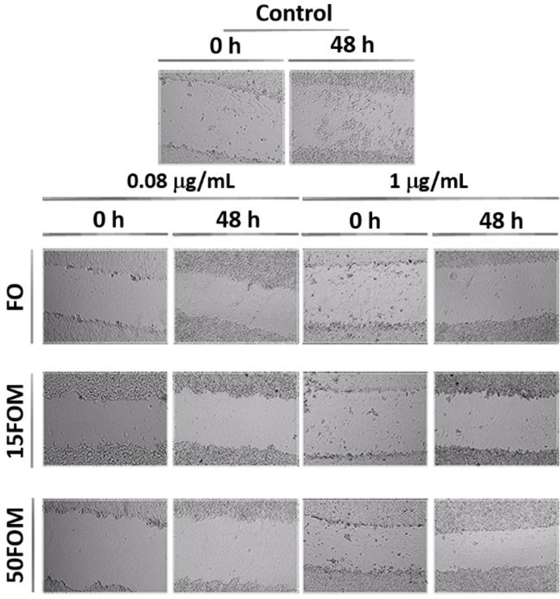
A cell migration assay was conducted to investigate the inhibitory effect of FOM on the migration ability of MCF-7 breast cancer cells. Following the creation of a physical wound as a scratch in the culture medium, FOM were applied at the concentrations at (0.08 μg/mL) and above (1 μg/mL) IC50 values [61]. In Figure 9, the distances between scratches were measured at 0 and 48 hours, and the wound closure percentage was calculated. According to the results, the highest migration of MCF-7 cells was observed in the control group with a wound closure percentage of 12% due to the aggressive nature of cancer cells. The wound closure percentages obtained for FO were 7.9% and 9.2% at 0.08 and 1 μg/mL, respectively, while 50FOM caused wound closure percentages of 8.5% and 8.7% at 0.08 and 1 μg/mL, respectively. 15FOM showed the lowest closure percentage at IC50 value. 15FOM showed inhibitory effect on MCF-7 cell migration by decreasing the wound closure percentages down to 1.7% and 4.5% at 0.08 and 1 μg/mL, respectively. As seen, the micelles loaded with FO exhibited higher anticancer activity compared to the free FO.
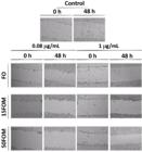
Images that show the effects of FO, 15FOM and 50FOM with the concentration of IC50 on cell migration ability of MCF-7 breast cancer cells at 0 and 48 hours.
CONCLUSION
In this study, we produced a drug delivery system by encapsulating FO into the polymeric micelles of PCL-b-PEG for potential breast cancer treatment and evaluated their anticancer activity on MCF-7 breast cancer cells as well as their biocompatibility with L929 fibroblast cells. The characterization studies, including size distribution, surface charge, and morphology, confirmed the successful formation of these polymeric micelles with different amounts of FO. The size of the micelles increased with higher amounts of encapsulated FO, as observed in both DLS and SEM analyses.
In vitro cytotoxicity assay results indicated that FOM, especially 15FOM, exhibited a concentration-dependent cytotoxic effect on MCF-7 cells, surpassing the activity of free FO. Moreover, the FOM demonstrated an inhibitory effect on the migration of MCF-7 cells, suggesting their potential for impeding cancer cell progression.
Overall, the study introduces the development of a phytochemical-based therapeutic system for breast cancer treatment. The FOMs show promise as an effective and biocompatible drug delivery system, providing a foundation for further research and development in the field of anticancer therapies.
-
Funding:
This research was funded by Scientific and Technological Research Council of Turkey (TUBITAK) Scientist Support Programs (BIDEB), grant number of 919B011903756.
-
Institutional Review Board Statement:
Not applicable.
-
Informed Consent Statement:
Not applicable.
Acknowledgments:
We would like to thank Yıldız Technical University for providing the laboratory and materials necessary to carry out the experiments.
Use of Generative Artificial Intelligence
The authors declare that large language models and other generative artificial intelligence (AI) or AI-assisted technologies cannot be credited as authors and have not been listed as authors of this paper.
The authors declare that generative artificial intelligence (AI) or AI-assisted tools were used under full human supervision. The tool(s) and version(s) used, and their purpose, are described here: ChatGPT (GPT-5, OpenAI) was only used for language revision. No confidential or sensitive data were uploaded to such tool(s), and all AI-assisted content was checked, corrected and approved by the authors, who take full responsibility for the integrity and originality of the manuscript.
Data Availability Statement:
Data are available on reasonable request for corresponding author.
REFERENCES
-
1 Current and future burden of breast cancer: global statistics for 2020 and 2040. [Internet]. Accessed Nov 16, 2025. Available from: https://www.iarc.who.int/fr/news-events/current-and-future-burden-of-breast-cancer-global-statistics-for-2020-and-2040
» https://www.iarc.who.int/fr/news-events/current-and-future-burden-of-breast-cancer-global-statistics-for-2020-and-2040 -
2 Cancer Statistics Center. American Cancer Society. [Internet]. Accessed Nov 16, 2025. Available from: https://cancerstatisticscenter.cancer.org/
» https://cancerstatisticscenter.cancer.org/ -
3 Breast cancer cases and deaths are projected to rise globally. [Internet]. Accessed Nov 16, 2025. Available from: https://www.iarc.who.int/news-events/breast-cancer-cases-and-deaths-are-projected-to-rise-globally
» https://www.iarc.who.int/news-events/breast-cancer-cases-and-deaths-are-projected-to-rise-globally - 4 Siegel RL, Miller KD, Wagle NS, Jemal A. Cancer statistics, 2023. CA Cancer J Clin. 2023;73(1):17-48.
-
5 Chhikara B, Parang K. Global cancer statistics 2022: The trends projection analysis. Pharm Fac Artic Res [Internet]. 2022 Oct 2; Available from: https://digitalcommons.chapman.edu/pharmacy_articles/938
» https://digitalcommons.chapman.edu/pharmacy_articles/938 - 6 Siddiqui JA, Singh A, Chagtoo M, Singh N, Godbole MM, Chakravarti B. Phytochemicals for breast cancer therapy: current status and future implications. Curr Cancer Drug Targets. 2015;15(2):116-35.
- 7 Prabhu RH, Patravale VB, Joshi MD. Polymeric nanoparticles for targeted treatment in oncology: current insights. Int J Nanomedicine. 2015;10:1001-18.
- 8 Choudhari AS, Mandave PC, Deshpande M, Ranjekar P, Prakash O. Phytochemicals in cancer treatment: from preclinical studies to clinical practice. Front Pharmacol. 2019;10:1614.
- 9 Buckner AL, Buckner CA, Montaut S, Lafrenie RM. Treatment with flaxseed oil induces apoptosis in cultured malignant cells. Heliyon. 2019;5(8):e02251.
- 10 Calado A, Neves PM, Santos T, Ravasco P. The effect of flaxseed in breast cancer: a literature review. Front Nutr. 2018;5:4.
- 11 Hu T, Linghu K, Huang S, Battino M, Georgiev MI, Zengin G, et al. Flaxseed extract induces apoptosis in human breast cancer MCF-7 cells. Food Chem Toxicol. 2019;127:188-96.
- 12 Mason JK, Chen J, Thompson LU. Flaxseed oil-trastuzumab interaction in breast cancer. Food Chem Toxicol. 2010;48(8-9):2223-6.
- 13 Guerriero E, Sorice A, Capone F, Storti G, Colonna G, Ciliberto G, et al. Combining doxorubicin with a phenolic extract from flaxseed oil: evaluation of the effect on two breast cancer cell lines. 2017;468.
- 14 Bak MJ, Das Gupta S, Wahler J, Suh N. Role of dietary bioactive natural products in estrogen receptor-positive breast cancer. Semin Cancer Biol. 2016;40-41:170-91.
-
15 Mangla B, Kaur A, Kumar P, Javed S, Ahsan W, Popli H. Development of nanobigel system combining apoptosis-inducing daidzein and flaxseed oil for the synergistic treatment of skin cancer. J Drug Deliv Sci Technol. 2024;105843. doi: 10.1016/j.jddst.2024.105843
» https://doi.org/10.1016/j.jddst.2024.105843 -
16 Harer Priyanka S, Harer Sunil L. Development and evaluation of a self-micro emulsifying omega-3 fatty acid drug delivery system using flaxseed oil. Futuristic Trends Pharm Nurs. 2024;3:128-40.doi: 10.58532/V3BAPN7P1CH11
» https://doi.org/10.58532/V3BAPN7P1CH11 -
17 Mazonde P, Khamanga SMM, Walker RB. Design, optimization, manufacture and characterization of efavirenz-loaded flaxseed oil nanoemulsions. Pharmaceutics. 2020;12(9):9. doi: 10.3390/pharmaceutics12090797
» https://doi.org/10.3390/pharmaceutics12090797 -
18 Song F, Tian S, Chen X, Sun S. Development of liposomes stabilized Pickering emulsions as a potential delivery system for flaxseed oil. LWT. 2024;195:115794. doi: 10.1016/j.lwt.2024.115794
» https://doi.org/10.1016/j.lwt.2024.115794 -
19 Song F, Tian S, Chen F, Sun X, Zhang B. Carboxymethyl chitosan-decorated proliposomes as carriers for improved stability and sustained release of flaxseed oil. J Food Sci. 2020;85(10):3237-43. doi: 10.1111/1750-3841.15406
» https://doi.org/10.1111/1750-3841.15406 -
20 Coc LMC, Lacatusu I, Badea N, Barbinta-Patrascu ME, Meghea A. Effective lipid nanocarriers based on linseed oil for delivery of natural polyphenolic active. J Nanomater. 2021;2021:8853941. doi: 10.1155/2021/8853941
» https://doi.org/10.1155/2021/8853941 -
21 Arshad M, Pradhan RA, Zubair M, Ullah A. Lipid-derived renewable amphiphilic nanocarriers for drug delivery, biopolymer-based formulations: biomedical and food applications. In: Biopolymer-Based Formulations. Elsevier; 2020. p. 283-310. doi: 10.1016/B978-0-12-816897-4.00013-8
» https://doi.org/10.1016/B978-0-12-816897-4.00013-8 -
22 Hosseini E, Rajaei A, Tabatabaei M, Mohsenifar A, Jahanbin K. Preparation of Pickering flaxseed oil-in-water emulsion stabilized by chitosan-myristic acid nanogels and investigation of its oxidative stability in presence of clove essential oil as antioxidant. Food Biophys. 2020;15(2):216-28. doi: 10.1007/s11483-019-09612-z
» https://doi.org/10.1007/s11483-019-09612-z - 23 Zhou Q, Zhang L, Yang T, Wu H. Stimuli-responsive polymeric micelles for drug delivery and cancer therapy. Int J Nanomedicine. 2018;13:2921-42.
- 24 Akram M, Iqbal M, Daniyal M, Khan AU. Awareness and current knowledge of breast cancer. Biol Res. 2017;50(1):33.
- 25 Ghosh B, Biswas S. Polymeric micelles in cancer therapy: state of the art. J Control Release. 2021;332:127-47.
- 26 Park IH, Sohn JH, Kim SB, Lee KS, Chung JS, Lee SH, et al. An open-label, randomized, parallel, phase III trial evaluating the efficacy and safety of polymeric micelle-formulated paclitaxel compared to conventional cremophor EL-based paclitaxel for recurrent or metastatic HER2-negative breast cancer. Cancer Res Treat. 2017;49(3):569-77.
- 27 Deshmukh AS, Chauhan PN, Noolvi MN, Chaturvedi K, Ganguly K, Shukla SS, et al. Polymeric micelles: basic research to clinical practice. Int J Pharm. 2017;532(1):249-68.
- 28 Liu G, Li Y, Yang L, Wei Y, Wang X, Wang Z, et al. Cytotoxicity study of polyethylene glycol derivatives. RSC Adv. 2017;7(30):18252-9.
- 29 Saad B, Suter UW. Biodegradable polymeric materials. Encyclopedia of Materials: Science and Technology. 2001.
- 30 Gea S, Surga M, Rahayu S, Marpongahtun, Hutapea YA, Piliang AF. The analysis of thermal and mechanical properties of biocomposite polycaprolactone/cellulose nanofiber from oil palm empty fruit bunches. AIP Conf Proc. 2018;2049(1):020063.
- 31 Shariff SHA, Abdul Khodir W, Hamid SA, Haris MS, Ismail MW. Poly(caprolactone)-b-poly(ethylene glycol)-based polymeric micelles as drug carriers for efficient breast cancer therapy: a systematic review. Polymers. 2022;14(22):4847.
- 32 Kim Y, Kwak J, Lim M, Lim SY, Chae S, Ha SJ, et al. Advances in PCL, PLA, and PLGA-based technologies for anticancer drug delivery. Pharmaceutics. 2025;17(10):1354.
- 33 Avramović N, Mandić B, Savić-Radojević A, Simić T. Polymeric nanocarriers of drug delivery systems in cancer therapy. Pharmaceutics. 2020;12(4):298.
- 34 Sze LP, Li HY, Lai KLA, Chow SF, Li Q, KennethTo KW, et al. Oral delivery of paclitaxel by polymeric micelles: a comparison of different block length on uptake, permeability and oral bioavailability. Colloids Surf B Biointerfaces. 2019;184:110554.
- 35 Washington KE, Kularatne RN, Biewer MC, Stefan MC. Combination loading of doxorubicin and resveratrol in polymeric micelles for increased loading efficiency and efficacy. ACS Biomater Sci Eng. 2018;4(3):997-1004.
- 36 Yadav H, Kumar P, Sharma V, Sharma G, Raza K, Katare OP. Enhanced efficacy and a better pharmacokinetic profile of tamoxifen employing polymeric micelles. RSC Adv. 2016;6(58):53351-7.
-
37 Kajla P, Sharma A, Sood DR. Flaxseed-a potential functional food source. J Food Sci Technol. 2015;52(4):1857-71. doi: 10.1007/s13197-014-1293-y
» https://doi.org/10.1007/s13197-014-1293-y -
38 Goyal A, Sharma V, Upadhyay N, Gill S, Sihag M. Flax and flaxseed oil: an ancient medicine & modern functional food. J Food Sci Technol. 2014;51(9):1633-53. doi: 10.1007/s13197-013-1247-9
» https://doi.org/10.1007/s13197-013-1247-9 - 39 Topuzoğulları NS, Topuzoğulları M, Mansuroğlu B. Investigation of the effect of pumpkin seed oil loaded PEG-PCL micelles on breast cancer and glioma cells. In: Proceedings of the 3rd International Eurasian Conference on Biological and Chemical Sciences (EurasianBioChem 2020); 2020 Nov 25-27; Ankara, Turkey. 2020;1:789-96
- 40 Wilhelm M, Zhao CL, Wang Y, Xu R, Winnik MA, Mura JL, et al. Poly(styrene-ethylene oxide) block copolymer micelle formation in water: a fluorescence probe study. Macromolecules. 1991;24(5):1033-40.
- 41 Mahmurat SA, Arayıcı PP, Bedir E, Akgül B, Özbek T, Abamor EŞ, Acar S. Enhancement of rifaximin bioavailability and antibacterial efficacy through mPEG-b-PCL micelles and chitosan-coated systems. J Drug Deliv Sci Technol. 2025;107615.
- 42 Kumar P, Nagarajan A, Uchil PD. Analysis of cell viability by the MTT assay. Cold Spring Harb Protoc. 2018;2018(6):pdb.prot095505.
- 43 Gerlier D, Thomasset N. Use of MTT colorimetric assay to measure cell activation. J Immunol Methods. 1986;94(1-2):57-63.
- 44 Rodriguez LG, Wu X, Guan JL. Wound-healing assay. Methods Mol Biol. 2005;294:23-9.
- 45 Majumder N, Das G, Das SK. Polymeric micelles for anticancer drug delivery. Ther Deliv. 2020;11(10):613-35.
- 46 Lu C, Guo S, Liu L, Zhang Y, Li Z, Gu J. Aggregation behavior of MPEG-PCL diblock copolymers in aqueous solutions and morphologies of the aggregates. J Polym Sci B Polym Phys. 2006;44(23):3406-17.
- 47 Ukawala M, Rajyaguru T, Chaudhari K, Manjappa AS, Pimple S, Babbar AK, et al. Investigation on design of stable etoposide-loaded PEG-PCL micelles: effect of molecular weight of PEG-PCL diblock copolymer on the in vitro and in vivo performance of micelles. Drug Deliv. 2012;19(3):155-67.
- 48 Balanuca B, Raluca S, Hanganu A, Iovu H. Novel linseed oil-based monomers: synthesis and characterization. UPB Sci Bull Ser B Chem Mater Sci. 2014;76:129-40.
- 49 Jain AK, Thareja S. In vitro and in vivo characterization of pharmaceutical nanocarriers used for drug delivery. Artif Cells Nanomed Biotechnol. 2019;47(1):524-39.
- 50 Granata G, Riccobene C, Napoli E, Geraci C. Polymeric nanocapsules containing fennel essential oil: their preparation, physicochemical characterization, stability over time and in simulated gastrointestinal conditions. Pharmaceutics. 2022;14(4):873.
- 51 Chroni A, Mavromoustakos T, Pispas S. Biocompatible PEO-b-PCL nanosized micelles as drug carriers: structure and drug-polymer interactions. Nanomaterials. 2020;10(9):1872.
-
52 Zhao Y, Wang Z, Liu S, Xie S, Xie Y, Li R, Oda H. Lyoprotectant formulation and optimization of the J-aggregates astaxanthin/BSA/chitosan nanosuspension. Biomolecules. 2023;13(3):496. doi: 10.3390/biom13030496
» https://doi.org/10.3390/biom13030496 -
53 Biological Evaluation - SCC Medical Devices. [Internet]. 2022 [cited 2024 Mar 26]. Available from: https://scc-medical-devices.com/biological-evaluation-biocompatibility-iso-10993/
» https://scc-medical-devices.com/biological-evaluation-biocompatibility-iso-10993/ -
54 Buckner, A. L., Buckner, C. A., Montaut, S., & Lafrenie, R. M. (2019). Treatment with flaxseed oil induces apoptosis in cultured malignant cells. Heliyon, 5(8), e02251. https://doi.org/10.1016/j.heliyon.2019.e02251
» https://doi.org/10.1016/j.heliyon.2019.e02251 - 55 Gregoriou Y, Gregoriou G, Yilmaz V, Kapnisis K, Prokopi M, Anayiotos A, et al. Resveratrol loaded polymeric micelles for theranostic targeting of breast cancer cells. Nanotheranostics. 2021;5(1):113-24.
- 56 Parthasarathy A, Vijayakumar S, Malaikozhundan B, Thangaraj MP, Ekambaram P, Murugan T, et al. Chitosan-coated silver nanoparticles promoted antibacterial, antibiofilm, wound-healing of murine macrophages and antiproliferation of human breast cancer MCF 7 cells. Polym Test. 2020;90:106675.
- 57 Zamani M, Rostamizadeh K, Kheiri Manjili H, Danafar H. In vitro and in vivo biocompatibility study of folate-lysine-PEG-PCL as nanocarrier for targeted breast cancer drug delivery. Eur Polym J. 2018;103:260-70.
- 58 Mason JK, Fu M, Chen J, Thompson LU. Flaxseed oil enhances the effectiveness of trastuzumab in reducing the growth of HER2-overexpressing human breast tumors (BT-474). J Nutr Biochem. 2015;26(1):16-23.
- 59 Mason JK, Chen J, Thompson LU. Flaxseed oil-trastuzumab interaction in breast cancer. Food Chem Toxicol. 2010;48(8-9):2223-6.
- 60 Choi JS, Cao J, Naeem M, Noh J, Hasan N, Choi HK, et al. Size-controlled biodegradable nanoparticles: preparation and size-dependent cellular uptake and tumor cell growth inhibition. Colloids Surf B Biointerfaces. 2014;122:545-51.
- 61 Poyraz FS, Ugraskan V, Mansuroglu B, Yazici O. Investigation of cytotoxic antiproliferative and antiapoptotic effects of nanosized boron phosphate filled sodium alginate composite on glioblastoma cancer cells. Mol Biol Rep. 2023;50(12):10257-70.
-
Editor-in-Chief:
Paulo Vitor Farago
-
Associate Editor:
Paulo Vitor Farago