Phytochemical Study of Astragalus onobrychis: Bioactive Compounds, Antioxidant Activity, in silico Molecular Docking, PASS and DFT Study
Abstract
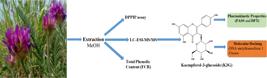
Astragalus species include rich bioflavonoids, triterpene glycosides, flavonoids, isoflavonoids and saponins. Astragalus species are traditionally used for diarrhea, dental treatment, diabetes treatment and as a cough remedy. In this study, the phytochemical content (total phenolic content and quantitative analysis of phenolics) and biological activity (DPPH˙) of the methanol extract of Astragalus onobrychis were investigated. The SPMB technique, a new method, was used for total phenolic and DPPH˙ analysis. Density functional theory (DFT) and theoretical in silico studies (molecular docking and PASS) were applied to the major component as kaempferol-3-glucoside (K3G) in the constituent analysis, which was performed by LC-ESI-MS/MS. Accordingly, the methanol extract of A. onobrychis was found to have high total phenolic content (FCR; 111.67 mg GA/g plant) and DPPH˙ (48.57%) analysis. Moreover, LC-ESI-MS/MS analysis revealed the K (285.011 µg/g extract) to be major constituent. In addition, the interactions of the K3G molecule with DNA mtyltransefraze 1 and urease enzyme were compared with the standard daunorubicin and thiourea. It was found that K3G may have inhibitory potential. These results can be substantiated in future studies by in vivo and in vitro studies.
Keywords:
Astragalus onobrychis; antioxidant activity; LC-ESI-MS/MS; kaempferol-3-glucoside.
HIGHLIGHTS
Astragalus onobrychis has high total phenolic content and DPPH˙.
In A. onobrychis, the main component is kaempferol-3-glucoside (K3G).
The K3G has high binding energy towards DNA methyltransefrase 1 and urease
INTRODUCTION
Plants have been used by humans for therapeutic purposes for centuries [1-3]. They have been presented to display significant biological activities due to their existence of secondary metabolites [4-7]. With the developments in the pharmaceutical industry, the use of herbal medicines declined, but due to the negative side effects of synthetic medicines, the tendency towards herbal medicines is increasing again[8-11]. To this end, more and more studies are being carried out every day looking at the therapeutic properties of plants found in nature and uncovering their unknown aspects (anti-cancer, antioxidant, antibacterial, antifungal, anti-inflammatory, etc.) [12-15]. As in all countries of the world, medically important plants have been used by the population in Turkey for centuries to treat illnesses [16].
The antimicrobial and antioxidant properties of secondary metabolites produced by plants have been extensively studied in recent years [17-19]. It is known that compounds with antioxidant activity in biological systems also have many biological functions such as antimutagenic, anticarcinogenic and anti-ageing functions [20-22]. Studies have shown that cheap synthetic antioxidants used in food are toxic and can cause cancer [23]. For this reason, the use of synthetic antioxidants has been restricted. The need for natural antioxidants has increased and the number of studies on the antioxidant properties of plants has also increased [24, 25].
Astragalus species which belongs to the Fabaceae family, is the genus with the most species in the world [26]. The genus Astragalus comprises almost 3000 species in the world [27]. In Turkey, it is widely known as "Geven". A. onobrychis L.; Sapphire Astragalus occurs in the Eastern Anatolia region of Turkey on stony slopes and steppes at an altitude of 1000-2400 m [28]. Astragalus species are also rich in bioflavonoids, amino acids, triterpene glycosides, flavonoids, isoflavonoids, and saponins [29, 30]. Astragalus species are known to be used in traditional medicine for the treatment of chronic diarrhea, acute hepatic edema, abnormal uterine bleeding, dental treatment, diabetes, and as a cough remedy [31]. In clinical studies, it was said to have liver-protecting, heart-strengthening, anti-cancer, antioxidant, immune system-stimulating, and anti-ageing properties [32].
In this study, phytochemical content analysis (LC-ESI-MS/MS and total phenolic content firstly by new approach biosensor method), biological activity (antioxidant activity firstly by new approach biosensor method) and in silico molecular docking study of A. onobrychis were performed. In analyzing the phytochemical content of the A. onobrychis, which is known to have anticancer and anti-urease activity, the theoretical interactions of the molecule, which was determined to be the main constituent, with the DNA methyltransferase 1 and urease enzyme and were calculated. The method used here for activity determination with the biosensor for the analysis of phenol content (FCR) and antioxidant (DPPH˙) activities is new and provides accurate results in a short time.
MATERIAL AND METHODS
Plant
Astragalus onobrychis (Herbarium: INWM00000118) was collected in June 2023 at Iğdır University Şehit Bülent Yurtseven campus. The plant identification was carried out by Dr. Belkıs Muca Yiğit, herbarium specialist at Iğdır University.
Extraction
A. onobrychis of flower was dried in an airy environment without sunlight. 50 grams of dried plants were ground into powder. It was transferred into a 1-liter conical flask and methanol solvent was added. The extraction process continued for 2 days. The solvent in the mixture was filtered with filter paper and then removed with the help of a rotary evaporator. As a result, methanol extract was obtained.
Total phenolic content determination test (FCR)
The total amount of phenolic compounds was determined using the FCR-SPMB method. This method is based on the measurement of the amount of FCR that remains undiminished after the reduction of FCR in the solution in the presence of antioxidants. In spectrometer measurements, the test results for the total amount of phenolic compounds are expressed as equivalent to gallic acid (mg/g extract). For this purpose, a GA-FCR calibration graph was constructed to determine the gallic acid equivalent, which corresponds to the amount of FCR reduced by the antioxidant species in the environment. The result was calculated using the obtained graph and the calculation equation [33, 34].
2,2-diphenyl-1-picrylhydrazyl (DPPH˙) activity test
For 2,2-diphenyl-1-picrylhydrazyl (DPPH˙) radical scavenging activity, 10 mL of the plant extract was taken and 10 mL of a 100 µg mL-1 DPPH˙ solution was added. The reaction was allowed to complete for 30 minutes and the potential values were measured by direct immersion of these samples in DPPH•-SPMB. The results were calculated as percentages according to the appropriate formula.
E0 is the potential value of the plant sample, E1 is the potential value of the DPPH˙ standard solution and E2 is the potential value of the DPPH˙ activity remaining in the medium after 30 minutes of reaction [35].
Characterization of phenolic profile by Liquid Chromatography-Electrospray Ionization Tandem Mass Spectrometry (LC-ESI-MS/MS)
The analysis was performed using a LC-ESI-MS/MS system consisting of an Agilent 1260 Infinity II, HPLC, and a 6460 triple quadrupole MS system. An Agilent Poroshell120 EC-C18 reversed-phase analytical column (100 mm × 3.0 mm, 2.7 μm) was used for chromatographic separation. For analyses, 4 mL of the samples were injected at a concentration of 1 mg/mL in full-loop injection mode and a flow rate of 0.4 mL/min, 25 ◦C HPLC temperature was achieved with a gradient solvent system. In ultrapure water and methanol (with 0.1% formic acid and 5 mM ammonium), an ideal separation of the components was achieved with a gradient system and a flow of methanol at 15, 12% for 1 min, 50% for 30 min, 90% for 30 min and 10% for 35 min, respectively. MS conditions were set as ESI negative and positive mode MS QqQ, 6.0 L/min dry gas flow rate, 300 ◦C dry gas temperature, 15 psi nebulizer pressure and capillary voltage of 3000 V, high pressure limit 400.00 bar; maximum flow gradient 100,000 mL/min2, the voltage (4 eV), the fragmentor (100 V), the scan time (200 ms) and the scan range of 50-1200 m/z [36, 37]. The screening was performed with 45 standards.
Molecular Docking application
The kaempferol-3-glucoside (K3G) structure was drawn with Chem-Draw Ultra 18.0 and refined to the lowest energy with Chem3D 18.0. The K3G with the lowest energy was plotted in Mol2 format. DNA metiltransefraze 1 (PBD: 3AV6) and urease (PBD: 4GY7) enzyme were taken from the Protein Data Bank (PDB). The K3G-enzyme interactions were analyzed using the Molegro Virtual Docker 6.0.1 (MVD) program. BIOVIA Discovery Studio Visualizer was used to create both twoand three-dimensional images to identify different types of these interactions [38].
Prediction of Activity Spectra for Substances (PASS) Prediction Analysis
The PASS analysis to determine the bioactivity spectra of compounds was performed via the PASS online web server (http://www.pharmaexpert.ru/passonline) [39]. The PASS prediction compares the Pa (probability of being active) and Pi (probability of being inactive) of compounds based on their canonical smile. With an accuracy of 90%, this tool is intended for the prediction of a wide range of biological activity. The probability for the active compound (Pa) and the probability for the inactive compound (Pi) indicate a PASS result. Activities for a particular chemical are only considered conceivable if Pa>Pi. Since Pa and Pi are probabilities, their values range from 0.000 to 1.000, and since they are calculated separately, Pa+Pi≠1. The PASS prediction results were interpreted and applied flexibly, yielding results such as (i) a higher probability of finding the activity experimentally when Pa>0.7, (ii) a lower probability of finding the activity experimentally when 0.5.
Density functional theory (DFT) and molecular electrostatic potentials (MEP) Studies
The Density functional theory (DFT) calculations in this paper were performed using the program Gaussian 09. The xs conformation was extensively optimized in the B3LYP functional using the 6-311++G (d, p) basis set [40, 41]. The geometry, the limiting molecular orbital energies, and the nonlinear optical (NLO) properties were optimized by applying DFT approaches. Gauss View 5 software was used to visualize the calculated structures, which included HOMO and LUMO representations as well as molecular electrostatic potential (MEP) representations [42].
RESULTS and DISCUSSION
The Antioxidant and Total Phenolic Content
In testing the total phenolic and antioxidant activity of the methanol extract of A. onobrychis, new methods presented in the literature were used. In our study, the DPPH˙ radical scavenger, the FRAP test for antioxidants, and the FCR test for total phenols were performed (Table 1). The method used measures the potential difference, in contrast to the commonly used UV spectrometer. One of the most advantageous aspects of this method is that it can be applied to all colorless, turbid, or non-turbid samples. It is called the potentiometric test of antioxidant activity. With this method, three different electrodes as selective PVC membrane biosensors (SPMD) were developed and used for three different tests. The method for preparing the electrodes was described in our previous study [33-35]
DPPH˙ and FCR content of A. onobrychis of flower methanol extract determined by potentiometric and spectroscopic methods
The DPPH˙-RSA value of the methanol extract of A. onobrychis at 500 ppm was measured to be 48.57%. In addition, the FCR content of 500 ppm A. onobrychis methanol extract was determined to be 111.68 mg GA/g plant. The extract was found to have significant activity compared to the standard at different concentrations (Table 1). In a study, the total phenolic content of A. onobrychis methanol extract was determined to be 15.30 mg GAEs/g extract [43].
Liquid Chromatography-Electrospray Ionization Tandem Mass Spectrometry (LC-ESI-MS/MS) Results
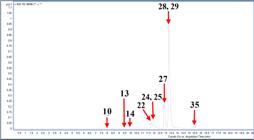
The phenolic content of A. onobrychis methanol extract was analyzed by LC-ESI-MS/MS. According to the analysis results, 10 phenolic compounds were detected (Figure 1). K3G (285.011 µg/g extract) and isoquercitrin (74.976 µg/g extract) were detected in the highest amounts (Table 2). In a similar study, it was reported that the major constituents in the content analysis of the methanol extract of A. onobrychis by LC-ESI-MS/MS were hesperidin (110863 μg/g extract) and hyperoside (2207.94 μg/g extract) [43].
The quantity and diversity of phenolic compounds in plants varies greatly depending on environmental factors such as soil quality, temperature, humidity, sunlight, time of harvest, climatic conditions, UV radiation and altitude [44]. In addition, different results are obtained due to the standards used in instrument such as HPLC, LC-ESI-MS/MS used in content analysis.
Molecular Docking Results
Epigenetic changes caused by methylation of deoxyribonucleic acid (DNA) are important for the development and progression of cancer cells. Hypermethylation of the promoter of tumor suppressor genes leads to transcriptional silencing and loss of gene function. DNA methylation is carried out by DNA methyltransferase. The activity of DNA methyltransferase 1 is sufficient to reactivate gene silencing and is a key enzyme in promoter hypermethylation [45]. DNA methyltransferase 1 is mainly involved in the maintenance of methylation during DNA replication [46]. In cancer therapy, new inhibitors must be developed that directly and specifically inhibit the activity of the DNA methyltransferase 1 enzyme [47]. Urease is produced by a variety of bacterial species [48]. Due to the enzymatic activity of urease, it has toxic effects on human cells. The presence of urease activity is an indicator of the presence of a number of bacterial infections. Urease is also an immunogenic protein and is recognized by the antibodies present in human serum. Antibodies of this type cause diseases such as rheumatoid arthritis, atherosclerosis or urinary tract infections [49].
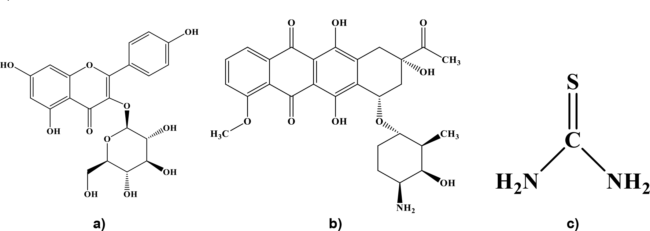
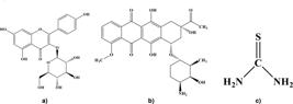
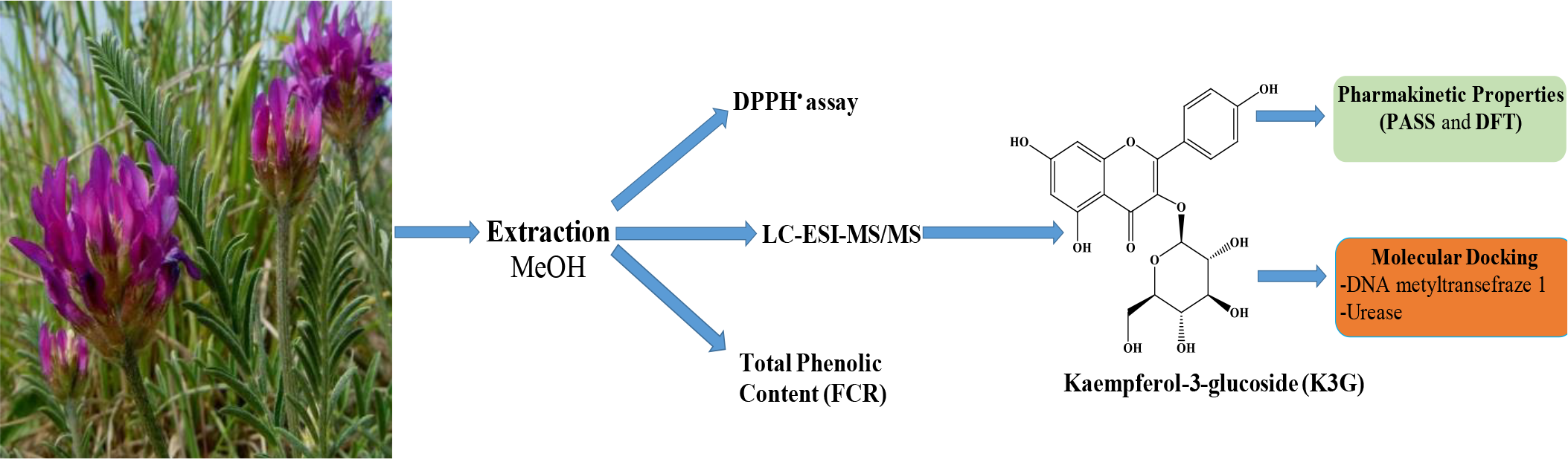
K3G (Figure 2), one of the naturally occurring flavonoids, is a bioactive component. It is known for its various pharmacological effects, such as anti-diabetic, anti-inflammatory, antioxidant, anti-cancer, and anti-ulcer properties [50]. Daunorubicin is a chemotherapeutic agent that is used to treat various types of cancer, particularly leukemia [51]. Thiourea derivatives are compounds that have a strong antitrypanosomal, antifungal, herbicidal and anticancer effect [52]. In this study, we theorised the interactions of the K3G molecule, which is reported to have anti-cancer and anti-urea activity, with DNA methyltransferase 1 and the enzyme urease. The results were compared with thiourea and daunorubicin, which are used as drugs (Figure 2).
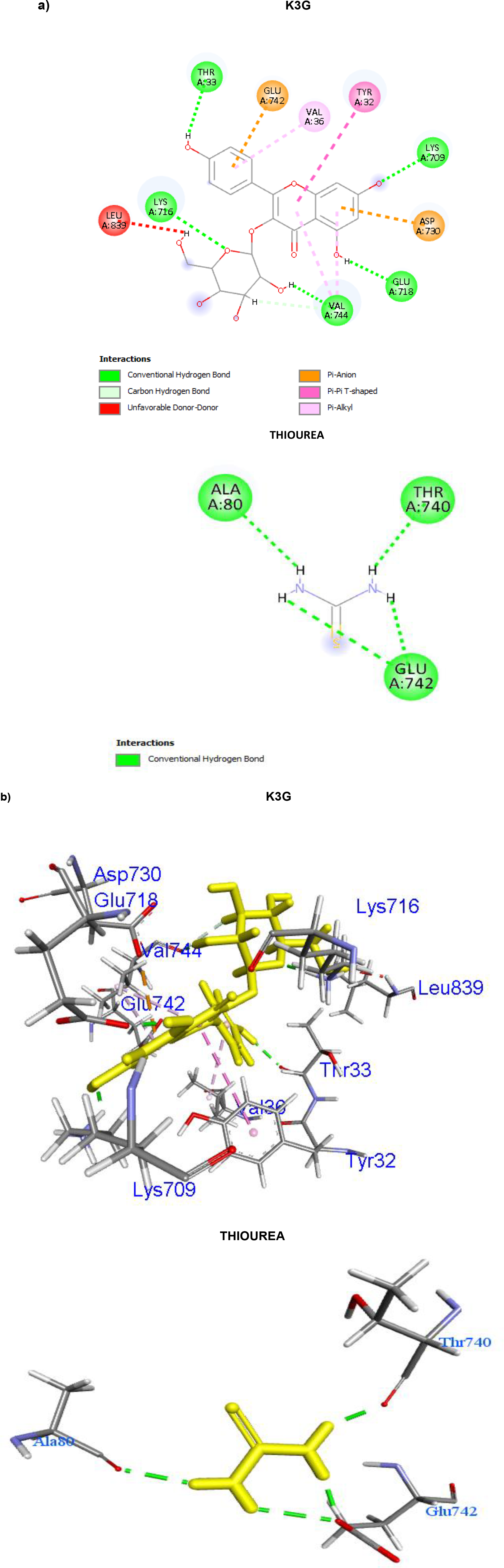
The K3G molecule interacted with urease by six conventional-hydrogen bonds formed with amino acid LYS709, LYS716, THR33, GLU718, VAL744, one carbon hydrogen bonds formed with amino acid VAL744, two pi-anion with amino acid ASP730, GLU742, one pi-pi T-shaped with amino acid TYR32, and three pi-alkyl with amino acid VAL744, VAL36 (Figure 3 - Supplementary material Table S1). K3G with urease interactions was determined as a MolDock score 124.48, binding energies -7.3 kcal/mol.
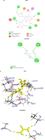
Thiourea molecule interacted with urease by four conventional-hydrogen bonds formed with amino acid GLU742, THR740, GLU742, ALA80 (Figure 3- Supplementary material Table S2). Thiourea with urease interactions was determined as a MolDock score -46.95, binding energies -3.3 kcal/mol.
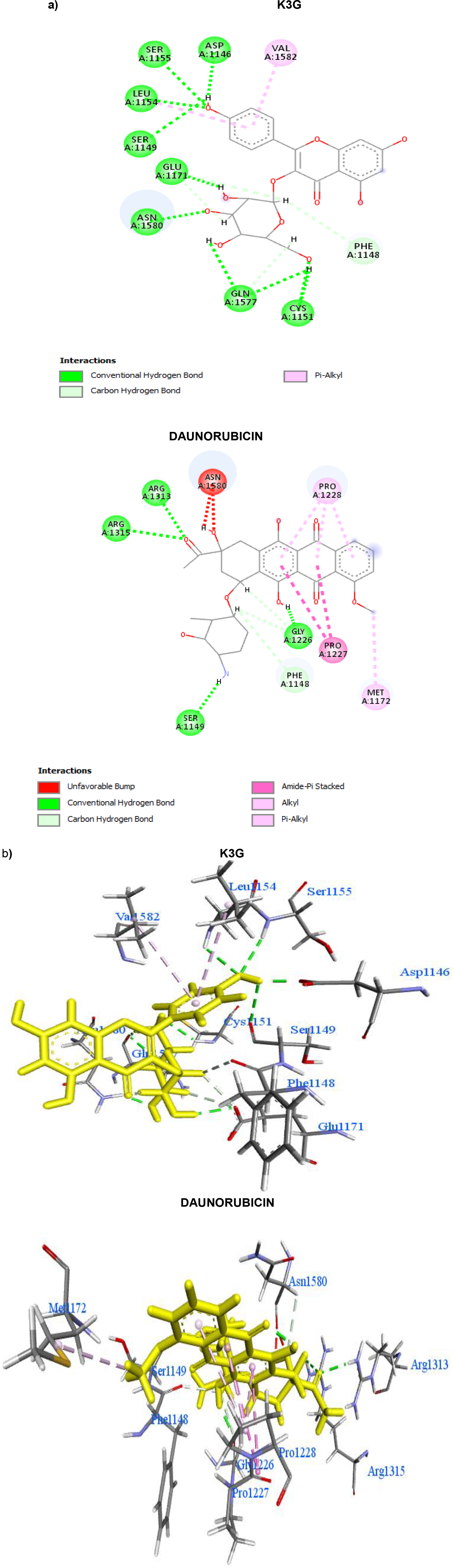
The K3G molecule interacted with DNA methyltransferase 1 by ten conventional-hydrogen bonds with amino acid CYS1151, LEU1154, SER1155, ASN1580, ASP1146, SER1149, GLU1171, GLN1577, CYS1151, five carbon hydrogen bonds formed with amino acid GLU1171, PHE1148, GLN1577 and two pi-alkyl with amino acid LEU1154, VAL1582 (Figure 4- Supplementary material Table S3). The K3G with DNA methyltransferase 1 interactions was determined as a MolDock score -143.88, binding energies -7.6 kcal/mol.
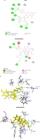
K3G and daunorubicin interaction with DNA methyltransferase 1, a) 2D images b) general view
Daunorubicin molecule interacted with DNA methyltransferase 1 by five conventional-hydrogen bonds with amino acid ARG1313, GLY1226, SER1149, and four carbon hydrogen bonds with amino acid ASN1580, GLY1226, PHE1148, two amide-pi-stacked with amino acid PRO1227-PRO1228, one alkyl with amino acid MET1172, and three pi-alkyl with amino acid PRO1228 (Figure 4- Supplementary material Table S4). Daunorubicin with DNA methyltransferase 1 interactions was determined as a MolDock score -97.16, binding energies -7.6 kcal/mol.
In molecular docking studies, K3G molecule was seen to have high MolDock score and binding energy. These results may be supported by in vivo activities.
Density functional theory (DFT) and molecular electrostatic potentials (MEP) Results
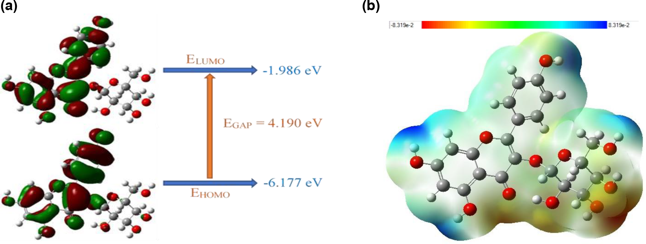
Density functional theory (DFT) has been shown to have an advantage over other computational methods in providing comprehensive insight into the molecular structure and chemical reactivity of the isolated molecule. Understanding the structural properties of compounds with biological activity can provide insight into this activity. The GAUSSIAN 09W program was used for the determination of molecular electrostatic potentials (MEP), FMO analysis, chemical reactivity parameters, and optimization of molecular geometry. In addition, Gauss View 5.0 was used to show the optimal structure of the investigated compounds [53, 54]. LUMO, an electron-accepting molecule, and HOMO, an electron-releasing molecule, are the two molecules that make up FMO. FMO uses these basic concepts to describe structure and explain why certain compounds are reactive. The energy difference between HOMO and LUMO is also very important for the chemical stability and reactivity of molecules and the characterization of electrical and optical properties. The least reactive and most stable molecule has the largest energy gap. The molecule with a lower energy gap is less stable and more reactive. FMOs are expressed by corresponding positive, negative, and negative regions shown in red and green colors.
In this study, DFT simulations were used to calculate HOMO and LUMO shapes and energies, as well as MEP surfaces. The HUMO-LUMO energy range of K3G is between 4.190 eV (Figure 5). As a result, K3G shows chemical reactivity.
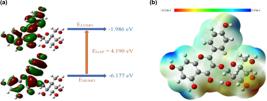
Frontier molecular orbitals and their calculated Egap value (a), Molecular electrostatic potential mapping and electron density (b) for the K3G
MEP maps are widely used to evaluate hydrogen bond interactions and identify reactive sites attacked by nucleophiles and electrophiles during chemical processes. MEP describes some molecular physical properties such as dipole moment, partial atomic charges, electronegativity, and chemical reactivity. It generally relates to how a chemical compound is charged. Different electrostatic potentials on the surface Areas of MEP are indicated by different colors such as blue, red and green respectively and represent the highest, lowest and highest positive-negative electrostatic potentials. The MEP map of the molecule (Figure 5) shows that the hydroxyl groups act as positive ESP positions on the hydrogen atoms, making them susceptible to nucleophilic attack. MEP is the chemical reactivity that makes it possible to study chemical reactions and develop synthetic processes to produce new materials. The mechanism of a reaction is linked to chemical reactivity.
After electrons are added to a saturated chemical system, energy shifts are calculated using the thermodynamic property electrophilicity index (ω). It is used to describe the chemical reactivity of a system. K3G can be classified as a strong electrophile according to the categorization created by Domingo, Aurell [55] (electrophiles: ω ≥ 1.50 eV - strong, 1.50 > ω > 0.80 eV - moderate, ω ≤ 0.80 eV - marginal). The minimal electrophilicity principle states that when hardness and chemical potential are both at their minimum, electrophilicity will be at its highest. K3G is strong electrophilic with an electrophilicity index of 3.976 eV and a low chemical potential of -4.082 eV, as shown in Table 3.
Prediction of Activity Spectra for Substances (PASS) Analysis results
Prediction of Activity Spectra for Substances (PASS) is an in silico method used to predict various biological activities that characterize substances. Table 4 gives the result of the findings showing the probability of activity (Pa) and the probability of inactivity (Pi). In PASS analysis, the probability of finding experimental activity is higher when Pa>0.7. Accordingly, the compounds have high hemostatic, cardioprotectant, free radical scavenger, lipid peroxidase inhibitor, anticarcinogenic, antioxidant, sugar-phosphatase inhibitor, antiprotozoal (leishmania), lactase inhibitor, antihemorrhagic, antihypercholesterolemic, kinase inhibitor, alpha glucosidase inhibitor, antineoplastic, histidine kinase inhibitor, antidote, antiinfective, antiinflammatory, antiviral (influenza), licheninase inhibitor, and antifungal effects.
CONCLUSION
Phytochemical content (total phenolic and phenolic content), antioxidant content (DPPH˙), as well as in silico (molecular docking and PASS) properties of the major component of the plant were investigated of the methanol extract of A. onobrychis flowers. Accordingly, total phenolic content was determined as 111.67 mg GA/g plant and DPPH˙ activity was determined as 48.57%. Moreover, according to the LC-ESI-MS/MS result, the main component was determined as K3G (285.011 µg/g extract). The interactions of K3G molecule, which has anticancer and antiurease activity, with DNA mtyltransefraze (anticancer) 1 and urease enzymes were determined. According to the results, the interactions of K3G with DNA mtyltransefrase (anticancer) 1 and urease were calculated as moldock score (-143.88, -124.48) and binding energies (-7.6 kcal/mol, -7.3 kcal/mol), respectively. The HUMO-LUMO energy range of K3G is between 4.190 eV, and K3G is strong electrophilic with an electrophilicity index of 3.976 eV and a low chemical potential of -4.082 eV in DFT studies. Additionally, K3G molecule showed high activity against 21 tests in PASS analysis.
-
Funding:
No funding.
Acknowledgments:
The authors would like to thank TUBA (Turkish Academy of Sciences).
Data Availability and Supplementary Material:
REFERENCES
- 1 Topçu G, Erenler R, Çakmak O, Johansson CB, Çelik C, Chai H-B, et al. Diterpenes from the berries of Juniperus excelsa Phytochemistry. 1999;50(7):1195-9.
- 2 Elmastas M, Ozturk L, Gokce I, Erenler R, Aboul-Enein HY. Determination of antioxidant activity of marshmallow flower (Althaea officinalis L.). Anal Lett. 2004;37(9):1859-69.
- 3 Yılmaz MA, Taslimi P, Kılıç Ö, Gülçin İ, Dey A, Bursal E. Unravelling the phenolic compound reserves, antioxidant and enzyme inhibitory activities of an endemic plant species, Achillea pseudoaleppica J Biomol Struct Dyn. 2023;41(2):445-56.
- 4 Demirtas I, Erenler R, Elmastas M, Goktasoglu A. Studies on the antioxidant potential of flavones of Allium vineale isolated from its water-soluble fraction. Food Chem. 2013;136(1):34-40.
- 5 Sahin Yaglioglu A, Akdulum B, Erenler R, Demirtas I, Telci I, Tekin S. Antiproliferative activity of pentadeca-(8E, 13Z) dien-11-yn-2-one and (E)-1,8-pentadecadiene from Echinacea pallida (Nutt.) Nutt. roots. Med Chem Res. 2013;22(6):2946-53.
- 6 Aksit H, Çelik SM, Sen Ö, Erenler R, Demirtas I, Telci I, et al. Complete isolation and characterization of polar portion of Mentha dumetorum water extract. Rec Nat Prod. 2014;8(3):277-80.
- 7 Zengin G, Mahomoodally MF, Aktumsek A, Ceylan R, Uysal S, Mocan A, et al. Functional constituents of six wild edible Silene species: A focus on their phytochemical profiles and bioactive properties. Food Bioscience. 2018;23:75-82.
- 8 Yaman C, Sarı Y, Atmaca S, Eroglu Z, Kayir O, Belguzar S, et al. Chemical Composition and Biological Effects of Essential Oils from Some Aromatic and Medicinal Plants. J Nat Prod. 2020;10.
- 9 Guzel A, Aksit H, Elmastas M, Erenler R. Bioassay-guided isolation and identification of antioxidant flavonoids from Cyclotrichium origanifolium (Labill.) Manden and Scheng. Pharmacogn Mag. 2017;13(50):316-20.
- 10 Elmastaş M, Telci İ, Akşit H, Erenler R. Comparison of total phenolic contents and antioxidant capacities in mint genotypes used as spices. Turk J Biochem. 2015;40(6):456-62.
- 11 Elmastas M, Erenler R, Isnac B, Aksit H, Sen O, Genc N, et al. Isolation and identification of a new neo-clerodane diterpenoid from Teucrium chamaedrys L. Nat Prod Res. 2016;30(3):299-304.
- 12 Koldaş S, Demirtas I, Özen T, Demirci M. Phytochemical Screening, Anticancer and Antioxidant Activities of Origanum vulgare L. subsp. viride (Boiss.) Hayek: A Plant of Traditional Usage. J Sci Food Agric. 2015;95.
- 13 Kooti W, Farokhipour M, Asadzadeh Z, Ashtary-Larky D, Asadi-Samani M. The role of medicinal plants in the treatment of diabetes: a systematic review. Electron Physician. 2016;8(1):1832-42.
- 14 Ceylan R, Zengin G, Mahomoodally MF, Sinan KI, Ak G, Jugreet S, et al. Enzyme inhibition and antioxidant functionality of eleven Inula species based on chemical components and chemometric insights. Biochem Syst Ecol. 2021;95:104225.
- 15 Uysal S, Zengin G, Mahomoodally MF, Yilmaz MA, Aktumsek A. Chemical profile, antioxidant properties and enzyme inhibitory effects of the root extracts of selected Potentilla species. S Afr J Bot. 2019;120:124-8.
- 16 Ozkan G, Kamiloglu S, Ozdal T, Boyacioglu D, Capanoglu E. Potential Use of Turkish Medicinal Plants in the Treatment of Various Diseases. Molecules. 2016;21(3):257.
- 17 Başar Y, Yenigün S, Behçet L, Ozen T, Demirtas İ. Antibacterial and Antioxidant Molecule Isolated from Nepeta aristata Boiss Et Kotschy Ex Boiss plant: 1,5,9-Epideoxyloganic Acid. IJCT. 2024;8(1):27-31.
- 18 Dabur R, Gupta A, Mandal TK, Singh DD, Bajpai V, Gurav AM, et al. Antimicrobial activity of some Indian medicinal plants. Afr J Tradit Complement Altern Med. 2007;4(3):313-8.
- 19 Yener I, Yilmaz MA, Olmez OT, Akdeniz M, Tekin F, Hasimi N, et al. A Detailed Biological and Chemical Investigation of Sixteen Achillea Species' Essential Oils via Chemometric Approach. Chem Biodivers. 2020;17(3):e1900484.
- 20 Başar Y, Karadağ M. Phytochemical Content Analysis of Different Lavandula Officinalis Extracts by LC-ESI-MS/MS and In Silico Molecular Docking Studies. TUBID. 2024;5(1):12-22.
- 21 Martelli G, Giacomini D. Antibacterial and antioxidant activities for natural and synthetic dual-active compounds. Eur J. Med Chem 2018;158:91-105.
- 22 Boga M, Alkan H, Ertaş A, Oral E, Yilmaz M, Yeşil Y, et al. Phytochemical profile and some biological activities of three Centaurea species from Turkey. Trop J Pharm Res. 2016;15:1865-75.
- 23 Singh BN, Malhotra BK. Effects of food on the clinical pharmacokinetics of anticancer agents: underlying mechanisms and implications for oral chemotherapy. Clin Pharmacokinet. 2004;43(15):1127-56.
- 24 Alma MH, Mavi A, Yildirim A, Digrak M, Hirata T. Screening chemical composition and in vitro antioxidant and antimicrobial activities of the essential oils from Origanum syriacum L. growing in Turkey. Biol Pharm Bull. 2003;26(12):1725-9.
- 25 Erenler R, Yaman C, Demirtas I, Mehmet A. Phytochemical Investigation of Hypericum heterophyllum Flowers: LC-ESI-MS/MS Analysis, Total Phenolic and Flavonoid Contents, Antioxidant Activity. Nat Prod J. 2023;13.
- 26 Li X, Qu L, Dong Y, Han L, Liu E, Fang S, et al. A Review of Recent Research Progress on the Astragalus Genus. Molecules. 2014;19(11):18850-80.
- 27 Zheng Y, Ren W, Zhang L, Zhang Y, Liu D, Liu Y. A Review of the Pharmacological Action of Astragalus Polysaccharide Front Pharmacol. 2020;11:349.
- 28 Plenk K, Willner W, Demina ON, Höhn M, Kuzemko A, Vassilev K, et al. Phylogeographic evidence for long-term persistence of the Eurasian steppe plant Astragalus onobrychis in the Pannonian region (eastern Central Europe). Flora. 2020;264:151555.
- 29 Sahin yaglioglu A, Erenler R, Geçer E, Genç N. Biosynthesis of Silver Nanoparticles Using Astragalus favesces Leaf: Identification, Antioxidant Activity, and Catalytic Degradation of Methylene Blue. J Inorg Organomet Polym. Mater. 2022;32:3700-7
- 30 Bagci E. Fatty acid composition of some Astragalus species from Turkey. Chem Nat Compd. 2006;42:645-8.
- 31 Erenler R, Geçer E. Green synthesis of silver nanoparticles from Astragalus logopodioides L. leaves. Turkish JAF Sci Tech. 2022;10:1112-5.
- 32 Lysiuk R, Darmohray R. Pharmacology and Ethnomedicine of the Genus Astragalus Int J Pharmacol, Phytochem Ethnomed. 2016;3:46-53.
- 33 Isildak Ö, Yıldız I. New Potentiometric Sensor for Total Phenolic Assay of Plant Extracts. SSRN Electronic Journal. 2022.
- 34 Isildak Ö, Yildiz I, Genc N. A new potentiometric PVC membrane sensor for the determination of DPPH radical scavenging activity of plant extracts. Food Chem. 2022;373(Pt A):131420.
- 35 Isildak Ö, Yildiz I, Genç N, Sabanci D, Işildak İ. New potentiometric PVC membrane electrode for Ferric Reduction Antioxidant Power assay. Food Chem. 2023;423:136261.
- 36 Yenigun S, Basar Y, Ipek Y, Gok M, Behcet L, Ozen T, et al. A potential DNA protector, enzyme inhibitor and in silico studies of daucosterol isolated from six Nepeta species. Process Biochem. 2024;143:234-47.
- 37 Atalar MN, Köktürk M, Altındağ F, Ozhan G, Özen T, Demirtas İ, et al. LC-ESI-MS/MS analysis of secondary metabolites of different St. John's wort (Hypericum perforatum) extracts used as food supplements and evaluation of developmental toxicity on zebrafish (Danio rerio) embryos and larvae. S Afr J Bot. 2023;159:580-7.
- 38 Yenigün S, Başar Y, Gül F, Marah S, Behcet L, Demirtas I, et al. Chemical Constituents and Bioactivities of Nepeta Taxa Essential Oils from Turkey: Principal Component Analysis, Molecular Docking Study, Molecular Dynamics, MM-PBSA and Drug-Likeness Estimation. ChemistrySelect. 2024;9.
- 39 Lagunin A, Stepanchikova A, Filimonov D, Poroikov V. PASS: prediction of activity spectra for biologically active substances. Bioinformatics. 2000;16(8):747-8.
- 40 Becke AD. Density-functional exchange-energy approximation with correct asymptotic behavior. Phys Rev A Gen Phys. 1988;38(6):3098-100.
- 41 Lee C, Yang W, Parr RG. Development of the Colle-Salvetti correlation-energy formula into a functional of the electron density. Phys Rev B Condens Matter. 1988;37(2):785-9.
- 42 İpek Y, Başar Y, Yenigün S, Behçet L, Özen T, Demirtas I. In vitro bioactivities and in silico enzyme interactions of abietatrien-3β-ol by bio-guided isolation from Nepeta italica subsp. italica J Biomol Struct Dyn. 2024.
- 43 Sarikurkcu C, Sahinler SS, Tepe B. Astragalus gymnolobus, A. leporinus var. hirsutus, and A. onobrychis: Phytochemical analysis and biological activity. Ind Crops Prod. 2020;150:112366.
- 44 Aydın S, Karaca Sanyürek N, Çakır A, Yılmaz Ö. The Effects of Different Altitudes on the Phenolic Contents and Antioxidant Activities of Some White Grape (Vitis Vinifera L.) Varieties. Res J Biol Sci. 2021:30-8.
- 45 Jung Y, Park J, Kim TY, Park JH, Jong HS, Im SA, et al. Potential advantages of DNA methyltransferase 1 (DNMT1)-targeted inhibition for cancer therapy. J. Mol. Med. (Berl). 2007;85(10):1137-48.
- 46 Mutze K, Langer R, Schumacher F, Becker K, Ott K, Novotny A, et al. DNA methyltransferase 1 as a predictive biomarker and potential therapeutic target for chemotherapy in gastric cancer. Eur J Cancer. 2011;47(12):1817-25.
- 47 Medina-Franco JL, Yoo J, Dueñas-González A. Chapter 13 - DNA Methyltransferase Inhibitors for Cancer Therapy. In: Zheng YG, editor. Epigenetic Technological Applications. Boston: Academic Press; 2015. p. 265-90.
- 48 Mobley HLT, Mendz GL, Hazell SL. Helicobacter pylori: Physiology and Genetics. Washington (DC): ASM Press Copyright © 2001, ASM Press.; 2001.
- 49 Konieczna I, Zarnowiec P, Kwinkowski M, Kolesinska B, Fraczyk J, Kaminski Z, et al. Bacterial urease and its role in long-lasting human diseases. Curr Protein Pept Sci. 2012;13(8):789-806.
- 50 Rashed, Khaled. "Kaempferol-3-od-glucoside bioactivities: a review." IJSIT. 9.4 (2020): 213-217.
- 51 Saleem T, Kasi A. Daunorubicin: StatPearls Publishing, Treasure Island (FL); 2023 2023.
- 52 Saeed A, Zaib S, Pervez A, Mumtaz A, Shahid M, Iqbal J. Synthesis, molecular docking studies, and in vitro screening of sulfanilamide-thiourea hybrids as antimicrobial and urease inhibitors. Med Chem Res. 2012;22.
- 53 Sameeh MY, Khowdiary MM, Nassar HS, Abdelall MM, Alderhami SA, Elhenawy AA. Discovery potent of thiazolidinedione derivatives as antioxidant, α-amylase inhibitor, and antidiabetic agent. Biomedicines. 2021;10(1):24.
- 54 Gökçimen S, İpek Y, Behçet L, Demirtaş İ, Özen T. Isolation, characterization and evaluation of oxypeucedanin and osthol from local endemic Prangos aricakensis Behçet and Yapar root as antioxidant, enzyme inhibitory, antibacterial and DNA protection: molecular docking and DFT approaches. J Biomol Struct Dyn. 2024:1-18.
- 55 Domingo LR, Aurell MJ, Pérez P, Contreras R. Quantitative characterization of the global electrophilicity power of common diene/dienophile pairs in Diels-Alder reactions. Tetrahedron. 2002;58(22):4417-23.
-
Editor-in-Chief:
Paulo Vitor Farago
-
Associate Editor:
Jane Manfron Budel

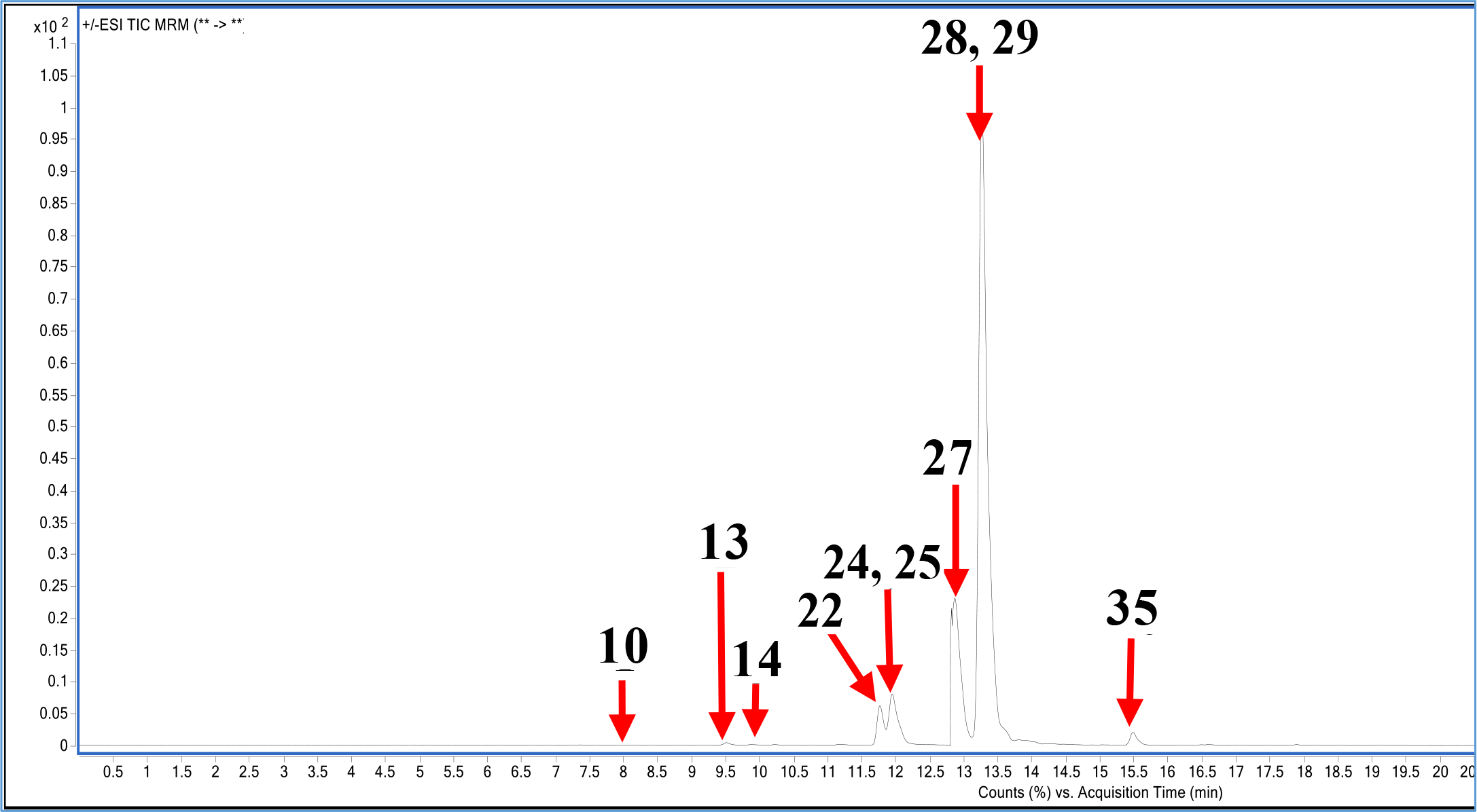 Shikimic acid (1), gallic acid (2), protocatechuic acid (3), epigallocatechin (4), catechin (5), chlorogenic acid (6), hydroxybenzaldeyde (7), vanillic acid (8), caffeic acid (9), syringic acid (10), caffein (11), vanillin (12), o-coumaric acid (13), salicylic acid (14), taxifolin (15), resveratrol (16), polydatine (17), trans-ferulic acid (18), sinapic acid (19), scutellarin (20), p-coumaric acid (21), coumarin (22), protocatehuic ethyl ester (23), hesperidin (24), isoquercitrin (25), rutin (26), quarcetin-3-xyloside (27), kaempferol-3-glucoside (28), fisetin (29), baicalin (30), chrysin (31), daidzein (32), trans-cinnamic acid (33), quercetin (34), naringenin (35), silibinin (36), hesperetin (37), morin (38), kaempferol (39), baicalein (40), luteolin (41), biochanin A (42), capcaicin (43), dihydrocapcaicin (44), diosgenin (45).