
 Activity of Silver Nitrate for Treatment of Amoebic Keratitis
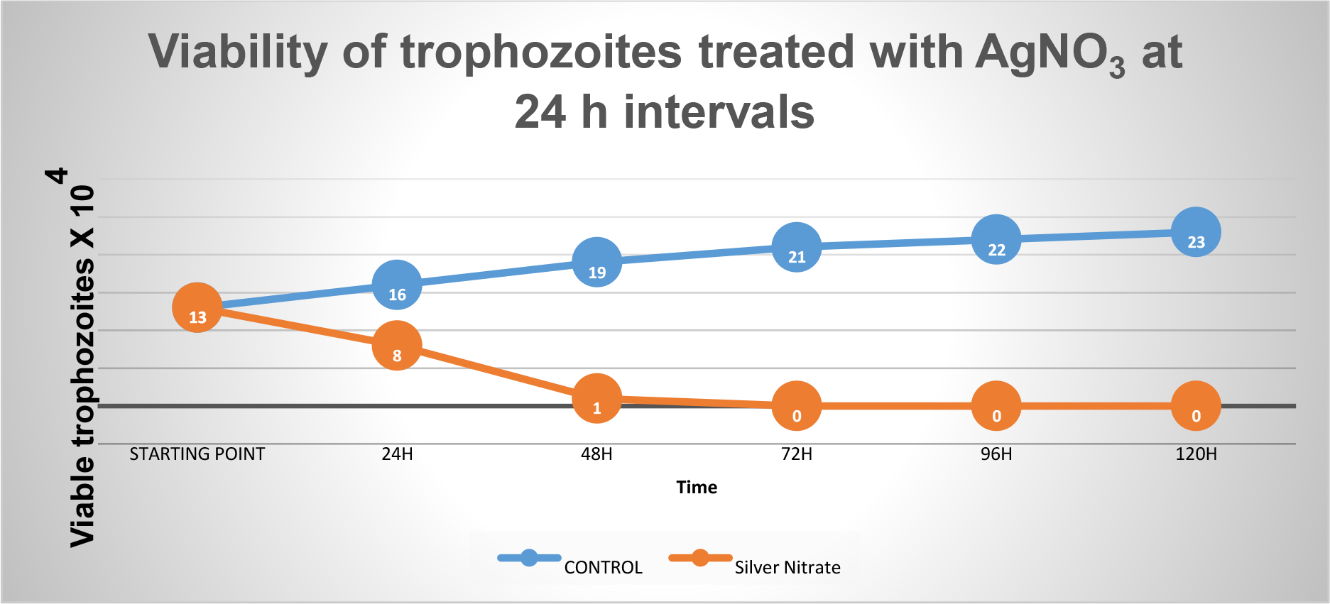
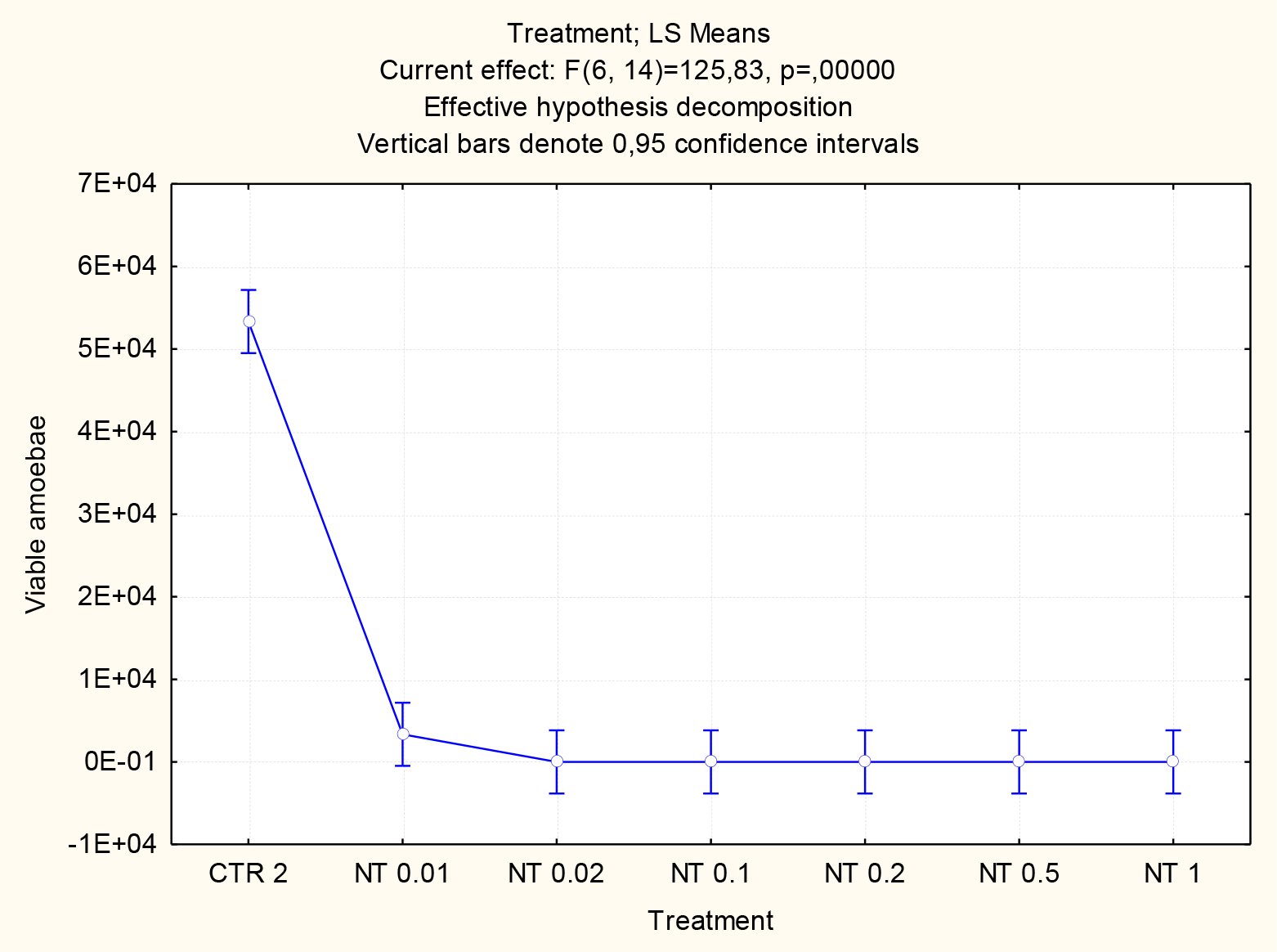
Silver nitrate is an inorganic salt that is active against a wide range of microorganisms, mainly bacteria and fungi. The emergence of antibiotics and new medications led to the abandonment of silver nitrate by modern medicine, however microbial resistance to existing medications led to the re-emergence of the use of silver nitrate by the scientific community. Amoebic keratitis is a serious condition associated to the use of contact lenses. Very often this disease results in blindness and its prevalence is increasing along with the number of lens wearers. It is caused by a free-living amoeba belonging to the genus Acanthamoeba. Currently, amoebic keratitis has no specific treatment and what it is available is quite prolonged and requires extreme discipline from the patient. In this study the trophozoites were cultured in PYG medium and subsequently incubated with different concentrations of silver nitrate. Trophozoite viability was assessed after 120 hours using the Trypan blue exclusion test. Morphological changes were evaluated by optical and transmission electron microscopy. .Concentrations above 0.1 mg/mL of silver nitrate have an amoebicidal effect in vitro in 5 days of incubation; a concentration of 1 mg/mL is capable of causing the death of more than 90% of the amoebas tested, in just 15 minutes. The results obtained suggest that silver nitrate-based eye drops can help or form part of a treatment against amoebic keratitis. In conclusion, silver nitrate showed an important amoebicidal effect, in the concentration range of 0.1 to 1 mg/mL.
Keywords:
Amoebic keratitis; Acanthamoeba; contact lenses.
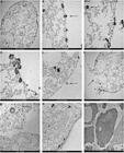
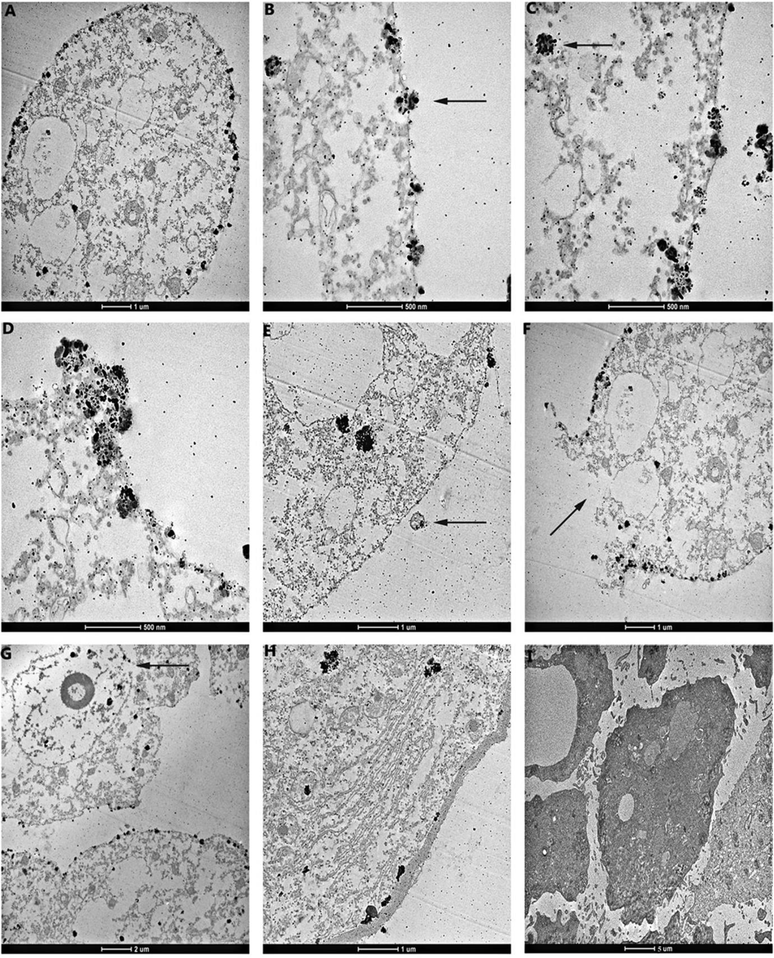
Electron microscopy has shown that silver nitrate promotes membrane rupture
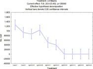
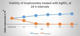
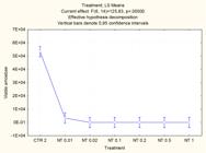
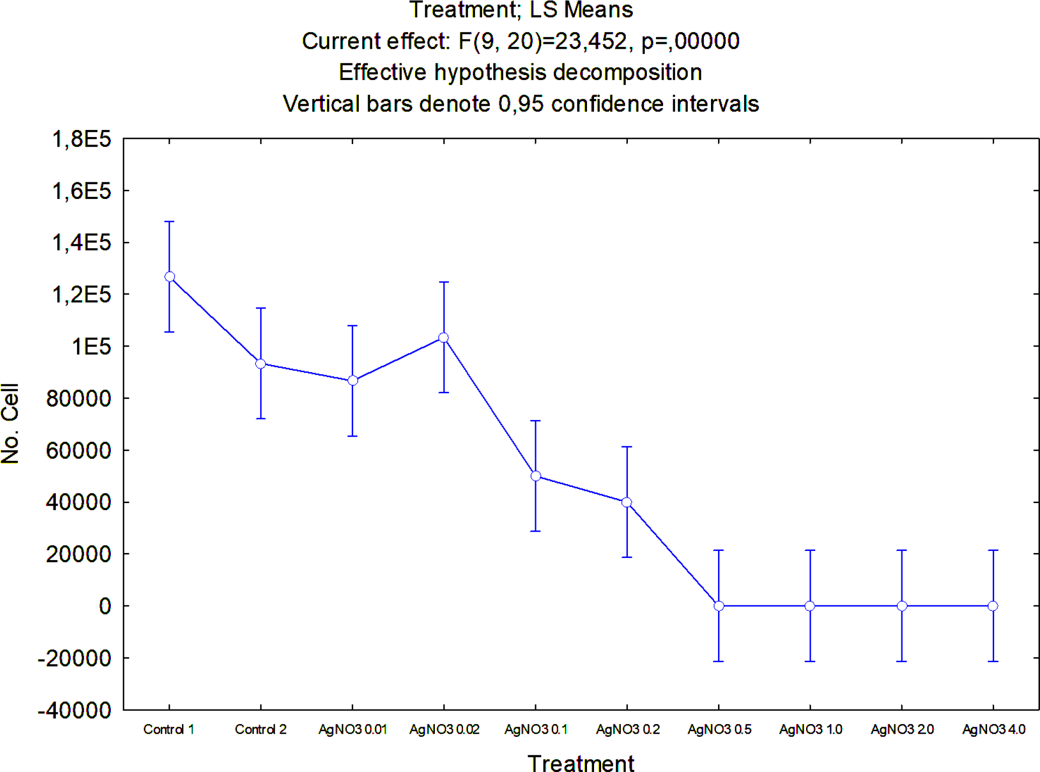
Concentrations above 0.1 mg/mL of silver nitrate have an amoebicidal effect;
1 mg/mL is capable of causing the death of more than 90% of the amoebas tested.
 Thumbnail
 Thumbnail
 Thumbnail
 Thumbnail
 Thumbnail
 Thumbnail
 Thumbnail
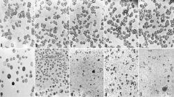
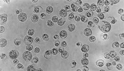
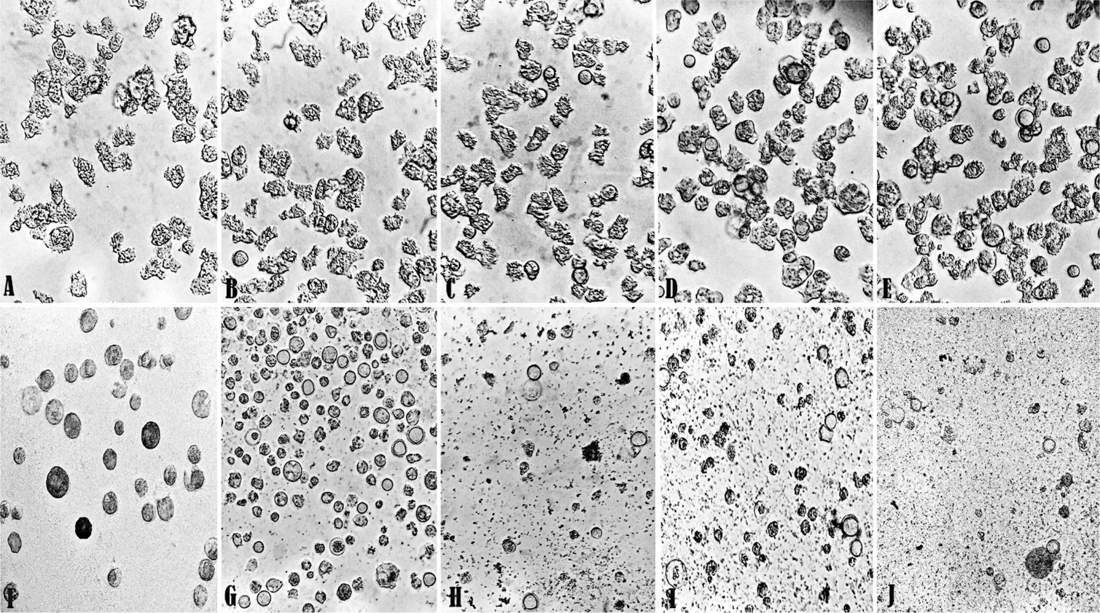 Legend: (A) control group at 24 hours; (B) control group at 48 hours; (C) control group at 72 hours; (D) control group at 96 hours; (E) control group at 120 hours; (F) trophozoites with 0.5 mg/mL of AgNO3 at 24 hours; (G) trophozoites with 0.5 mg/mL of AgNO3 at 48 hours; (H) trophozoites with 0.5 mg/mL of AgNO3 at 72 hours; (I) trophozoites with 0.5 mg/mL of AgNO3 at 96 hours; (J) trophozoites with 0.5 mg/mL of AgNO3 at 120 hours.
 Legend:
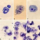
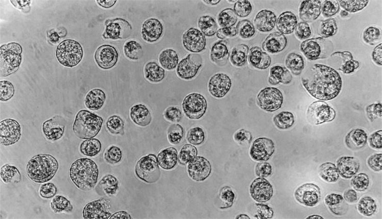
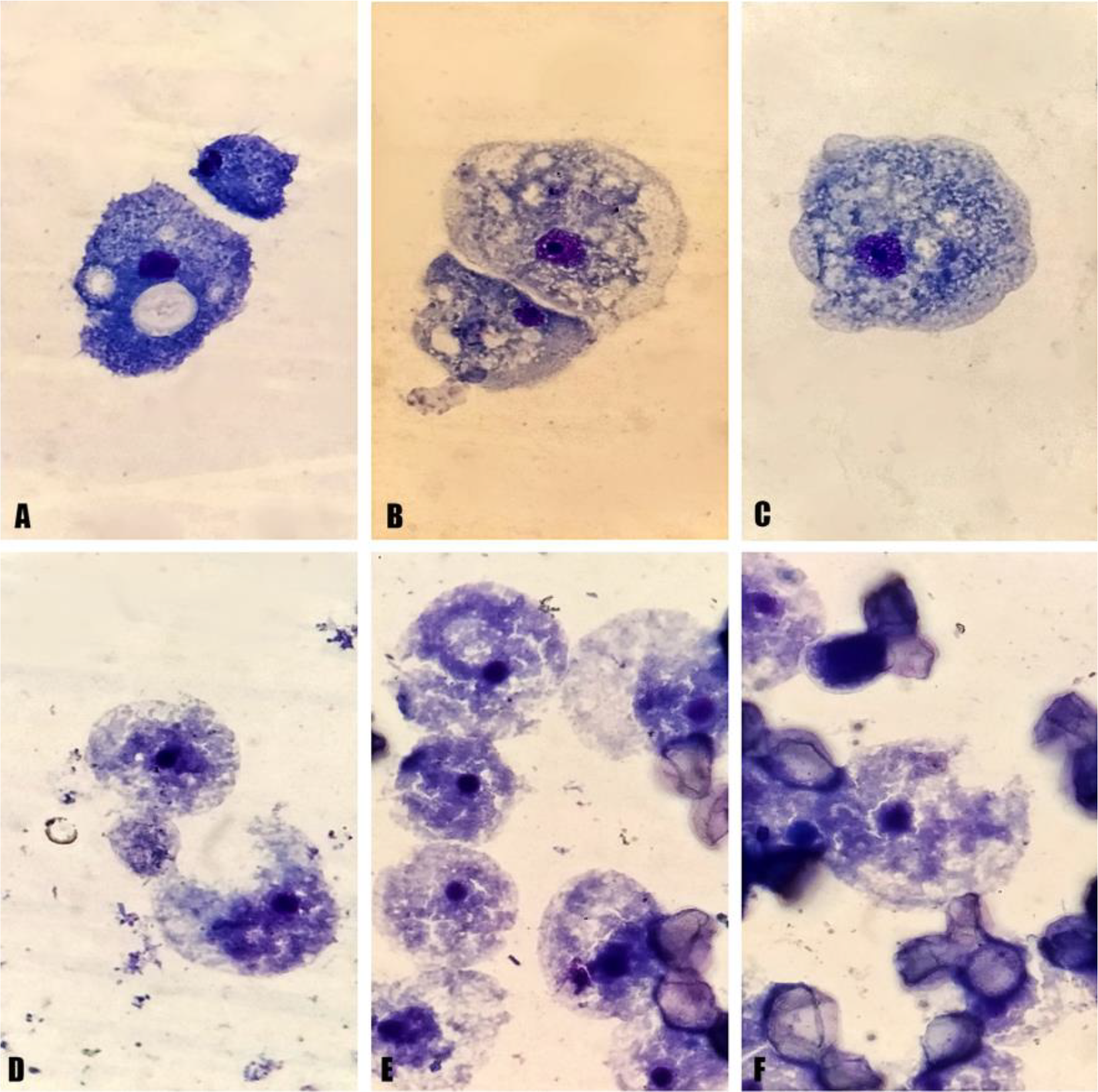
Sections A to C represent untreated trophozoites (CTR) with their morphology unchanged. Sections D to F represent trophozoites, after treatment with AgNO3 at 1 mg/mL, with ruptured cell membranes and cytoplasm; the cysts apparently show no changes.
Legend:
Sections A to C represent untreated trophozoites (CTR) with their morphology unchanged. Sections D to F represent trophozoites, after treatment with AgNO3 at 1 mg/mL, with ruptured cell membranes and cytoplasm; the cysts apparently show no changes.
 Legend
: (A) trophozoite showing complete cellular degradation after 30 minutes of incubation with AgNO3. (B) section showing a fragment of the trophozoite membrane with silver impregnation in specific sites (arrow). (C) vesicle impregnated with silver inside the trophozoite (arrow). (D) membrane protuberances impregnated with silver. (E) membrane vesicle impregnated with silver inside, in the extracellular environment (arrow). (F) rupture of the cell membrane caused by AgNO3 (arrow). (G) rupture of the nuclear membrane (arrow). (H) cyst showing cytoplasmic degradation and accumulation of silver in the cytoplasm. (I) untreated trophozoite (control) showing perfect cellular structure.
Legend
: (A) trophozoite showing complete cellular degradation after 30 minutes of incubation with AgNO3. (B) section showing a fragment of the trophozoite membrane with silver impregnation in specific sites (arrow). (C) vesicle impregnated with silver inside the trophozoite (arrow). (D) membrane protuberances impregnated with silver. (E) membrane vesicle impregnated with silver inside, in the extracellular environment (arrow). (F) rupture of the cell membrane caused by AgNO3 (arrow). (G) rupture of the nuclear membrane (arrow). (H) cyst showing cytoplasmic degradation and accumulation of silver in the cytoplasm. (I) untreated trophozoite (control) showing perfect cellular structure.