Synthesis and Characterization of Membranes Produced from Gelatin and Poly (ethylene glycol)
Abstract
This study focuses on the development and characterization of gelatin-based membranes with varying concentrations of poly(ethylene glycol) (PEG) as potential cost-effective skin substitutes for burn wound treatment. In lowand middle-income countries, where burn injuries pose a significant public health challenge, affordable solutions are crucial. Gelatin/PEG membranes were prepared with PEG concentrations ranging from 3% to 30%. Fourier Transform Infrared Spectroscopy (FT-IR) analysis demonstrated an interaction between gelatin and PEG, indicating cohesive integration through hydrogen bonding, while mechanical tests showed an initial enhancement in Young’s modulus up to a 10% PEG concentration, suggesting improved interaction. Swelling tests revealed that higher PEG concentrations led to increased swelling capacity and reduced mass loss. Scanning electron micrographs supported these findings, showing an irregular surface and the formation of PEG agglomerates, which contributed to improved mechanical resistance. These gelatin-PEG membranes offer a promising avenue for cost-effective skin substitutes, addressing the need for accessible solutions in burn wound care, especially in resource-constrained settings. Further research and clinical validation are essential to assess their efficacy and safety in practical applications, potentially revolutionizing burn wound treatment.
Keywords:
Polyethylene glycols; Gelatin; Burns; Wound healing.
HIGHLIGHTS
Gelatin/PEG membranes for skin substitutes.
Cost-effective solution for burn wound treatment.
INTRODUCTION
Burns represent a serious public health issue with significant social and economic impacts. According to the World Health Organization (WHO), a large proportion of fatal burn injuries occur in lowand middle-income countries. Although most burns are caused by open flames, a substantial number of cases also result from scalds (hot liquids) and chemical exposure [1,2].
The treatment of burn injuries is generally highly complex, as burns are classified into three degrees of severity: first, second, and third. Each degree requires a specific therapeutic approach. Common treatments range from the application of medications to reduce pain and hydrate the affected area, to the use of skin grafts, which may be either partial or full-thickness. Currently, a wide variety of skin substitutes are available, with different applications. These can be of biological, biosynthetic, or synthetic origin (tissue engineering) and serve to replace the skin either temporarily or permanently, thereby reducing the risk of infection [1-3].
Several polymers are used in the manufacture of skin dressings, including poly(glycolic acid), poly(lactic acid), poly(acrylic acid), poly(ɛ-caprolactone), poly(vinyl pyrrolidone), poly(vinyl alcohol), and poly(ethylene glycol), due to their biocompatible properties. In the present study, commercially available gelatin, commonly found in supermarkets, was used. This type of gelatin contains a small amount of collagen and can promote dermal regeneration in burned areas at a low cost. Gelatin-based materials are known for their low toxicity and low antigenicity [4,5]. Another advantage of gelatin is its ability to be combined with other natural polymers, such as chitosan, to produce bioactive composites for use as skin dressings. Furthermore, poly(ethylene glycol) was incorporated to enhance the mechanical properties of the gelatin-based membrane [6].
According to the WHO [7], the number of deaths caused by burns is estimated at around 300,000 per year worldwide, resulting from fires and accidents involving electrical, chemical, radiation, and other agents. Burn wounds are among the most difficult to treat due to issues such as dehydration of the injured area and extensive tissue damage, which compromise several vital functions of the skin [8].
Wound healing is the primary focus in treating skin injuries caused by burns, given the wide variety of clinical cases. The general goal is to achieve physiological healing and repair the injuries without compromising the patient’s functional abilities and aesthetics [8].
Currently, numerous devices for partial or total replacement of the dermis have been developed to assist in tissue repair and regeneration. These dressings are designed to absorb exudate from wounds, thereby maintaining a moist environment that facilitates the debridement of necrotic tissue and promotes spontaneous re-epithelialization of the skin [9,10]. Membranes of poly(ethylene glycol) and gelatin have been shown to improve wound healing in rats, maintaining wound hydration and gas permeability [11].
INTEGRA® is a commercially available dermal regeneration membrane that can be applied in plastic and reconstructive surgeries. It consists of permanent internal components and temporary external components, composed of a porous matrix of collagen and glycosaminoglycan. The temporary layer is made of silicone and functions to prevent fluid loss from wounds, while also acting as a barrier against microbial entry. Despite its effectiveness in treating skin lesions, the high cost of this material limits its feasibility for use within public health systems [12,13]. Previous studies often used more complex and consequently more expensive formulations, such as adding other polymers, like chitosan, and employing acrylates, thiol-acrylate or even enzymatic crosslinking [14,15]. Another factor to consider is the use of commercial-grade gelatin instead of reagent-grade gelatin, further helping to reduce costs, while also avoiding toxic solvents and presenting a single-step synthesis limited to non-toxic and biodegradable gelatin and poly(ethylene glycol) [16,17]. These characteristics show good potential for scalability [14,15,18]. Therefore, the main focus of this study is to obtain a low-cost material and features that make it suitable for use as a skin substitute, thus enabling its application.
MATERIAL AND METHODS
The gelatin used was commercial, powdered, colorless and flavorless (Dr.Oetker®). Poly(ethylene glycol) (PEG) used with a molar mass value of 6000 in analytical grade (Fluka® analytical).
The membranes were produced from commercially available food-grade gelatin, adapted from [19], with varying concentrations of PEG 6000. Initially, 15 g of gelatin were dissolved in 70 mL of water at 60 °C. The solution was homogenized and poured into a petri dish to form the pure gelatin membrane. The material was then dried in an oven at 35 °C for approximately 12 hours. After this step, the membranes were packaged and cooled for 24 hours. The gelatin/PEG membranes were prepared following the same procedure as the pure gelatin membrane, with PEG added at concentrations of 3, 5, 7, 10, 20, and 30% by weight relative to the gelatin.
Characterization of membranes
Fourier Transform Infrared Spectroscopy (FTIR)
The absorption spectra of the membranes in the Fourier Transform Infrared (FTIR) region were obtained using a Perkin Elmer Spectrum 65 spectrophotometer with a resolution of 4 cm⁻1, over the range of 4000 to 600 cm⁻1. The measurements were performed using the attenuated total reflectance (ATR) technique with a ZnSe crystal
Mechanical Testing
For the mechanical tensile test, an Instron EMIC 23-30 testing machine equipped with 30 kN load cells was used. The test was conducted at a traction speed of 20 mm/min, with a clamp separation of 30 mm, at room temperature. The specimens measured 50 mm in length, 10 mm in width, and approximately 2 mm in thickness, and were held by a pair of articulated front clamping jaws, model GR018.
Scanning electron microscopy (SEM)
Photomicrographs of the membrane surfaces were obtained using a ZEISS LEO 400 scanning electron microscope (Cambridge, England) equipped with an OXFORD detector (model 7060), operating at an accelerating voltage of 20 kV. The samples were sputter-coated with gold prior to imaging.
Swelling test
Swelling tests were performed in duplicate. Initially, the dry samples were weighed and transferred to Falcon tubes containing phosphate-buffered saline (PBS) solution. The tubes were then placed in a water bath at 30 °C, and the sample masses were periodically measured over the course of 1 hour.
RESULTS AND DISCUSSION
Fourier Transform Infrared Spectroscopy (FTIR)
Figure 1 illustrates the spectra obtained for the pure gelatin membrane, the gelatin/PEG membrane, and the pure PEG membrane. In the spectra of gelatin, a peak near 3350 cm⁻1 can be observed, which is attributed to the stretching of the OH bond [20-22]. The broad stretching band observed at 3287 cm⁻1 is mainly attributed to N-H and O-H stretching vibrations. Its broadening and slight shift are indicative of hydrogen bonding between gelatin and PEG, where hydroxyl groups from PEG interact with amide and amino groups in the gelatin matrix [19,23]. Considering that gelatin contains collagen in its structure, characteristic bands can be observed at 1658 cm⁻1 (amide I), 1552 cm⁻1 (amide II), and 1235 cm⁻1, attributed to C-N stretching [24,25]. In turn, PEG exhibits characteristic bands near 3000 cm⁻1 corresponding to the C-H bonds of the CH group [26,27]. The band at 2885 cm⁻1 corresponds to the C-H stretching of the CH₂ group, while the band at 1762 cm⁻1 is attributed to the stretching of the C=O group [26-31]. It is noted that in the spectra corresponding to concentrations of 7%, 10%, and 20%, a small peak appears near 1720 cm⁻1, indicating an interaction between gelatin and PEG at these concentrations. The appearance of the shoulder at ~1720 cm⁻1 suggests interaction between the C=O group of PEG and the amide groups of gelatin, consistent with partial miscibility in the polymer blend. Similar behavior was observed by Kolhe and Kannan [27], who reported hydrogen bonding and good miscibility in PEG/chitosan systems analyzed by FTIR spectroscopy. This supports the cohesive integration of PEG within the gelatin matrix up to 10% content.
FTIR spectra of pure gelatin membrane, pure polyethylene glycol (PEG), and gelatin membranes with different PEG concentrations.
Mechanical testing
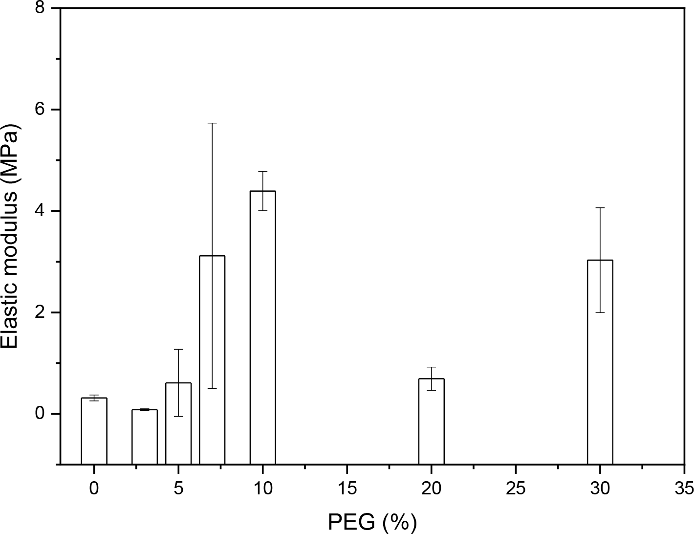
An important requirement for an ideal membrane used as a dressing is that it must exhibit high elongation at break and good tensile strength [11]. Figure 2 shows that, as the PEG concentration in the gelatin matrix increases, the elastic modulus of the membranes also increases compared to the pure gelatin membrane, up to a PEG concentration of 10%, after which it decreases.
This increase may be related to improved interaction between PEG and gelatin [32,33], as evidenced in Figure 1 by the appearance of a peak at 1720 cm⁻1 in gelatin membranes with 7% and 10% PEG, this interaction is present. However, the sample with 20% PEG did not exhibit this behavior, which could be related to improper homogenization with the gelatin and the absence of interaction between them.
According to Zaman and coauthors [11] wound dressings require an optimal balance between tensile strength (typically above 0.5 MPa) and elasticity to accommodate body movements without mechanical failure. The membrane containing 10% PEG exhibited a modulus within this desirable range, indicating potential clinical applicability. When compared to commercial products such as Integra® or biopolymer-based membranes like PEG/chitosan blends, the developed material demonstrated comparable or even superior swelling capacity (swelling ratio >500%), which is advantageous for exudate absorption and for maintaining a moist environment conducive to wound healing.
Swelling test
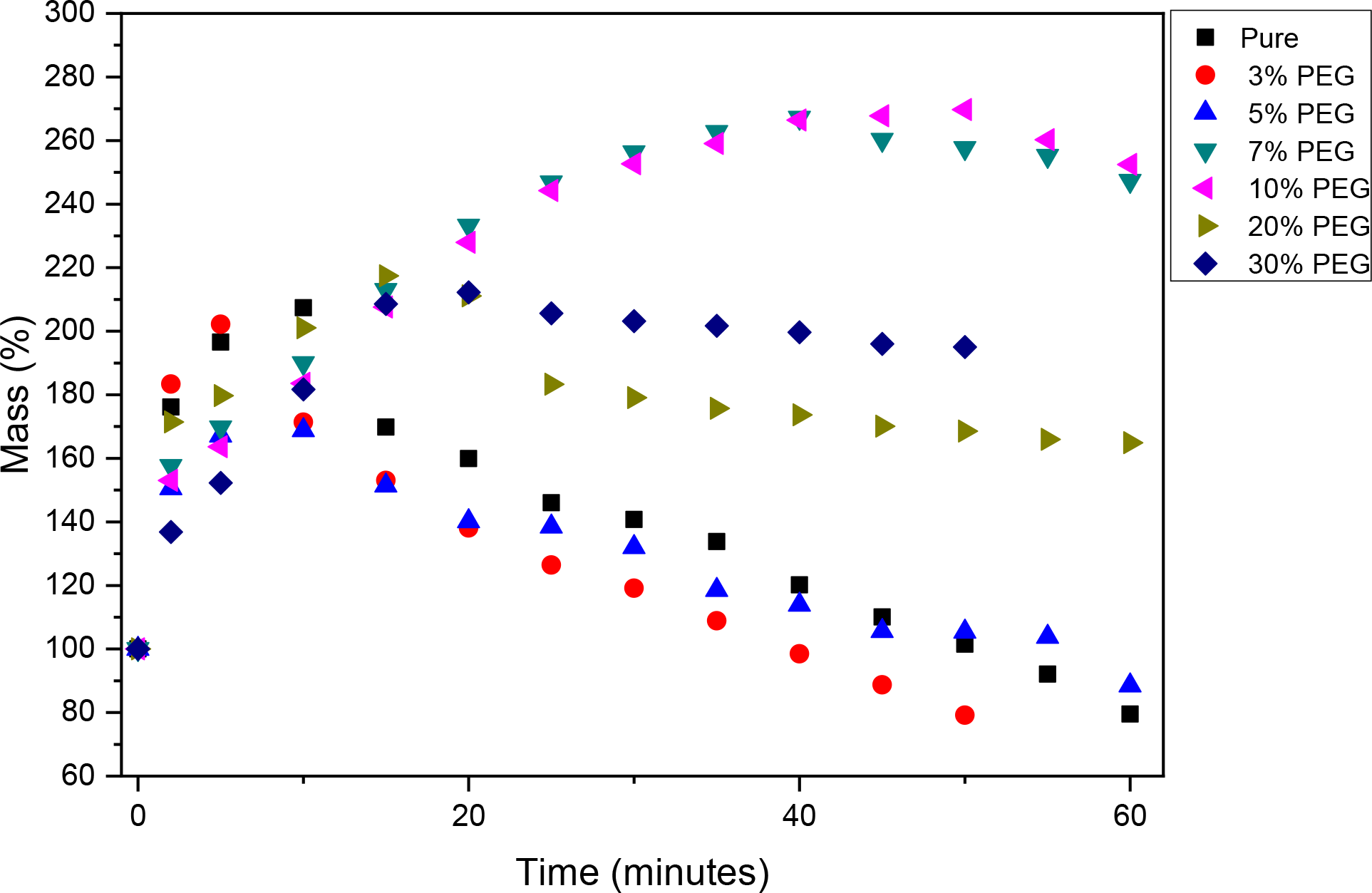
Figure 3 illustrates the membrane swelling test. It is observed that membranes with the lowest PEG concentrations (3% and 5%) showed the greatest mass loss, following the behavior of the pure gelatin membrane. This occurs because gelatin is quite soluble in water and, with PEG present at low concentrations and lacking interaction with gelatin as shown in Figure 1, the gelatin is quickly leached into the PBS solution [34-36]. In turn, the membranes with the highest PEG concentrations (7%, 10%, and 30%) exhibited the greatest swelling capacity due to better interaction between gelatin and PEG, thereby preventing the gelatin from being leached into the PBS solution.

These results also provide indirect evidence of the membranes’ hydrophilicity, as the high swelling capacity observed, particularly for the 10% PEG formulation, suggests an enhanced affinity for aqueous environments. This is a desirable feature for wound dressings, as it allows efficient absorption of exudates and contributes to maintaining a moist environment favorable for healing. Furthermore, the irregular surface observed in SEM analysis may enhance surface area and permeability, which are important for the transport of fluids and nutrients. Although direct measurements of hydrophilicity (e.g., contact angle) and transport properties were not performed in the present study, future work will address these aspects to further validate the clinical potential of the membranes.
Scanning electron microscopy (SEM)
The scanning electron micrographs of the material surfaces are shown in Figure 4. They reveal that the addition of poly(ethylene glycol) made the surface of the pure gelatin membrane less smooth. Furthermore, it is observed that PEG forms small agglomerates within the gelatin matrix, which act as fillers, particularly in the sample with 10% PEG, working synergistically with the interaction between gelatin and PEG [32], as mentioned in Figure 2, this promotes an increase in the mechanical resistance of the membrane [37,38]. It is also observed that the 10% PEG sample exhibited greater surface irregularity, having the largest surface area among the microscopically analyzed samples. This finding corroborates the swelling test results, which showed that the 10% PEG sample had the highest swelling capacity. Thus, a correlation was established between the greater swelling capacity and the increased surface irregularity of the sample.
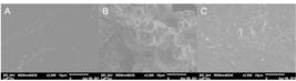
Scanning electron micrographs of gelatin membranes: (A) pure membrane without PEG; (B) membrane with 10% PEG; (C) membrane with 30% PEG.
Considering both the swelling behavior and the mechanical performance of the membranes, the sample containing 10% PEG proved to be the most promising formulation for potential biomedical applications. This is due to its high elastic modulus, which indicates greater mechanical integrity, as well as its superior swelling capacity among the investigated samples, a desirable feature in applications involving wound dressings. Furthermore, SEM images showed a significant increase in surface irregularities, as well as the formation of agglomerates in the sample containing 10% PEG, which may have contributed to the aforementioned properties. Taken together, these characteristics suggest that the membrane with 10% PEG has potential for use as a temporary skin wound dressing, such as for burn treatment, especially in resource-limited settings [39,40].
CONCLUSION
In conclusion, the development and characterization of gelatin-based membranes with varying PEG concentrations demonstrated promising potential as cost-effective and efficient skin substitutes. Fourier Transform Infrared Spectroscopy (FT-IR) analysis confirmed the interaction between gelatin and PEG, indicating cohesive integration of the materials. Mechanical tests showed an initial increase in elastic modulus with PEG concentrations up to 10%, suggesting improved interaction between components. However, concentrations above 10% exhibited a decline, possibly due to inadequate homogenization or reduced interaction.
This study contributes to ongoing efforts in wound healing and skin regeneration by addressing the need for affordable solutions, particularly in lowand middle-income countries where burns represent a significant public health concern. Further research and clinical trials are necessary to validate the efficacy and safety of these membranes in real-world applications, ultimately paving the way for accessible and effective burn wound treatments. It is worth mentioning that the work focuses on the physicochemical characterization of the membranes and their mechanical properties, with great potential, being a preliminary step for future studies. We recommend that biocompatibility tests such as cytotoxicity, cell adhesion and proliferation, and antimicrobial tests be performed, as we have limitations to perform these tests.
-
Funding:
This study was fnanced in part by the Coordenação de Aperfeiçoamento de Pessoal de Nível Superior -Brasil (CAPES) - Finance Code 001.
Acknowledgments:
The authors would like to thank the Pontifical Catholic University of São Paulo (PUC-SP), Sorocaba campus, for supporting the development of the work in the laboratory, as well as the characterizations.
Data Availability Statement:
Research data are only available upon request for corresponding author.
REFERENCES
-
1 Peck MD. Epidemiology of burns throughout the world. Part I: Distribution and risk factors. Burns. 2011;37:1087-100. https://doi.org/10.1016/j.burns.2011.06.005
» https://doi.org/10.1016/j.burns.2011.06.005 -
2 Peck MD. Epidemiology of burns throughout the world. Part II: Intentional burns in adults. Burns. 2012;38:630-7. https://doi.org/10.1016/j.burns.2011.12.028
» https://doi.org/10.1016/j.burns.2011.12.028 -
3 Ferreira MC, Paggiaro AO, Isaac C, Teixeira Neto N, Santos GB. Skin substitutes: current concepts and a new classification system. Rev Bras Cir Plást. 2011;26:696-702. https://doi.org/10.1590/S1983-51752011000400028
» https://doi.org/10.1590/S1983-51752011000400028 -
4 Ponticiello M, Schinagl R, Kadiyala S, Barry F. Gelatin-based resorbable sponge as a carrier matrix for human mesenchymal stem cells in cartilage regeneration therapy. J Biomed Mater Res. 2000;52:246-55. https://doi.org/10.1002/1097-4636(200011)52:23.0.CO;2-W
» https://doi.org/10.1002/1097-4636(200011)52:23.0.CO;2-W -
5 Alves LR, Carriello GM, Pegoraro GM, Filho JF. [Applications of hydrogels as biomaterials: a review of Brazilian dissertations and theses since 2017]. Discip Sci Nat Technol. 2021;22:53-79. https://doi.org/10.37779/nt.v22i2.4031
» https://doi.org/10.37779/nt.v22i2.4031 -
6 Mogoşanu GD, Grumezescu AM. Natural and synthetic polymers for wounds and burns dressing. Int J Pharm. 2014;463:127-36. https://doi.org/10.1016/j.ijpharm.2013.12.015
» https://doi.org/10.1016/j.ijpharm.2013.12.015 -
7 Peck M, Molnar J, Swart D. A global plan for burn prevention and care. Bull World Health Organ. 2009;87:802-3. https://doi.org/10.1590/S0042-96862009001000018
» https://doi.org/10.1590/S0042-96862009001000018 -
8 Jeschke MG, van Baar ME, Choudhry MA, Chung KK, Gibran NS, Logsetty S. Burn injury. Nat Rev Dis Primers. 2020;6:11. https://doi.org/10.1038/s41572-020-0145-5
» https://doi.org/10.1038/s41572-020-0145-5 -
9 Rousselle P, Braye F, Dayan G. Re-epithelialization of adult skin wounds: Cellular mechanisms and therapeutic strategies. Adv Drug Deliv Rev. 2019;146:344-65. https://doi.org/10.1016/j.addr.2018.06.019
» https://doi.org/10.1016/j.addr.2018.06.019 -
10 Abdelrahman T, Newton H. Wound dressings: principles and practice. Surgery (Oxford). 2011;29:491-5. https://doi.org/10.1016/j.mpsur.2011.06.007
» https://doi.org/10.1016/j.mpsur.2011.06.007 -
11 Zaman HU, Islam JMM, Khan MA, Khan RA. Physico-mechanical properties of wound dressing material and its biomedical application. J Mech Behav Biomed Mater. 2011;4:1369-75. https://doi.org/10.1016/j.jmbbm.2011.05.007
» https://doi.org/10.1016/j.jmbbm.2011.05.007 -
12 Ozgonul C, Grisolia ABD, Demirci H. The use of Integra® dermal regeneration template for the orbital exenteration socket: a novel technique. Ophthalmic Plast Reconstr Surg. 2018;34:64-7. https://doi.org/10.1097/IOP.0000000000000869
» https://doi.org/10.1097/IOP.0000000000000869 -
13 Schiavon M, Francescon M, Drigo D, Salloum G, Baraziol R, Tesei J, et al. The use of Integra dermal regeneration template versus flaps for reconstruction of full-thickness scalp defects involving the calvaria: a cost-benefit analysis. Aesthet Plast Surg. 2016;40:901-7. https://doi.org/10.1007/s00266-016-0703-0
» https://doi.org/10.1007/s00266-016-0703-0 -
14 Xu K, Cantu DA, Fu Y, Kim J, Zheng X, Hematti P, et al. Thiol-ene Michael-type formation of gelatin/poly(ethylene glycol) biomatrices for three-dimensional mesenchymal stromal/stem cell administration to cutaneous wounds. Acta Biomater. 2013;9:8802-14. https://doi.org/10.1016/j.actbio.2013.06.021
» https://doi.org/10.1016/j.actbio.2013.06.021 -
15 Hwang J, Thi PL, Lee S, Park EH, Lee E, Kim E, et al. Injectable gelatin-poly(ethylene glycol) adhesive hydrogels with highly hemostatic and wound healing capabilities. J Ind Eng Chem. 2022;109:372-83. https://doi.org/10.1016/j.jiec.2022.02.019
» https://doi.org/10.1016/j.jiec.2022.02.019 -
16 Gaballa SA, Naguib Y, Mady FM, Khaled KA. Polyethylene glycol: properties, applications, and challenges. J Adv Biomed Pharm Sci. 2024;7:26-36. https://doi.org/10.21608/jabps.2023.241685.1205
» https://doi.org/10.21608/jabps.2023.241685.1205 -
17 Said NS, Howell N, Sarbon NM. A review on potential use of gelatin-based film as active and smart biodegradable films for food packaging application. Food Rev Int. 2023;39:1063-85. https://doi.org/10.1080/87559129.2021.1929298
» https://doi.org/10.1080/87559129.2021.1929298 -
18 Ravanfar K, Amniattalab A, Mohammadi R. Curcumin-polyethylene glycol loaded on chitosan-gelatin nanoparticles enhances burn wound healing in rat. J Burn Care Res. 2022;43:1399-409. https://doi.org/10.1093/jbcr/irac048
» https://doi.org/10.1093/jbcr/irac048 -
19 Biscarat J, Charmette C, Masquelez N, Sanchez J, Pochat-Bohatier C. Crosslinking of gelatin membranes with ferulic acid or glutaraldehyde: relationship between gas permeability and renaturation level of gelatin triple helices. J Polym Sci B Polym Phys. 2015;53:280-7. https://doi.org/10.1002/polb.23628
» https://doi.org/10.1002/polb.23628 -
20 Das RS, Warkhade SK, Kumar A, Wankhade AV. Graphene oxide-based zirconium oxide nanocomposite for enhanced visible light-driven photocatalytic activity. Res Chem Intermed. 2019;45:1689-705. https://doi.org/10.1007/s11164-018-3699-z
» https://doi.org/10.1007/s11164-018-3699-z -
21 Chan SY, Choo WS, Young DJ, Loh XJ. Thixotropic supramolecular pectin-poly(ethylene glycol) methacrylate (PEGMA) hydrogels. Polymers (Basel). 2016;8:404. https://doi.org/10.3390/polym8110404
» https://doi.org/10.3390/polym8110404 -
22 Rutnakornpituk M, Ngamdee P, Phinyocheep P. Synthesis, characterization and properties of chitosan modified with poly(ethylene glycol)-polydimethylsiloxane amphiphilic block copolymers. Polymer (Guildf). 2005;46:9742-52. https://doi.org/10.1016/j.polymer.2005.08.051
» https://doi.org/10.1016/j.polymer.2005.08.051 -
23 Afshari M, Pourmadadi M, Shabestari SM, Abdouss H, Abdouss M. Development and characterization of biocompatible gelatin/polyethylene glycol/copper-doped carbon quantum dots@curcumin nanocarriers for pH-sensitive curcumin delivery in glioblastoma treatment. Inorg Chem Commun. 2025;179:114782. https://doi.org/10.1016/j.inoche.2025.114782
» https://doi.org/10.1016/j.inoche.2025.114782 -
24 Yaylayan VA, Harty-Majors S, Ismail AA. Monitoring carbonyl-amine reaction and enolization of 1-hydroxy-2-propanone (acetol) by FTIR spectroscopy. J Agric Food Chem. 1999;47:2335-40. https://doi.org/10.1021/jf9812836
» https://doi.org/10.1021/jf9812836 -
25 Deus VL, Resende LM, Bispo ES, Franca AS, Gloria MBA. FTIR and PLS-regression in the evaluation of bioactive amines, total phenolic compounds and antioxidant potential of dark chocolates. Food Chem. 2021;357:129754. https://doi.org/10.1016/j.foodchem.2021.129754
» https://doi.org/10.1016/j.foodchem.2021.129754 -
26 Alves LR, Carriello GM, Pegoraro GM, Lopes HSM, Janolla TA, Dias ANC, et al. Synthesis and characterization of polyurethane and samarium(III) oxide and holmium(III) oxide composites. Polímeros. 2023;33:e20230039. https://doi.org/10.1590/0104-1428.20230023
» https://doi.org/10.1590/0104-1428.20230023 -
27 Kolhe P, Kannan RM. Improvement in ductility of chitosan through blending and copolymerization with PEG: FTIR investigation of molecular interactions. Biomacromolecules. 2003;4:173-80. https://doi.org/10.1021/bm025689+.
» https://doi.org/10.1021/bm025689 -
28 Pegoraro GM, Mourato AS, Carriello GM, Mambrini GP. Use of FTIR for preliminary analysis of the conversion rate of oleic acid to ethyl oleate. Disciplinarum Scientia Nat Tecnol. 2022;23:85-97. https://doi.org/10.37779/nt.v23i2.4209
» https://doi.org/10.37779/nt.v23i2.4209 -
29 Xing J, Deng L, Xie C, Xiao L, Zhai Y, Jin F, et al. Methoxy poly(ethylene glycol)-b-poly(octadecanoic anhydride)-b-methoxy poly(ethylene glycol) amphiphilic triblock copolymer nanoparticles as delivery vehicles for paclitaxel. Polym Adv Technol. 2011;22:669-74. https://doi.org/10.1002/pat.1563
» https://doi.org/10.1002/pat.1563 -
30 Zamiri MA, Kargari A, Sanaeepur H. Ethylene vinyl acetate/poly(ethylene glycol) blend membranes for CO2/N2 separation. Greenh Gases Sci Technol. 2015;5:668-81. https://doi.org/10.1002/ghg.1513
» https://doi.org/10.1002/ghg.1513 -
31 Jiao Y, Fu Y, Jiang Z. The synthesis and characterization of poly(ethylene glycol) grafted on pullulan. J Appl Polym Sci. 2004;91:1217-21. https://doi.org/10.1002/app.13238
» https://doi.org/10.1002/app.13238 -
32 Zhang J, Wang J, Zhang H, Lin J, Ge Z, Zou X. Macroporous interpenetrating network of polyethylene glycol (PEG) and gelatin for cartilage regeneration. Biomed Mater. 2016;11:035014. https://doi.org/10.1088/1748-6041/11/3/035014
» https://doi.org/10.1088/1748-6041/11/3/035014 -
33 Su J, Satchell SC, Wertheim JA, Shah RN. Poly(ethylene glycol)-crosslinked gelatin hydrogel substrates with conjugated bioactive peptides influence endothelial cell behavior. Biomaterials. 2019;201:99-112. https://doi.org/10.1016/j.biomaterials.2019.02.001
» https://doi.org/10.1016/j.biomaterials.2019.02.001 -
34 Gullapalli RP, Mazzitelli CL. Polyethylene glycols in oral and parenteral formulations-a critical review. Int J Pharm. 2015;496:219-39. https://doi.org/10.1016/j.ijpharm.2015.11.015
» https://doi.org/10.1016/j.ijpharm.2015.11.015 -
35 Lee JM, Chan ES, Nagasundara Ramanan R, Ooi CW. Liquid-liquid equilibria of aqueous two-phase systems made of polyethylene glycol and gelatin systems and their application in emulsion formation. Fluid Phase Equilibria. 2020;508:112441. https://doi.org/10.1016/j.fluid.2019.112441
» https://doi.org/10.1016/j.fluid.2019.112441 -
36 Yanagisawa M, Yamashita Y, Mukai S, Annaka M, Tokita M. Phase separation in binary polymer solution: gelatin/poly(ethylene glycol) system. J Mol Liq. 2014;200:2-6. https://doi.org/10.1016/j.molliq.2013.12.035
» https://doi.org/10.1016/j.molliq.2013.12.035 -
37 Mu C, Li X, Guo J, Bi C, Li D. Effects of montmorillonite on the structure and properties of gelatin-polyethylene glycol composite fibers. J Appl Polym Sci. 2013;129:773-8. https://doi.org/10.1002/app.38808
» https://doi.org/10.1002/app.38808 -
38 Raut PW, Shitole AA, Khandwekar A, Sharma N. Engineering biomimetic polyurethane using polyethylene glycol and gelatin for blood-contacting applications. J Mater Sci. 2019;54:10457-72. https://doi.org/10.1007/s10853-019-03643-0
» https://doi.org/10.1007/s10853-019-03643-0 -
39 Sonthithai P, Kaewkong P, Channasanon S, Tanodekaew S. 3D-printed PEG-PLA/gelatin hydrogel: characterization toward in vitro chondrocyte redifferentiation. ACS Biomater Sci Eng. 2025;11:2157-66. https://doi.org/10.1021/acsbiomaterials.4c02409
» https://doi.org/10.1021/acsbiomaterials.4c02409 -
40 Charron PN, Braddish TA, Floreani R. PVA-gelatin hydrogels formed using combined theta-gel and cryo-gel fabrication techniques. J Mech Behav Biomed Mater. 2019;92:90-6. https://doi.org/10.1016/j.jmbbm.2019.01.002
» https://doi.org/10.1016/j.jmbbm.2019.01.002
-
Editor-in-Chief:
Alexandre Rasi Aoki
-
Associate Editor:
Andressa Novatski