Tissue-specific validation of reference genes for accurate qRT-PCR analysis during sex differentiation in Amphiprion ocellaris
Abstract
Quantitative real-time PCR (qRT-PCR) is a widely used method for measuring gene expression, but its reliability depends on using stable reference genes for normalization. This study assessed the expression stability of four commonly used housekeeping genes—18S rRNA, ef1α, rpl8, and β-actin—in Amphiprion ocellaris, a socially regulated protandrous hermaphrodite. Brain and gonadal tissues were sampled from individuals at different developmental stages and sex types, including juveniles, transitional individuals, and paired breeders. To further evaluate tissue-wide stability, 11 different tissues were also analyzed in adult breeders. Gene expression stability was evaluated using RefFinder, which integrates four established algorithms. Among the candidates, 18S rRNA was consistently ranked as the most stable across tissues and analysis methods, while β-actin showed the highest variability and was the least reliable. Validation using two sex differentiation genes, cyp19a1a and cyp19a1b, normalized with 18S rRNA, confirmed biologically consistent and tissue-specific expression patterns. These findings identify 18S rRNA as the most suitable reference gene for qRT-PCR studies in A. ocellaris, with ef1α and rpl8 as potential alternatives in gonadal tissues. Using validated reference genes is essential for accurate gene expression analysis in studies of sex differentiation and reproductive plasticity in sequentially hermaphroditic teleosts.
Keywords:
Amphiprion ocellaris; housekeeping genes; qRT-PCR; reference gene
Resumo
A PCR em tempo real quantitativa (qRT-PCR) é um método amplamente utilizado para medir a expressão gênica, mas sua confiabilidade depende do uso de genes de referência estáveis para a normalização. Este estudo avaliou a estabilidade de expressão de quatro genes de manutenção comumente utilizados — 18S rRNA, ef1α, rpl8 e β-actin — em Amphiprion ocellaris, um hermafrodita protândrico regulado socialmente. Tecidos cerebrais e gonadais foram amostrados de indivíduos em diferentes estágios de desenvolvimento e tipos sexuais, incluindo juvenis, indivíduos em transição e casais reprodutores. Para avaliar mais amplamente a estabilidade entre tecidos, 11 tecidos diferentes também foram analisados em adultos reprodutores. A estabilidade da expressão gênica foi avaliada usando o RefFinder, que integra quatro algoritmos consagrados. Entre os candidatos, o 18S rRNA foi consistentemente classificado como o mais estável entre os tecidos e métodos de análise, enquanto a β-actin apresentou a maior variabilidade e foi o gene menos confiável. A validação utilizando dois genes relacionados à diferenciação sexual, cyp19a1a e cyp19a1b, normalizados com 18S rRNA, confirmou perfis de expressão biologicamente consistentes e específicos por tecido. Esses resultados identificam o 18S rRNA como o gene de referência mais adequado para estudos de qRT-PCR em A. ocellaris, com ef1α e rpl8 como alternativas potenciais para tecidos gonadais. O uso de genes de referência validados é essencial para análises precisas de expressão gênica em estudos sobre diferenciação sexual e plasticidade reprodutiva em teleósteos hermafroditas sequenciais.
Palavras-chave:
Amphiprion ocellaris; genes constitutivos; qRT-PCR; gene de referência
1. Introduction
The false clown anemonefish (Amphiprion ocellaris Cuvier, 1830) is a reef-associated teleost found throughout the Indo-Pacific. Known for its striking coloration, social hierarchy, and ease of captive breeding, it has become a model organism for studies in evolutionary genomics and social behavior (Herrera et al., 2023). One of the most interesting traits of this species is protandrous hermaphroditism. This means the fish starts life as a male and can later become a female. The change usually happens when the dominant female is lost. In a stable group, the top-ranking fish becomes the female. The next in line becomes the breeding male, while the others stay immature (Buston, 2003; Madhu and Madhu, 2006; Parker et al., 2022).
Although interest is growing in understanding the molecular basis of sex change in sequential hermaphrodites, no study has thoroughly validated stable reference genes for gene expression analysis in these species. Without reliable internal controls, qRT-PCR results may be inaccurate, especially in tissues undergoing active transcriptional changes during sex differentiation. In A. ocellaris, this process is regulated by a complex interplay of hormones and genes that drive internal changes, even though males and females often look externally similar, particularly when unpaired or outside a social hierarchy.
Key genes involved in this process include SRY-box transcription factor 9 (sox9), anti-Müllerian hormone (amh), and two aromatase isoforms: cytochrome P450 family 19 subfamily A member 1a (cyp19a1a) and cytochrome P450 family 19 subfamily A member 1b (cyp19a1b). cyp19a1a encodes the gonadal aromatase enzyme responsible for ovarian estrogen production. In contrast, cyp19a1b encodes the brain-specific aromatase, which is mainly expressed in radial glial cells and is involved in the neuroendocrine regulation of sex change (Diotel et al., 2010; Liew et al., 2012).
Quantitative real-time PCR (qRT-PCR) is widely used to quantify gene expression due to its high sensitivity, specificity, and broad dynamic range (Bustin et al., 2009). However, the accuracy of qRT-PCR relies heavily on selecting appropriate reference genes to normalize expression levels across different tissues and experimental conditions. Ideally, reference genes should be stably expressed regardless of tissue type, developmental stage, or treatment (Vandesompele et al., 2002; Small et al., 2008; De Santis et al., 2011; Du et al., 2024). Commonly used candidates include 18S ribosomal RNA (18S rRNA), eukaryotic translation elongation factor 1 alpha (ef1α), ribosomal protein L8 (rpl8), and beta-actin (β-actin), which are often assumed to be uniformly expressed. However, increasing evidence shows that their expression can vary in response to physiological or environmental factors, potentially leading to inaccurate normalization if not empirically validated (Boonphakdee et al., 2019; Dharmaratnam et al., 2021; Jiang et al., 2023; Chen et al., 2024).
Although interest in the molecular mechanisms of sex change in clownfish is increasing, no systematic study has evaluated reference gene stability in A. ocellaris. To address this gap, we assessed the expression stability of four widely used reference genes—18S rRNA, ef1α, rpl8, and β-actin—in brain and gonadal tissues across key developmental stages and social ranks. Stability was evaluated using RefFinder, which integrates four established algorithms: geNorm, NormFinder, BestKeeper, and the comparative ΔCt method. The most stable genes were then used to normalize the expression of cyp19a1a and cyp19a1b, which serve as tissue-specific markers of aromatase activity in gonads and brain, respectively. This study provides a validated framework for accurate gene expression analysis in A. ocellaris and offers valuable insights into the molecular basis of sex differentiation and reproductive plasticity in sequentially hermaphroditic teleosts.
2. Materials and Methods
2.1. Fish sample preparation
False clown anemonefish (A. ocellaris) used in this study were bred from a single founder pair obtained from the Samut Sakhon Coastal Fisheries Research and Development Center, Thailand. All individuals were maintained in a closed recirculating seawater system at the Department of Aquatic Science, Faculty of Science, Burapha University. Water temperature was controlled at 27 ± 1 °C, salinity was maintained between 33 and 35 ppt, and lighting followed a 12:12 h light–dark cycle. Fish were fed twice daily with a combination of commercial marine pellets and live Artemia nauplii, according to the protocol of Thamnawasolos and Boonphakdee (2023).
To investigate reference gene expression across developmental stages and social hierarchies, individuals were sampled at specific time points. The sampling design included: juveniles at 3 months of age (n = 3); unpaired, immature fish at 8 months (n = 9); and 13-month-old adults, consisting of unpaired individuals, females paired for five months, and their male partners (n = 3 per category; total n = 9). A fourth group included 24-month-old adults, comprising unpaired individuals, reproductively active females that had spawned continuously for at least six months, and their male partners (n = 3 per category; total n = 9). Sex and social status were determined through sustained behavioral observations, including assessments of dominance interactions, nesting activity, and reproductive behavior.
Fish were anesthetized using tricaine methanesulfonate (MS-222; 100 mg/L) and euthanized in accordance with institutional animal care protocols. In juveniles, immature individuals, and unpaired adults at both 13 and 24 months of age, only brain and gonadal tissues were collected. In contrast, for reproductively active breeding pairs, an expanded tissue panel was sampled, including liver, stomach, intestine, spleen, caecum, heart, gill, muscle, kidney, brain, and gonad. All tissues were immediately preserved in RNAlater™ (Thermo Fisher Scientific, USA) and stored at −80 °C until RNA extraction. All procedures were approved by the Animal Care and Use Committee of Burapha University (Approval ID: IACUC 040-2561) and conducted in accordance with national animal welfare guidelines (Thamnawasolos and Boonphakdee, 2023).
2.2. RNA extraction and cDNA synthesis
Total RNA was extracted from brain and gonadal tissues of all fish and from 11 tissues in adult breeders using TRIzol® Reagent (Invitrogen, USA), following the manufacturer's protocol. RNA integrity was confirmed via 1% agarose gel electrophoresis, and purity was assessed using a NanoDrop™ spectrophotometer (Thermo Scientific, USA). Only samples with A260/280 ratios between 1.8 and 2.1 were used. Genomic DNA contamination was removed with DNase I (Thermo Scientific, USA). First-strand cDNA was synthesized from 1 µg of RNA using the High-Capacity cDNA Reverse Transcription Kit (Applied Biosystems, USA) and diluted 10-fold with nuclease-free water for qRT-PCR.
2.3. Primer design and validation
Primers for cyp19a1a and cyp19a1b were adopted from Thamnawasolos and Boonphakdee (2023), while primers for 18S rRNA and β-actin were obtained from Boonphakdee et al. (2019). New primers for ef1α and rpl8 were designed using Primer3 based on GenBank sequences (XM_023263215 and XM_055015586, respectively). Primer specificity and efficiency were validated via melting curve analysis, agarose gel electrophoresis, and standard curve generation. Primer details are summarized in Table 1.
Primer sequences and qRT-PCR parameters for reference and target genes in Amphiprion ocellaris. Forward and reverse primer sequences, amplicon sizes (bp), melting temperatures (°C), qRT-PCR amplification efficiencies (%), and correlation coefficients (R2) for all genes analyzed.
2.4. Quantitative real-time PCR (qRT-PCR)
qRT-PCR was performed using a StepOnePlus™ Real-Time PCR System (Applied Biosystems, USA) and Maxima SYBR Green/ROX qPCR Master Mix (Thermo Fisher Scientific, USA). Each 15 µL reaction contained 1 µL of diluted cDNA, 0.3 µM of each primer, and master mix. Cycling conditions were: 95 °C for 10 min, followed by 40 cycles at 95 °C for 15 s, 55 °C for 20 s, and 72 °C for 20 s. Specificity was confirmed through melting curves and gel electrophoresis. Standard curves generated from pooled cDNA revealed amplification efficiencies between 91.3% and 103.2%, with R2 > 0.99.
2.5. Gene expression normalization and stability analysis
Reference gene stability was assessed using RefFinder (https://www.heartcure.com.au/reffinder/), an integrative tool that combines four widely used algorithms: geNorm, NormFinder, BestKeeper, and the comparative ΔCt method. Each of these algorithms applies a distinct mathematical principle to evaluate gene stability. geNorm calculates pairwise variation among genes to identify the most stably expressed candidates (Vandesompele et al., 2002). NormFinder uses a model-based approach that accounts for intra- and inter-group variation to estimate stability scores (Andersen et al., 2004). BestKeeper evaluates the standard deviation and correlation of Ct values to rank gene consistency (Pfaffl et al., 2004). The comparative ΔCt method determines stability by analyzing pairwise differences in Ct values across samples. A comprehensive ranking of candidate genes was generated by calculating the geometric mean of stability scores from all four methods (Švec et al., 2015).
To provide an additional quantitative measure of stability, the coefficient of variation (CV%) was calculated based on raw Ct values. Lower CV% values reflect uniform gene expression across different biological samples (Pfaffl et al., 2004). The formula used for this calculation, CV% = (standard deviation / mean Ct) × 100, is provided in Figure 1.
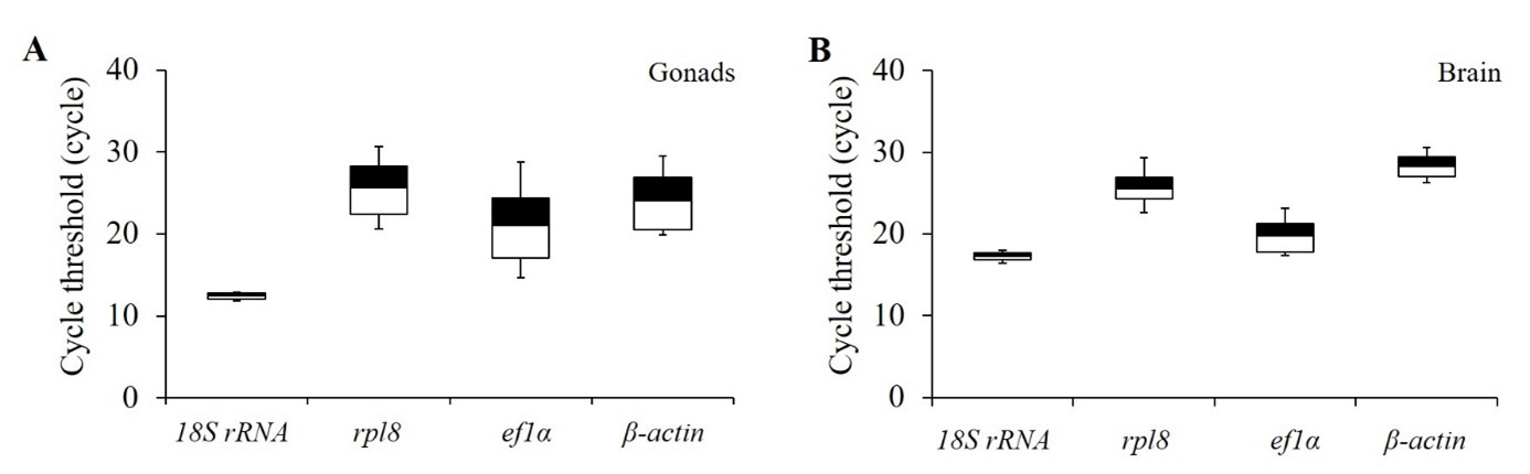
Ct value distribution of candidate reference genes in Amphiprion ocellaris. (A) Distribution of Ct values in gonadal tissue; (B) Distribution of Ct values in brain tissue. Boxplots indicate interquartile range (25th–75th percentiles), medians (horizontal lines), and whiskers representing the 10th and 90th percentiles. Lower Ct variability reflects greater expression stability.
To validate the stability of the selected reference gene, the expression of two key sex-related genes—gonadal aromatase (cyp19a1a) and brain aromatase (cyp19a1b)—was assessed. Gene expression levels were normalized using 18S rRNA, the most stable reference gene identified by RefFinder. Relative expression was calculated using the 2–ΔCt method (Schmittgen and Livak, 2008), and fold changes between groups were determined using the 2–ΔΔCt method (Livak and Schmittgen, 2001), following the approach described in Boonphakdee et al. (2019).
2.6. Statistical analysis
Quantitative real-time PCR (qRT-PCR) results are presented as means ± standard error (SE). Differences in gene expression among experimental groups were analyzed using one-way ANOVA, followed by Duncan’s multiple range test for post hoc comparisons, with P < 0.05 considered statistically significant. All analyses were performed using SPSS version 23.0 (IBM Corp., USA).
To complement the RefFinder rankings, coefficient of variation (CV%) values were also calculated for each candidate reference gene using the formula (Equation 1):
Lower CV% values indicate greater expression stability across samples, offering an additional measure of gene reliability (Pfaffl et al., 2004).
3. Results
3.1. Expression profiles of candidate reference genes
To identify suitable reference genes for sex differentiation studies in the false clownfish, we focused on brain and gonadal tissues, which are central to neuroendocrine signaling and reproductive function. Expression levels of four candidate reference genes—18S rRNA, ef1α, rpl8, and β-actin—were analyzed across developmental stages and social ranks (Table 1). In gonadal tissue, quantification cycle (Ct) values ranged from 11.21 to 31.61. Among the candidates, 18S rRNA showed the lowest mean Ct (12.45 ± 0.50), indicating the highest transcript abundance. This was followed by ef1α (21.04 ± 5.06), β-actin (24.10 ± 3.86), and rpl8 (25.62 ± 3.74). A similar trend was observed in brain tissue, where Ct values ranged from 15.30 to 34.91, and 18S rRNA again showed the lowest variability (17.30 ± 0.63), followed by ef1α, rpl8, and β-actin. Notably, β-actin exhibited the widest Ct range and highest coefficient of variation (CV%), indicating unstable expression across samples.
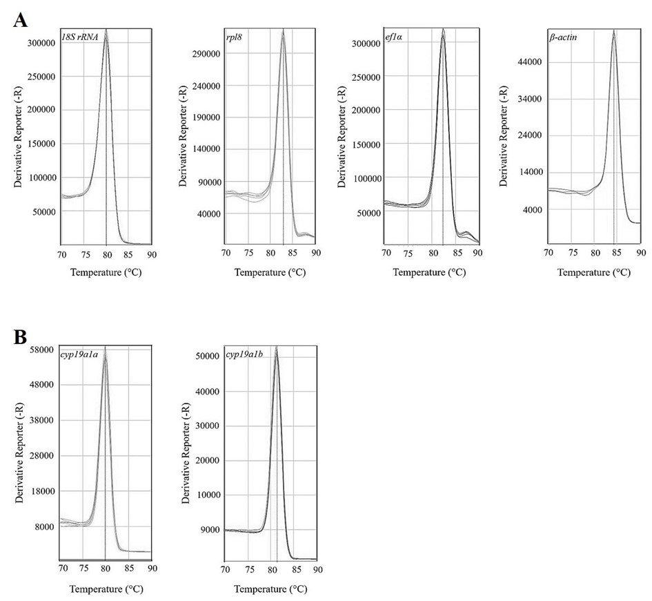
Amplification specificity was confirmed by melting curve analysis, which showed single, sharp peaks with no primer-dimer artifacts (Figures 2A and 2B). Ct value distributions and descriptive statistics (mean, SD, minimum, maximum, range, and CV%) for each gene are provided in Table 2 and visualized in Figure 1. These data served as the foundation for subsequent stability analyses.
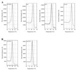
Amplicon specificity and melting curve analysis of candidate and target genes. (A) Melting curves for the four candidate reference genes (18S rRNA, rpl8, ef1α, and β-actin) confirm specific amplification without primer-dimer formation; (B) Melting curves for the two target genes (cyp19a1a and cyp19a1b) also show product specificity, supporting their suitability for gene expression analysis.
Summary statistics of Ct values for candidate reference genes in brain and gonadal tissues of Amphiprion ocellaris.
3.2. Reference gene stability and overall ranking
Gene stability in brain and gonadal tissues was assessed using RefFinder, which integrates four established algorithms: the ΔCt method, geNorm, NormFinder, and BestKeeper. Across all methods, 18S rRNA consistently emerged as the most stable gene in both tissues. In the gonads, 18S rRNA ranked first in the ΔCt, NormFinder, and BestKeeper algorithms and had the lowest overall geometric mean score (GM = 1.32). Although geNorm slightly favored ef1α and rpl8, their performance was less consistent across the two tissues (Table 3).
Stability ranking of reference genes based on RefFinder analysis. Ranking results for each gene derived from the ΔCt, geNorm, NormFinder, and BestKeeper algorithms. Genes are ranked from most (1) to least (4) stable for each algorithm.
In the brain, 18S rRNA again achieved the top rank across all four methods, with the lowest GM (1.00) and narrowest Ct range, supporting its strong expression stability (Table 3, Figure 1). By contrast, β-actin was the least stable gene in both tissues, showing the highest Ct variability and lowest rankings across all algorithms. Together, these results establish 18S rRNA as the most reliable reference gene for qRT-PCR normalization in A. ocellaris, suggesting that β-actin should be avoided due to its inconsistent expression.
3.3. Validation of reference gene with sex-related target genes
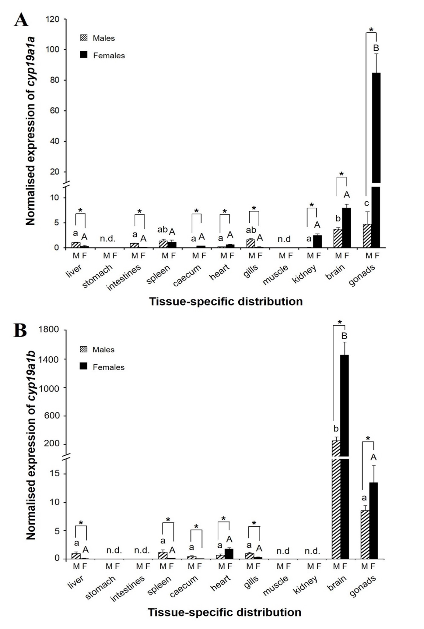
To validate the suitability of 18S rRNA as a reference gene, we analyzed the expression of two key sex differentiation markers—cyp19a1a and cyp19a1b—in 24-month-old breeding males and females across 11 different tissues (Figure 3). As shown in Figure 3A, cyp19a1a expression was significantly higher in female gonads (84.69 ± 12.64) compared to males (4.72 ± 2.47) (P< 0.05), consistent with its established role in ovarian estrogen synthesis. Low expression levels were also detected in the brain, spleen, and heart, while other tissues exhibited negligible or undetectable expression. In contrast, cyp19a1b was predominantly expressed in the brain, where females exhibited significantly higher levels (1452.73 ± 176.39) than males (253.28 ± 54.01) (P< 0.05) (Figure 3B), supporting its role in brain-specific aromatization.
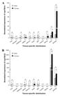
Tissue-specific expression of cyp19a1a and cyp19a1b in Amphiprion ocellaris. (A) Relative expression of cyp19a1a; (B) Relative expression of cyp19a1b, normalized to 18S rRNA, across eleven tissues: liver, stomach, intestine, spleen, caecum, heart, gill, muscle, kidney, brain, and gonad. Expression levels were calculated using the ΔΔCt method. Error bars represent standard deviation (SD) from three biological replicates (n = 3 per sex). Differences among tissues and sexes were tested using one-way ANOVA followed by Duncan’s multiple range test (P < 0.05). Different lowercase letters indicate significant differences among tissues within males; uppercase letters indicate differences among tissues within females; asterisks (*) denote significant sex differences within the same tissue. “n.d.” = not detected.
Other tissues showed little to no expression of this isoform. Statistical annotations in Figure 3 clarify these differences: lowercase letters represent variation among tissues within males, uppercase letters denote differences among tissues within females, and asterisks (*) indicate significant sex differences within the same tissue (P < 0.05). These results highlight the distinct tissue-specific roles of the two aromatase isoforms—cyp19a1a in the gonads and cyp19a1b in the brain. The consistent and biologically meaningful expression profiles obtained after normalization with 18S rRNA confirm its reliability as a reference gene and reinforce its suitability for gene expression studies related to sex differentiation in A. ocellaris.
4. Discussion
Accurate normalization is essential for interpreting qRT-PCR data, particularly in dynamic biological processes like sex differentiation, where gene expression is often tissue- and stage-specific. In this study, we evaluated the expression stability of four commonly used reference genes—18S rRNA, ef1α, rpl8, and β-actin—in brain and gonadal tissues of A. ocellaris, a socially controlled protandrous hermaphrodite. We aimed to identify the most stable internal controls for use across different life stages and social contexts.
To capture this biological complexity, we included fish from key developmental points—juveniles (3 months), unpaired immature fish (8 months), and mature adults (13 and 24 months)—under varying social conditions. These stages are known to trigger transcriptional shifts that can affect the reliability of housekeeping genes (Bustin et al., 2009). By encompassing this range, we ensured robust assessment of reference gene performance across biologically relevant conditions.
Among the four candidate reference genes, 18S rRNA consistently exhibited the highest expression stability in both brain and gonadal tissues of A. ocellaris. This was confirmed by the RefFinder analysis, which integrates four widely used algorithms—geNorm, NormFinder, BestKeeper, and the comparative ΔCt method—to rank gene stability. The robustness of 18S rRNA was further supported by its low coefficient of variation (CV%) in Ct values, indicating consistent expression across samples. In contrast, ef1α and rpl8 demonstrated acceptable stability in gonadal tissue but were more variable in brain samples, reflected in their higher CV% values. This suggests they may be better suited for tissue-specific normalization.
On the other hand, β-actin consistently showed the highest Ct variability and CV%, and was ranked the least stable gene across all algorithms. This makes it unsuitable for normalization under the conditions tested in this study. Despite this, β-actin remains one of the most frequently used reference genes in fish molecular studies. The continued reliance on β-actin likely reflects its historical use, commercial availability, and the common—but often untested—assumption of stable expression. However, our results and growing evidence from other species suggest that β-actin expression is highly variable, depending on tissue type, developmental stage, and physiological stress.
For example, in Oreochromis niloticus, both gapdh and β-actin ranked among the least stable reference genes following immune challenge across multiple tissues (Yang et al., 2013). Similar findings were reported in Danio rerio, where ef1α and 18S rRNA showed greater stability than gapdh and β-actin (McCurley and Callard, 2008; Rassier et al., 2020). These studies highlight the risks of assuming universal gene stability without empirical validation.
Our findings in A. ocellaris are consistent with broader teleost trends, where reference gene stability varies by species, tissue type, and experimental condition, as shown in Table 4. In the protogynous hermaphrodite Epinephelus akaara, transcriptomic analysis during sex reversal identified colg5 and arfgef1 as the most stable genes (Wang et al., 2017). Similarly, in Scatophagus argus, ef1α and hprt1 were found to be stable in the ovary, while rpl4 was more suitable for the pituitary (Liu et al., 2023). In Scophthalmus maximus, ef1α, ctsd, and gapdh showed stable expression across different tissues of the hypothalamus–pituitary–gonad–liver axis (Gao et al., 2020). In Paralichthys olivaceus, non-traditional genes such as gatd1 and rpl6 outperformed commonly used reference genes under conditions of acute thermal stress (Han et al., 2025).
Validated reference genes in teleost fish across reproductive strategies, tissue types, and experimental contexts.
Nonetheless, unvalidated reference gene use continues in reproductive studies. For example, O. niloticus studies used β-actin and U6 sRNA for miRNA expression without verifying their stability, raising concerns about data reliability (Jiang et al., 2024). Although some studies report stable β-actin expression under certain conditions, such as early development or metal exposure in Oncorhynchus mykiss (Shekh et al., 2017), even these cases report tissue-specific instability, for instance, in gills exposed to copper.
Taken together, these findings emphasize that no single reference gene is universally stable. Our results confirm that 18S rRNA is the most reliable gene for qRT-PCR normalization in A. ocellaris, while β-actin should be avoided due to its context-sensitive variability. This underscores the importance of empirical validation over historical convention when selecting internal controls for gene expression studies in teleosts.
To validate 18S rRNA as a reference gene, we analyzed the expression of cyp19a1a and cyp19a1b, two aromatase genes critical to estrogen synthesis in gonads and brain. Normalization with 18S rRNA revealed expected tissue- and sex-specific patterns—cyp19a1a was predominantly expressed in female gonads, while cyp19a1b showed higher expression in female brains. These results are consistent with the known roles of aromatase in teleost sex differentiation and reinforce the reliability of 18S rRNA for qRT-PCR normalization in A. ocellaris (Thamnawasolos and Boonphakdee, 2023). Similar findings have been reported in other protandrous hermaphrodites, such as yellowfin seabream (Acanthopagrus latus), where cyp19a1a and foxl2 were significantly upregulated in ovaries, further supporting the conserved role of aromatase in ovarian differentiation (Li et al., 2020).
This study further supports A. ocellaris as a valuable model for reproductive biology and genomic research in reef fishes (Herrera et al., 2023). Among the tested genes, 18S rRNA was consistently the most stable across tissues and conditions. Ef1α and rpl8 were also reliable in gonadal tissues, while β-actin showed high variability and is not recommended. These findings highlight the importance of validating reference genes for each experimental context rather than relying on traditional housekeeping genes.
5. Conclusion
This study identifies 18S rRNA as the most stable reference gene for qRT-PCR normalization in A. ocellaris, supporting its use in gene expression studies related to sex differentiation. Ef1α and rpl8 are also suitable for gonad-specific applications, while β-actin is not recommended due to high variability. Validation with cyp19a1a and cyp19a1b confirmed that normalization with 18S rRNA yields consistent and biologically relevant results. These findings highlight the importance of selecting reference genes based on species, tissue, and developmental context to ensure accurate and reproducible expression analyses in sex-changing teleosts.
Acknowledgements
The authors gratefully acknowledge the staff of the Wet Laboratory, Department of Aquatic Science, Faculty of Science, Burapha University, for their technical support and assistance with fish maintenance throughout the study. Burapha University financially supported this research under Grant No. 171/2561 awarded to C. Boonphakdee.
Data Availability Statement
The entire data set that supports the results of this study was published in the article itself.
References
-
ANDERSEN, C.L., JENSEN, J.L. and ØRNTØFT, T.F., 2004. Normalization of real-time quantitative reverse transcription–PCR data: A model-based variance estimation approach to identify genes suited for normalization, applied to bladder and colon cancer data sets. Cancer Research, vol. 64, no. 15, pp. 5245-5250. https://doi.org/10.1158/0008-5472.CAN-04-0496 PMid:15289330.
» https://doi.org/10.1158/0008-5472.CAN-04-0496 -
BOONPHAKDEE, C., OCHAROEN, Y., SHINN, A.P., SUANLA, S. and THAMNAWASOLOS, J., 2019. 18S rRNA, a potential reference gene in the qRT-PCR measurement of bisphenol A contamination in green mussels (Perna viridis) collected from the Gulf of Thailand. Agriculture and Natural Resources, vol. 53, no. 6, pp. 652-661. https://doi.org/10.34044/j.anres.2019.53.6.13
» https://doi.org/10.34044/j.anres.2019.53.6.13 -
BUSTON, P., 2003. Social hierarchies: size and growth modification in clownfish. Nature, vol. 424, no. 6945, pp. 145-146. https://doi.org/10.1038/424145a
» https://doi.org/10.1038/424145a -
BUSTIN, S.A., BENEŠ, V., GARSON, J.A., HELLEMANS, J., HUGGETT, J.F., KUBISTA, M., MUELLER, R., NOLAN, T., PFAFFL, M.W., SHIPLEY, G.L., VANDESOMPELE, J. and WITTWER, C.T., 2009. The MIQE guidelines: minimum information for publication of quantitative real-time PCR experiments. Clinical Chemistry, vol. 55, no. 4, pp. 611-622. https://doi.org/10.1373/clinchem.2008.112797 PMid:19246619.
» https://doi.org/10.1373/clinchem.2008.112797 -
CHEN, L., LIANG, Q., LAI, Z., CUI, H., XU, Z., CHEN, Z., DONG, Z., WANG, Z. and GUO, Y., 2024. Systematic selection of suitable reference genes for quantitative real-time PCR normalization studies of gene expression in Lutjanus erythropterus. Scientific Reports, vol. 14, no. 1, pp. 13323. https://doi.org/10.1038/s41598-024-63335-x PMid:38858385.
» https://doi.org/10.1038/s41598-024-63335-x -
DE SANTIS, C., SMITH-KEUNE, C. and JERRY, D.R., 2011. Normalizing RT-qPCR data: are we getting the right answers? An appraisal of normalization approaches and internal reference genes from a case study in the finfish Lates calcarifer. Marine Biotechnology, vol. 13, no. 2, pp. 170-180. https://doi.org/10.1007/s10126-010-9277-z PMid:20309600.
» https://doi.org/10.1007/s10126-010-9277-z -
DHARMARATNAM, A., SUDHAGAR, A., NITHIANANTHAM, S.R., DAS, S. and SWAMINATHAN, T.R., 2021. Evaluation of candidate reference genes for quantitative RT-qPCR analysis in goldfish (Carassius auratus L.) in healthy and CyHV-2 infected fish. Veterinary Immunology and Immunopathology, vol. 237, pp. 110270. https://doi.org/10.1016/j.vetimm.2021.110270 PMid:34015681.
» https://doi.org/10.1016/j.vetimm.2021.110270 -
DIOTEL, N., LE PAGE, Y., MOURIEC, K., TONG, S.K., PELLEGRINI, E., VAILLANT, C., ANGLADE, I., BRION, F., PAKDEL, F., CHUNG, B.C. and KAH, O., 2010. Aromatase in the brain of teleost fish: expression, regulation and putative functions. Frontiers in Neuroendocrinology, vol. 31, no. 2, pp. 172-192. https://doi.org/10.1016/j.yfrne.2010.01.003 PMid:20116395.
» https://doi.org/10.1016/j.yfrne.2010.01.003 -
DU, X., ZHANG, H., ZHU, L., CAO, Z., ZHANG, C., WU, Y., ZHOU, Y. and SUN, Y., 2024. Selection of reference genes by quantitative real-time PCR in different cell lines from humpback grouper (Cromileptes altivelis). Fishes, vol. 9, no. 12, pp. 491. https://doi.org/10.3390/fishes9120491
» https://doi.org/10.3390/fishes9120491 -
GAO, Y., GAO, Y.-T., HUANG, B., MENG, Z. and JIA, Y., 2020. Reference gene validation for quantification of gene expression during ovarian development of turbot (Scophthalmus maximus). Scientific Reports, vol. 10, no. 1, pp. 823. https://doi.org/10.1038/s41598-020-57633-3 PMid:31964949.
» https://doi.org/10.1038/s41598-020-57633-3 -
HAN, P., CHEN, J., SUN, Z., REN, S. and WANG, X., 2025. Evaluation of reference genes for gene expression analysis in Japanese flounder (Paralichthys olivaceus) under temperature stress. BMC Genomics, vol. 26, no. 1, pp. 117. https://doi.org/10.1186/s12864-025-11285-7 PMid:39920593.
» https://doi.org/10.1186/s12864-025-11285-7 -
HERRERA, M., RAVASI, T. and LAUDET, V., 2023. Anemonefishes: A model system for evolutionary genomics. F1000 Research, vol. 12, pp. 204. https://doi.org/10.12688/f1000research.130752.2 PMid:37928172.
» https://doi.org/10.12688/f1000research.130752.2 -
JIANG, B., LI, Q., ZHANG, Z., HUANG, Y., WU, Y., LI, X., HUANG, M., HUANG, Y. and JIAN, J., 2023. Selection and evaluation of stable reference genes for quantitative real-time PCR in the head kidney leukocyte of Oreochromis niloticus. Aquaculture Reports, vol. 31, pp. 101660. https://doi.org/10.1016/j.aqrep.2023.101660
» https://doi.org/10.1016/j.aqrep.2023.101660 -
JIANG, B., LU, S., LI, Y., BADRAN, M.F., DONG, Y., XU, P., QIANG, J. and TAO, Y., 2024. Integrative analysis of miRNA-mRNA expression in the brain during high temperature-induced masculinization of female Nile tilapia (Oreochromis niloticus). Genomics, vol. 116, no. 3, pp. 110856. https://doi.org/10.1016/j.ygeno.2024.110856 PMid:38734154.
» https://doi.org/10.1016/j.ygeno.2024.110856 -
LI, S., LIN, G., FANG, W., HUANG, P., GAO, D., HUANG, J., XIE, J. and LÜ, J., 2020. Gonadal transcriptome analysis of sex-related genes in the protandrous yellowfin seabream (Acanthopagrus latus). Frontiers in Genetics, vol. 11, pp. 709. https://doi.org/10.3389/fgene.2020.00709 PMid:32765585.
» https://doi.org/10.3389/fgene.2020.00709 -
LIANG, J., XU, J., XIE, S., CAO, J. and TAN, B., 2022. Selection of appropriate housekeeping genes for gene expression normalization in hybrid grouper (Epinephelus fuscoguttatus ♀ × E. lanceolatus ♂). Turkish Journal of Fisheries and Aquatic Sciences, vol. 22, no. 9. https://doi.org/10.4194/TRJFAS20646
» https://doi.org/10.4194/TRJFAS20646 -
LIEW, W.C., BARTFAI, R., LIM, Z., SREENIVASAN, R., SIEGFRIED, K.R. and ORBAN, L., 2012. Polygenic sex determination system in zebrafish. PLoS One, vol. 7, no. 4, pp. e34397. https://doi.org/10.1371/journal.pone.0034397 PMid:22506019.
» https://doi.org/10.1371/journal.pone.0034397 -
LIU, Y.-F., CHENG, J.-X., FAN, K.-P., XIA, Y.-Q., ZHANG, Z.-Q., LIU, Y. and LIU, P.-F., 2021. Evaluation of potential reference genes by quantitative RT-qPCR analysis of Takifugu rubripes under normal conditions and after Cryptocaryon irritans infection. Aquaculture Research, vol. 52, no. 11, pp. 5814-5828. https://doi.org/10.1111/are.15456
» https://doi.org/10.1111/are.15456 -
LIU, Z., WANG, T., LIU, P., JIANG, D., LIU, X., DENG, S., WU, T., HUANG, Y., ZHU, C., LI, G. and JIANG, M., 2023. Selection of reference gene for expression studies in the ovary and pituitary of spotted scat (Scatophagus argus) at different ovarian stages. Fishes, vol. 8, no. 2, pp. 120. https://doi.org/10.3390/fishes8020120
» https://doi.org/10.3390/fishes8020120 -
LIVAK, K.J. and SCHMITTGEN, T.D., 2001. Analysis of relative gene expression data using real-time quantitative PCR and the 2−ΔΔCT method. Methods : A Companion to Methods in Enzymology, vol. 25, no. 4, pp. 402-408. https://doi.org/10.1006/meth.2001.1262 PMid:11846609.
» https://doi.org/10.1006/meth.2001.1262 - MADHU, K. and MADHU, R., 2006. Protandrous hermaphroditism in the clownfish Amphiprion percula from Andaman and Nicobar Islands. Indian Journal of Fisheries, vol. 53, no. 4, pp. 373-382.
-
MCCURLEY, A.T. and CALLARD, G.V., 2008. Characterization of housekeeping genes in zebrafish: male-female differences and effects of tissue type, developmental stage and chemical treatment. BMC Molecular Biology, vol. 9, no. 1, pp. 102. https://doi.org/10.1186/1471-2199-9-102 PMid:19014500.
» https://doi.org/10.1186/1471-2199-9-102 -
PARKER, C.G., LEE, J.S., HISTED, A.R., CRAIG, S.E. and RHODES, J.S., 2022. Stable and persistent male-like behavior during male-to-female sex change in the common clownfish Amphiprion ocellaris. Hormones and Behavior, vol. 145, pp. 105239. https://doi.org/10.1016/j.yhbeh.2022.105239 PMid:35926412.
» https://doi.org/10.1016/j.yhbeh.2022.105239 -
PFAFFL, M.W., TICHOPAD, A., PRGOMET, C. and NEUVIANS, T.P., 2004. Determination of stable housekeeping genes, differentially regulated target genes and sample integrity: BestKeeper–Excel-based tool using pair-wise correlations. Biotechnology Letters, vol. 26, no. 6, pp. 509-515. https://doi.org/10.1023/B:BILE.0000019559.84305.47 PMid:15127793.
» https://doi.org/10.1023/B:BILE.0000019559.84305.47 -
RASSIER, G.T., SILVEIRA, T., REMIÃO, M.H., DANELUZ, L.O., MARTINS, A.W.S., DELLAGOSTIN, E.N., ORTIZ, H.G., DOMINGUES, W.B., KOMNINOU, E.R., KÜTTER, M.T., MARINS, L.F.F. and CAMPOS, V.F., 2020. Evaluation of qPCR reference genes in GH-overexpressing transgenic zebrafish (Danio rerio). Scientific Reports, vol. 10, no. 1, pp. 12692. https://doi.org/10.1038/s41598-020-69423-y PMid:32728128.
» https://doi.org/10.1038/s41598-020-69423-y -
SCHMITTGEN, T.D. and LIVAK, K.J., 2008. Analyzing real-time PCR data by the comparative CT method. Nature Protocols, vol. 3, no. 6, pp. 1101-1108. https://doi.org/10.1038/nprot.2008.73 PMid:18546601.
» https://doi.org/10.1038/nprot.2008.73 -
SHEKH, K., TANG, S., NIYOGI, S. and HECKER, M., 2017. Expression stability and selection of optimal reference genes for gene expression normalization in early life stage rainbow trout exposed to cadmium and copper. Aquatic Toxicology, vol. 190, pp. 217-227. https://doi.org/10.1016/j.aquatox.2017.07.009 PMid:28763741.
» https://doi.org/10.1016/j.aquatox.2017.07.009 -
SMALL, B.C., MURDOCK, C.A., BILODEAU‐BOURGEOIS, A.L., PETERSON, B.C. and WALDBIESER, G.C., 2008. Stability of reference genes for real-time PCR analyses in channel catfish (Ictalurus punctatus) tissues under varying physiological conditions. Comparative Biochemistry and Physiology. Part B, Biochemistry & Molecular Biology, vol. 151, no. 3, pp. 296-304. https://doi.org/10.1016/j.cbpb.2008.07.010 PMid:18692590.
» https://doi.org/10.1016/j.cbpb.2008.07.010 -
ŠVEC, D., TICHOPÁD, A., NOVOSADOVÁ, V., PFAFFL, M.W. and KUBISTA, M., 2015. How good is a PCR efficiency estimate: recommendations for precise and robust qPCR efficiency assessments. Biomolecular Detection and Quantification, vol. 3, pp. 9-16. https://doi.org/10.1016/j.bdq.2015.01.005 PMid:27077029.
» https://doi.org/10.1016/j.bdq.2015.01.005 -
THAMNAWASOLOS, J. and BOONPHAKDEE, C., 2023. Cyp19 expression and sex change timing in captive-bred false clownfish. Brazilian Journal of Biology = Revista Brasileira de Biologia, vol. 83, pp. e273824. https://doi.org/10.1590/1519-6984.273824 PMid:37585931.
» https://doi.org/10.1590/1519-6984.273824 -
VANDESOMPELE, J., DE PRETER, K., PATTYN, F., POPPE, B., VAN ROY, N., DE PAEPE, A. and SPELEMAN, F., 2002. Accurate normalization of real-time quantitative RT-PCR data by geometric averaging of multiple internal control genes. Genome Biology, vol. 3, no. 7, pp. RESEARCH0034. https://doi.org/10.1186/gb-2002-3-7-research0034 PMid:12184808.
» https://doi.org/10.1186/gb-2002-3-7-research0034 -
WANG, H., ZHANG, X., LIU, Q., LIU, X. and DING, S., 2017. Selection and evaluation of new reference genes for RT-qPCR analysis in Epinephelus akaara based on transcriptome data. PLoS One, vol. 12, no. 2, pp. e0171646. https://doi.org/10.1371/journal.pone.0171646 PMid:28182746.
» https://doi.org/10.1371/journal.pone.0171646 -
YANG, C.G., WANG, X.L., TIAN, J., LIU, W., WU, F., JIANG, M. and WEN, H., 2013. Evaluation of reference genes for quantitative real-time RT-PCR analysis of gene expression in Nile tilapia (Oreochromis niloticus). Gene, vol. 527, no. 1, pp. 183-192. https://doi.org/10.1016/j.gene.2013.06.013 PMid:23792389.
» https://doi.org/10.1016/j.gene.2013.06.013
Edited by
-
Editor:
Takako Matsumura Tundisi