Asexual ascomycetes associated with Elaeis guineensis Jacq. (Arecaceae): new records for Brazil, South America and American continent
Abstract
The oil palm (Elaeis guineensis Jacq.) holds significant economic value in the Amazon region due to the versatility of its extracted oil; however, information on the funga associated with this palm species in the Amazon is limited. During an investigation of decomposing asexual ascomycetes on debris from E. guineensis in plantation areas in the Eastern Amazon, some interesting fungi were found. Among them, three species represent new records of asexual ascomycetes for the Amazon biome, one species represents a new record of an asexual ascomycete for Brazil (Helminthosporium longisinuatum), one for South America (Dictyocheirospora gigantica), and seven for the American continent (Cacumisporium rugosum, Dictyocheirospora suae, Distoseptispora appendiculata, Endocalyx indumentum, Pseudoberkleasmium chiangmaiense, Sporidesmium antidesmatis, and Trichocladium palmae). This study contributes to understanding the diversity of asexual ascomycetes on E. guineensis in Brazil and highlights the potential of Arecaceae as a model group for investigating fungal diversity.
Keywords:
oil palm; diversity; funga; saprobes; taxonomy
Resumo
O dendezeiro (Elaeis guineensis Jacq.) apresenta grande valor econômico na região amazônica, devido à versatilidade de usos de seu óleo extraído, mas com poucas informações disponíveis sobre a funga associada a essa palmeira na Amazônia. Durante uma investigação de ascomicetos assexuais decompositores de folhas de E. guineensis, coletados em áreas de plantações na Amazônia Oriental, alguns interessantes fungos foram encontrados. Dentre eles, três espécies representam novos registros de ascomicetos assexuais para o bioma Amazônia, um para o Brasil (Helminthosporium longisinuatum), um para América do Sul (Dictyocheirospora gigantica) e sete para o Continente Americano (Cacumisporium rugosum, Dictyocheirospora suae, Distoseptispora appendiculata, Endocalyx indumentum, Pseudoberkleasmium chiangmaiense, Sporidesmium antidesmatis e Trichocladium palmae). Este estudo contribui para o entendimento da diversidade de ascomicetos assexuais associados com E. guineensis no Brasil e destaca o potencial das plantas da família Arecaceae como um grupo modelo para a investigação da diversidade fúngica.
Palavras-chave:
dendezeiro; diversidade; funga; sapróbios; taxonomia
1. Introduction
In Brazil, oil palm (Elaeis guineensis Jacq.), locally known as dendezeiro, was naturalized in the state of Bahia at the end of the 16th century. It is currently cultivated along the south-eastern and north-eastern coasts and especially in the eastern Amazon region, which has the largest plantation areas in the country (Homma, 2016). Oil palm stands out for the versatile oil’s industrial uses, including the use in butter, oils, cleaning products, and biodiesel production. The oil also stands out for its antioxidant capacity, making it valuable to the pharmaceutical and cosmetic industries (Alhaji et al., 2024).
Studies addressing the fungal diversity associated with oil palm have reported a high predominance of phytopathogenic fungi, mainly in Africa and Asia (Turner, 1971; Azeez et al., 2024), as well as several saprobic fungi (Pirozynski, 1972). In Brazil, some studies on fungi associated with E. guineensis have focused on phytopathogens (Mendes et al., 2019), whereas for saprobic fungi, Vitória et al. (2014, 2016) identified several new records and new ascomycetes species in North-eastern Brazil.
Particularly in the eastern Amazon region, several studies on saprobic microfungi associated with palm trees have been conducted, including Euterpe oleracea Mart., Bactris gasipaes Kunth, Astrocaryum gynacanthum Mart., and A. murumuru Mart. (Castro et al., 2012; Monteiro et al., 2019; Silva et al., 2022). Despite the relevance of palm trees and the socioeconomic importance of oil palm (Lima et al., 2026), studies specifically addressing fungi associated with E. guineensis remain scarce. Only a few have documented phytopathogenic fungi, such as Rhizoctonia solani J.G. Kühn, which causes leaf necrosis (Boari et al., 2017), and Helminthosporiella stilbacea Konta & K.D. Hyde, responsible for leaf spot symptoms (Rosado et al., 2019).
Therefore, given the limited information available, this study aimed to describe and illustrate new records of asexual ascomycetes associated with the decomposition of leaves and fruit bunches of E. guineensis in plantations in the Eastern Amazon.
2. Material and Methods
Three field collections were carried out between December 2022 and August 2023 in three municipalities with E. guineensis plantations in the state of Pará: a) Abaetetuba (1°43’24”S-48°52’54”W); b) Moju (1°53’05”S-48°45’55”W); and c) Santa Bárbara do Pará (1°13’27”S-48°17’38”W).
Fifteen E. guineensis individuals were randomly selected in each study area, and five decomposing substrates (bunch, leaflet, petiole, rachis, and sheath) were collected per individual. The substrates were subjected to the washing technique in running water and processed according to Castañeda-Ruiz et al. (2016). Observation of the incubated material began on the third day, and the samples were examined periodically for up to 45 days. The reproductive structures of the asexual ascomycetes were mounted in a semi-permanent lacto-glycerol slide (distilled water + lactic acid + glycerin) (Neergaard et al., 2000).
Species identification was based on observation and measurement of taxonomically relevant structures using an optical microscope and comparison with species described in the specialized literature (Ellis, 1971, 1976; Matsushima, 1975; Seifert et al., 2011). The nomenclature and taxonomic status of the identified species were verified using Index Fungorum and MycoBank, as well as recent taxonomic literature (Hyde et al., 2024). Species distribution was assessed through the analysis of published scientific articles and consultation of the databases SpeciesLink (CRIA, 2025), Flora e Funga do Brasil (JBRJ, 2025), and GBIF (2025). Photomicrographs were taken in a Leica DM6B microscope equipped with a digital camera, using LAS V4.12 software. Slides and dried specimens were incorporated into the fungal collection of the João Murça Pires Herbarium (MG), at the Emílio Goeldi Museum of Pará. We provide descriptions, taxonomic comments, and illustrations for the new records for Brazil, South America, and the American continent.
3. Results
A total 79 species and 62 genera of asexual ascomycetes associated with E. guineensis were recorded in collections carried out in the state of Pará (Table 1). The highest number of fungal records was found in the plantation areas of the municipality of Santa Bárbara do Pará, followed by Moju and Abaetetuba. Among the E. guineensis substrates sampled, the greatest number of fungi was observed on leaflets (37), followed by petioles (33), sheaths (26), rachises (23), and fruit bunches (6).
List of asexual ascomycetes species associated with Elaeis guineensis with their respective Family/Order, substrates and collection locations.
Of the asexual ascomycetes identified, three species are new records for the Brazilian Amazon, one for Brazil, one for South America, and seven represent new occurrences for the American continent (Table 1).
3.1. Taxonomic treatment
Cacumisporium rugosum C.K.M. Tsui, Goh, K.D. Hyde & Hodgkiss, Mycologia 93(2): 389 (2001). Figure 1AC
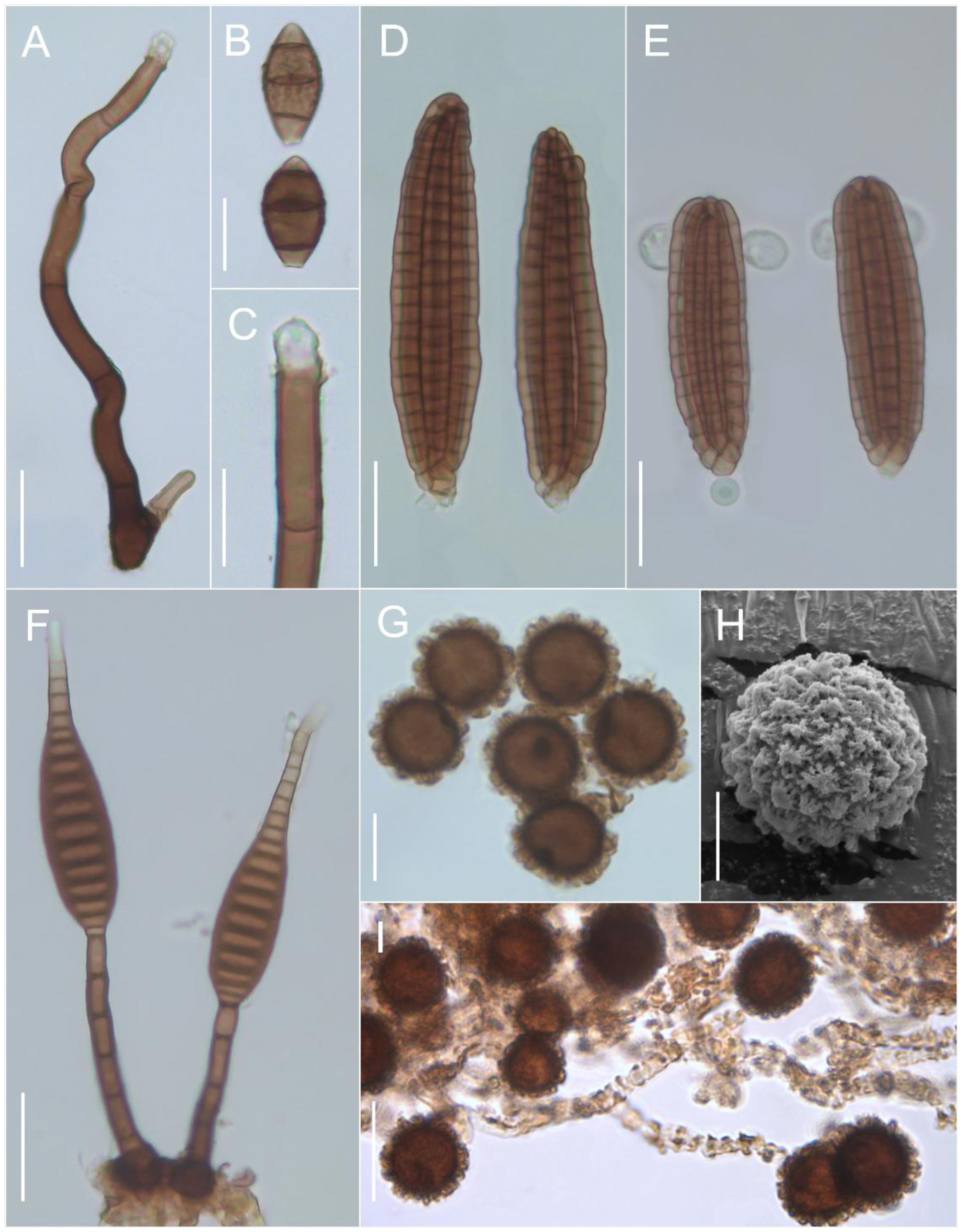
A-C. Cacumisporium rugosum. A. Conidiophore. B. Conidia. C. Conidiogenous cell. D. Dictyocheirospora gigantica. D. Conidia. E. Dictyocheirospora suae. E. Conidia with subglobose appendages. F. Distoseptispora appendiculata. F. Conidiophores and conidia. G-I. Endocalyx indumentum. G. Conidia. H. Superficial view of the ornamented conidial wall (SEM). I. Conidia mixed with peridial hyphae. Scale bars: A, D, E, F = 25 µm; B, C, I = 20 µm; G, H = 10 µm.
Conidiophores macronematous, mononematous, solitary, erect, straight, simple, cylindrical, with a swollen base, septate, smooth, dark brown, paler toward the apex, 175-250 × 7.5-10 μm. Conidiogenous cells polyblastic, integrated, terminal, producing conidia in sympodial succession with multiple conidiogenous loci, above hyaline, cylindrical collarettes. Conidia acrogenous, solitary, obovoidal to broadly ellipsoidal, 3-euseptate, with two central dark brown cells and pale brown basal and apical cells, rugose to verrucose except at the end cells, 32.5-35 × 15-20 μm; frills on the conidial wall were not observed.
Material examined: BRAZIL, Pará, Santa Bárbara do Pará, Assentamento Abril Vermelho, on decaying bunch of Elaeis guineensis (Arecaceae), 20-I-2023, M.C.S. Ferreira & J.S. Monteiro, MF-161 (MG 253148).
Known distribution: China (Tsui et al., 2001), Brazil (this study).
Comments:Cacumisporium Preuss was established by Preuss (1851), with C. tenebrosum Preuss (= C. capitulatum (Corda) S. Hughes) as the type species. The genus is characterized by solitary, macronematous, mononematous, erect, brown conidiophores with heterogeneous (holoblastic denticulate and/or phialidic) conidiogenous cells, and producing phragmosporous, euseptate, (sub)hyaline or brown, smooth or ornamented conidia (Xu et al., 2019; Réblová and Nekvindová, 2023). Currently, the genus has nine species included in Chaetosphaeriaceae (Chaetosphaeriales), with DNA sequence for only four species (Réblová and Nekvindová, 2023). Cacumisporium rugosum is morphologically similar to C. capitulatum in that it has 3-septate conidia, but it differs by the presence of rugose ornamentation and larger conidia with dark brown central cells (Réblová and Gams, 1999; Tsui et al., 2001). In the Brazilian specimens, conidiophores shorter in length than those of the type material were recorded (175-250 vs. 200-350 μm). This represents the first record of C. rugosum for the American continent.
Dictyocheirospora gigantica (Goh & K.D. Hyde) M.J. D'souza, Boonmee & K.D. Hyde, Fungal Diversity 80(1): 469 (2016). Figure 1D
Conidiomata sporodochial, superficial, compact, scattered, dark brown. Conidiophores not observed. Conidiogenous cells holoblastic, monoblastic, determinate, subspherical to doliiform, smooth, hyaline. Conidia solitary, cylindric-clavate, not complanate, euseptate, consisting of 7-8 rows of cells tightly appressed together, each row with 17-22 cells, smooth, dark brown, 90-102.5 × 20-25 μm, truncate basal cell, pale brown, apical portion slightly incurved, conidial appendages absent.
Material examined: BRAZIL, Pará, Moju, Fazenda Kimura, on decaying petiole of Elaeis guineensis (Arecaceae), 29-IV-2023, M.C.S. Ferreira & J.S. Monteiro, MF-290 (MG 253277).
Known distribution: South Africa, Solomon Islands, and Thailand (Goh et al., 1999), Mexico (Arias et al., 2018), Brazil (this study).
Comments:Dictyocheirospora M.J. D'souza, Boonmee & K.D. Hyde was established by Boonmee et al. (2016). The genus was proposed with six species in addition to the type species D. rotunda D’souza, Bhat & Hyde. The genus is characterized by the formation of punctiform sporodochia with micronematous, septate, hyaline conidiophores and holoblastic, integrated, terminal conidiogenous cells producing cheroid, complanate or not, euseptate or distoseptate conidia, with or without appendages (Boonmee et al., 2016; Shen et al., 2022). Currently, Dictyocheirospora has 36 epithets listed in Index Fungorum (2025). Dictyocheirospora is morphologically similar to Dictyosporium, except for its cheroid, non-complanate, cylindrical conidia (Boonmee et al., 2016; Yang et al., 2018a).
Dictyocheirospora gigantica was described by Goh et al. (1999) as Dictyosporium giganticum Goh & K.D. Hyde and later combined into Dictyocheirospora M.J. D'souza, Boonmee & K.D. Hyde after a phylogenetic review and analysis of Dictyosporium Corda (Boonmee et al., 2016). The species resembles D. heptaspora (Garov.) M.J. D'souza, Boonmee & K.D. Hyde, but it has larger conidia (Goh et al., 1999). The Brazilian material presented smaller conidia than those of the type material (90-102.5 × 20-25 μm vs. 105-121 × 25-32 μm). This is the first record of the species for South America.
Dictyocheirospora suae H.W. Shen & Z.L. Luo, Journal of Fungi 8 (11, no. 1200): 10 (2022). Figure 1E
Conidiomata sporodochial, superficial, punctiform, scattered, brown. Conidiophores not observed. Conidiogenous cells holoblastic, monoblastic, determinate, cylindrical, smooth, hyaline. Conidia solitary, cheiroid, ellipsoid to cylindrical, not complanate, euseptate, consisting of 5-6 rows of cells tightly clustered at the apex of rows, unseparated, each row with 12-14 cells, smooth, brown, 62.5-72.5 × 17.5-22.5 μm, truncate basal cell, pale brown, with globose to subglobose apical appendages, 4-7 × 4-6 μm.
Material examined: BRAZIL, Pará, Abaetetuba, Fazenda Capim Grosso, on decaying petiole of Elaeis guineensis (Arecaceae), 29-XII-2022, M.C.S. Ferreira & J.S. Monteiro, MF-134 (MG 253121).
Known distribution: China (Shen et al., 2022), Brazil (this study).
Comments:Dictyocheirospora suae was described by Shen et al. (2022) and it is morphologically similar to D. multiappendiculata Shen & Luo, but has larger conidia, a greater number of cells in each branch, and fewer apical appendages (usually two) (Shen et al., 2022). Other comparable species are D. indica (Prasher & R.K. Verma) J. Yang & K.D. Hyde, which has smaller conidia (33-48 × 13-18 µm) and appendages attached to subapical cells (Prasher and Verma, 2015); D. musae (Photita) J. Yang, K.D. Hyde & Z.Y. Liu that has seven rows of cells with clavate to obovoid appendages measuring 12-28 × 3-9 µm and attached to the central cells; and D. tetraploides (L. Cai & K.D. Hyde) J. Yang & K.D. Hyde that has five rows with cylindrical to clavate, subapical appendages measuring 10-25 × 5-10 µm (Photita et al., 2002; Cai et al., 2003). The Brazilian material presented conidia slightly smaller than those of the type species (62.5-72.5 × 17.5-22.5 vs. 72-79 × 20-25 μm). This is the first record of the species for the American continent.
Distoseptispora appendiculata D.F. Bao, Z.L. Luo & H.Y. Su, Fungal Diversity 99: 484 (2019). Figure 1F
Conidiophores macronematous, mononematous, solitary, erect, straight or slightly flexuous, cylindrical, unbranched, septate, smooth, dark brown, paler toward the apex, 50-82.5 × 5-5.5 μm. Conidiogenous cells holoblastic, monoblastic, integrated, terminal, determinate, cylindrical, smooth, brown, 7.5-12.5 × 5-5.5 μm. Conidia acrogenous, solitary, obpyriform to obclavate, tapering toward the apex, thick-walled, 17-distoseptate, smooth, dark brown at the base, hyaline toward the apex, truncate at base, slender and rounded at apex, 60-100 × 10-17.5 μm, with a gelatinous, hyaline sheath around tip.
Material examined: BRAZIL, Pará, Moju, Fazenda Kimura, on decaying petiole of Elaeis guineensis (Arecaceae), 29-XII-2022, M.C.S. Ferreira & J.S. Monteiro, MF-86 (MG 253073).
Known distribution: Thailand (Luo et al., 2019), Brazil (this study).
Comments: K.D. Hyde and McKenzie established the family Distoseptisporaceae to accommodate the type genus Distoseptispora K.D. Hyde, McKenzie & Maharachch., comprising four species in addition to the type species D. fluminicola McKenzie, Hong Y. Su, Z.L. Luo & K.D. Hyde (Su et al., 2016). The genus is characterized by macronematous, mononematous, unbranched conidiophores with monoblastic or polyblastic, integrated, terminal, cylindrical conidiogenous cells producing cylindrical, distoseptate conidia with rounded apices and truncate bases, showing morphological similarity to Ellisembia Subram. and Sporidesmium Link (Su et al., 2016; Yang et al., 2018b). Through phylogenetic analyses, Luo et al. (2019) placed Distoseptisporaceae within the order Distoseptisporales. Currently, 124 species are listed in Index Fungorum (2025). Distoseptispora appendiculata differs from other species of the genus by having a conspicuous, hyaline, gelatinous sheath at the apex of the conidium (Luo et al., 2019). The Brazilian material exhibited only slightly evident mucilage compared to the type species, as well as slightly larger conidia (60-100 × 10-17.5 μm vs. 67-89 × 10-16 μm) than those reported by Luo et al. (2019). This species represents a new record for the American continent.
Endocalyx indumentum G. Okada & Tubaki, Mycologia 76(2): 305 (1984). Figure 1GI
Conidiomata scattered, arising from pustules slightly elevated on host surface, circular, discolored, blackish, cupulate to cylindrical, with a dark brown conidial mass; peridial hyphae sterile, interwoven, verrugose, dark brown, 4-5 μm diam., enclosing the inner conidial mass. Conidiophores micronematous, septate, slightly geniculate, smooth, hyaline to pale brown. Conidiogenous cells holoblastic, monoblastic, integrated, terminal, determinate, smooth, hyaline. Conidia solitary, sphaerical to oval, aseptate, ornamented, punctate outer wall layer, smooth, dark brown, 9-15 μm diam.; spore wall ornamentation by light microscopy with a thick layer of densely packed, pale brown, curved or straight, hair-like projections; by scanning electron microscopy (SEM), the projections are filamentous, slightly flexuous, and irregularly adherent to one another.
Material examined: BRAZIL, Pará, Moju, Fazenda Kimura, on decaying rachis of Elaeis guineensis (Arecaceae), 29-XII-2022, M.C.S. Ferreira & J.S. Monteiro, MF-63 (MG 253050).
Known distribution: Japan (Okada and Tubaki, 1984), Thailand (herbarium record, 1993), Brazil (this study).
Comments:Berkeley and Broome (1876) established the genus Endocalyx Berk. & Broome with the type E. thwaitesii Berk. & Broome (= E. psilostoma Berkeley & Broome). The genus is characterized by the formation of cupulate to cylindrical synnemata or sporodochia, basauxic, hyaline to sub-hyaline conidiophores with integrated monoblastic or polyblastic conidiogenous cells producing unicellular, elliptical to spherical conidia, frequently with a hyaline cleft (Delgado et al., 2022). Konta et al. (2021) allocated the genus to the family Cainiaceae (Xylariales), which currently has 12 species, most of which found on palm trees (Arecaceae) (Senanayake et al., 2023). Endocalyx indumentum differs from other species of the genus by the filamentous ornamentations of the conidia wall. The materials from Brazil showed larger conidia diameters (9-15 μm) than the type material (8-12 μm) (Okada and Tubaki, 1984). This is the first record of this species for the American continent.
Helminthosporium longisinuatum Matsush., Matsush. Mycol. Mem. 7: 53 (1993). Figure 2AC
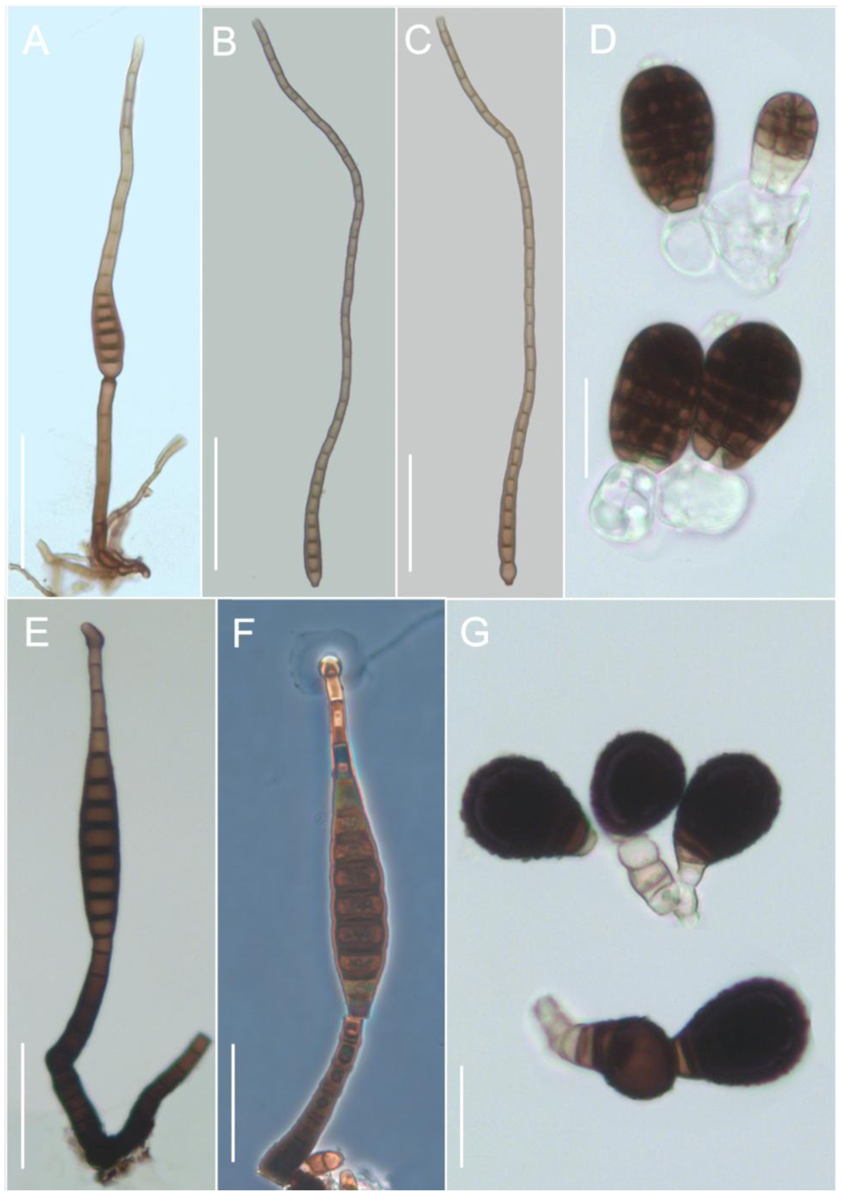
A-C. Helminthosporium longisinuatum. A. Conidiophore and conidium. B-C. Conidia. D. Pseudoberkleasmium chiangmaiense. D. Conidia and subglobose conidiogenous cells. E-F. Sporidesmium antidesmatis. E. Conidiophores and conidium. F. Detail of the subglobose mucilage at the apex of the conidium. G. Trichocladium palmae. G. Conidia and cylindrical conidiogenous cells. Scale bars: A, B, C, E, F = 50 µm; D, G = 20 µm.
Conidiophores macronematous, mononematous, solitary, erect, straight or slightly flexuous, cylindrical, unbranched, septate, smooth, brown, 35-87.5 × 4-6 μm. Conidiogenous cells polytretic, integrated, terminal, determinate, cylindrical, smooth, pale brown, 15-17.5 × 5-5.5 μm. Conidia acropleurogenous, solitary, narrowly obclavate, sinuous, often curved in an S-shape, until 31-distoseptate, slightly constricted at the septa, smooth, brown, paler toward the apex, highly variable length, 75-580 μm long, truncate at base, brown, 7.5-10 μm wide, elongated apex, pale brown, 5-7 μm wide.
Material examined: BRAZIL, Pará, Santa Bárbara do Pará, Assentamento Abril Vermelho, on decaying petiole of Elaeis guineensis (Arecaceae), 20-I-2023, M.C.S. Ferreira & J.S. Monteiro, MF-164 (MG 253151).
Known distribution: Peru (Matsushima, 1993), Poland (Czeczuga et al., 2007), Brazil (this study).
Comments:Helminthosporium was established by Link (1809) with H. velutinum Link as the type species. It is characterized by its marcronematous, mononematous, cylindrical conidiophores with polytretic conidiogenous cells producing solitary conidia or conidia in short chains, which are clavate or obclavate and distoseptate (Ellis, 1971; Seifert et al., 2011). In Index Fungorum (2025) there are more than 790 epithets for Helminthosporium, but most of the recorded species are not congeneric with the type species and have been relocated to other genera (Voglmayr and Jaklitsch, 2017). Currently, the genus has 75 accepted species (Sun et al., 2025; Liao et al., 2025) belonging to the family Massarinaceae (Pleosporales). Helminthosporium longisinuatum differs from other species of the genus by having long conidia, generally curved in an S shape. The Brazilian specimen had dimensions very similar to the type material (Matsushima, 1993). This is the first record of this species for Brazil.
Pseudoberkleasmium chiangmaiense Y.Z. Lu & K.D. Hyde, Fungal Diversity 96: 62 (2019). Figure 2D
Conidiomata sporodochial, superficial, compact, scattered, black, shining. Conidiophores micronematous, mononematous, reduced, erect, cylindrical, smooth, hyaline. Conidiogenous cells holoblastic, monoblastic, integrated, terminal, determinate, globose to subglobose, guttulate, smooth, hyaline, 15-17.5 × 12.5-17.5 μm. Conidia acrogenous, solitary, ellipsoidal to obovoid, dictyosporous, muriform, apex rounded, smooth, dark brown to black, paler toward the base, 22.5-32.5 × 12.5-22.5 μm, usually with conidiogenous cell attached.
Material examined: BRAZIL, Pará, Santa Bárbara do Pará, Assentamento Abril Vermelho, on decaying sheath of Elaeis guineensis (Arecaceae), 20-I-2023, M.C.S. Ferreira & J.S. Monteiro, MF-160 (MG 253147).
Known distribution: Thailand (Hyde et al., 2019), China (Bao et al., 2021), Brazil (this study).
Comments:Pseudoberkleasmium Tibpromma & K.D. Hyde was described by Tibpromma et al. (2018), with P. pandanicola Tibpromma & Hyde as the type species. The genus is characterized by the formation of micronematous, fasciculate, hyaline or subhyaline conidiophores and monoblastic, globose conidiogenous cells, with or without guttules, producing muriform conidia, generally with the conidiogenous cell attached to the base of the conidium (Tibpromma et al., 2018; Tian et al., 2022). Hyde et al. (2019) proposed the family Pseudoberkleasmiaceae (Pleosporales) to accommodate the representatives of this genus, in which currently four species are accepted (Tian et al., 2022). Pseudoberkleasmium chiangmaiense is morphologically similar to P. pandanicola Tibpromma & K.D. Hyde, but differs in the size of the conidiogenous cells, which are larger (Hyde et al., 2019). The Brazilian material has slightly smaller and wider conidia than those described in the type material (22.5-32.5 × 12.5-22.5 μm vs. 30-35 × 15-20 μm) by Hyde et al. (2019). This species represents a new record for the American continent.
Sporidesmium antidesmatis Jian Ma & X.G. Zhang, Mycotaxon 119: 19 (2012). Figure 2EF
Conidiophores macronematous, mononematous, solitary or in groups, erect, straight or slightly flexuous, cylindrical, unbranched, septate, smooth, brown to dark brown, 70-102.5 × 10⁻¹⁵ μm. Conidiogenous cells holoblastic, monoblastic, integrated, terminal, determinate, cylindrical, smooth, brown, 7.5-12.5 × 7.5-8 μm. Conidia acrogenous, solitary, straight or slightly curved, obclavate, tapering toward the apex, 11-12-euseptate, smooth, brown, 132-180 × 17.5-20 μm, truncate at base, rounded at apex, with a subglobose drop of mucilage.
Material examined: BRAZIL, Pará, Abaetetuba, Capim Grosso, on decaying petiole of Elaeis guineensis (Arecaceae), 29-XII-2022, M.C.S. Ferreira & J.S. Monteiro, MF-141 (MG 253128).
Known distribution: China (Ma et al., 2012), Brazil (this study).
Comments:Sporidesmium Link was described by Link (1809), who designated S. atrum Link as the type species. The genus is characterized by macronematous, unbranched conidiophores with monoblastic, terminal, determinate or extending conidiogenous cells that produce euseptate or distoseptate conidia of variable shapes, sometimes surrounded by a mucilaginous sheath (Ellis, 1971; Seifert et al., 2011). Currently, more than 500 epithets are listed under the genus in Index Fungorum (2025); however, many species have been revised and transferred to more than 30 genera (Delgado et al., 2018). Su et al. (2016) reassigned the genus to Sporidesmiaceae (Sporidesmiales, Sordariomycetes) (established by Fries in 1849), including eight species morphologically similar to S. ehrenbergii M.B. Ellis, the lectotype of the genus (Delgado et al., 2024). Sporidesmium antidesmatis superficially resembles S. fragilissimum (Berk. & M.A. Curtis) M.B. Ellis but differs by its larger, smooth-walled conidia and the presence of a mucilaginous sheath (Ma et al., 2012). The examined specimen presented conidiophores (70-102.5 × 10⁻¹⁵ vs. 17-70 × 6-7.5 μm) and conidia (132-180 × 17.5-20 vs. 108-150 × 9-11 μm) that were longer and wider than those of the type material (Ma et al., 2012). This is the first record of this species for the American continent.
Trichocladium palmae Manohar., N.K. Rao, Kunwar & D.K. Agarwal, Indian Phytopath. 59(3): 357 (2006). Figure 2G
Conidiophores micronematous, mononematous, solitary, cylindrical, unbranched, smooth, hyaline. Conidiogenous cells monoblastic, integrated, terminal, determinate, cylindrical, smooth, hyaline. Conidia acrogenous, solitary, dry, broadly obovoid, with rounded apical cell, truncate base, thick-walled, 3-euseptate, thick septa, verrucose, dark brown to black large apical cell, dark brown intermediate cell, paler basal cell than other cells, 21-35 × 19-22 μm.
Material examined: BRAZIL, Pará, Moju, Fazenda Kimura, on decaying sheath of Elaeis guineensis (Arecaceae), 29-XII-2022, M.C.S. Ferreira & J.S. Monteiro, MF-87 (MG 253074).
Known distribution: India (Manoharachary et al., 2006), Brazil (this study).
Comments:Trichocladium Harz was established by Harz (1871) to accommodate the type species T. asperum Harz. It is characterized by solitary, septate or non-septate, (sub)hyaline to brown conidia produced by monoblastic or polyblastic conidiogenous cells on micronematous conidiophores (Seifert et al., 2011). Hambleton et al. (2005) established the genus in the family Chaetomiaceae (Sordariales) and Wang et al. (2019) described four subclades in Trichocladium. Currently, this genus includes 42 valid species listed in Index Fungorum (2025). Trichocladium palmae is easily differentiated by its micronematous conidiophores and broadly obovoid, verrucose conidia with three septa dividing the conidium unequally (Manoharachary et al., 2006). The specimen recorded in Brazil has dimensions similar to the type material. This is the first record of this species for the American continent.
4. Discussion
This study presents a list of asexual ascomycetes associated with the decomposition of E. guineensis substrates in plantations in the Brazilian Amazon. The oil palm tree is one of the palms with the highest number of fungi recorded on several continents, but information on that is limited in Brazil, despite the fact that E. guineensis represents one of the most important monocultures in the country. Most of the recorded species are mainly allocated within Sordariomycetes and Dothideomycetes, which include various phytopathogenic, saprobic, and endophytic fungi, a pattern also observed in B. gasipaes (peach palm) and other Amazonian palms (Monteiro et al., 2019; Silva et al., 2022).
Asexual ascomycetes present in the leaf litter of Amazonian environments, especially those associated with palms, have considerable potential for producing lignocellulolytic enzymes capable of degrading agro-industrial residues (Sousa and Santos, 2025). Considering that E. guineensis plantations generate large quantities of organic waste, investigations of the fungal communities associated with this palm may represent promising sources of species with a high capacity to transform these residues (Rupani et al., 2022; Lau et al., 2024).
In this study, we expanded the known geographic distribution of several fungal species. Cacumisporium rugosum, D. suae, D. appendiculata, S. antidesmatis, and T. palmae were previously known only from their type localities in Asian countries, making this the second global record of these species. The remaining species (D. gigantica, E. indumentum, H. longisinuatum, and P. chiangmaiense) have been reported only a few times, highlighting the need for additional collections. The report of these new records demonstrates that classical identification methods based on morphological characteristics remain fundamental for taxonomic and diversity studies, especially when fresh isolates or suitable material for DNA sequencing are not available (Gautam et al., 2022). Furthermore, morphological analyses continue to underpin fungal taxonomy, also providing important functional traits for ecological studies across diverse fungal groups (Golan and Pringle, 2017; Calhim et al., 2018).
Elaeis guineensis is one of the palm trees with the highest number of fungal associations recorded to date (Pereira and Phillips, 2023). Even in plantation areas, such as those located in the Amazon, this palm seems to be a potential reservoir of fungal diversity that deserves further, more comprehensive investigation addressing other fungal and ecological groups.
5. Conclusion
The occurrence of asexual ascomycetes species associated with E. guineensis in plantations of the Brazilian Amazon indicates that this palm serves as a suitable host and provides favorable substrates for the establishment and development of these fungi.
The data presented suggest the need for further studies addressing asexual ascomycetes and other fungal and ecological groups, such as basidiomycetes and endophytic fungi, which remain little explored in palms and hold great ecological and biotechnological importance.
Acknowledgements
We are grateful to the Emilio Goeldi Museum of Pará and the Universidade Federal Rural da Amazônia for the infrastructure provided to conduct the study. This work was supported by the Coordenação de Aperfeiçoamento de Pessoal de Nível Superior (CAPES) – financing code 001.
Data Availability Statement
The entire data set that supports the results of this study was published in the article itself.
References
-
ALHAJI, A.M., ALMEIDA, E.S., CARNEIRO, C.R., SILVA, C.A.S., MONTEIRO, S. and COIMBRA, J.S.D.R., 2024. Palm Oil (Elaeis guineensis): a journey through sustainability, processing, and utilization. Foods, vol. 13, no. 17, pp. 2814. https://doi.org/10.3390/foods13172814 PMid:39272579.
» https://doi.org/10.3390/foods13172814 -
ARIAS, R.M., HEREDIA-ABARCA, G. and CASTAÑEDA-RUIZ, R., 2018. Checklist of saprobic asexual microfungi from the tropical montane cloud forest of Veracruz, Mexico. Mycotaxon, vol. 132, pp. 986. https://doi.org/10.5248/132.985
» https://doi.org/10.5248/132.985 -
AZEEZ, A.A., ESIEGBUYA, D.O., LATEEF, A.A. and ASIEGBU, F.O., 2024. Mycobiome analysis of leaf, root, and soil of symptomatic oil palm trees (Elaeis guineensis Jacq.) affected by leaf spot disease. Frontiers in Microbiology, vol. 15, pp. 1422360. https://doi.org/10.3389/fmicb.2024.1422360 PMid:39712899.
» https://doi.org/10.3389/fmicb.2024.1422360 -
BAO, D.F., HYDE, K.D., MCKENZIE, E.H., JEEWON, R., SU, H.Y., NALUMPANG, S. and LUO, Z.L., 2021. Biodiversity of lignicolous freshwater hyphomycetes from China and Thailand and description of sixteen species. Journal of Fungi, vol. 7, no. 8, pp. 669. https://doi.org/10.3390/jof7080669 PMid:34436208.
» https://doi.org/10.3390/jof7080669 -
BERKELEY, M.J. and BROOME, C.E., 1876. Supplement to the enumeration of fungi of Ceylon. Journal of the Linnean Society of London, Botany, vol. 15, no. 82, pp. 82-86. https://doi.org/10.1111/j.1095-8339.1876.tb00225.x
» https://doi.org/10.1111/j.1095-8339.1876.tb00225.x -
BOARI, A.J., QUADROS, A.F.F. and NECHET, K.L., 2017. Rhizoctonia solani AG 1-IA causing leaf blight in oil palm seedlings in Brazil. Australasian Plant Disease Notes, Australasian Plant Pathology Society, vol. 12, no. 1, pp. 42. https://doi.org/10.1007/s13314-017-0269-y
» https://doi.org/10.1007/s13314-017-0269-y -
BOONMEE, S., D’SOUZA, M.J., LUO, Z., PINRUAN, U., TANAKA, K., SU, H., BHAT, D.J., MCKENZIE, E.H.C., JONES, E.B.G., TAYLOR, J.E., PHILLIPS, A.J.L., HIRAYAMA, K., EUNGWANICHAYAPANT, P.D. and HYDE, K.D., 2016. Dictyosporiaceae fam. nov. Fungal Diversity, vol. 80, no. 1, pp. 457-482. https://doi.org/10.1007/s13225-016-0363-z
» https://doi.org/10.1007/s13225-016-0363-z - CAI, L., ZHANG, K.Q., MCKENZIE, E.H.C. and HYDE, K.D., 2003. New species of Dictyosporium and Digitodesmium from submerged wood in Yunnan, China. Sydowia, vol. 55, pp. 129-135.
-
CALHIM, S., HALME, P., PETERSEN, J.H., LÆSSØE, T., BÄSSLER, C. and HEILMANN-CLAUSEN, J., 2018. Fungal spore diversity reflects substrate-specific deposition challenges. Scientific Reports, vol. 8, no. 1, pp. 5356. https://doi.org/10.1038/s41598-018-23292-8 PMid:29599480.
» https://doi.org/10.1038/s41598-018-23292-8 -
CASTAÑEDA-RUIZ, R.F., HEREDIA, G., GUSMÃO, L.F.P. and LI, D.W., 2016. Fungal diversity of Central and South America. In: D.W. LI, ed. Biology of microfungi: fungal biology. Cham: Springer, pp. 197-217. https://doi.org/10.1007/978-3-319-29137-6_9
» https://doi.org/10.1007/978-3-319-29137-6_9 -
CASTRO, C.C., GUTIÉRREZ, A.H. and SOTÃO, H.M.P., 2012. Fungos conidiais em Euterpe oleracea Mart. (açaizeiro) na Ilha do Combu, Pará-Brasil. Acta Botanica Brasílica, vol. 26, no. 4, pp. 761-771. https://doi.org/10.1590/S0102-33062012000400005
» https://doi.org/10.1590/S0102-33062012000400005 -
CENTRO DE REFERÊNCIA E INFORMAÇÃO AMBIENTAL – CRIA, 2025 [viewed 20 September 2025]. Specieslink [online]. Available from: https://specieslink.net/search/
» https://specieslink.net/search/ - CZECZUGA, B., MUSZYŃSKA, E., GODLEWSKA, A. and MAZALSKA, B., 2007. Aquatic fungi and straminipilous organisms on decomposing fragments of wetland plants. Mycologia Balcanica, vol. 4, pp. 31-44.
-
DELGADO, G., MILLER, A.N. and PIEPENBRING, M., 2018. South Florida microfungi: Castanedospora, a new genus to accommodate Sporidesmium pachyanthicola (Capnodiales, Ascomycota). Cryptogamie. Mycologie, vol. 39, no. 1, pp. 109-127. https://doi.org/10.7872/crym/v39.iss1.2018.109
» https://doi.org/10.7872/crym/v39.iss1.2018.109 -
DELGADO, G., MILLER, A.N., HASHIMOTO, A., LIDA, T., OHKUMA, M. and OKADA, G., 2022. A phylogenetic assessment of Endocalyx (Cainiaceae, Xylariales) with E. grossus comb. et stat. nov. Mycological Progress, vol. 21, no. 1, pp. 221-242. https://doi.org/10.1007/s11557-021-01759-9
» https://doi.org/10.1007/s11557-021-01759-9 -
DELGADO, G., KOUKOL, O., MACIÁ-VICENTE, J.G., COLBERT, W. and PIEPENBRING, M., 2024. Redefining Ellisembia sensu stricto with a reassessment of related taxa in Sordariomycetes. Mycological Progress, vol. 23, no. 1, pp. 32. https://doi.org/10.1007/s11557-024-01967-z
» https://doi.org/10.1007/s11557-024-01967-z -
ELLIS, M.B., 1971. Dematiaceous Hyphomycetes Kew: Commonwealth Mycological Institute. https://doi.org/10.1079/9780851986180.0000
» https://doi.org/10.1079/9780851986180.0000 -
ELLIS, M.B., 1976. More dematiaceous Hyphomycetes Kew: Commonwealth Mycological Institute. https://doi.org/10.1079/9780851983653.0000
» https://doi.org/10.1079/9780851983653.0000 -
GAUTAM, A.K., RAJNISH, K.V., SHUBHI, A., SUSHMA, Y.B., BANDARUPALLI, D., MEKALA, N. and NAKARIN, S., 2022. Current insight into traditional and modern methods in fungal diversity estimates. Journal of Fungi, vol. 8, no. 3, pp. 226. https://doi.org/10.3390/jof8030226 PMid:35330228.
» https://doi.org/10.3390/jof8030226 -
GLOBAL BIODIVERSITY INFORMATION FACILITY – GBIF [online], 2025 [viewed 12 September 2025]. Available from: https://www.gbif.org/
» https://www.gbif.org/ - GOH, T.K., HYDE, K.D., HO, W.H. and YANNA, 1999. A revision of the genus Dictyosporium, with descriptions of three new species. Fungal Diversity, vol. 2, pp. 65-100.
-
GOLAN, J.J. and PRINGLE, A., 2017. Long-distance dispersal of fungi. Microbiology Spectrum, vol. 5, no. 4, pp. FUNK-0047-2016. https://doi.org/10.1128/microbiolspec.FUNK-0047-2016 PMid:28710849.
» https://doi.org/10.1128/microbiolspec.FUNK-0047-2016 -
HAMBLETON, S., NICKERSON, N.L. and SEIFERT, K.A., 2005. Leohumicola, a new genus of heat-resistant hyphomycetes. Studies in Mycology, vol. 53, pp. 29-52. https://doi.org/10.3114/sim.53.1.29
» https://doi.org/10.3114/sim.53.1.29 - HARZ, C.O., 1871. Einige neue Hyphomyceten. Bulletin de la Société Impériale des Naturalistes de Moscou, vol. 44, pp. 88-147.
- HOMMA, A.K.O., 2016. Cronologia do cultivo do dendezeiro na Amazônia. Belém: Embrapa Amazônia Oriental, 48 p.
-
HYDE, K.D., NOORABADI, M.T., THIYAGARAJA, V., HE, M.Q., JOHNSTON, P.R., WIJESINGHE, S.N., ARMAND, A., BIKETOVA, A.Y., CHETHANA, K.W.T., ERDOĞDU, M., 2024. The 2024 outline of Fungi and fungus-like taxa. Mycosphere : Journal of Fungal Biology, vol. 15, no. 1, pp. 5146-6239. https://doi.org/10.5943/mycosphere/15/1/25
» https://doi.org/10.5943/mycosphere/15/1/25 -
HYDE, K.D., TENNAKOON, D.S., JEEWON, R., BHAT, D.J., MAHARACHCHIKUMBURA, S.S.N., ROSSI, W., LEONARDI, M., LEE, H.B., MUN, H.Y., HOUBRAKEN, J., NGUYEN, T.T.T., JEON, S.J., FRISVAD, J.C., WANASINGHE, D.N., LÜCKING, R., APTROOT, A., CÁCERES, M.E.S., KARUNARATHNA, S.C., HONGSANAN, S., PHOOKAMSAK, R., DE SILVA, N.I., THAMBUGALA, K.M., JAYAWARDENA, R.S., SENANAYAKE, I.C., BOONMEE, S., CHEN, J., LUO, Z.-L., PHUKHAMSAKDA, C., PEREIRA, O.L., ABREU, V.P., ROSADO, A.W.C., BART, B., RANDRIANJOHANY, E., HOFSTETTER, V., GIBERTONI, T.B., SOARES, A.M.S., PLAUTZ JUNIOR, H.L., SOTÃO, H.M.P., XAVIER, W.K.S., BEZERRA, J.D.P., DE OLIVEIRA, T.G.L., DE SOUZA-MOTTA, C.M., MAGALHÃES, O.M.C., BUNDHUN, D., HARISHCHANDRA, D., MANAWASINGHE, I.S., DONG, W., ZHANG, S.-N., BAO, D.-F., SAMARAKOON, M.C., PEM, D., KARUNARATHNA, A., LIN, C.-G., YANG, J., PERERA, R.H., KUMAR, V., HUANG, S.-K., DAYARATHNE, M.C., EKANAYAKA, A.H., JAYASIRI, S.C., XIAO, Y., KONTA, S., NISKANEN, T., LIIMATAINEN, K., DAI, Y.-C., JI, X.-H., TIAN, X.-M., MEŠIĆ, A., SINGH, S.K., PHUTTHACHAROEN, K., CAI, L., SORVONGXAY, T., THIYAGARAJA, V., NORPHANPHOUN, C., CHAIWAN, N., LU, Y.-Z., JIANG, H.-B., ZHANG, J.-F., ABEYWICKRAMA, P.D., ALUTHMUHANDIRAM, J.V.S., BRAHMANAGE, R.S., ZENG, M., CHETHANA, T., WEI, D., RÉBLOVÁ, M., FOURNIER, J., NEKVINDOVÁ, J., DO NASCIMENTO BARBOSA, R., DOS SANTOS, J.E.F., DE OLIVEIRA, N.T., LI, G.-J., ERTZ, D., SHANG, Q.-J., PHILLIPS, A.J.L., KUO, C.-H., CAMPORESI, E., BULGAKOV, T.S., LUMYONG, S., JONES, E.B.G., CHOMNUNTI, P., GENTEKAKI, E., BUNGARTZ, F., ZENG, X.-Y., FRYAR, S., TKALČEC, Z., LIANG, J., LI, G., WEN, T.-C., SINGH, P.N., GAFFOROV, Y., PROMPUTTHA, I., YASANTHIKA, E., GOONASEKARA, I.D., ZHAO, R.-L., ZHAO, Q., KIRK, P.M., LIU, J.-K., YAN, J.Y., MORTIMER, P.E., XU, J. and DOILOM, M., 2019. Fungal diversity notes 1036-1150: taxonomic and phylogenetic contributions on genera and species of fungal taxa. Fungal Diversity, vol. 96, no. 1, pp. 1-242. https://doi.org/10.1007/s13225-019-00429-2
» https://doi.org/10.1007/s13225-019-00429-2 -
INDEX FUNGORUM, 2025 [viewed 20 August 2025]. Names database [online]. Available from: https://www.indexfungorum.org/names/Names.asp
» https://www.indexfungorum.org/names/Names.asp -
JARDIM BOTÂNICO DO RIO DE JANEIRO – JBRJ, 2025 [viewed 12 September 2025]. Flora e Funga do Brasil – Reflora [online]. Rio de Janeiro: JBRJ. Available from: https://floradobrasil.jbrj.gov.br/consulta/
» https://floradobrasil.jbrj.gov.br/consulta/ -
KONTA, S., HYDE, K.D., EUNGWANICHAYAPANT, P.D., KARUNARATHNA, S.C., SAMARAKOON, M.C., XU, J., DAUNER, L.A.P., ALUTHWATTHA, S.T., LUMYONG, S. and TIBPROMMA, S., 2021. Multigene phylogeny reveals Haploanthostomella elaeidis gen. et sp. nov. and familial replacement of Endocalyx (Xylariales, Sordariomycetes, Ascomycota). Life, vol. 11, no. 6, pp. 486. https://doi.org/10.3390/life11060486 PMid:34073589.
» https://doi.org/10.3390/life11060486 -
LAU, G.W., KING, P.J.H., CHUBO, J.K., KING, I.C., ONG, K.H., ISMAIL, Z., ROBIN, T. and SHAMSI, I.H., 2024. The potential benefits of palm oil waste-derived compost in embracing the circular economy. Agronomy, vol. 14, no. 11, pp. 2517. https://doi.org/10.3390/agronomy14112517
» https://doi.org/10.3390/agronomy14112517 -
LIAO, M.-G., LUO, X.-X., XIA, J.-W., HU, Y.-F., ZHANG, X.-G., ZHANG, L.-H., ZHANG, X.-P., XU, Z.-H. and MA, J., 2025. Six novel species of Distoseptispora (Distoseptisporaceae, Distoseptisporales) and Helminthosporium (Massarinaceae, Pleosporales) isolated from terrestrial habitats in southern China. Journal of Fungi, vol. 11, no. 7, pp. 494. https://doi.org/10.3390/jof11070494 PMid:40985377.
» https://doi.org/10.3390/jof11070494 -
LIMA, L.S., FEITOSA, H.M.R., RAMOS, M.A., JESUS, E.L., SOUZA, B.M. and COSTA-NETO, E.M., 2026. Knowledge and uses of Arecaceae in Brazil: a systematic review. Brazilian Journal of Biology, vol. 85, e299028. https://doi.org/10.1590/1519-6984.299028 PMid:41615107.
» https://doi.org/10.1590/1519-6984.299028 - LINK, H.F., 1809. Observationes in ordines plantarum naturales. Dissertatio I. Magazin der Gesellschaft Naturforschender Freunde Berlin, vol. 3, pp. 3-42.
-
LUO, Z.L., HYDE, K.D., LIU, J.K., MAHARACHCHIKUMBURA, S.S.N., JEEWON, R., BAO, D.F., BHAT, D.J., LIN, C.-G., LI, W.-L., YANG, J., LIU, N.G., LU, Y.-Z., JAYAWARDENA, R.S., LI, J.-F. and SU, H.-Y., 2019. Freshwater Sordariomycetes. Fungal Diversity, vol. 99, no. 1, pp. 451-660. https://doi.org/10.1007/s13225-019-00438-1
» https://doi.org/10.1007/s13225-019-00438-1 -
MA, J., MA, L.-G., ZHANG, Y.-D., XIA, J.-W. and ZHANG, X.-G., 2012. New species and record of Sporidesmium from Southern China. Mycotaxon, vol. 119, no. 1, pp. 17-25. https://doi.org/10.5248/119.17
» https://doi.org/10.5248/119.17 - MANOHARACHARY, C., RAO, N.K., KUNWAR, I.K. and AGARWAL, D.K., 2006. Two new species of Trichocladium Harz. from India. Indian Phytopathology, vol. 59, no. 3, pp. 356-358.
- MATSUSHIMA, T., 1975. Icones microfungorum a Matsushima lectorum Kobe: Matsushima.
- MATSUSHIMA, T., 1993. Matsushima Mycological Memoirs. Kobe: Matsushima, 53 p. Matsushima Fungus Collection, no. 7.
- MENDES, M.A.S., URBEN, A.F. and DIANESE, J.C., 2019. Fungos em plantas no Brasil 2. ed. Brasília: Escola Nacional de Gestão Agropecuária, 970 p.
-
MONTEIRO, J.S., SARMENTO, P.S.M. and SOTÃO, H.M.P., 2019. Saprobic conidial fungi associated with palm leaf litter in eastern Amazon, Brazil. Anais da Academia Brasileira de Ciências, vol. 91, no. 3, e20180545. https://doi.org/10.1590/0001-3675201920180545 PMid:31365651.
» https://doi.org/10.1590/0001-3675201920180545 -
NEERGAARD, E., LYSHEDE, O.B., GAHOONIA, T.S., CARE, D. and HOOKER, J.E., 2000. Anatomy and histology of roots and root-soil boundary. In: A.L. SMIT, A.G. BENGOUGH, C. ENGELS, M. NOORDWIJK, S. PELLERIN and S.C. GEIJN, eds. Root methods: a handbook Berlin: Springer-Verlag, pp. 33-73. https://doi.org/10.1007/978-3-662-04188-8_2
» https://doi.org/10.1007/978-3-662-04188-8_2 -
OKADA, G. and TUBAKI, K., 1984. A new species and a new variety of Endocalyx (Deuteromycotina) from Japan. Mycologia, vol. 76, no. 2, pp. 305-309. https://doi.org/10.1080/00275514.1984.12023839
» https://doi.org/10.1080/00275514.1984.12023839 -
PEREIRA, D.S. and PHILLIPS, A.J.L., 2023. Palm fungi and their key role in biodiversity surveys: a review. Journal of Fungi, vol. 9, no. 11, pp. 1121. https://doi.org/10.3390/jof9111121 PMid:37998926.
» https://doi.org/10.3390/jof9111121 -
PHOTITA, W., LUMYONG, P., MCKENZIE, E.H.C., HYDE, K.D. and LUMYONG, S., 2002. A new Dictyosporium species from Musa acuminata in Thailand. Mycotaxon, vol. 82, pp. 415-419. https://doi.org/10.5962/p.414984
» https://doi.org/10.5962/p.414984 - PIROZYNSKI, K.A., 1972. Microfungi of Tanzania. I. Miscellaneous fungi on oil palm. II. New hyphomycetes. Mycological Papers, no. 129, pp. 1-64.
-
PRASHER, I.B. and VERMA, R.K., 2015. Two new species of Dictyosporium from India. Phytotaxa, vol. 204, no. 3, pp. 193-202. https://doi.org/10.11646/phytotaxa.204.3.2
» https://doi.org/10.11646/phytotaxa.204.3.2 - PREUSS, C.G.T., 1851. Uebersicht untersuchter Pilze, besonders aus der Umgegend von Hoyerswerda. Linnaea, vol. 24, pp. 99-153.
- RÉBLOVÁ, M. and GAMS, W., 1999. Teleomorph-anamorph connections in Ascomycetes. 1. Cylindrotrichum and Cacumisporium anamorphs of Chaetosphaeria. Czech Mycology, vol. 51, pp. 1-40.
-
RÉBLOVÁ, M. and NEKVINDOVÁ, J., 2023. New genera and species with Chloridium-like morphotype in the Chaetosphaeriales and Vermiculariopsiellales. Studies in Mycology, vol. 106, no. 1, pp. 199-258. https://doi.org/10.3114/sim.2023.106.04 PMid:38298574.
» https://doi.org/10.3114/sim.2023.106.04 -
ROSADO, A.W.C., BOARI, A.J., CUSTÓDIO, F.A., QUADROS, A.F.F., BATISTA, I.C.A. and PEREIRA, O.L., 2019. Helminthosporiella stilbacea associated with African oil palm (Elaeis guineensis) in Brazil. Forest Pathology, vol. 49, no. 5, e12529. https://doi.org/10.1111/efp.12529
» https://doi.org/10.1111/efp.12529 -
RUPANI, P.F., EMBRANDIRI, A., REZANIA, S., WANG, S., SHAO, W., DOMÍNGUEZ, J. and APPELS, L., 2022. Changes in the microbiota during biological treatment of palm oil mill waste: a critical review. Journal of Environmental Management, vol. 320, pp. 115772. https://doi.org/10.1016/j.jenvman.2022.115772 PMid:35944317.
» https://doi.org/10.1016/j.jenvman.2022.115772 - SEIFERT, K., MORGAN-JONES, G., GAMS, W. and KENDRICK, B., 2011. The genera of hyphomycetes Utrecht: CBS-KNAW Fungal Biodiversity Centre, 997 p. CBS Biodiversity Series, no. 9.
-
SENANAYAKE, I.C., ROSSI, W., LEONARDI, M., WEIR, A., MCHUGH, M., RAJESHKUMAR, K.C., VERMA, R.K., KARUNARATHNA, S.C., TIBPROMMA, S., ASHTEKAR, N., ASHTAMOORTHY, S.K., RAVEENDRAN, S., KOUR, G., SINGH, A., DE LA PEÑA-LASTRA, S., MATEOS, A., KOLAŘÍK, M., ANTONÍN, V., ŠEVČÍKOVÁ, H., ESTEVE-RAVENTÓS, F., LARSSON, E., PANCORBO, F., MORENO, G., ALTÉS, A., TURÉGANO, Y., DU, T.-Y., LU, L., LI, Q.-R., KANG, J.-C., GUNASEELAN, S., KEZO, K., KALIYAPERUMAL, M., FU, J., SAMARAKOON, M.C., GAFFOROV, Y., TESHABOEVA, S., KUNJAN, P.C., CHAMAPARAMBATH, A., FLAKUS, A., ETAYO, J., RODRIGUEZ-FLAKUS, P., ZHURBENKO, M.P., DE SILVA, N.I., TENNAKOON, D.S., LATHA, K.P.D., MANIMOHAN, P., RAJ, K.N.A., CALABON, M.S., AHMADPOUR, A., HEIDARIAN, Z., ALAVI, Z., ALAVI, F., GHOSTA, Y., AZIZI, R., LUO, M., ZHAO, M.-P., KULARATHNAGE, N.D., HUA, L., YANG, Y.-H., LIAO, C.-F., ZHAO, H.-J., LESTARI, A.S., JAYASIRI, S.C., YU, F.-M., LEI, L., LIU, J.-W., KARIMI, O., TANG, S.-M., SUN, Y.-R., WANG, Y., ZENG, M., HTET, Z.H., LINALDEDDU, B.T., ALVES, A., PHILLIPS, A.J.L., BREGANT, C., MONTECCHIO, L., DE KESEL, A., HUSTAD, V.P., MILLER, A.N., FEDOSOVA, A.G., KUČERA, V., RAZA, M., HUSSAIN, M., CHEN, Y.-P., THIYAGARAJA, V., GOMDOLA, D., RATHNAYAKA, A.R., DISSANAYAKE, A.J., SUWANNARACH, N., HONGSANAN, S., MAHARACHCHIKUMBURA, S.S.N., DISSANAYAKE, L.S., WIJAYAWARDENE, N.N., PHOOKAMSAK, R., LUMYONG, S., JONES, E.B.G., YAPA, N., WANASINGHE, D.N., XIE, N., DOILOM, M., MANAWASINGHE, I.S., LIU, J.-K., ZHAO, Q., XU, B., HYDE, K.D. and SONG, J., 2023. Fungal diversity notes 1611-1716: taxonomic and phylogenetic contributions on fungal genera and species with emphasis in South China. Fungal Diversity, vol. 122, no. 1, pp. 161-403. https://doi.org/10.1007/s13225-023-00523-6
» https://doi.org/10.1007/s13225-023-00523-6 -
SHEN, H., BAO, D., WANASINGHE, D.N., BOONMEE, S., LIU, J. and LUO, Z., 2022. Novel species and records of Dictyosporiaceae from freshwater habitats in China and Thailand. Journal of Fungi, vol. 8, no. 11, pp. 1200. https://doi.org/10.3390/jof8111200 PMid:36422021.
» https://doi.org/10.3390/jof8111200 -
SILVA, A.P., SOTÃO, H.M.P., GUTIÉRREZ, A.H. and MONTEIRO, J.S., 2022. Fungi associated with Bactris gasipaes Kunth (Arecaceae) in Brazil: checklist and new records of Didymostilbe capsici, Ellisembia antillana and Aculeata aquatica (anamorphic Ascomycota). Nova Hedwigia, vol. 114, no. 1-2, pp. 197-220. https://doi.org/10.1127/nova_hedwigia/2022/0679
» https://doi.org/10.1127/nova_hedwigia/2022/0679 -
SOUSA, I.A.L. and SANTOS, A.S., 2025. Fungi from litter in Amazonian forest ecosystems and biocatalytic aspects in the biotransformation of residues: a review. Caderno Pedagógico, vol. 22, no. 8, e17641. https://doi.org/10.54033/cadpedv22n8-297
» https://doi.org/10.54033/cadpedv22n8-297 -
SU, H., HYDE, K.D., MAHARACHCHIKUMBURA, S.S.N., ARIYAWANSA, H.A., LUO, Z., PROMPUTTHA, I., TIAN, Q., LIN, C., SHANG, Q., ZHAO, Y., CHAI, H., LIU, X., BAHKALI, A.H., BHAT, J.D., MCKENZIE, E.H.C. and ZHOU, D., 2016. The families Distoseptisporaceae fam. nov., Kirschsteiniotheliaceae, Sporormiaceae and Torulaceae, with new species from freshwater in Yunnan Province, China. Fungal Diversity, vol. 80, no. 1, pp. 375-409. https://doi.org/10.1007/s13225-016-0362-0
» https://doi.org/10.1007/s13225-016-0362-0 -
SUN, Y.-R., HYDE, K.D., LIU, N.-G., JAYAWARDENA, R.S., WIJAYAWARDENE, N.N., MA, J., ZHANG, Q., AL-OTIBI, F. and WANG, Y., 2025. Micro-fungi in southern China and northern Thailand: emphasis on medicinal plants. Fungal Diversity, vol. 131, no. 1, pp. 99-299. https://doi.org/10.1007/s13225-024-00549-4
» https://doi.org/10.1007/s13225-024-00549-4 -
TIAN, X., TIBPROMMA, S., KARUNARATHNA, S.C., DAI, D., LU, Y., MAPOOK, A. and JAYAWARDENA, R.S., 2022. A new species and a new host record of Pseudoberkleasmium (Pseudoberkleasmiaceae, Dothideomycetes) from Cocos nucifera and Zea mays in northern Thailand. Phytotaxa, vol. 547, no. 3, pp. 232-242. https://doi.org/10.11646/phytotaxa.547.3.2
» https://doi.org/10.11646/phytotaxa.547.3.2 -
TIBPROMMA, S., HYDE, K.D., MCKENZIE, E.H.C., BHAT, D.J., PHILLIPS, A.J.L., WANASINGHE, D.N., SAMARAKOON, M.C., JAYAWARDENA, R.S., DISSANAYAKE, A.J. and TENNAKOON, D.S., 2018. Fungal diversity notes 840-928: micro-fungi associated with Pandanaceae. Fungal Diversity, vol. 93, pp. 1-160. https://doi.org/10.1007/s13225-018-0408-6
» https://doi.org/10.1007/s13225-018-0408-6 -
TSUI, C.K.M., GOH, T.K., HYDE, K.D. and HODGKISS, I.J., 2001. New species or records of Cacumisporium, Helicosporium, Monotosporella and Bahusutrabeeja on submerged wood in Hong Kong streams. Mycologia, vol. 93, no. 2, pp. 389-397. https://doi.org/10.1080/00275514.2001.12063170
» https://doi.org/10.1080/00275514.2001.12063170 - TURNER, P.D., 1971. Micro-organisms associated with oil palm (Elaeis guineensis Jacq.). Kew: Commonwealth Mycological Institute, 58 p. Phytopathological Papers, no. 14.
-
VITÓRIA, N.S., CAVALCANTI, M.A.Q., SANTOS, M.V.O., SILVERIO, M. and BEZERRA, J.L., 2014. Ascomycota em palmeiras: novos registros e novos hospedeiros para o Nordeste Brasileiro. Agrotropica, vol. 26, pp. 35-42. https://doi.org/10.21757/0103-3816.2014v26n1p35-42
» https://doi.org/10.21757/0103-3816.2014v26n1p35-42 -
VITÓRIA, N.S., CAVALCANTI, M.A.Q. and BEZERRA, J.L., 2016. Species of Astrosphaeriella and Fissuroma from palms: new records for South America and Brazil. Nova Hedwigia, vol. 102, no. 1-2, pp. 129-140. https://doi.org/10.1127/nova_hedwigia/2015/0293
» https://doi.org/10.1127/nova_hedwigia/2015/0293 -
VOGLMAYR, H. and JAKLITSCH, W.M., 2017. Corynespora, Exosporium and Helminthosporium revisited: new species and generic reclassification. Studies in Mycology, vol. 87, no. 1, pp. 43-76. https://doi.org/10.1016/j.simyco.2017.05.001 PMid:28649153.
» https://doi.org/10.1016/j.simyco.2017.05.001 -
WANG, X.W., YANG, F.Y., MEIJER, M., KRAAK, B., SUN, B.D., JIANG, Y.L., WU, Y.M., BAI, F.Y., SEIFERT, K.A., CROUS, P.W., SAMSON, R.A. and HOUBRAKEN, J., 2019. Redefining Humicola sensu stricto and related genera in the Chaetomiaceae. Studies in Mycology, vol. 93, no. 1, pp. 65-153. https://doi.org/10.1016/j.simyco.2018.07.001 PMid:30210181.
» https://doi.org/10.1016/j.simyco.2018.07.001 -
XU, Z.H., ZHANG, K., ZHANG, X.G., CASTAÑEDA-RUÍZ, R.F. and MA, J., 2019. Cacumisporium fusiforme sp. nov. from Jiangxi, China. Mycotaxon, vol. 134, no. 2, pp. 275-280. https://doi.org/10.5248/134.275
» https://doi.org/10.5248/134.275 -
YANG, J., LIU, J.K., HYDE, K.D., JONES, E.G. and LIU, Z.Y., 2018a. New species in Dictyosporium, new combinations in Dictyocheirospora and an updated backbone tree for Dictyosporiaceae. MycoKeys, vol. 36, no. 36, pp. 83-105. https://doi.org/10.3897/mycokeys.36.27051 PMid:30057482.
» https://doi.org/10.3897/mycokeys.36.27051 -
YANG, J., MAHARACHCHIKUMBURA, S.S.N., LIU, J.K., HYDE, K.D., JONES, E.B.G., AL-SADI, A.M. and LIU, Z.Y., 2018b. Pseudostanjehughesia aquitropica gen. et sp. nov. and Sporidesmium sensu lato species from freshwater habitats. Mycological Progress, vol. 17, no. 5, pp. 591-616. https://doi.org/10.1007/s11557-017-1339-4
» https://doi.org/10.1007/s11557-017-1339-4
Edited by
-
Editor:
Takako Matsumura Tundisi