Regulation of obesity and fatty liver by Moringa oleifera: insights into inflammatory pathways
Abstract
Obesity and fatty liver initially present as relatively benign conditions but can progress to severe metabolic disorders when accompanied by chronic inflammation. These conditions frequently precede diabetes mellitus, cardiovascular disease, and certain cancers. This study investigated the therapeutic potential of methanolic extract of Moringa oleifera (Me.MO) in managing obesity, fatty liver, and associated inflammatory states. Using complementary in vitro and in vivo approaches, we confirmed the presence of bioactive flavonoids and phenolic acids through HPLC analysis. Wistar albino rats were fed either a normal diet (ND) or high-fat diet (HFD) with streptozotocin (STZ) administration, with or without Me.MO (250 mg/kg or 500 mg/kg) or metformin (70 mg/kg) for 12 weeks. Rats receiving 500 mg/kg Me.MO showed significant (p < 0.01) reductions in body weight, liver weight, and plasma glucose levels. Laboratory analyses revealed significant (p < 0.05) inhibitory effects of Me.MO on pro-inflammatory mediators (IL-1β and TNF-α) and increases in modulatory markers (IL-10, IL-6, and COX-2) across treatment groups. Histopathological examination showed no significant structural or functional alterations in liver and adipose tissues in treatment groups compared to the HFD group, which displayed marked steatosis and inflammation. These findings suggest that Me.MO effectively ameliorates diet-induced obesity, fatty liver, and inflammatory stress through modulation of the immunometabolic axis. Further investigations are warranted to establish the safety and efficacy of Moringa oleifera for clinical applications.
Keywords:
obesity; inflammation; fatty liver; Moringa oleifera; immunometabolism
Resumo
A obesidade e a esteatose hepática inicialmente se apresentam como condições relativamente benignas, mas podem progredir para distúrbios metabólicos graves quando acompanhadas de inflamação crônica. Essas condições em geral precedem o diabetes mellitus, doenças cardiovasculares e certos tipos de câncer. Este estudo investigou o potencial terapêutico do extrato metanólico de Moringa oleifera (Me.MO) no tratamento da obesidade, da esteatose hepática e dos estados inflamatórios associados. Utilizando abordagens complementares in vitro e in vivo, confirmou-se a presença de flavonoides e ácidos fenólicos bioativos por meio de análise por HPLC. Ratos Wistar albinos foram alimentados com uma dieta normal (DN) ou uma dieta rica em gordura (DRG) com administração de estreptozotocina (STZ), com ou sem Me.MO (250 mg/kg ou 500 mg/kg) ou metformina (70 mg/kg) durante 12 semanas. Ratos que receberam 500 mg/kg de Me.MO apresentaram reduções significativas (p < 0,01) no peso corporal, peso do fígado e níveis de glicose plasmática. Análises laboratoriais revelaram efeitos inibitórios significativos (p < 0,05) do Me.MO sobre mediadores pró-inflamatórios (IL-1β e TNF-α) e aumentos em marcadores moduladores (IL-10, IL-6 e COX-2) em todos os grupos de tratamento. O exame histopatológico não mostrou alterações estruturais ou funcionais significativas no fígado e nos tecidos adiposos dos grupos de tratamento, em comparação com o grupo HFD, que apresentou esteatose e inflamação acentuadas. Esses achados sugerem que o Me.MO melhora efetivamente a obesidade induzida pela dieta, a esteatose hepática e o estresse inflamatório por meio da modulação do eixo imunometabólico. Investigações adicionais são necessárias para estabelecer a segurança e a eficácia da Moringa oleifera em aplicações clínicas.
Palavras-chave:
obesidade; inflamação; esteatose hepática; Moringa oleifera; imunometabolismo
1. Introduction
The paradigm shift in viewing obesity and related metabolic complications as chronic low-grade inflammatory conditions rather than simple lipid overload states has fundamentally transformed our understanding of these disorders. Current evidence demonstrates that obesity activates the immune system (Saltiel and Olefsky, 2017), which undergoes metabolic reprogramming in response to excess lipids (Flock et al., 2013; Green and Beck, 2017; Michailidou et al., 2022), initiating a detrimental cycle of immunometabolic dysregulation (Hotamisligil, 2017; Gleeson et al., 2021). This chronic inflammatory state often leads to more serious conditions including fatty liver disease (Bosserhoff and Hellerbrand, 2011), serum lipid abnormalities (Liu et al., 2020), and potentially fatal cardiovascular complications (Koliaki et al., 2019; Oikonomou and Antoniades, 2023).
Obesity is frequently accompanied by non-alcoholic fatty liver disease (NAFLD), which can progress to non-alcoholic steatohepatitis (NASH) and eventually cirrhosis (Rohm et al., 2022). This progression is driven by continuous inflammatory stress that promotes hepatocellular damage, fibrosis, and functional impairment (Vulchi et al., 2023). The interconnection between adipose tissue inflammation and hepatic steatosis underscores the systemic nature of obesity-related metabolic disorders and highlights therapeutic approaches that simultaneously address both conditions (Friedman et al., 2018).
Currently, several FDA-approved medications for chronic obesity exist, including orlistat, phentermine-topiramate, naltrexone-bupropion, liraglutide, semaglutide, and the recently approved tirzepatide (Munawar et al., 2025). These medications primarily focus on suppressing appetite by targeting GLP-1 receptors or reducing fat absorption from the intestine (Tak and Lee, 2021). Recent additions to this pharmacological arsenal, particularly tirzepatide, have shown promising effects on both weight loss and glycemic control through dual GIP/GLP-1 receptor agonism (Giorgino et al., 2024). Despite these advancements, limitations persist in addressing systemic inflammation, regulating lipid metabolism holistically, and managing varied pharmacokinetic profiles and adverse reactions, leaving room for more comprehensive therapeutic options.
Moringa oleifera (MO), commonly known as the “Miracle Tree”, belongs to the family Moringaceae and is native to Asia and Africa. It is extensively cultivated in tropical and subtropical regions worldwide. Various parts of MO, including leaves, roots, pods, flowers, and seeds, possess both nutritional and medicinal properties (Vergara-Jimenez et al., 2017). Traditionally, MO has been employed for treating various conditions including malaria, typhoid, hematological, cardiovascular, and gastrointestinal disorders (Albrahim and Binobead, 2018). Phytochemical analyses have revealed the presence of numerous bioactive compounds in MO, including vitamins, alkaloids, glycosides, flavonoids, tannins, saponins, and terpenoids (Pareek et al., 2023). Specific flavonoids identified in MO leaves and seeds, such as myricetin, quercetin, kaempferol, isorhamnetin, and rutin, have demonstrated antimicrobial, antiproliferative (inhibiting S and G2M pathways), and anti-apoptotic effects (downregulating nuclear factor kappa-B) (Nizioł-Łukaszewska et al., 2020).
Recent systematic reviews have further confirmed the antidiabetic, antioxidant, and anti-inflammatory properties of MO across multiple experimental models (Pareek et al., 2023). Studies have also demonstrated its hepatoprotective effects against drug-induced and metabolic liver injuries (Hassaan et al., 2026). The polyphenolic compounds in MO have been shown to modulate inflammatory signaling pathways including NF-κB, MAPK, and JAK-STAT (Silva Parente et al., 2025), which are crucial in obesity-related inflammation. While the anti-inflammatory potential of MO has been reported in cancer (Cuellar-Núñez et al., 2021) and renal damage (Abou-Zeid et al., 2021), its effects on inflammation under lipid overload conditions and the detailed mechanisms involved remain to be fully elucidated.
While Moringa oleifera has been previously studied for metabolic dysfunction, most reports examined isolated outcomes without integrating inflammation, phytochemistry, and histopathology. This study advances the field by providing a comprehensive immunometabolic evaluation—linking five HPLC-identified bioactive compounds to dose-dependent reductions in pro-inflammatory cytokines (IL-1β, TNF-α), elevations in regulatory markers (IL-6, COX-2), and graded histopathological improvements in both liver and adipose tissue, using metformin as a positive control.
This study aims to investigate the potential of methanolic extract of Moringa oleifera (Me.MO) in ameliorating obesity-induced fatty liver and associated inflammatory states. We hypothesize that Me.MO exerts its beneficial effects through modulation of the immunometabolic axis, which is pivotal for developing holistic approaches to obesity treatment. By examining the relationship between inflammatory markers and metabolic parameters, this study provides a foundation for further exploration of MO as a therapeutic agent for obesity-related metabolic disorders.
2. Materials and Methods
2.1. Extract preparation
Fresh leaves of Moringa oleifera were washed with water and dried in air shade at 25 ± 2 °C for two weeks. After cleaning, dried leaves were ground, and fine powder (500 g) was macerated with methanol (2.5 L) in an airtight container for three days. The container was shaken manually every 8 hours to ensure thorough mixing, and the marc was separated through filtration (Whatman filter paper #01, pore size 11 μm). The residue (marc) was re-macerated twice with fresh methanol (1.5 L each time) for 24 hours to maximize yield. All filtrates were combined and concentrated under reduced pressure using a rotary evaporator (Büchi R-300, Switzerland) at 40 °C and 120 rpm until a solvent-free semisolid extract was obtained. The final extract (yield: 12.4% w/w) was transferred into sterile glass vials, lyophilized to remove residual moisture, and preserved in a refrigerator (-4 °C) for further use and labeled as Me.MO.
2.2. In vivo activities
2.2.1. Animals and treatment
This study adhered strictly to the guidelines outlined in the Guide for Research Ethical Committee. The protocol received approval from the University's Committee on the Ethics of Animal Experiments (reference number: A-H-F-8 Jun).
Twenty-five male Wistar albino rats (10-12 weeks old) were purchased from the animal house of Riphah International University Lahore. The animals were housed in polypropylene cages under standard laboratory conditions (12 h light/dark cycle, 22 ± 2 °C, 55 ± 5% humidity) with ad libitum access to food and water. All experimental protocols were approved by the Animals Ethical Committee of COMSATS University Islamabad, Lahore campus (reference number: A-H-F-8 Jun, date 8 Jun 2023). The animals were acclimatized for one week prior to experimentation.
Animals were divided into five groups (n=5 per group): Group I (control, normal diet), Group II (HFD only), Group III (HFD + Me.MO 250 mg/kg), Group IV (HFD + Me.MO 500 mg/kg), and Group V (HFD + metformin 70 mg/kg). The HFD composition (24% fat, 24% protein, 41% carbohydrates) was maintained consistently throughout the 12-week experimental period, while the control group received standard laboratory chow.
2.2.2. Induction of obesity and lipid dysregulation
At the beginning of the experiment (week 0), body weight and blood glucose levels were measured for all animals using a digital weighing scale and glucose oxidase reagent strips (Accu-Chek®), respectively. Diabetogenic agent streptozotocin (STZ) (30 mg/kg) was administered intraperitoneally in addition to HFD to all animals except those in Group I (Rohm et al., 2022). Blood glucose levels were measured 48 hours post-STZ administration to confirm hyperglycemia. Treatment with plant extract or metformin commenced thereafter and continued for the subsequent 8 weeks.
2.2.3. Physical parameters
Body weight and blood glucose levels were monitored weekly throughout the study period. All treatment groups continued to receive HFD throughout the experiment, except for the control group. At the end of the experimental period, animals were euthanized under anesthesia, blood samples were collected for biochemical and hematological analyses, and liver and adipose tissues were harvested for weight measurement and histopathological examination.
2.2.4. Biochemical parameters
Serum samples were analyzed for random blood glucose, lipid profile components (low-density lipoproteins [LDL], high-density lipoproteins [HDL], triglycerides [TG], total cholesterol), and liver function markers (alanine aminotransferase [ALT], aspartate aminotransferase [AST]) using commercial kits according to the manufacturer's instructions (Oltman et al., 2008).
2.2.5. Inflammatory parameters
Serum concentrations of inflammatory biomarkers including tumor necrosis factor alpha (TNF-α), interleukin-6 (IL-6), interleukin-1 beta (IL-1β), nuclear factor kappa B (NF-κB), and interleukin-10 (IL-10) were determined using enzyme-linked immunosorbent assay (ELISA) kits following the manufacturer's protocols (Saleem et al., 2021).
2.2.6. Histopathological analysis
Liver and adipose tissues were fixed in 10% neutral buffered formalin, processed for paraffin embedding, sectioned at 5 μm thickness, and stained with hematoxylin and eosin (H&E). Histopathological examination was performed under a light microscope by an experienced pathologist blinded to the experimental groups. Tissue sections were evaluated for steatosis, inflammation, fibrosis, and structural alterations according to standardized scoring systems for NAFLD/NASH.
2.3. In vitro analysis
2.3.1. High-Performance Liquid Chromatography (HPLC)
Phytochemical composition of Me.MO was analyzed using HPLC. The analysis employed two mobile phases: phase-A (water and acetic acid in a ratio of 94:6, pH 2.27) and phase-B (acetonitrile at 15% from 0-15 minutes, 45% from 15-30 minutes, and 100% from 30-45 minutes). Separation was performed using a Shim-pack HPLC column (CLC-ODS; C-18; Shimadzu, Japan; 25 cm × 4.6 mm, 5 μm particle size). Compounds were detected using an ultraviolet detector at 280 nm wavelength. Chromatograms were generated by plotting detector response (voltage) against retention time, and peaks were identified by comparison with authentic standards (Yaseen et al., 2020).
2.4. Statistical analysis
Data were analyzed using GraphPad Prism software (version 9.0) and Microsoft Excel. Results are presented as mean ± standard error of the mean (SEM). One-way analysis of variance (ANOVA) followed by Tukey's post-hoc test was used for multiple group comparisons. Statistical significance was set at p < 0.05. For correlation analyses between inflammatory markers and metabolic parameters, Pearson's correlation coefficient was calculated.
3. Results
3.1. HPLC analysis
HPLC analysis of Me.MO revealed the presence of five major bioactive compounds: quercetin, vanillic acid, chlorogenic acid, syringic acid, and m-coumaric acid. Table 1 presents the retention times, concentrations (ppm), and relative peak areas (%) of these compounds. Syringic acid was found in the highest concentration (371.96 ppm), followed by chlorogenic acid (170.13 ppm), vanillic acid (125.08 ppm), m-coumaric acid (92.11 ppm), and quercetin (13.30 ppm). The HPLC chromatogram with identified peaks is shown in Table 1.
3.2. Me.MO reduces body weights and liver weights
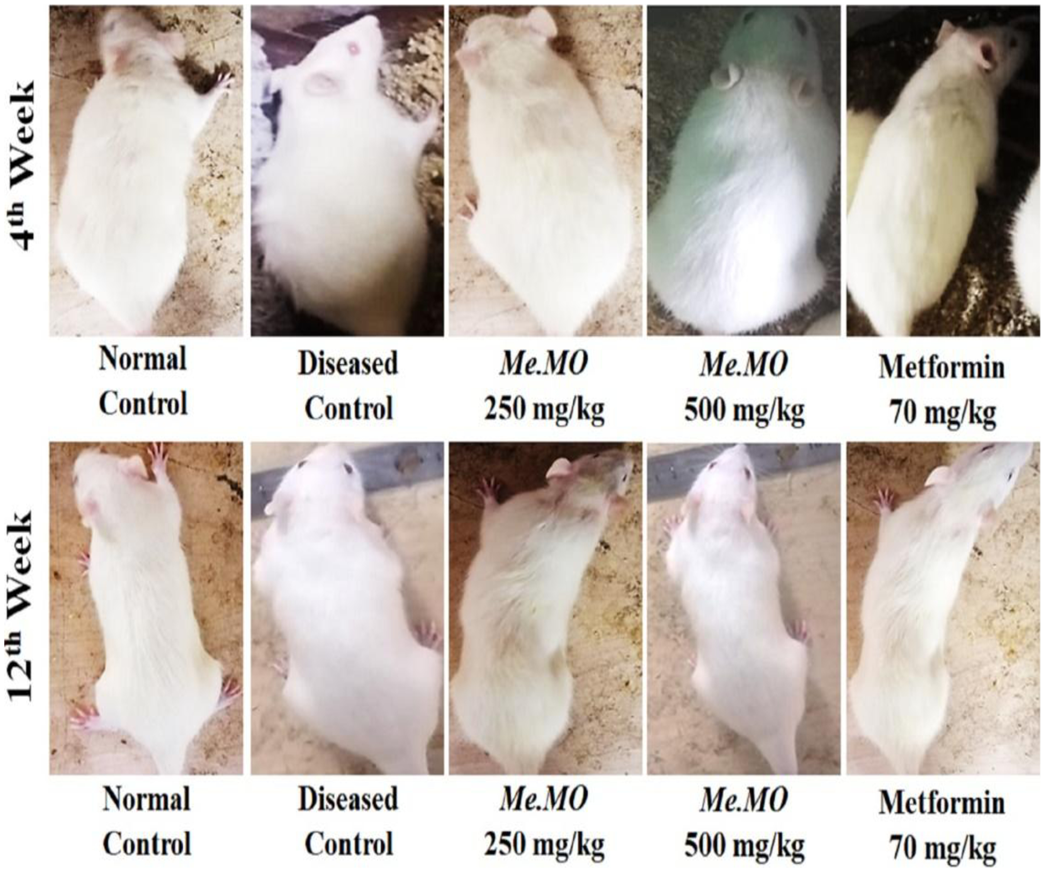
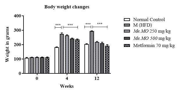
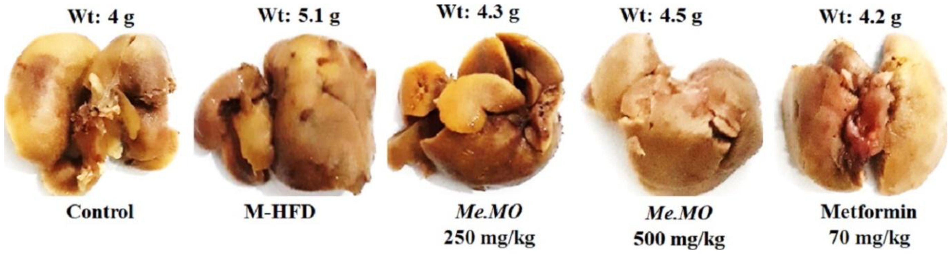
Figures 1and 2 represent the effect of Me.MO on the body weight of animals before, during, and after the study. At week 0, no significant (p > 0.05) difference was observed in body weights across all groups. However, by week 4, animal weights increased dramatically after HFD administration. Statistical analysis showed a significant (p < 0.001) increase in animal weights after receiving the HFD. At week 12, the mean weights for control, HFD, Me.MO 250 mg/kg, Me.MO 500 mg/kg, and metformin (70 mg/kg) groups were 205.0 ± 0.55, 291.0 ± 1.00, 206.0 ± 0.15, 215.0 ± 0.30, and 186.0 ± 0.90 g, respectively. Macroscopic analysis also showed marked increase and decrease in body weight of rats at the initial (week 4) and final stage (week 12) of the study (Figure 2). Similarly, liver weight increased in the HFD-induced group, while administration of Me.MO significantly reversed the elevated liver weights (Figure 3).
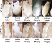
Macroscopic analysis of body weight variations on 0, 4th and 12th week in different study groups shown in representative pictographic form. Animals were selected blindly and randomly for macroscopic analysis. Animals were weighed after the pictures were taken and found consistent with the macroscopic analysis.
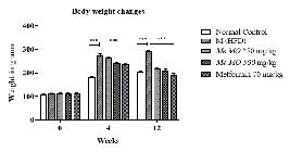
The variation in body weights at 0, 4th, and 12th week of study. The data shown represent the means ± SEM. ***P < 0.001.
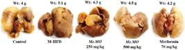
Macroscopic analysis and weight measurement of rats. The average liver weight of that group (n = 5) is given above the respective representative images.
3.3. Random blood glucose levels reversed by Me.MO
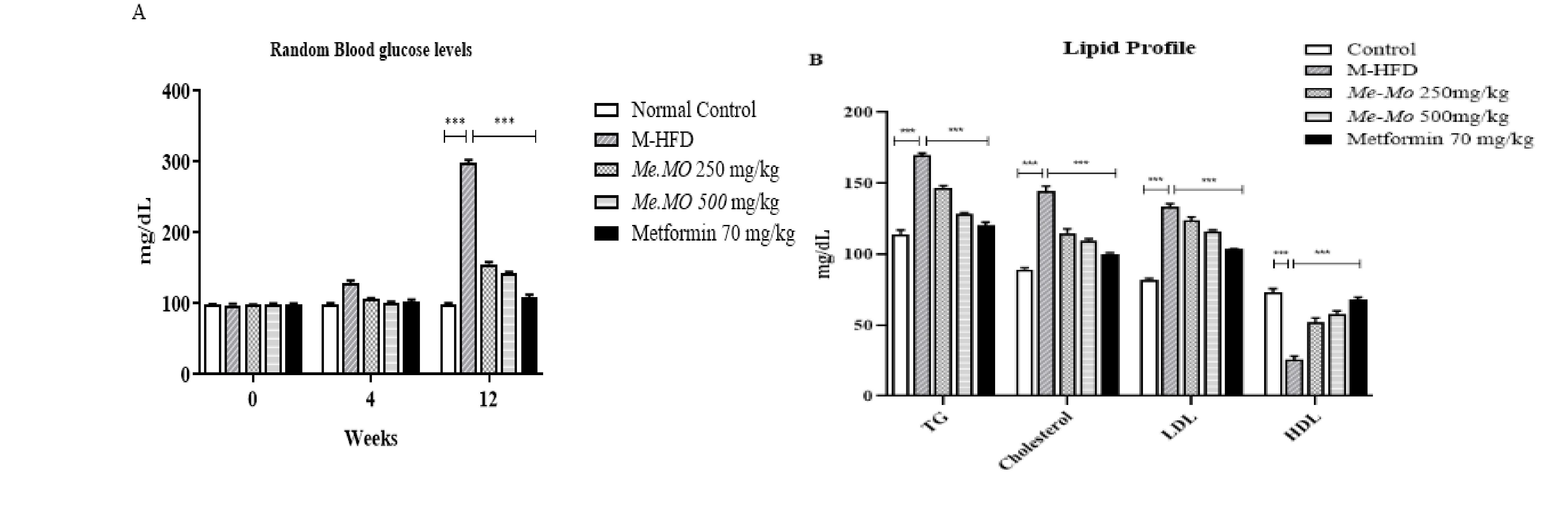
Figure 4A shows the effects of Me.MO on random blood glucose levels. At baseline (week 0), glucose levels were comparable across all groups. Following HFD and STZ administration, glucose levels increased significantly in all treatment groups compared to the control group. At week 12, random blood glucose levels were significantly elevated in the HFD group (305.0 ± 0.50 mg/dL) compared to the control. Treatment with Me.MO at 250 mg/kg (143.0 ± 0.40 mg/dL), Me.MO at 500 mg/kg (157.0 ± 1.65 mg/dL), and metformin (111.0 ± 0.60 mg/dL) significantly reduced glucose levels (p < 0.001) compared to the HFD group.
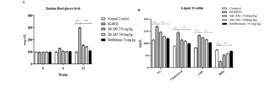
(A) Section A shows graphical representation of random blood glucose levels in HFD and STZ induced model at 0, 4th and 12th week of treatment respectively.The data shown represent the means ± SEM. ***P < 0.01; (B) Section B shows a graphical representation of the lipid profile after a twelve-week study. The data shown represent the means ± SEM. ***P < 0.01.
3.4. Assessment of lipid profile
Figure 4B illustrates the effects of Me.MO on serum lipid parameters. The HFD group showed significant increases in total cholesterol (145 ± 2.06 mg/dL), triglycerides (171 ± 4.35 mg/dL), and LDL-cholesterol (137 ± 4.09 mg/dL), with a concomitant decrease in HDL-cholesterol (36.3 ± 2.90 mg/dL) compared to the control group (p < 0.001). Treatment with Me.MO at both 250 mg/kg and 500 mg/kg doses significantly reduced total cholesterol (112 ± 1.32 mg/dL and 112 ± 1.19 mg/dL, respectively), triglycerides (138 ± 3.14 mg/dL and 143 ± 3.23 mg/dL, respectively), and LDL-cholesterol (114 ± 2.39 mg/dL and 118 ± 3.20 mg/dL, respectively) compared to the HFD group (p < 0.001). Additionally, Me.MO treatment significantly increased HDL-cholesterol levels compared to the HFD group (p < 0.001). These lipid-modulating effects were comparable to those observed with metformin treatment.
3.5. Histopathological analysis
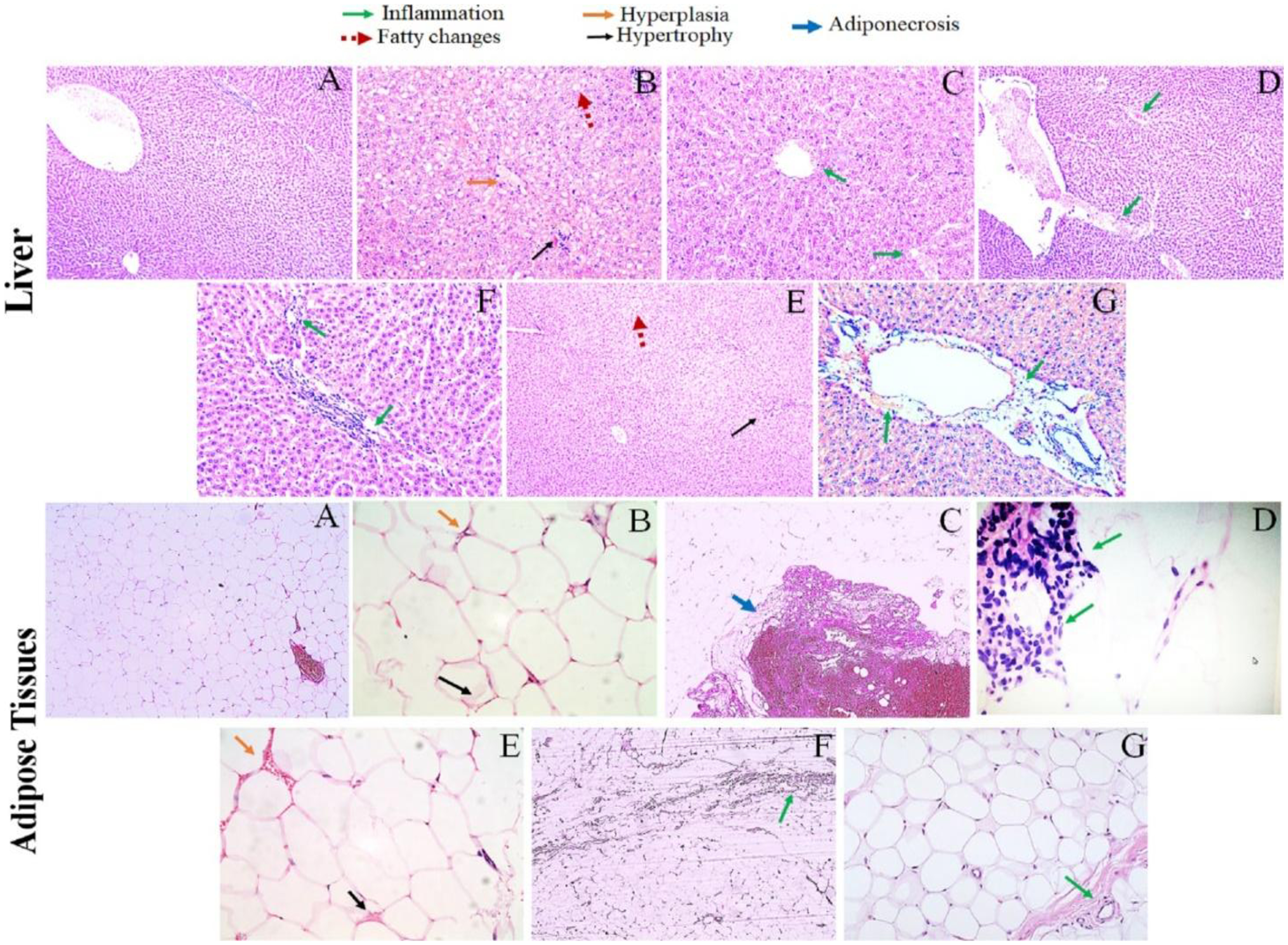
Histopathological examination of liver tissues revealed marked steatosis, hepatocellular ballooning, and inflammatory cell infiltration in the HFD group (Figure 5). Liver sections from the Me.MO 500 mg/kg group showed mild steatosis and inflammation, while the Me.MO 250 mg/kg group displayed moderate improvements compared to the HFD group. The metformin group exhibited histological features similar to the Me.MO 250 mg/kg group, with mild inflammatory changes (indicated by green arrows).
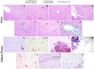
Histopathological analysis of HFD and STZ induced diabetic rats. A (Control), B and C (M-HFD), D (250 Me.MO), E and F (500 Me.MO) and G (Metformin 70 mg/kg). Representative micrographs show tissue sections stained with hematoxylin and eosin (H&E). Green square‑tipped arrows indicate hepatocellular hyperplasia (increased cell density). Blue circle‑tipped arrows indicate inflammation (presence of mononuclear or mixed inflammatory cell infiltrates). Red diamond‑tipped arrows indicate fatty changes (macrovesicular or microvesicular steatosis). Black arrowheads indicate adiponecrosis (loss of adipocyte architecture with necrotic debris and surrounding inflammatory reaction).
Adipose tissue examination revealed adipocyte hypertrophy and evidence of adiponecrosis (indicated by blue arrows) in the HFD group. Treatment with Me.MO at both doses resulted in reduced adipocyte size and decreased inflammatory changes, with the 500 mg/kg dose showing more pronounced effects. These histopathological findings correlated with the observed reductions in body weight and improvements in metabolic parameters.
3.6. Measurement of pro- and anti-inflammatory cytokines
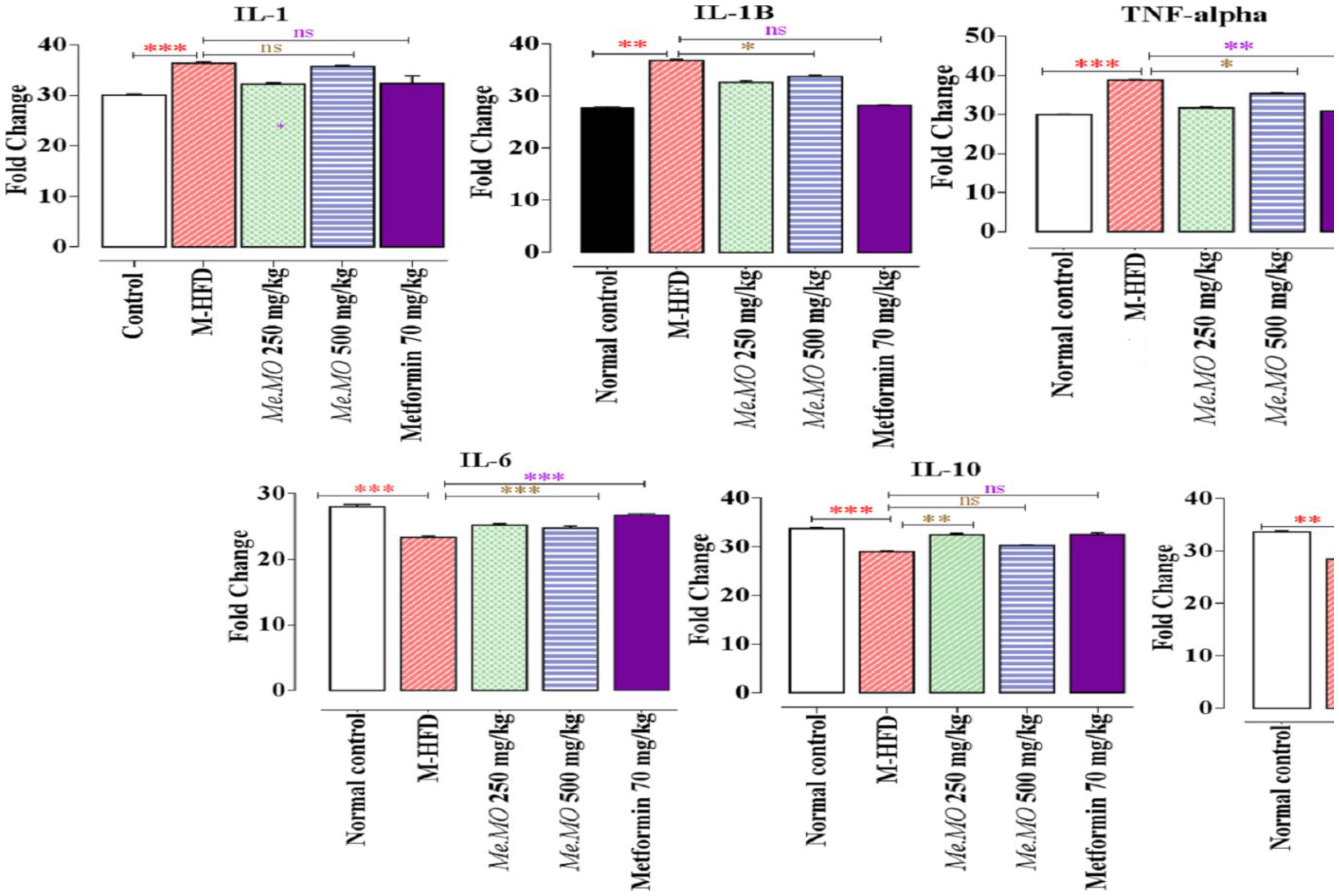
Figure 6 presents the effects of Me.MO on inflammatory cytokines. Treatment with Me.MO at both doses and metformin significantly reduced pro-inflammatory cytokines IL-1β and TNF-α compared to the HFD group (p < 0.001). Interestingly, levels of the anti-inflammatory cytokine IL-10 showed no significant differences between treatment groups and the HFD group (p > 0.05). However, other anti-inflammatory/regulatory markers including IL-6 and COX-2 were significantly elevated in the Me.MO and metformin treatment groups compared to the HFD group (p < 0.001). These findings suggest that Me.MO exerts its beneficial effects through modulation of the inflammatory response, potentially shifting the balance from pro-inflammatory to anti-inflammatory states.
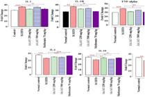
Effects of MHTD on inflammatory cytokine expression, COX-2 levels, and platelet counts. Data are presented as fold change relative to the control group (set to 1000 for Platelets, and normalized for other markers). Groups include Control, Normal control, MHTD, and MHTD + MHTD (likely a combination or repeated treatment). The following parameters were assessed: IL-1, IL-1B, TNF-alpha, Platelets, IL-6, IL-10, and COX-2. MHTD treatment alone or in combination resulted in a dose- or time-dependent reduction in pro-inflammatory cytokines (IL-1, IL-1B, TNF-alpha, IL-6) and COX-2 expression compared to controls, while levels in Normal control groups remained consistently lower. Platelet counts remained unchanged across all experimental conditions (constant fold change of 1000), indicating no effect of MHTD on platelet levels under these experimental settings. Anti-inflammatory cytokine IL-10 showed a similar pattern of reduction. All values represent mean fold change (n=…, error bars omitted for clarity).
4. Discussion
Obesity and fatty liver disease represent increasingly prevalent metabolic disorders with significant health implications. While these conditions initially present as relatively benign, their progression is associated with chronic inflammation and metabolic dysregulation. This situation potentially leads to more severe complications including type 2 diabetes, cardiovascular disease, and hepatic cirrhosis (Friedman et al., 2018). The present study investigated the therapeutic potential of methanolic extract of Moringa oleifera (Me.MO) in ameliorating obesity, fatty liver, and associated inflammatory states.
Our findings demonstrate that Me.MO administration significantly reduced body weight and liver weight in rats fed a high-fat diet supplemented with streptozotocin. These effects were dose-dependent, with the higher dose (500 mg/kg) showing more pronounced benefits. Recent advances in understanding the immunometabolic interface in obesity have highlighted the role of adipose tissue macrophages in perpetuating chronic inflammation (Jang et al., 2025). Our results align with emerging evidence that plant-derived polyphenols can modulate this inflammatory milieu, potentially interrupting the vicious cycle of lipid accumulation and immune activation (Mamun et al., 2024).
The HFD-STZ model employed in this study effectively mimics the pathophysiological changes observed in obesity and associated metabolic disorders, including insulin resistance, hyperglycemia, and dyslipidemia (Zhang et al., 2008; Glastras et al., 2016). The development of obesity and fatty liver in this model involves complex interactions between excessive caloric intake, lipid accumulation, and inflammatory processes (Vulchi et al., 2023). Our observation that Me.MO treatment attenuated these changes suggests its multi-faceted effects on metabolic regulation and inflammation.
Lipid profile abnormalities, including elevated LDL-cholesterol, triglycerides, and reduced HDL-cholesterol, are key contributors to cardiovascular risk in obesity and metabolic syndrome (Estrada-Luna et al., 2018). Our observation that Me.MO treatment significantly improved lipid profiles aligns with recent clinical evidence suggesting that Moringa supplementation can reduce total cholesterol and LDL levels in patients with dyslipidemia (Crișan et al., 2025). The mechanisms may involve both decreased cholesterol synthesis through inhibition of HMG-CoA reductase and enhanced cholesterol efflux through upregulation of reverse cholesterol transport pathways (Hassaan et al., 2026).
Hyperglycemia is a hallmark of metabolic dysfunction in obesity, often resulting from insulin resistance in peripheral tissues (Chang et al., 2015). In our study, Me.MO treatment significantly reduced blood glucose levels in HFD-STZ rats, suggesting improved insulin sensitivity and glucose homeostasis. These findings are consistent with previous reports on the antidiabetic effects of Moringa (Sholapur and Patil, 2013; Efiong et al., 2013). The insulin-sensitizing effects of Me.MO may be attributed to bioactive compounds such as quercetin and chlorogenic acid, which have been shown to enhance insulin signaling pathways and glucose uptake in skeletal muscle (Santana-Gálvez et al., 2017).
The anti-inflammatory effects of Me.MO observed in our study are consistent with recent research demonstrating that bioactive compounds in Moringa, particularly isothiocyanates and flavonoids, can inhibit NF-κB signaling and reduce the production of pro-inflammatory cytokines in multiple tissue types (Silva Parente et al., 2025). These effects may be particularly relevant in the context of diet-induced metabolic inflammation, where adipose tissue macrophages shift toward a pro-inflammatory M1 phenotype (Toprak et al., 2026). By modulating the balance of pro- and anti-inflammatory mediators, Me.MO may interrupt the self-perpetuating cycle of inflammation and metabolic dysfunction.
Histopathological findings further support the beneficial effects of Me.MO on liver and adipose tissue health. The reduction in hepatic steatosis, inflammation, and adipocyte hypertrophy observed in Me.MO-treated animals correlates with improved metabolic parameters and reduced inflammatory markers. These structural improvements likely contribute to the functional benefits observed, including enhanced insulin sensitivity and lipid metabolism.
HPLC analysis confirmed the presence of several bioactive compounds in Me.MO, including quercetin, chlorogenic acid, syringic acid, vanillic acid, and m-coumaric acid. These polyphenolic compounds have well-documented metabolic and anti-inflammatory effects (Vogel et al., 2014). Quercetin has been shown to attenuate metabolic syndrome symptoms by reducing oxidative stress and inflammation (Rochlani et al., 2017; Hassan et al., 2014). Vanillic acid can ameliorate hyperinsulinemia and improve antioxidant status, thereby reducing cardiovascular risk (Chang et al., 2015). Chlorogenic acid plays dual roles in metabolic regulation by inhibiting glucose-6-phosphatase expression and enhancing glucose uptake in skeletal muscle, thus improving insulin sensitivity and lipid metabolism (Santana-Gálvez et al., 2017). Syringic acid can regulate metabolic enzymes and promote β-cell regeneration (Srinivasulu et al., 2018), while m-coumaric acid inhibits reactive oxygen species production, thereby preventing hyperglycemia-induced vascular damage (Moselhy et al., 2018). The synergistic actions of these compounds likely contribute to the comprehensive therapeutic effects of Me.MO observed in this study.
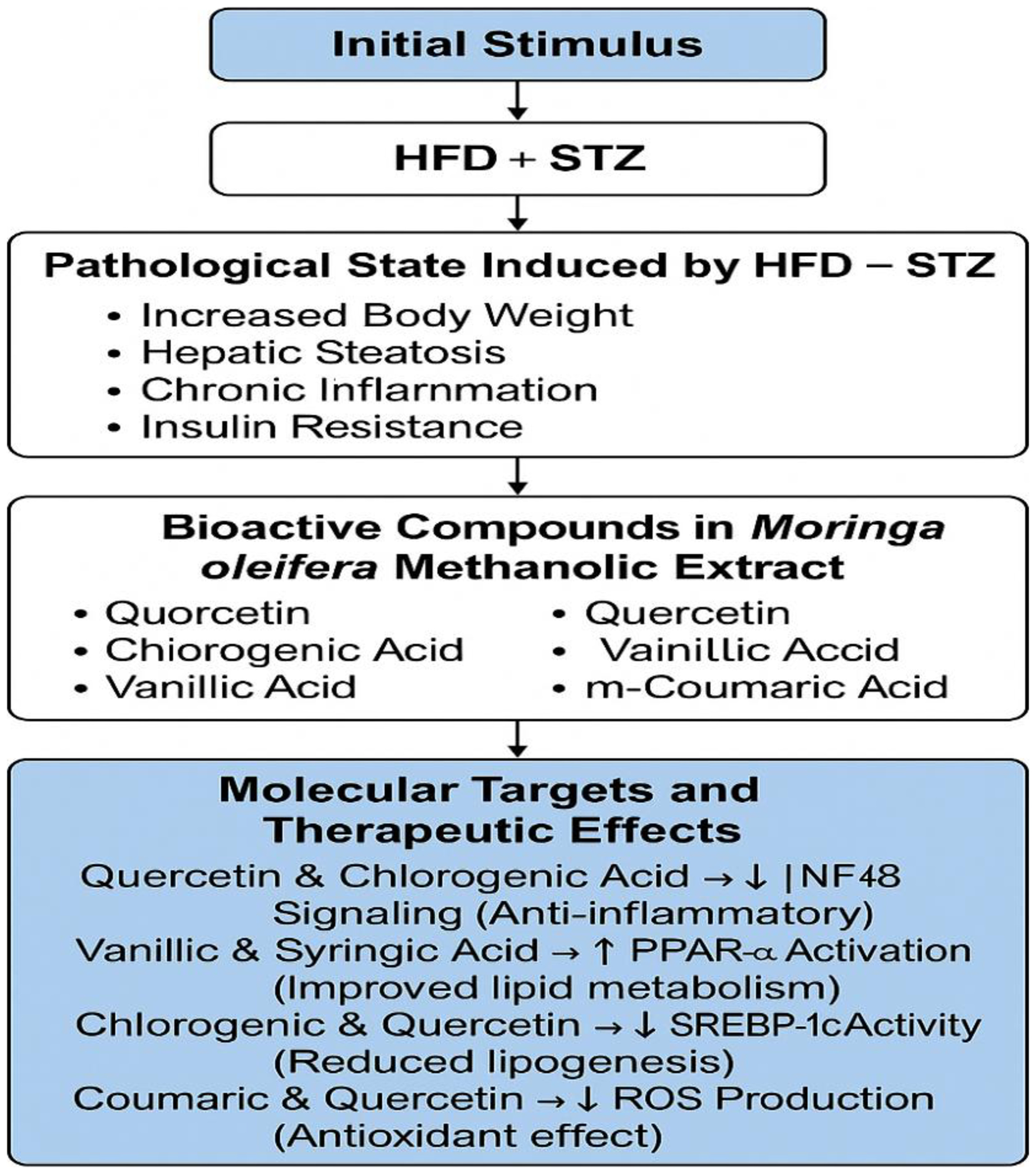
Based on our findings and existing literature, we propose a potential mechanism of action for Me.MO in obesity and fatty liver (Figure 7). The bioactive compounds in Me.MO target multiple pathways, including inflammatory signaling (NF-κB, MAPK), lipid metabolism (PPAR-α, SREBP-1c), and insulin signaling, resulting in reduced adiposity, improved hepatic function, modulated immune responses, and enhanced metabolic parameters. This multi-target approach addresses the complex pathophysiology of obesity and fatty liver, offering advantages over conventional single-target pharmaceuticals.
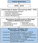
Illustrates the comprehensive mechanism by which Moringa oleifera methanolic extract exerts therapeutic effects. Starting from the initial stimulus (HFD + STZ), it outlines the induced pathological states and highlights the key bioactive compounds within the extract. These compounds target molecular pathways associated with inflammation, oxidative stress, lipid metabolism, and insulin secretion, contributing to the mitigation of metabolic dysfunction.
While our study provides compelling evidence for the therapeutic potential of Me.MO, several limitations should be acknowledged. The sample size was relatively small, and the experimental duration may not fully capture long-term effects or safety profiles. Additionally, the mechanisms underlying the observed effects require further molecular characterization. Future studies should address these limitations and explore the potential for clinical translation.
5. Conclusions
This study demonstrates that the methanolic extract of Moringa oleifera effectively ameliorates HFD-induced obesity, fatty liver, and associated inflammatory stress in rats. The dual action of Me.MO on metabolic parameters and inflammatory pathways suggests a promising mechanistic basis for its therapeutic effects. The immunometabolic axis appears to be a key target for Me.MO's anti-obesity potential, highlighting the importance of addressing inflammation in metabolic disease management. While these preclinical findings are encouraging, further investigations, including toxicological assessments, dose optimization studies, and eventually clinical trials, are necessary to establish the safety and efficacy of Moringa oleifera for human applications. Future research should also explore the specific bioactive compounds responsible for these effects and their molecular mechanisms of action.
Acknowledgements
The study was supported by a research grant from the Deanship of Research & Graduate Studies, Ajman University #2023-IRG-PH-11.
Data Availability Statement
The datasets used and/or analyzed during the current study are available from the corresponding author upon reasonable request.
References
-
ABOU-ZEID, S.M., AHMED, A.I., AWAD, A., MOHAMMED, W.A., METWALLY, M.M.M., ALMEER, R., ABDEL-DAIM, M.M. and KHALIL, S.R., 2021. Moringa oleifera ethanolic extract attenuates tilmicosin-induced renal damage in male rats via suppression of oxidative stress, inflammatory injury, and intermediate filament proteins mRNA expression. Biomedicine & Pharmacotherapy = Biomédecine & Pharmacothérapie, vol. 133, pp. 110997. https://doi.org/10.1016/j.biopha.2020.110997 PMid:33197759.
» https://doi.org/10.1016/j.biopha.2020.110997 -
ALBRAHIM, T. and BINOBEAD, M.A., 2018. Roles of Moringa oleifera leaf extract in improving the impact of high dietary intake of monosodium glutamate-induced liver toxicity, oxidative stress, genotoxicity, DNA damage, and PCNA alterations in male rats. Oxidative Medicine and Cellular Longevity, vol. 2018, no. 1, pp. 4501097. https://doi.org/10.1155/2018/4501097 PMid:30647808.
» https://doi.org/10.1155/2018/4501097 -
BOSSERHOFF, A. and HELLERBRAND, C., 2011. Obesity and fatty liver are ‘grease’ for the machinery of hepatic fibrosis. Digestive Diseases, vol. 29, no. 4, pp. 377-383. https://doi.org/10.1159/000329800 PMid:21894008.
» https://doi.org/10.1159/000329800 -
CHANG, W.C., WU, J.S.B., CHEN, C.W., KUO, P.L., CHIEN, H.M., WANG, Y.T. and SHEN, S.C., 2015. Protective effect of vanillic acid against hyperinsulinemia, hyperglycemia and hyperlipidemia via alleviating hepatic insulin resistance and inflammation in high-fat diet (HFD)-fed rats. Nutrients, vol. 7, no. 12, pp. 9946-9959. https://doi.org/10.3390/nu7125514 PMid:26633482.
» https://doi.org/10.3390/nu7125514 -
CRIȘAN, D., GAVRILAȘ, L., PĂLTINEAN, R., FRUMUZACHI, O., MOCAN, A. and CRIȘAN, G., 2025. Effects ofMoringa oleiferaLam. supplementation on cardiometabolic outcomes: a meta-analysis of randomized controlled trials with GRADE assessment. Nutrients, vol. 17, no. 22, pp. 3501. https://doi.org/10.3390/nu17223501
» https://doi.org/10.3390/nu17223501 -
CUELLAR-NÚÑEZ, M.L., GONZALEZ DE MEJIA, E. and LOARCA-PIÑA, G., 2021. Moringa oleifera leaves alleviated inflammation through downregulation of IL-2, IL-6, and TNF-α in a colitis-associated colorectal cancer model. Food Research International, vol. 144, pp. 110318. https://doi.org/10.1016/j.foodres.2021.110318 PMid:34053523.
» https://doi.org/10.1016/j.foodres.2021.110318 - EFIONG, E.E., IGILE, G.O., MGBEJE, B.I.A., OTU, E.A. and EBONG, P.E., 2013. Hepatoprotective and anti-diabetic effect of combined extracts of Moringa oleifera and Vernonia amygdalina. Journal of Diabetes and Endocrinology, vol. 4, no. 4, pp. 45-50. http://doi.org/10.5897/JDE2013.0069.
-
ESTRADA-LUNA, D., ORTIZ-RODRIGUEZ, M.A., MEDINA-BRISEÑO, L., CARREÓN-TORRES, E., IZQUIERDO-VEGA, J.A., SHARMA, A., CANCINO-DÍAZ, J.C., PÉREZ-MÉNDEZ, O., BELEFANT-MILLER, H. and BETANZOS-CABRERA, G., 2018. Current therapies focused on high-density lipoproteins associated with cardiovascular disease. Molecules, vol. 23, no. 11, pp. 2730. https://doi.org/10.3390/molecules23112730 PMid:30360466.
» https://doi.org/10.3390/molecules23112730 -
FLOCK, M.R., ROGERS, C.J., PRABHU, K.S. and KRIS-ETHERTON, P.M., 2013. Immunometabolic role of long-chain omega-3 fatty acids in obesity-induced inflammation. Diabetes/Metabolism Research and Reviews, vol. 29, no. 6, pp. 431-445. https://doi.org/10.1002/dmrr.2414 PMid:23592441.
» https://doi.org/10.1002/dmrr.2414 -
FRIEDMAN, S.L., NEUSCHWANDER-TETRI, B.A., RINELLA, M. and SANYAL, A.J., 2018. Mechanisms of NAFLD development and therapeutic strategies. Nature Medicine, vol. 24, no. 7, pp. 908-922. https://doi.org/10.1038/s41591-018-0104-9 PMid:29967350.
» https://doi.org/10.1038/s41591-018-0104-9 -
GIORGINO, F., FRANCO, D.R., NICOLAY, C., HEMMINGWAY, A., RODRÍGUEZ, Á. and WIESE, R.J., 2024. Effects of tirzepatide versus basal insulins in people with type 2 diabetes and different baseline glycemic patterns: post hoc analyses of the SURPASS-3 and SURPASS-4 trials. Diabetes Care, vol. 47, no. 6, pp. 1020-1027. https://doi.org/10.2337/dc23-2366
» https://doi.org/10.2337/dc23-2366 -
GLASTRAS, S.J., CHEN, H., TEH, R., MCGRATH, R.T., CHEN, J., POLLOCK, C.A., WONG, M.G. and SAAD, S., 2016. Mouse models of diabetes, obesity and related kidney disease. PLoS One, vol. 11, no. 8, e0162131. https://doi.org/10.1371/journal.pone.0162131 PMid:27579698.
» https://doi.org/10.1371/journal.pone.0162131 -
GLEESON, L.E., ROCHE, H.M. and SHEEDY, F.J., 2021. Obesity, COVID-19 and innate immunometabolism. The British Journal of Nutrition, vol. 125, no. 6, pp. 628-632. https://doi.org/10.1017/S0007114520003529 PMid:32892755.
» https://doi.org/10.1017/S0007114520003529 -
GREEN, W.D. and BECK, M.A., 2017. Obesity altered T cell metabolism and the response to infection. Current Opinion in Immunology, vol. 46, pp. 1-7. https://doi.org/10.1016/j.coi.2017.03.008 PMid:28359913.
» https://doi.org/10.1016/j.coi.2017.03.008 -
HASSAAN, A.I., EBEED, N.M., FATOUH, A. and ELHARIRY, H., 2026. Metabolic and molecular evaluation of Moringa oleifera-supplemented ketogenic meal replacement in healthy C57BL/6 mice. Scientific Reports, vol. 16, no. 1, pp. 4091. https://doi.org/10.1038/s41598-025-34443-z
» https://doi.org/10.1038/s41598-025-34443-z -
HASSAN, W., RONGYIN, G., DAOUD, A., DING, L., WANG, L., LIU, J. and SHANG, J., 2014. Reduced oxidative stress contributes to the lipid lowering effects of isoquercitrin in free fatty acids induced hepatocytes. Oxidative Medicine and Cellular Longevity, vol. 2014, pp. 313602. https://doi.org/10.1155/2014/313602 PMid:25404990.
» https://doi.org/10.1155/2014/313602 -
HOTAMISLIGIL, G.S., 2017. Foundations of immunometabolism and implications for metabolic health and disease. Immunity, vol. 47, no. 3, pp. 406-420. https://doi.org/10.1016/j.immuni.2017.08.009 PMid:28930657.
» https://doi.org/10.1016/j.immuni.2017.08.009 -
JANG, J.H., SUNG, J.H. and HUH, J.Y., 2025. Diverse functions of macrophages in obesity and metabolic dysfunction-associated steatotic liver disease: bridging inflammation and metabolism. Immune Network, vol. 25, no. 1, e12. https://doi.org/10.4110/in.2025.25.e12
» https://doi.org/10.4110/in.2025.25.e12 -
KOLIAKI, C., LIATIS, S. and KOKKINOS, A., 2019. Obesity and cardiovascular disease: revisiting an old relationship. Metabolism: Clinical and Experimental, vol. 92, pp. 98-107. https://doi.org/10.1016/j.metabol.2018.10.011 PMid:30399375.
» https://doi.org/10.1016/j.metabol.2018.10.011 -
LIU, R., WANG, M., LI, E., YANG, Y., LI, J., CHEN, S., SHEN, W.J., AZHAR, S., GUO, Z. and HU, Z., 2020. Dysregulation of microRNA-125a contributes to obesity-associated insulin resistance and dysregulates lipid metabolism in mice. Biochimica et Biophysica Acta. Molecular and Cell Biology of Lipids, vol. 1865, no. 5, pp. 158640. https://doi.org/10.1016/j.bbalip.2020.158640 PMid:31988048.
» https://doi.org/10.1016/j.bbalip.2020.158640 -
MAMUN, M.A.A., RAKIB, A., MANDAL, M., KUMAR, S., SINGLA, B. and SINGH, U.P., 2024. Polyphenols: role in modulating immune function and obesity. Biomolecules, vol. 14, no. 2, pp. 221. https://doi.org/10.3390/biom14020221
» https://doi.org/10.3390/biom14020221 -
MICHAILIDOU, Z., GOMEZ-SALAZAR, M. and ALEXAKI, V.I., 2022. Innate immune cells in the adipose tissue in health and metabolic disease. Journal of Innate Immunity, vol. 14, no. 1, pp. 4-30. https://doi.org/10.1159/000515117 PMid:33849008.
» https://doi.org/10.1159/000515117 -
MOSELHY, S.S., RAZVI, S.S., ALSHIBILI, F.A., KUERBAN, A., HASAN, M.N., BALAMASH, K.S., HUWAIT, E.A., ABDULAAL, W.H., AL-GHAMDI, M.A., KUMOSANI, T.A., ABULNAJA, K.O., AL-MALKI, A.L., ASAMI, T. and ISMAIL, I.M., 2018. m-Coumaric acid attenuates non-catalytic protein glycosylation in the retinas of diabetic rats. Journal of Pesticide Science, vol. 43, no. 3, pp. 180-185. https://doi.org/10.1584/jpestics.D17-091 PMid:30363123.
» https://doi.org/10.1584/jpestics.D17-091 -
MUNAWAR, N., MAHATO, A., RAWAT, A., GILL, F.S., KUMAR, D., KATWAL, S., WEI, C.R. and ALI, N., 2025. Tirzepatide versus semaglutide for weight loss in overweight and obese adults: a systematic review and meta-analysis of direct comparative studies. Cureus, vol. 17, no. 6, e86080. https://doi.org/10.7759/cureus.86080
» https://doi.org/10.7759/cureus.86080 -
NIZIOŁ-ŁUKASZEWSKA, Z., FURMAN-TOCZEK, D., BUJAK, T., WASILEWSKI, T. and HORDYJEWICZ-BARAN, Z., 2020. Moringa oleifera L. extracts as bioactive ingredients that increase safety of body wash cosmetics. Dermatology Research and Practice, vol. 2020, pp. 8197902. https://doi.org/10.1155/2020/8197902 PMid:32695156.
» https://doi.org/10.1155/2020/8197902 -
OIKONOMOU, E.K. and ANTONIADES, C., 2023. The role of adipose tissue in cardiovascular health and disease. Nature Reviews. Cardiology, vol. 20, pp. 55-72. https://doi.org/10.1038/s41569-022-00735-4 PMid:30287946.
» https://doi.org/10.1038/s41569-022-00735-4 -
OLTMAN, C.L., DAVIDSON, E.P., COPPEY, L.J., KLEINSCHMIDT, T.L., LUND, D.D. and YOREK, M.A., 2008. Attenuation of vascular/neural dysfunction in Zucker rats treated with enalapril or rosuvastatin. Obesity, vol. 16, no. 1, pp. 82-89. https://doi.org/10.1038/oby.2007.19 PMid:18223617.
» https://doi.org/10.1038/oby.2007.19 -
PAREEK, A., PANT, M., GUPTA, M.M., KASHANIA, P., RATAN, Y., JAIN, V., PAREEK, A. and CHUTURGOON, A.A., 2023. Moringa oleifera: an updated comprehensive review of its pharmacological activities, ethnomedicinal, phytopharmaceutical formulation, clinical, phytochemical, and toxicological aspects. International Journal of Molecular Sciences, vol. 24, no. 3, pp. 2098. https://doi.org/10.3390/ijms24032098
» https://doi.org/10.3390/ijms24032098 -
ROCHLANI, Y., POTHINENI, N.V., KOVELAMUDI, S. and MEHTA, J.L., 2017. Metabolic syndrome: pathophysiology, management, and modulation by natural compounds. Therapeutic Advances in Cardiovascular Disease, vol. 11, no. 8, pp. 215-225. https://doi.org/10.1177/1753944717711379 PMid:28639538.
» https://doi.org/10.1177/1753944717711379 -
ROHM, T.V., MEIER, D.T., OLEFSKY, J.M. and DONATH, M.Y., 2022. Inflammation in obesity, diabetes, and related disorders. Immunity, vol. 55, no. 1, pp. 31-55. https://doi.org/10.1016/j.immuni.2021.12.013
» https://doi.org/10.1016/j.immuni.2021.12.013 -
SALEEM, M., IFTIKHAR, A., ASIF, M., HUSSAIN, K., ALAMGEER, SHAH, P.A., SALEEM, A., AKHTAR, F., AMIR, R.M. and QAMAR, M.M., 2021. Asphodelus tenuifolius extracts arrested inflammation and arthritis through modulation of TNF-α, NF-κB, ILs, and COX-2 activities in in vivo models. Inflammopharmacology, vol. 29, no. 2, pp. 483-497. https://doi.org/10.1007/s10787-020-00761-z PMid:33064243.
» https://doi.org/10.1007/s10787-020-00761-z -
SALTIEL, A.R. and OLEFSKY, J.M., 2017. Inflammatory mechanisms linking obesity and metabolic disease. The Journal of Clinical Investigation, vol. 127, no. 1, pp. 1-4. https://doi.org/10.1172/JCI92035 PMid:28045402.
» https://doi.org/10.1172/JCI92035 -
SANTANA-GÁLVEZ, J., CISNEROS-ZEVALLOS, L. and JACOBO-VELÁZQUEZ, D.A., 2017. Chlorogenic acid: recent advances on its dual role as a food additive and a nutraceutical against metabolic syndrome. Molecules, vol. 22, no. 3, pp. 358. https://doi.org/10.3390/molecules22030358 PMid:28245635.
» https://doi.org/10.3390/molecules22030358 -
SHOLAPUR, H. and PATIL, B., 2013. Effect of Moringa oleifera bark extracts on dexamethasone-induced insulin resistance in rats. Drug Research, vol. 63, no. 10, pp. 527-531. https://doi.org/10.1055/s-0033-1347238 PMid:23780503.
» https://doi.org/10.1055/s-0033-1347238 -
SILVA PARENTE, T.S.J., SARANDY, M.M., DE ARAÚJO, E.R.D., GONÇALVES, R.V. and ZUCOLOTTO, S.M., 2025. Effect of Moringa oleifera on inflammatory diseases: an umbrella review of 26 systematic reviews. Frontiers in Pharmacology, vol. 16, pp. 1572337. https://doi.org/10.3389/fphar.2025.1572337
» https://doi.org/10.3389/fphar.2025.1572337 -
SRINIVASULU, C., RAMGOPAL, M., RAMANJANEYULU, G., ANURADHA, C. and KUMAR, C.S., 2018. Syringic acid (SA)‒a review of its occurrence, biosynthesis, pharmacological and industrial importance. Biomedicine & Pharmacotherapy = Biomédecine & Pharmacothérapie, vol. 108, pp. 547-557. https://doi.org/10.1016/j.biopha.2018.09.069 PMid:30243088.
» https://doi.org/10.1016/j.biopha.2018.09.069 -
TAK, Y.J. and LEE, S.Y., 2021. Long-term efficacy and safety of anti-obesity treatment: where do we stand? Current Obesity Reports, vol. 10, no. 1, pp. 14-30. https://doi.org/10.1007/s13679-020-00422-w PMid:33410104.
» https://doi.org/10.1007/s13679-020-00422-w -
TOPRAK, K., YILDIRIM, Z., KAPLAN, D.N., SENTURK, G. and SANLIER, N., 2026. Phytochemicals and obesity: a mini review from the dietary phytochemical index perspective. Current Nutrition Reports, vol. 15, no. 1, pp. 37. https://doi.org/10.1007/s13668-026-00763-3
» https://doi.org/10.1007/s13668-026-00763-3 -
VERGARA-JIMENEZ, M., ALMATRAFI, M.M. and FERNANDEZ, M.L., 2017. Bioactive components in Moringa oleifera leaves protect against chronic disease. Antioxidants, vol. 6, no. 4, pp. 91. https://doi.org/10.3390/antiox6040091 PMid:29144438.
» https://doi.org/10.3390/antiox6040091 -
VOGEL, P., KASPER MACHADO, I., GARAVAGLIA, J., ZANI, V.T., DE SOUZA, D. and MORELO DAL BOSCO, S., 2014. Polyphenols benefits of olive leaf (Olea europaea L) to human health. Nutrición Hospitalaria, vol. 31, no. 3, pp. 1427-1433. https://doi.org/10.3305/nh.2015.31.3.8400 PMid:25726243.
» https://doi.org/10.3305/nh.2015.31.3.8400 -
VULCHI, J., SURYADEVARA, V., MOHAN, P., KAMALANATHAN, S., SAHOO, J., NAIK, D. and SELVARAJAN, S., 2023. Obesity and metabolic dysfunction-associated fatty liver disease: understanding the intricate link. Journal of Translational Gastroenterology, vol. 1, no. 2, pp. 74-86. https://doi.org/10.14218/JTG.2023.00043
» https://doi.org/10.14218/JTG.2023.00043 -
YASEEN, H.S., ASIF, M., SAADULLAH, M., MAHRUKH, M., ASGHAR, S., SHAMS, M.U., BAZMI, R.R., SALEEM, M., YOUSAF, H.M. and YASEEN, M., 2020. Methanolic extract of Ephedra ciliata promotes wound healing and arrests inflammatory cascade in vivo through downregulation of TNF-α. Inflammopharmacology, vol. 28, no. 6, pp. 1691-1704. https://doi.org/10.1007/s10787-020-00713-7 PMid:32385747.
» https://doi.org/10.1007/s10787-020-00713-7 -
ZHANG, M., LV, X.Y., LI, J., XU, Z.G. and CHEN, L., 2008. The characterization of high-fat diet and multiple low-dose streptozotocin induced type 2 diabetes rat model. Experimental Diabetes Research, vol. 2008, no. 1, pp. 704045. https://doi.org/10.1155/2008/704045 PMid:19132099.
» https://doi.org/10.1155/2008/704045
Edited by
-
Editor:
Marcelo A.M. Esquisatto