Exploring the anticancer potential of Costus speciosus: a comprehensive review
Abstract
Costus speciosus is a medicinal plant with a long history in Indian Ayurvedic medicine, recognized for its diverse bioactive properties, including antibacterial, anticancer, anti-inflammatory, and antidiabetic effects. This review highlights its therapeutic potential, particularly in cancer treatment, where its bioactive compounds exhibit cytotoxic effects against breast, ovarian, and uterine cancers. These compounds have shown the ability to induce apoptosis, regulate the cell cycle, and inhibit cancer progression, offering a promising alternative to conventional chemotherapy with potentially fewer side effects. Additionally, C. speciosus demonstrates antioxidant, antimicrobial, and antidiabetic properties, expanding its clinical applications. Despite its promising pharmacological profile, further research is required to understand the molecular mechanisms underlying its therapeutic effects and ensure the safety and efficacy of its various extracts for therapeutic use.
Keywords:
Costus specious; anticancer; antioxidant; antimicrobial; antidiabetic; anti-inflammatory; therapeutic medications
Resumo
Costus speciosus é uma planta medicinal com uma longa história na medicina ayurvédica indiana, reconhecida por suas diversas propriedades bioativas, incluindo efeitos antibacterianos, anticâncer, anti-inflamatórios e antidiabéticos. Esta revisão destaca seu potencial terapêutico, particularmente no tratamento do câncer, onde seus compostos bioativos exibem efeitos citotóxicos contra cânceres de mama, ovário e útero. Esses compostos demonstraram a capacidade de induzir apoptose, regular o ciclo celular e inibir a progressão do câncer, oferecendo uma alternativa promissora à quimioterapia convencional com potencialmente menos efeitos colaterais. Além disso, C. speciosus demonstra propriedades antioxidantes, antimicrobianas e antidiabéticas, expandindo suas aplicações clínicas. Apesar de seu perfil farmacológico promissor, mais pesquisas são necessárias para entender os mecanismos moleculares subjacentes aos seus efeitos terapêuticos e garantir a segurança e eficácia de seus vários extratos para uso terapêutico.
Palavras-chave:
Costus speciosus; anticâncer; antioxidante; antimicrobiano; antidiabético; anti-inflamatório; medicamentos terapêuticos
1. Introduction
Uncontrolled, aberrant cells are a hallmark of cancer, a group of malignant disorders (Gurunanselage Don and Yap, 2019). Despite improvements in diagnosis, treatment, and preventative tools, cancer has a relatively high fatality rate in humans (Mohd-Salleh et al., 2020). Even with new chemotherapeutic techniques and other therapies, the incidence of cancer continues to rise and it remains a major global public health concern (Mahmod et al., 2020). According to global cancer estimates, 1 in 5 men and 1 in 6 women are likely to develop cancer, and 1 in 8 men and 1 in 10 women will die from the disease (Ali et al., 2021). At present, cancer represents the main cause of death for humans and animals throughout the world.
Cancer is characterised by uncontrolled cell cycle progression, apoptosis suppression, and increased angiogenic potential as well as proliferation, invasion, and metastatic capacities. Genetic instability and changes inside cells and tissues lead to the unchecked proliferation of normal cells, turning them into cancerous cells (Stadlbauer et al., 2019). The multi-step process of carcinogenesis is initiated by alterations in the expression of oncogenes and transcriptional factors involved in cell proliferation, cell cycle regulation, cell apoptosis, differentiation, angiogenesis, invasion, and metastasis (Ramasamy and Agarwal, 2008). Indeed, a myriad of genes can be involved in cancer, including oncogenes, tumour suppressor genes, DNA repair genes, and genes involved in cell growth metabolism (Mota et al., 2020; Negrao et al., 2021). This genetic instability is caused by both external and internal factors (Abdullahi et al., 2019). Therefore, the best cancer chemopreventive medicines should be those that target one or more of these mechanisms (Liu et al., 2014). According to Geiger et al. (2020), malignancies such as breast cancer, ovarian, colorectal, bladder, kidney cancer, and other tumour diseases.
Although great advancements have allowed clinicians to treat cancer more effectively, several adverse effects are associated with modern chemotherapy. Natural remedies, such as the use of medicinal plant or their products, to treat cancer may decrease these side effects. Recently, innovative screening practices utilising herbs or plants have enabled the discovery of phytochemicals with anticancer properties (Abd El-Hack et al., 2023). Because of their unmatched chemical variety, natural medicines derived from medicinal plants as opposed to pure chemicals or crude extracts offer endless sources of novel pharmaceuticals (Mat Jusoh et al., 2023).
Herbal medicine is the study of employing medicinal herbs that exert pharmacologic effects (Sharma et al., 2020). There has been great research interest on medicinal plants (Darkhor et al., 2018). According to Larki et al. (2020), a medicinal plant is any plant that contains compounds in one or more of its organs that can be utilised therapeutically. The utilisation of medicinal plants is not a recent phenomenon. For millennia, they have been used to treat and prevent illnesses. Various chemical families of natural phytochemicals, acting as natural antioxidants, and other substances are responsible for the efficacy of these therapeutic medications made from natural products (Larki et al., 2020). According to World Health Organization (WHO), the illnesses of an estimated 1.5 billion people are treated with medicinal, primarily herbal, plants (Dutta et al., 2020). More specifically, approximately 80% of people in developing nations use traditional plant-based therapies, and roughly 25% of the medications provided today are derived from medicinal plants (Dutta et al., 2020).
In addition to being safer than manufactured medications, medicinal plants contain a variety of bioactive compounds that can be utilised to prevent and treat a number of illnesses (Rajashree et al., 2012). Herbal medicine aims to enhance human well-being and to delay the course of diseases. Examples of medicinal plants that have been used as herbal treatments include ginseng, ginger, and creep ginger (C. speciosus). They contain metabolites that can contribute to halt the progression of many diseases (Oladeji et al., 2020; Maji et al., 2020). There are two types of metabolites in medicinal plants: primary and secondary (Dhaniaputri et al., 2022). While secondary metabolites are byproducts of a plant’s metabolism rather than a contributor to its growth, primary metabolites contribute directly to the development of a plant (Saltveit, 2017). Primary metabolites include hormones and carbohydrates, whereas secondary metabolites include polyphenols, flavonoids, steroids, alkaloids, terpenoids, and tannins (Aristina et al., 2019).
A common medicinal herb used to treat and prevent a variety of ailments is C. speciosus (Ahmad et al., 2018), a perennial rhizomatous herb that is also known as spiral ginger, crepe ginger, or the insulin plant. It contains a variety of important bioactive phytochemicals that have antimicrobial, insecticidal, antioxidant, anticancer, and antidiabetic qualities. This plant has many ethnobotanical uses and is extensively dispersed throughout India (Bahshwan and Aljehany, 2020).
2. Phytochemicals Found in Costus speciosus
C. speciosus is employed in the production of several medications as well as the synthesis of oral contraceptives, sex hormones, and cortisone. About 3.4% of C. speciosus rhizomes are composed of diosgenin, an asteroidal sapogenin (Singh et al., 2017). C. speciosus has a variety of medicinal advantages (Table 1). While its leaves are utilized to treat fever, its rhizome juice is employed to relieve headaches (Srivastava et al., 2011). According to Bavarva and Narasimhacharya (2008), the rhizomes of C. speciosus have tonic, expectorant, astringent, bitter, and digestion-improving properties. Additionally, the rhizomes have anabolic and antifertility effects (Bhattacharya and Nagaich, 2010). According to Lijuan et al. (2011), the rhizome extract also promotes uterine contraction. The rhizomes have also demonstrated depressive effects on the central nervous system (CNS) and heart. Fever and dysentery are treated with a decoction of the stems (Pawar and Pawar, 2014). According to Ariharan et al. (2012), young stems are used to treat intestinal worms, bronchitis, asthma, anaemia, and antiemetic medications.
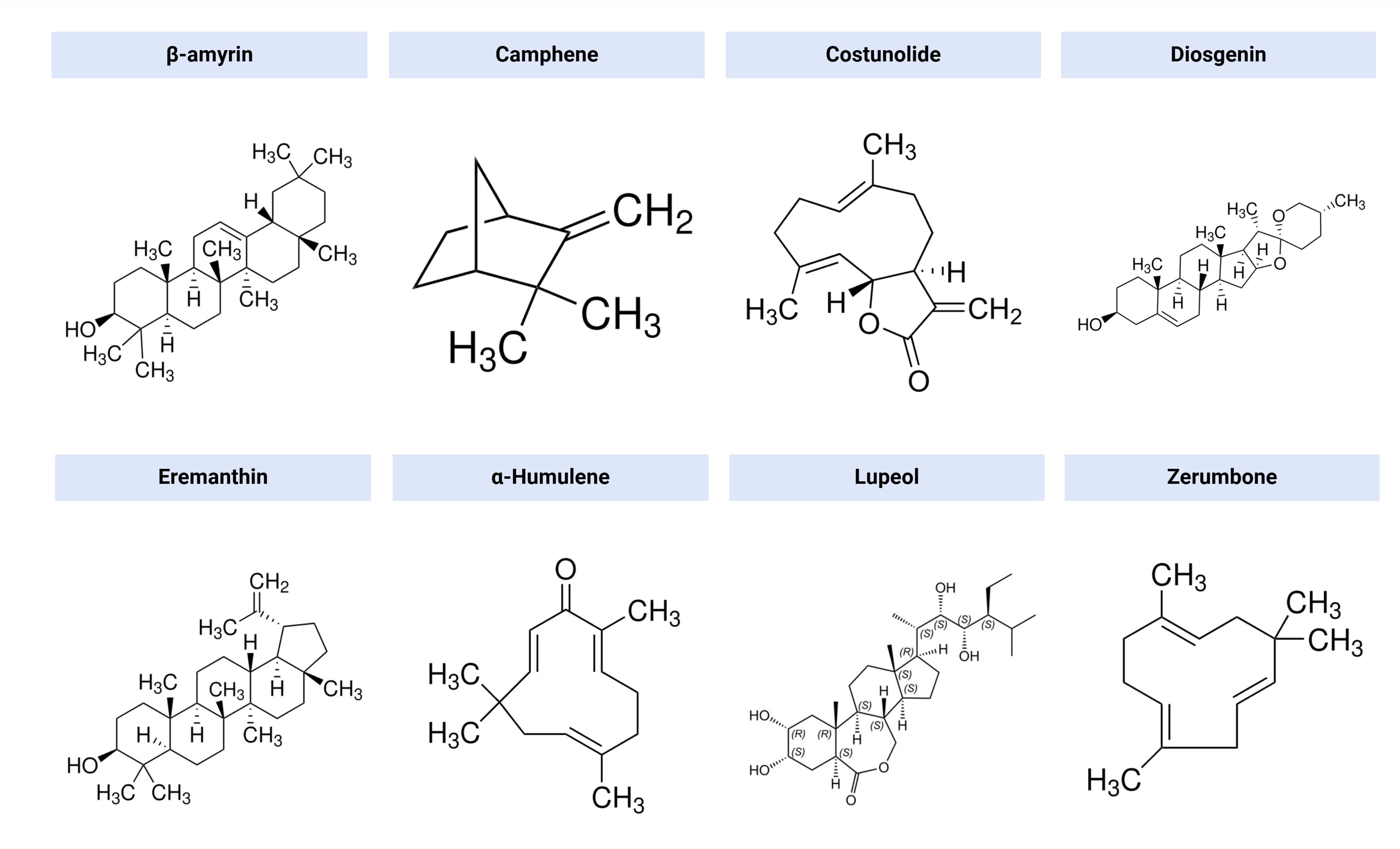
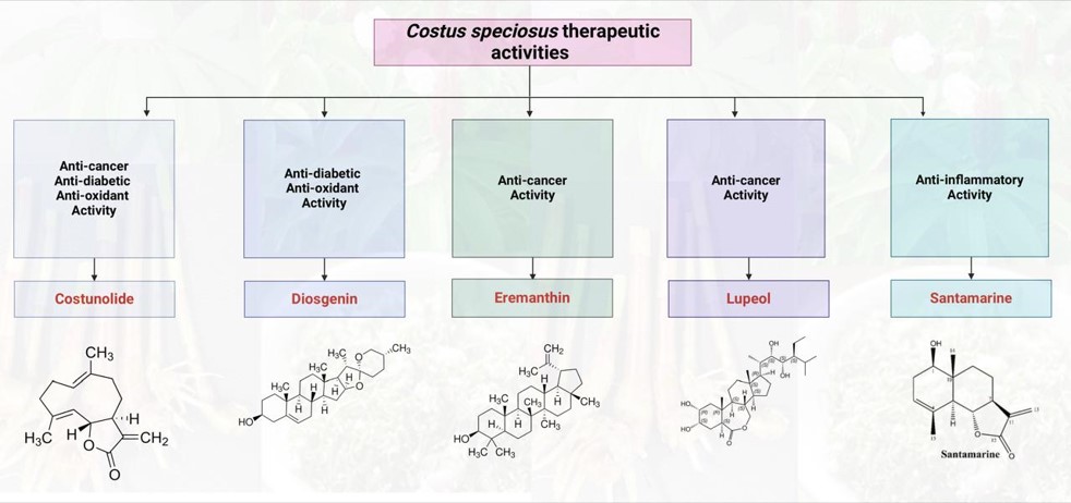
Based on phytochemical screening, alkaloids, glycosides, steroids, natural antioxidants, tannins, and vitamin B have been found in C. speciosus (Singh et al., 2014). Figure 1 displays the chemical structures of a few active principles that have been identified in various C. speciosus components (Duraipandiyan et al., 2012; Rasul et al., 2013).
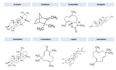
Examples of active compounds found in Costus speciosus. Source: Figure is designed by authors.
3. The Activity of Costus speciosus
3.1. Antioxidant activity
Antioxidants are a class of chemicals that significantly inhibit oxidation processes by scavenging free radicals and inducing cellular antioxidant enzymes (Daisy et al., 2008). Reactive oxygen species (ROS) are produced as byproducts of cellular metabolism and enzymatic reactions, and they can also be induced by a wide range of chemicals (Navrot et al., 2007). According to Nehete et al. (2010), ROS are crucial in the oxidative stress linked to diabetes, cirrhosis, cancer, and atherosclerosis. Antioxidant systems—which include nonenzymatic molecules such as ascorbic acid, α-tocopherol, glutathione, and β-carotene, as well as a range of enzymes such as superoxide dismutase (SOD), glutathione peroxidase (GPx), catalase, and glutathione S-transferases—combat oxidative stress (Gottfredsen et al., 2013). Phenols and flavonoids, which are found in medicinal plants, may offer living organisms a defence against ROS risks due to their redox characteristics, including strong metal ion chelation and free radical scavenging (Govindarajan et al., 2005).
Researchers have extracted the compounds present in C. speciosus leaf, stem peel, peeled stem, and roots in different solvents, and have demonstrated their antioxidant activity in vitro by using the 2,2-diphenyl-1-picrylhydrazyl (DPPH) radical scavenging and thiobarbituric acid (TBA) techniques (Vijayalakshmi and Sarada, 2008). The traditional use of C. speciosus root and stem peel for a variety of illnesses and ailments has been validated. Jha et al. (2010) found that C. speciosus rhizomes exhibited a stronger ability to scavenge nitric oxide (NO) compared with vitamin C and quercetin, which are typically used as standards in procedures such as the DPPH radical scavenging assays. The authors proposed that the presence of glycosides, flavonoids, triterpenoids, tannins, and steroids in the methanolic extract of C. speciosus may be responsible for the antioxidant action. In the same setting, several solvents have been used to produce C. speciosus rhizome extracts and assessed in vitro for their antioxidant capacity. The benzene extract presented the highest levels of phenolics (4.38%), flavonoids, and some vitamins (Devi and Urooj, 2010), and thus better antioxidant activity (Nehete et al., 2010).
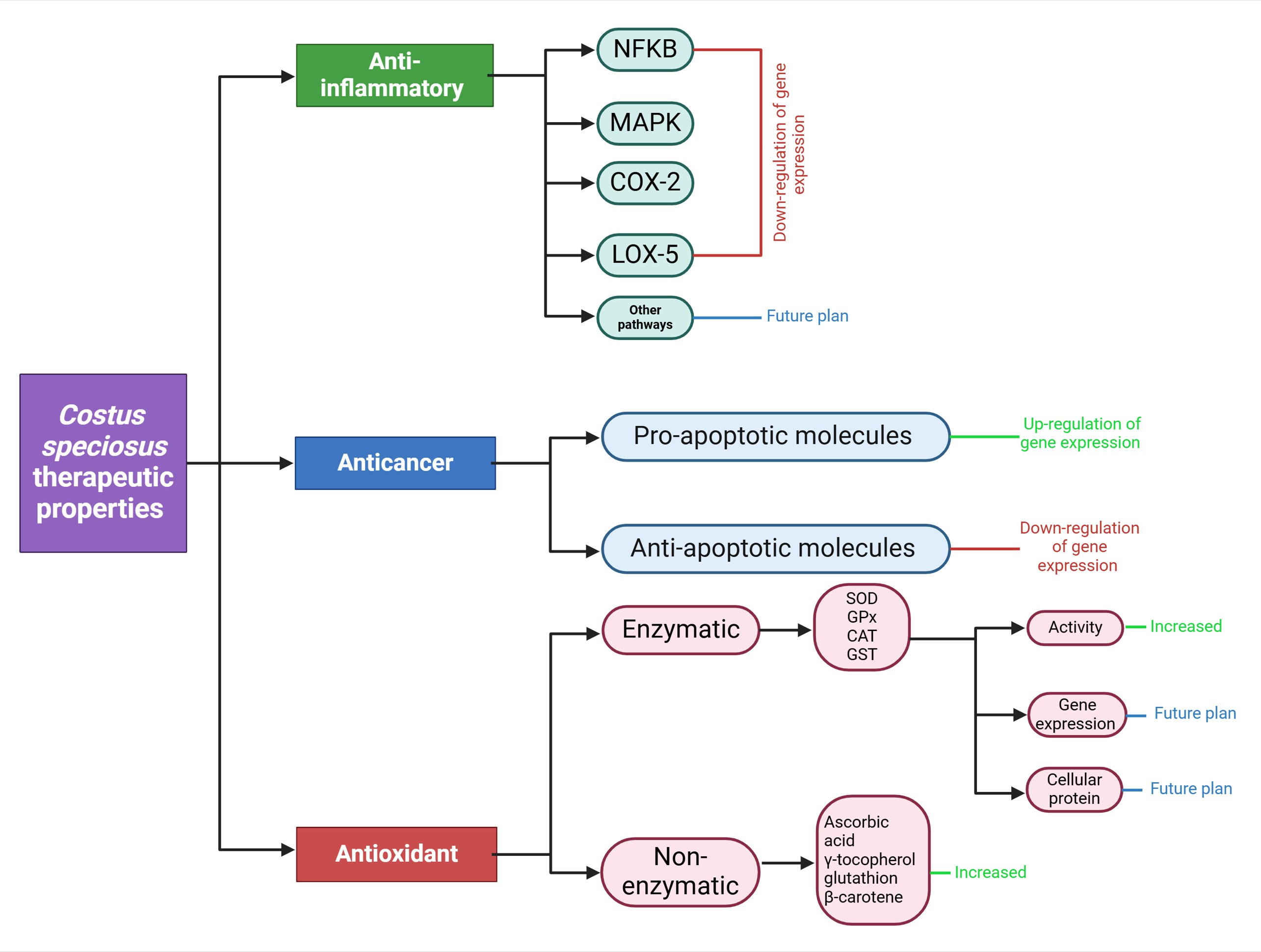
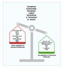
In an in vivo study, researchers administered a component of C. speciosus, costunolide, orally via an intragastric tube to rats (20 mg/kg/day) for 2 months (Eliza et al., 2010). The rats had been treated with streptozotocin to induce diabetes. The authors noted a significant decrease in TBA reactive substances and an increase in enzyme activities in the brain, liver, heart, kidney, and pancreas. In another study, buffalo calves were fed ground C. speciosus rhizomes. Compared with the control group, which was fed the basal diet, there was a significant drop in malondialdehyde (MDA), a product of lipid peroxidation, and an increase in the total antioxidant capacity (TAC) of erythrocytes of the C. speciosus–supplemented animals (El-Far and Abou-Ghanema, 2013) (Figure 2).
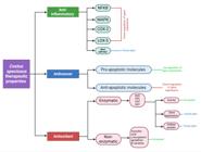
An overview of the anti-inflammatory, anticancer, and antioxidant properties of Costus speciosus. Source: Figure is designed by authors.
3.2. Anti-inflammatory effects
According to Vazquez et al. (2011), inflammation is a pathophysiological reaction to tissue damage and is intimately linked to the aetiology of a number of inflammatory disorders. A number of medicinal plants have shown anti-inflammatory properties, including Zinger officinale, Curcuma longa, and C. speciosus (Singh et al., 2013). Traditional folk forages have utilised C. speciosus to cure conditions such as headaches, rheumatism, fever, and bronchitis. The bioactive components of C. speciosus, including diosgenin and costunolide, have been used to treat a number of inflammatory diseases. They can inhibit the activity of tumour necrosis factor alpha (TNF-α), a cytokine that is released in response to infections and amplifies inflammation. In RAW 264.7 cells, a mouse macrophage cell line, diosgenin showed activity similar to the popular immune system suppressor methotrexate. It had an inhibitory or suppressive impact on the virus (Selim and Al Jaouni, 2016). Costunolide inhibits TNF-α, as well as cyclooxygenase (COX) and interleukins (e.g., interleukin 6 [IL-6] and IL-1), by blocking nuclear factor kappa-light-chain-enhancer of activated B cells (NF-κB) in mitogen-driven protein kinase pathways and activated microglia (Rayan et al., 2011; Al Attas et al., 2015). A number of other bioactive compounds, including dehydrodihydrocostus lactone, reynosin, arbusculin A, santamarine, and stigmasterol, were separated from the chloroform extract of C. speciosus and showed anti-inflammatory activity on isolated blood mononuclear cells (Al-Attas et al., 2015).
Singh et al. (2013) reported the in vivo anti-inflammatory qualities of a methanolic C. speciosus extract. There was notable anti-inflammatory effects 5 hours after administering 400 or 800 mg/kg of the methanolic extract. In a pilot study conducted at King Abdulaziz University in Saudi Arabia, 15 patients who had acute tonsillitis and pharyngitis were treated with an aqueous C. speciosus extract as nasal drops (0.75 mL every 8 hours for patients aged 2–6 years, and 1.5 mL every 8 hours for patients aged >6 years). Sixty percent of patients treated with the extract presented an improvement within in first 24 hours, and 93% showed remission by day 5 (Bakhsh et al., 2015). Figure 2 summarises the anti-inflammatory properties of C. speciosus.
3.3. Antidiabetic activity
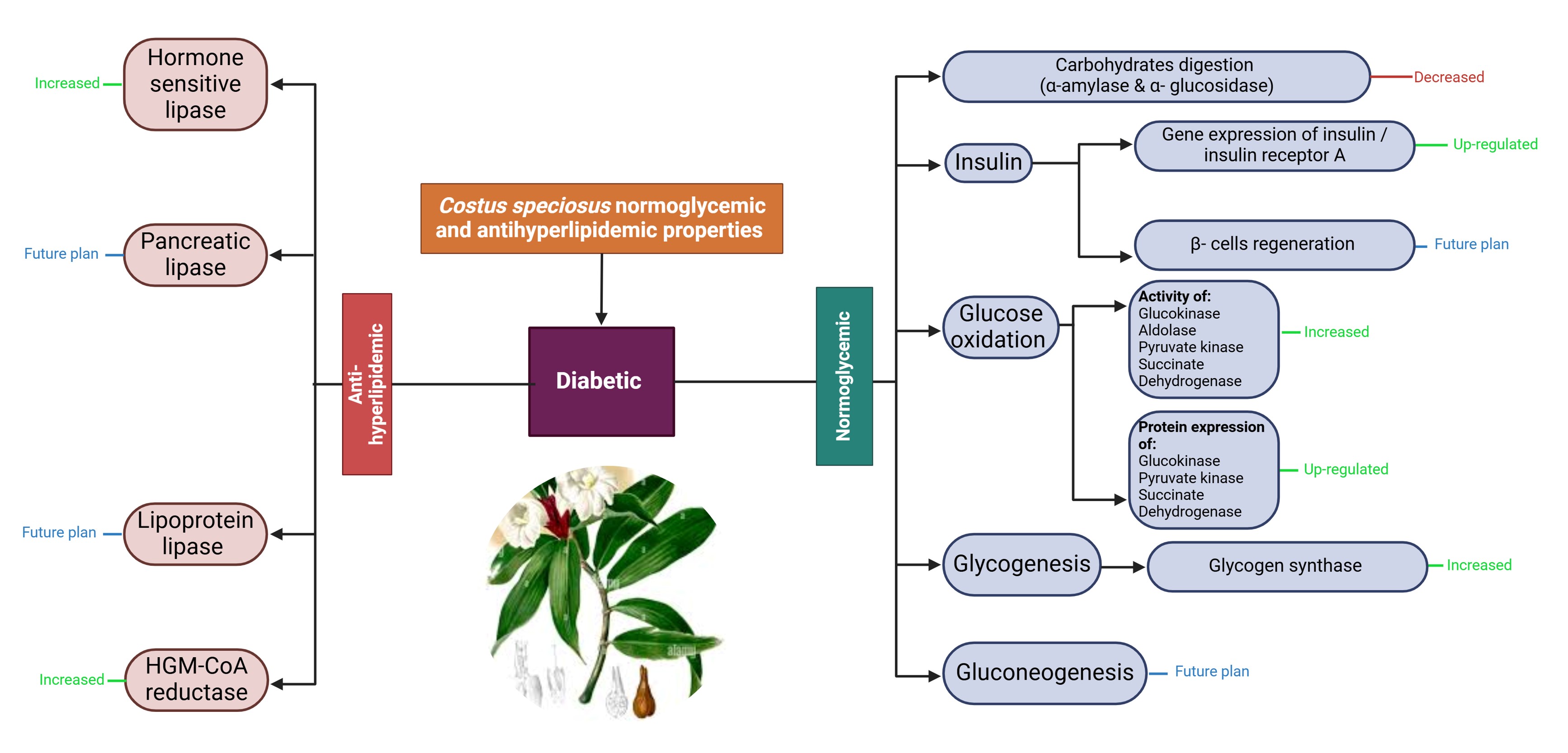
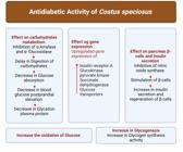
Diabetes mellitus is a metabolic disorder in which a relative or absolute lack of insulin raises blood glucose levels. The global prevalence of this condition is approximately 4% (Rani et al., 2012; Sulakshana and Rani, 2015). In addition to medications such as insulin, sulfonylureas, biguanides, and thiazolidinediones that are frequently used to treat diabetes, a number of medicinal plant species have been identified as normoglycemic agents; they are more efficacious, have fewer adverse effects, and are comparatively less expensive (Medagama and Bandara, 2014). One of these therapeutic plants is C. speciosus, which is known as the insulin plant because it contains the crucial antidiabetic compound diosgenin (Rani et al., 2012; Sulakshana and Rani, 2015). Figure 3 illustrates the normoglycemic impact of C. speciosus .
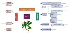
An overview of the normoglycemic and antihyperlipidemic properties of Costus speciosus. Source: Figure is designed by authors.
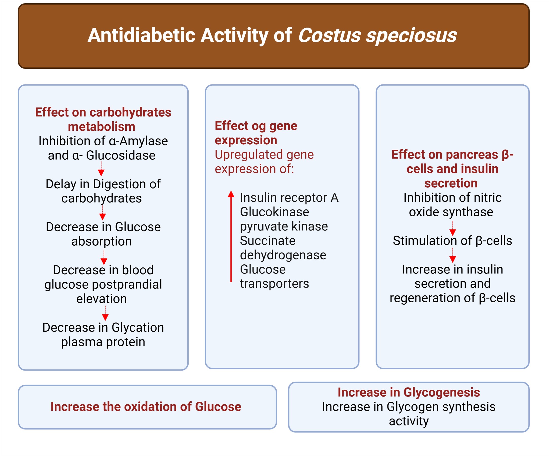
Daisy et al. (2008) examined the antidiabetic potential of C. speciosus in diabetic rats. Administering the methyl acetate, hexane, or methanolic extracts reduced the blood glucose levels in these rats. The hexane extract had the strongest hypoglycaemic action and antidiabetic potential of these three extracts. Furthermore, there was a reduction in the glycosylated haemoglobin and cholesterol levels. Mosihuzzaman et al. (1994) examined the antihyperlipidemic, antidiabetic, and antioxidant qualities of an ethanolic C. speciosus extract. It significantly reduced glucose levels, perhaps due to elevated glycogenesis and reduced gluconeogenesis. Furthermore, it decreased cholesterol, plasma lipids, and triacylglycerol, and improved the activity of liver enzymes. In another study, a methanolic leaf extract inhibited α-glucosidase and α-amylase, which ultimately lowered protein glycation (Perera et al., 2016). Moreover, costunolide stimulates beta cells, which results in the release of insulin (Eliza et al., 2009). Figure 4 displays the antidiabetic effects of active ingredients of C. speciosus, such as costunolide and eremanthin.
3.4. Antimicrobial activity
When antibiotics are administered orally, undesirable side effects can occur. For example, taking penicillin orally can result in heartburn, nausea, vomiting, and diarrhoea. As a result, researchers have evaluated the use of spices and herbs as replacements for antibiotics (Lai and Roy, 2004). The hexane and methanolic extracts of C. speciosus leaves and rhizomes could inhibit Salmonella spp., Pseudomonas spp., Salmonella spp., Escherichia coli, Shigella spp., Staphylococcus aureus, and Klebsiella pneumoniae (Malabadi, 2005; Ariharan et al., 2012). A hexane extract demonstrated notable antifungal effects, with a minimal inhibitory concentration of 31.25–250 μg/mL against Trichophyton mentagrophytes, Trichophyton simii, Epidermophyton floccosum, Trichophyton rubrum, Curvularia lunata, Scopulariopsis sp., Aspergillus niger, and Magnaporthe grisea (Duraipandiyan et al., 2012). The authors also separated eremanthin and costunolide from the rhizome extract and found that at extremely low concentrations, they inhibited the growth of numerous fungi. According to Al-Ameri and Falah (2014), a methanolic rhizome extract inhibited Aspergillus spp. that can cause lung infections. Additionally, Aspergillus fumigatus was well suppressed in experimental mice by the ethanolic rhizome extract (Al-Ameri and Azaaz, 2013). Finally, Salim et al. (2019) examined the antibacterial and antifungal properties of C. speciosus ethanolic and aqueous extracts. The aqueous extract exerted an antibacterial effect, whereas the ethanolic extract possessed strong antifungal and antibacterial properties.
4. Anticancer Effects of Costus speciosus
It is believed that the secondary metabolites of medicinal plants are the source of their numerous biological activities. There have been reports of anticancer properties in plants that contain bioactive chemicals with heterocyclic structures (Ramazani et al., 2014). Flavonoids and terpenoids are present in the nonpolar and semipolar fractions of methanolic extracts of C. speciosus, whereas the water-soluble fractions contain saponins (Gheraibia et al., 2020). Triterpenes have been extracted from the roots of C. speciosus. Flavonoids have also been extracted from C. speciosus (Silva et al., 2000). Other polyphenolic compounds and beneficial compounds have been found in a number of C. speciosus tissues (Binny et al., 2010).
Interestingly, a large number of cancer cells have been demonstrated to be susceptible to the cytotoxic effects of C. speciosus. Methanolic extracts have been shown to inhibit the growth of triple-negative breast cancer (TNBC), human colon adenocarcinoma cells (Baskar et al., 2012), metastatic breast cancer cells (Gheraibia et al., 2020), and hepatoblastoma (Bawakid et al., 2021). The nonpolar components of these extracts include compounds that inhibit the cell cycle, downregulate mutant p53, and induce apoptosis (Gheraibia et al., 2020). Furthermore, crude ethanolic Costus cuspidatus extracts demonstrated cytotoxic effects on human T cells and human leukemia (Siqueira et al., 2016). Although these compounds have relevant biological activity, the mechanisms of action remain poorly understood. Only a few molecular docking simulations of bioactive chemicals from C. speciosus have been published, including caspases (Pawar and Pawar, 2014) and COXs (Naznin et al., 2022).
When tested against human colon cancer cell lines, a number of C. speciosus rhizome extracts showed dose-dependent antioxidant and antiproliferative effects (Baskar et al., 2012). Moreover, 100 μg/mL of a methanolic C. speciosus leaf extract dramatically reduced the viability of hepatocellular carcinoma cells (Nair et al., 2014). In another study, diosgenin demonstrated strong cytotoxic effect on hepatocellular carcinoma and breast cancer cells. Furthermore, diosgenin exerted cytotoxic effects that were similar to those of paclitaxel. It upregulated the expression of death receptor 4 and caspase-3 to induced apoptosis of MCF-7 cells, a human breast cancer cell line (Selim and Al Jaouni, 2015). The effects of C. speciosus rhizome extracts were evaluated using human colon adenocarcinoma cell lines. They exerted notable antiproliferative and antioxidant effects in a dose- and time-dependent manner (Baskar et al., 2012). Nair et al. (2014) found that treatment with 100 μg/mL of a methanolic C. speciosus extract for 24 h significantly reduced the viability of breast cancer cells. Figure 5 summarises the anti-cancer properties of C. speciosus.
A summary of the anticancer properties of the active components in Costus speciosus. Source: Figure is designed by authors.
4.1. Mechanisms that underlie the anticancer activity of Costus speciosus
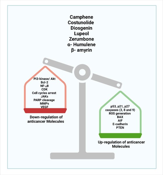
Natural, plant-derived molecules have structurally favourable molecular properties compared with synthetic molecules and represent a prominent source of anticancer drugs (Pan et al., 2010). The main plant-derived molecules that are currently utilised in medicine as single entities are alkaloids, lignans, and alkaloid derivatives. Researchers have reported several mechanisms by which the components of C. speciosus exert anticancer effects. Figure 6 summarises the anti-inflammatory, anticancer, and antioxidant properties of C. speciosus.
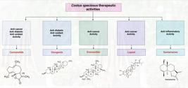
An overview of the anti-inflammatory, anticancer, and antioxidant properties of Costus speciosus. Source: Figure is designed by authors.
Elkady (2019) evaluated the anticancer effectiveness C. speciosus extracts against the cell cycle distribution of human prostate cancer cells. The findings demonstrated that by inducing apoptosis, C. speciosus extracts prevented PC-3 cell clonal development, invasion, proliferation, and migration
A methanolic C. speciosus extract disrupted progression of the cell cycle, as evidenced by elevated casapase-3 activity in the treated cells. Research on how this extract affects cellular pro-apoptotic and anti-apoptotic molecules using molecular methods is still required.
The tumour suppressor p53 is a transcription factor that reacts to various forms of cellular stress. It is acknowledged as the genome protector (Lyle et al., 2014). The expression of growth arrest genes such as p21 is promoted by p53. According to Masgras et al. (2012), p21 is a tumour suppressor that can stop the growth of cancer cells. PUMA, Noxa, BAX, and p53AIP1 are examples of pro-apoptotic genes that localise to the mitochondria and encourage cytochrome c release and reduce mitochondrial membrane potential. Furthermore, p53 controls elements that are involved in the extrinsic apoptotic pathway, such as Fas or DR5/KILLER. Lastly, p53 induces the generation of ROS, which harm the mitochondria and trigger apoptosis (Polyak et al., 1997).
Pitchai et al. (2014) examined the cytotoxicity of costunolide to human breast adenocarcinoma cells (MDA-MB-231, a TNBC cell line). It led to cell cycle arrest, as determined by flow cytometry. Additionally, at 20 and 40 μM, costunolide inhibited the overexpression of NF-κB subunits. Roy and Manikkam (2015) reported a half-maximal inhibitory concentration (IC50) of 40 μM for costunolide to induce apoptosis of MCF-7 cells. Moreover, there was upregulated expression of cyclin, caspase-3, and caspase-9 in costunolide-treated MCF-7 cells compared with normal breast cells (MCF-10A).
Cyclin-dependent kinase inhibitor 1B (p27Kip1) is an enzyme inhibitor that binds to and stops the activity of the cyclin E–CDK2 and cyclin D–CDK4 complexes. According to Møller (2000), this causes G1-phase arrest, which may stop or slow the growth of cancer cells.
Caspases are endoproteases that hydrolyse peptide bonds in cell proteins to carry out their functions. There are two groups of apoptotic caspases: executioners and initiators (McIlwain et al., 2013). They are triggered in the two primary apoptotic pathways—intrinsic, in which mitochondria are essential, and extrinsic, which is regulated by death receptors. Together with cytochrome c, apoptotic protease activating factor 1 (Apaf-1), and deoxyadenosine triphosphate (dATP), caspase-9 is activated by the mitochondrial route and forms an apoptosome in the cytosol. Caspase-3 is activated by the apoptosome (Zou et al., 1997). On the other hand, Fas ligand interaction with Fas complexes triggers caspase-3 and apoptosis, activating the extrinsic pathway (Wajant, 2002).
Changes in cytosolic calcium (Ca2+) levels support a variety of cellular processes, including myofibril contraction, hormone production, and metabolic control (Berridge et al., 2003). Too much Ca2+ in the cell can lead to cytotoxicity and apoptosis (Mattson and Chan, 2003).
A Ca2+-dependent cytosolic enzyme called nitric oxide synthase converts l-arginine to NO, which then combines with the free superoxide radical to generate the hazardous free peroxynitrite radical. These free radicals increase the risk of harming intracellular proteins, enzymes, DNA, and cellular membranes. When arachidonic acid is converted to prostaglandin G2, COX2-dependent processes produce ROS, which cause direct oxidative damage to DNA and promote apoptosis (Nikolic and van Breemen, 2001).
A mitochondrial intermembrane flavoprotein known as apoptosis-inducing factor (AIF) causes DNA breakage and chromatin condensation. Through mitochondrial membrane permeabilisation, AIF can also play a role in controlling apoptosis (Candé et al., 2002).
Cell–cell adhesion is influenced by E-cadherin. Downregulation of E-cadherin reduces cell–cell adhesion and promotes metastasis; it enhances invasiveness and favours circulation access, dispersion to distant anatomic sites, extravasation, and colonisation. E-cadherin expression can be increased by combining diosgenin with RNAs that silence hypoxia-inducible factor 1-alpha (HIF-1α) (Berx et al., 1995). By blocking phosphorylated protein kinase B (p-Akt) and mouse double minute 2 homolog, phosphatase and tensin homolog (PTEN) increases p53 levels, which causes G1-phase arrest and apoptosis. According to Haas-Kogan et al. (1998), PTEN works by dephosphorylating phosphatidyl inositol 3-phosphate (PIP3) and inhibiting survival signalling that is mediated by Akt.
According to Youle and Strasser (2008), the B cell lymphoma-2 (BCL2) protein family includes important regulators of apoptosis. It can be divided into three subgroups: pro-apoptotic (BAX, BAK, and BOK), anti-apoptotic (BCL2, BCL-xL, and BCL2L10), and BH3-only pro-apoptotic members. BAX is a tumour suppressor gene, while BCL2 is an oncogene. BCL2 overexpression enhances cell survival both in vivo and in vitro. BAX overexpression accelerates cell death; indeed, cell survival or death is determined by the BCL2/BAX ratio (Chao and Korsmeyer, 1998). Additionally, NF-κB p65–p52 signalling mediated the effects of glial cell line–derived neurotrophic factor on BCL2 and BCL2-w expression (Cao et al., 2013).
NF-κB activates groups of pro-apoptotic and anti-apoptotic genes in addition to its own inhibitor, IκB (Perkins, 2007). NF-κB inhibits the action of the caspase cascade and stimulates the transcription of the inhibitor of apoptosis protein (IAP) gene. IκB is broken down once the cell is stimulated by different substances, which enables NF-κB to move to the nucleus and attach to the promoter regions of its numerous target genes to support cell survival (Park and Hong, 2016).
Diosgenin inhibits the growth of cancer cells by inducing apoptosis. Diosgenin presented an IC50 of 32.62 μg/mL for HepG2 cells (a hepatocellular carcinoma cell line), more efficacious than paclitaxel, and 11.03 μg/mL for MCF-7 cells. Selim and Al Jaouni (2015) claimed that diosgenin increases the levels of caspase-3 and death receptor 4, which trigger apoptosis in MCF-7 cells. The active components of C. speciosus may have anticancer properties through upregulating pro-apoptotic and downregulating antiapoptotic molecules, which simultaneously reduce the growth and spread of cancer cells (El-Far et al., 2016).
Dehydrocostus lactone and other sesquiterpene lactones that are found in C. speciosus also have notable anticancer effects (DeVita et al., 2008; Kumar et al., 2024). According to Hua et al. (2016), in human lung squamous carcinoma cells, dehydrocostus lactone triggered mitochondria-mediated apoptosis and induced G1/S-phase arrest. Dehydrocostus lactone may be useful in the treatment of cancer because it reduces osteoclastogenesis and osteoclast-induced bone loss (Hu et al., 2019), perhaps by altering the NF-κB pathway (Lu et al., 2023). Hoesel and Schmid (2013) reported that dehydrocostus lactone targets the phosphoinositide 3-kinase (PI3K)/Akt signalling pathway to suppress cell growth and to encourage apoptosis in a variety of cancer cells.
Another substance found in C. speciosus, cyanaropicrin, has also demonstrated encouraging anticancer activity. Cynaropicrin efficiently suppresses leukocyte cancer cell proliferation and induces apoptosis and cell cycle arrest (Lau et al., 2008). Zimmermann et al. (2012) stated that cynaropicrin may be a cytotoxic substance because it demonstrated cytotoxicity against a variety of cell types. De Cicco et al. (2021) investigated how cynaropicrin inhibited the growth of human melanoma and emphasised its potential lethal effects. Pulito et al. (2015) validated the cytotoxic actions of cynaropicrin by showing that it promotes apoptosis.
Diosgenin has the ability to inhibit proliferation by producing abundant ROS. This oxidative stress causes HepG2 cells to undergo apoptosis by activating the c-Jun N-terminal kinase (JNK/p38) MAPK pathway (Kim et al., 2012).
Numerous medicinal plants, including Laurus nobilis, Saussurea lappa, and C. speciosus, contain costunolide (El-Far et al., 2018). Costunolide prevents the growth, invasion, metastasis, and angiogenesis of cancer cells (Liu et al., 2011). By upregulating p53, costunolide prevented human colorectal cancer (HCT116) cells from proliferating (Hu et al., 2018). Additionally, by activating p53 and generating ROS, costunolide caused human oesophageal carcinoma (Eca-109) cells to undergo apoptosis (Hua et al., 2016). In addition to inducing p21WAF1-related G2/M-phase arrest and Fas-mediated apoptosis, ROS production caused MDA-MB-231 cells to undergo apoptosis (Choi et al., 2012). Free costunolide reduced BCL2 in HCT116 and MDA-MB-231 cells and increased BAX in MDA-MB-231 cells, causing both cell lines to undergo apoptosis. In HCT116 cells, costunolide induced apoptosis by suppressing mammalian target of rapamycin (mTOR) phosphorylation (Hu et al., 2018) and promoting the upregulation of p53 and thioredoxin reductase 1 (TrxR1) (Zhuge et al., 2018). By producing ROS and causing cell cycle arrest, costunolide caused MDA-MB-231 to undergo apoptosis (Choi et al., 2012). It caused prostate cancer cells to undergo apoptosis by activating MAPKs and ROS production (Chen et al., 2017), and it inhibited the PI3K/Akt pathway to induce apoptosis in doxorubicin-resistant chronic myeloid leukemia (K562/ADR) cells (Cai et al., 2019).
4.2. The potential benefits of Costus speciosus for breast cancer
According to Siegel et al. (2017), approximately 30% of all new cancer diagnoses are for breast cancer, the second most lethal cancer. According to recent data, breast cancer has overtaken lung cancer as the most common cancer throughout the world. It poses a serious risk to women’s health and lives due to its very high mortality rate. Histopathological examination is the most commonly used method to diagnose breast cancer. Automated histopathological image categorisation could expedite a diagnosis and reduce the possibility of errors in breast cancer diagnosis (Lakhani et al., 2012). A biopsy is used in histopathology to acquire pictures of the afflicted tissue (Das et al., 2021). Treatment of sickness and the prognosis depend on early identification (WHO, 2022).
According to Halimah et al. (2024), aC. speciosus leaf extract reduced the growth of MCF-7 cells due to the induction of apoptosis. An ethanolic extract induced necrosis of HeLa cells. Furthermore, diosgenin and beta-sitosterol may interact with Phe247 and Phe250 in the caspase-3 catalytic site.
C.speciosus contains secondary metabolites, including beta-sitosterol, tigogenin, diosgenin, and 5α-stigmast-9, that could be useful to treat breast cancer (Bawakid et al., 2021; Thambi and Cherian, 2023). Researchers have examined the binding mechanism and affinity of beta-sitosterol and diosgenin in the catalytic domain of cysteineaspartyl protease-3 (caspase-3) by using molecular docking simulation. Diosgenin exerts anticancer activity through a number of mechanisms. It has been shown to alter the cell cycle and to promote apoptosis. Diosgenin-exposed cells exhibited p53 activation and cell cycle arrest (Corbiere et al., 2004). Diosgenin may cause human hepatocellular carcinoma cells to undergo G2/M-phase arrest and die (Li et al., 2015). In osteosarcoma cells (1547), diosgenin increased COX activity and led to cell cycle arrest and apoptosis (Moalic et al., 2001). Diosgenin promoted the overexpression of caspase-3 (Raju et al., 2004), caspase-8 (Li et al., 2015), and caspase-9 (Cailleteau et al., 2009). According to Srinivasan et al. (2009), diosgenin could downregulate BCL2 and BCL-xL. According to Selim and Al Jaouni (2015), diosgenin isolated from C. speciosus also increased the proliferation of MCF-7 cells and caspase-3 levels. Widastuti et al. (2017) found that diosgenin and taurine reduce the quantity of spermatogonia, spermatocytes, and spermatid cells that form. The combination of doxorubicin and hesperidin could increase MC-7 apoptosis and may represent a novel chemotherapeutic (Hermawan et al., 2010).
Vegetables, grains, nuts, seeds, and olive oil are rich sources of beta-sitosterol. This phytosterol reduces the viability of MCF-7 and MDA-MB-231 cells by altering the PI3K/Akt/mTOR pathway (Zhu et al., 2018). It alters the shape of human cervical cancer cells by increasing the electron density in the cell membrane and decreasing the number of organelles (Baek et al., 2017). In U937 cells, beta-sitosterol downregulated BCL2 but did not alter the expression BCL-xL or BAX, indicating a link between caspase-3 activation and BCL2 downregulation (Sharmila and Sindhu, 2017).
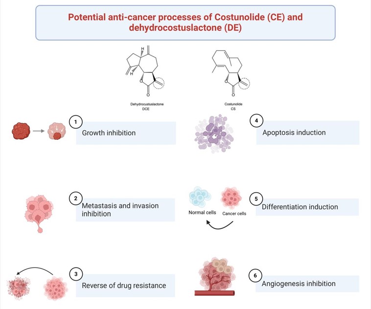
Dehydrocostus lactone and costunolide exert anticancer activity through a number of mechanisms: the inhibition of angiogenesis (Hao et al., 2010), the induction of apoptosis and differentiation in cancer cells (Choi and Ahn, 2009), the inhibition of invasion and metastasis (Kim et al., 2008), the reversal of multidrug resistance (Yang et al., 2011), and the inhibition of cancer cell proliferation (Liu et al., 2011) (Figure 7). Costunolide causes breast cancer cells to undergo apoptosis (Roy and Manikkam, 2015). This compound interacts with SOD, catalase, and GPx in human breast cancer cells (Rajalakshmi et al., 2014). Additionally, it worked in concert with Rhaphidophora pinnata, which inhibits MCF-7 cell proliferation and death (MASFRIA et al., 2013).
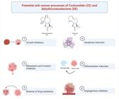
The chemical structures of costunolide (CE) and dehydrocostus lactone (DE) and their potential anticancer effects through six main mechanisms: growth suppression, cell cycle regulation, apoptosis induction, inhibition of angiogenesis, inhibition of invasion and metastasis, differentiation induction, and reversal of treatment resistance. Source: Figure is designed by authors.
4.3. The potential benefits of Costus speciosus for ovarian cancer
Among the gynaecological cancers, ovarian cancer has the highest fatality rate (Kuroki and Guntupalli, 2020). Ovarian cancer is frequently referred to as a silent killer because its symptoms are rarely detected in the early stages, so the majority of new cases are detected in advanced stages (III and IV) (Nersesian et al., 2019; Pokhriyal et al., 2019). Each year, up to 314,000 women receive an ovarian cancer diagnosis each year, and 207,000 of them die as a result of the disease. Female patients with ovarian cancer have the lowest probability of surviving a malignancy (WHO, 2020).
To make surgery easier, chemotherapy is used to reduce the tumour size. The majority of ovarian tumours respond well to platinum treatment; nevertheless, platinum resistance occurs in some of these cases, and survival is only marginally increased (Binju et al., 2019). Furthermore, 70% of those with more aggressive tumours recur 6–12 months after finishing chemotherapy (Huang et al., 2020). Cancer chemotherapy is limited by nonspecific or dose-related cellular toxicity and multidrug resistance (Harsono, 2020). Chemotherapy can induce apoptosis by increasing the levels of proapoptotic transcription factors such as p53 and BAX (Nurmaulawati, 2021).
Apoptosis happens frequently to preserve homeostatic equilibrium (Singh et al., 2019). The protein groups p53 and BCL2 are crucial in controlling apoptosis. According to Manne et al. (2021), p53 is a tumour suppressor gene, while proapoptotic BCL2 family proteins make tumours more sensitive to treatment. Apoptosis and aberrant cell proliferation may result from an imbalance between these proteins. Numerous alterations have been found in both the intrinsic and extrinsic apoptotic pathways, and cancer cells employ a variety of tactics to evade apoptotic processes (Liu et al., 2017). Whereas p53 controls BAX, an increase in BH3 alone can initiate the activation of BAX/BAKp53, which has essential roles in regulating the cell cycle and apoptosis, and preserving genomic stability (Wang et al., 2023). Activation-induced oligomerisation of BAX and BAK leads to mitochondrial outer membrane permeabilisation (MOMP) (Dadsena et al., 2021). Myeloid cell leukemia 1 (MCL1) has a lot of promise as a target for tumour treatment. Selective MCL1 inhibitors have emerged as novel anticancer medications (Wang et al., 2021).When a specific inhibitor binds to MCL1, the pro-apoptotic proteins BAX/BAK are released, and apoptosis is triggered.
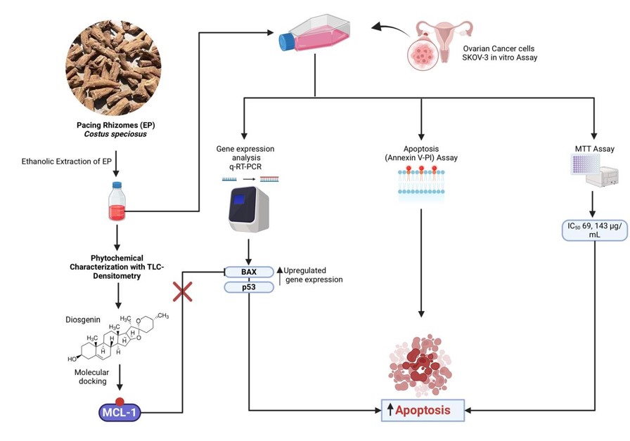
A potential alternative treatment for ovarian cancer is C. speciosus. The rhizomes and roots contain gracilin, quinine, dioscin, dioscinprosapogenin A and B, and sitosterol-β-D-glucoside (Nafisah et al., 2022). The pharmacological properties of diosgenin mostly relate to its anticancer action based on preclinical animal models and in vitro experiments. According to Sethi et al. (2018), diosgenin primarily functions by altering a number of cell signalling pathways that are involved in cell cycle regulation, cell differentiation, and cell death. In SKOV-3 cells, an ovarian epithelial cell line, the ethanol extract of rhizome pacing (EP) was cytotoxic, with a notable IC50 of 69.143 µg/mL. Flow cytometry showed that apoptosis occurred in SKOV-3 cells treated with the extract (Hidayah et al., 2023). Diosgenin might be the reason why EP triggered apoptosis.
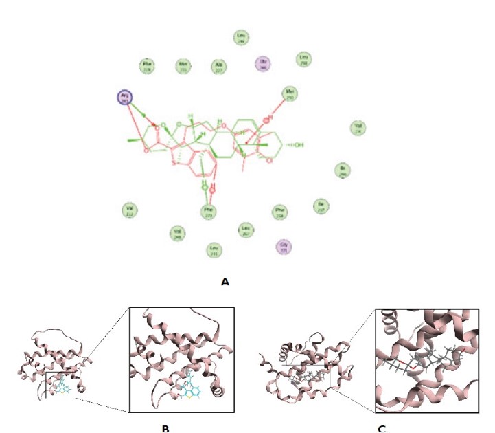
Molecular docking is a computational simulation that pairs a ligand receptor’s active site to anticipate the binding between drugs. As seen in Figure 8, molecular docking is frequently employed to identify novel medications that are more efficacious (Pratama et al., 2017). Based on molecular docking, diosgenin can bind more efficaciously to MCL1 compared with 19G, a native ligand. Of note, when binding to MCL1, diosgenin can interact with that crucial residue with which 19G interacts. Therefore, diosgenin may represent an MCL1 inhibitor.
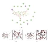
(A) Overlay of the diosgenin (green) and the native ligand complex; (B) and (C) three-dimensional (3D) visualisation of the native ligand and 4HW3 (Hidayah et al., 2023).
Yang et al. (2011) stated that costunolide induced the apoptosis of platinum-resistant ovarian cancer cell lines—MPSC1(PT), A2780(PT), and SKOV3(PT)—in a time- and dose-dependent manner and suppressed tumour growth in a SKOV3(PT)-bearing mouse model through activation of caspase-3, caspase-8, and caspase-9, and downregulation of BCL2. In a xenografted tumour of human gastric adenocarcinoma BGC-823 cells, costunolide increased cleaved caspase-9, cleaved caspase-3, and Bax levels, and decreased BCL2 (Yan et al., 2019).
In SKOV-3 cells, overexpression of the BAX gene leads to MOMP and thus the release of molecules that lead to the initiation of apoptosis. Elevated p53 gene expression resulting from DNA damage can cause cancer cells to undergo apoptosis (Aubrey et al., 2017). As shown in Figure 9, increased p53 gene expression can control BAX expression and activity, further encouraging apoptosis. This anti-apoptotic protein plays a crucial role in blocking the apoptosis pathway by preventing BAX activation.
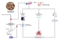
The mechanism by which p53 and BAX regulate the apoptosis of SKOV-3 cancer cells. Source: Figure is designed by authors.
4.4. The potential benefits of Costus speciosus for uterine disorders
Lijuan et al. (2011) evaluated the influence of an ethanolic C. specious rhizome extract on spontaneous phasic uterine contractions. The experiment involved the use of longitudinal myometrial strips to assess isometric force. Along with a considerable increase in basal tension, the amplitude and frequency of phasic contraction also increased. By inhibiting myosin light chain kinase (MLCK) or L-type calcium channels, the force was eliminated in the presence of the extract. Fulvestrant, an oestrogen receptor blocker, had no effect on the extract’s effects. The extract included a considerable amount of diosgenin, but diosgenin alone had no impact on force or even inhibited force, depending on the amount that was added. The extract produced a considerable amount of force without extracellular Ca2+, which was prevented by blocking the sarcoplasmic reticulum calcium-ATPase (SERCA), while fulvestrant did not. Based on these findings, non-estrogenic actions rather than the action of diosgenin are responsible for the uterotonic effect.
Menstruation cramps can produce significant pain that impairs the quality of life of many women. Even in the absence of an underlying infection or other medically recognised illness states, severe uterine contractions are believed to cause moderate to severe pain in cases of primary dysmenorrhea, a condition associated with premenstrual syndrome (PMS). The release of prostaglandins, leukotrienes, and leukocytes that typically accompany the disintegration of the endometrial lining are thought to be responsible for the associated pain and the uterine hypercontractility that is evocative of labour. According to Röhrl et al. (2016), standardised extracts of Vitex agnus-castus berries and C. speciosus are clinically useful in alleviating PMS symptoms.
Flavonoids, low-molecular-weight phenolic compounds (Middleton-Junior et al., 2000), exert antiviral, anti-inflammatory, and antioxidant properties. Flavonoid extracted from C. speciosus demonstrated spasmolytic effect on the rat aorta and uterus as well as the guinea pig ileum and trachea (Macêdo et al., 2011).
Our understanding of uterine pathophysiology, including embryo implantation and disorders such as dysmenorrhea and colic, which are caused by uncontrollable uterine contractions, has advanced significantly in recent years (Aguilar and Mitchell, 2010). According to Bortoleto (1995), excessive contractions cause pain that can potentially result in hypoxia, ischemia, and decreased uterine vascular flow. Certain flavonoids, including genistein, kaempferol, quercetin (Revuelta et al., 1997), and isoliquiritigenin, have demonstrated spasmolytic activity in the rat uterus. These flavonoids also demonstrated analgesic activity in mice (Shi et al., 2012), and the flavonoid galetin 3,6-dimethyl ether (Macêdo et al., 2011) demonstrated anti-inflammatory and antinociceptive properties in mice (Queiroz et al., 2010).
There has been almost no investigation regarding the mechanisms by which C. speciosus affects uterine cancer; thus, additional research is needed in this area.
5. Comprehensive review:
-
This study provides a thorough overview of the biological activities, chemical compositions, and potential therapeutic uses of C. speciosus, a critically endangered medicinal plant.
-
Diverse applications: This review highlights the wide range of pharmacological properties of C. speciosus, including its antimicrobial, anti-inflammatory, antioxidant, anti-cancer, antiviral, and hepatoprotective effects, demonstrating its versatility as a potential therapeutic agent.
-
Detailed analysis: This study investigated the specific bioactive compounds present in C. speciosus, such as costunolide, dehydrocostus lactone, and cynaropicrin, and explored their mechanisms of action and contribution to the plant’s diverse biological activities.
Overall, our comprehensive study of C. speciosus presents a robust and informative review, highlighting the significant potential of this medicinal plant and acknowledging the need for further research, standardized methodologies, and conservation efforts to fully realize its therapeutic applications.
6. Conclusion
This review has demonstrated the diverse therapeutic potential of C. speciosus in many disorders, opening up new avenues for clinical investigation. Future research should identify the specific compounds present in C. speciosus that exert the therapeutic effects and their mechanisms of action. Moreover, safety evaluations of C. speciosus extracts are necessary. In the future, methanolic, ethanolic, hexane, and chloroform extracts of C. speciosus could be used either alone or in conjunction with pharmaceuticals to treat numerous diseases.
Future research should focus on validating the potential of C. speciosus in clinical settings and further investigate its biotechnological and pharmaceutical applications. Standardized research methodologies and rigorous clinical trials are vital for confirming its effectiveness and safety in various medical fields.
Acknowledgements
We would like to express our great appreciation to the Deanship of Scientific Research & Science College & Biology department at Imam Abdulrahman bin Faisal University, Kingdom of Saudi Arabia. This research was funded by a grant from the The Deanship of Scientific Research, Imam Abdulrahman bin Faisal University, Kingdom of Saudi Arabia Grant No. (Sci-2019- 397).
Ethics and consent
References
-
ABD EL-HACK, M.E., ALFWUAIRES, M.A., JGHEF, M.M., KHAFAGA, A.F., ABDELNOUR, S.A., ABDEL-HAMID, M., ALAGAWANY, M., ALGEFARE, A.I., ALNAMSHAN, M.M., IMAM, M.S., GAMAL, M., ELESAWI, A.E. and ABUKHALIL, M.H., 2023. The efficacy of applying some plants and herbs in cancer therapy for humans and animals: a comprehensive review. Annals of Animal Science, vol. 23, no. 2, pp. 315-338. http://doi.org/10.2478/aoas-2022-0078
» http://doi.org/10.2478/aoas-2022-0078 -
ABDULLAHI, S., ISMAIL, A.F. and SAMAT, S., 2019. Determination of indoor doses and excess lifetime cancer risks caused by building materials containing natural radionuclides in Malaysia. Nuclear Engineering and Technology, vol. 51, no. 1, pp. 325-336. http://doi.org/10.1016/j.net.2018.09.017
» http://doi.org/10.1016/j.net.2018.09.017 -
AGUILAR, H. and MITCHELL, B., 2010. Physiological pathways and molecular mechanisms regulating uterine contractility. Human Reproduction Update, vol. 16, no. 6, pp. 725-744. http://doi.org/10.1093/humupd/dmq016 PMid:20551073.
» http://doi.org/10.1093/humupd/dmq016 - AHMAD, M.S., SHAWKY, A., GHOBASHY, M.O. and FELIFEL, R.H., 2018. Effect of some medicinal plants on life cycle of citrus brown mites (Eutetranychusorientalis). International Journal of Pharmaceutical Research and Allied Sciences, vol. 7, no. 4, pp. 13-17.
- AL-AMERI, N.O. and AZAAZ, Z.F., 2013. Effect of alcoholic extract of Costus speciosus Koen on Aspergillus fumigatus in lab rats (II). Journal of Natural Sciences Research, vol. 3, no. 15, pp. 80-87.
- AL-AMERI, N.O. and FALAH, A.Z., 2014. Morphological effects of alcoholic extract of Costus speciosus Koen on Aspergillus sp that causing pulmonary infections (III). Journal of Natural Sciences Research, vol. 4, no. 3, pp. 98-101.
-
AL-ATTAS, A.A., EL-SHAER, N.S., MOHAMED, G.A., IBRAHIM, S.R. and ESMAT, A., 2015. Anti-inflammatory sesquiterpenes from Costus speciosus rhizomes. Journal of Ethnopharmacology, vol. 176, pp. 365-374. http://doi.org/10.1016/j.jep.2015.11.026 PMid:26593213.
» http://doi.org/10.1016/j.jep.2015.11.026 -
ALI, S., KHAN, M.R., IQBAL, J., BATOOL, R., NAZ, I., YASEEN, T., ABBASI, B.A., NASIR, J.A. and EL-SEREHY, H.A., 2021. Chemical composition and pharmacological bio-efficacy of Parrotiopsis jacquemontiana (Decne) Rehder for anticancer activity. Saudi Journal of Biological Sciences, vol. 28, no. 9, pp. 4969-4986. http://doi.org/10.1016/j.sjbs.2021.07.072 PMid:34466072.
» http://doi.org/10.1016/j.sjbs.2021.07.072 -
AMBROŽ, M., BOUŠOVÁ, I., SKARKA, A., HANUŠOVA, V., KRÁLOVÁ, V., MATOUŠKOVÁ, P., SZOTÁKOVA, B. and SKÁLOVÁ, L., 2015. The influence of sesquiterpenes from Myrica rubra on the antiproliferative and pro-oxidative effects of doxorubicin and its accumulation in cancer cells. Molecules, vol. 20, no. 8, pp. 15343-15358. http://doi.org/10.3390/molecules200815343 PMid:26307963.
» http://doi.org/10.3390/molecules200815343 - ARIHARAN, V.N., MEENA DEVI, V.N., RAJAKOKILA, M. and NAGENDRA PRASAD, P., 2012. Antibacterial activity of Costus speciosus rhizome extract on some pathogenic bacteria. International Journal of Advanced Life Sciences, vol. 4, pp. 24-27.
- ARISTINA, R.F., ASTUTI, W. and PRATIWI, D.R., 2019. Screening and phytochemicals test of extract endhophytes bacteria from stem of pacing (Costus sp). Jurnal Atomik, vol. 4, no. 1, pp. 21-24.
-
AUBREY, B.J., KELLY, G.L., JANIC, A., HEROLD, M.J. and STRASSER, A., 2017. How does p53 induce apoptosis and how does this relate to p53- mediated tumour suppression? Cell Death and Differentiation, vol. 25, no. 1, pp. 104-113. http://doi.org/10.1038/cdd.2017.169 PMid:29149101.
» http://doi.org/10.1038/cdd.2017.169 -
BAEK, J., ROH, H.-S., CHOI, C.-I., BAEK, K.-H. and KIM, K.H., 2017. Raphanus sativus sprout causes selective cytotoxic effect on p53- deficient human lung cancer cells in vitro. Natural Product Communications, vol. 12, no. 2, pp. 237-240. http://doi.org/10.1177/1934578X1701200224 PMid:30428220.
» http://doi.org/10.1177/1934578X1701200224 - BAHSHWAN, S.M. and ALJEHANY, B.M., 2020. A review on the therapeutic and medicinal activities of Costus Speciosus Pharmacophore, vol. 11, no. 3, pp. 124-129.
-
BAKHSH, Z.A., AL-KHATIB, T.A., AL-MUHAYAWI, S.M., ELASSOULI, S.M., ELFIKY, I.A. and MOURAD, S.A., 2015. Evaluating the therapeutic efficacy, tolerability, and safety of an aqueous extract of Costus speciosus rhizome in acute pharyngitis and acute tonsillitis: a pilot study. Saudi Medical Journal, vol. 36, no. 8, pp. 997-1000. http://doi.org/10.15537/smj.2015.8.11377 PMid:26219454.
» http://doi.org/10.15537/smj.2015.8.11377 -
BASKAR, A.-A., AL NUMAIR, K.-S., ALSAIF, M.-A. and IGNACIMUTHU, S., 2012. In vitro antioxidant and antiproliferative potential of medicinal plants used in traditional Indian medicine to treat cancer. Redox Report, vol. 17, no. 4, pp. 145-156. http://doi.org/10.1179/1351000212Y.0000000017 PMid:22776996.
» http://doi.org/10.1179/1351000212Y.0000000017 -
BAVARVA, J.H. and NARASIMHACHARYA, A.V.R.L., 2008. Antihyperglycemic and hypolipidemic effects of Costus speciosus in alloxan induced diabetic rats. Phytotherapy Research, vol. 22, no. 5, pp. 620-626. http://doi.org/10.1002/ptr.2302 PMid:18444247.
» http://doi.org/10.1002/ptr.2302 -
BAWAKID, N.O., ABDEL-LATEFF, A., EL-SENDUNY, F.F. and ALARIF, W.M., 2021. Costus speciosus J Koenig (Costaceae) exerts antiproliferative effect on breast cancer cells via induction of cell cycle arrest and inhibition of activity of metalloproteinase-2. Tropical Journal of Pharmaceutical Research, vol. 20, no. 7, pp. 1365-1372. http://doi.org/10.4314/tjpr.v20i7.7
» http://doi.org/10.4314/tjpr.v20i7.7 -
BERRIDGE, M.J., BOOTMAN, M.D. and RODERICK, H.L., 2003. Calcium signaling: dynamics, homeostasis and remodeling. Nature Reviews. Molecular Cell Biology, vol. 4, no. 7, pp. 517-529. http://doi.org/10.1038/nrm1155 PMid:12838335.
» http://doi.org/10.1038/nrm1155 -
BERX, G., CLETON-JANSEN, A.M., NOLLET, F., DE LEEUW, W.J., VAN DE VIJVER, M., CORNELISSE, C. and VAN ROY, F., 1995. E-cadherin is a tumour/invasion suppressor gene mutated in human lobular breast cancers. The EMBO Journal, vol. 14, no. 24, pp. 6107-6115. http://doi.org/10.1002/j.1460-2075.1995.tb00301.x PMid:8557030.
» http://doi.org/10.1002/j.1460-2075.1995.tb00301.x -
BHATTACHARYA, S. and NAGAICH, U., 2010. Assessment of anti-nociceptive efficacy of Costus speciosus rhizome in Swiss albino mice. Journal of Advanced Pharmaceutical Technology & Research, vol. 1, no. 1, pp. 34-40. http://doi.org/10.4103/2231-4040.70519 PMid:22247830.
» http://doi.org/10.4103/2231-4040.70519 -
BINJU, M., AMAYA-PADILLA, M.A., WAN, G., GUNOSEWOYO, H., SURYORAHMANTO, Y. and YU, Y., 2019. Therapeutic inducers of apoptosis in ovarian cancer. Cancers, vol. 11, no. 11, pp. 1786. http://doi.org/10.3390/cancers11111786 PMid:31766284.
» http://doi.org/10.3390/cancers11111786 - BINNY, K., KUMAR, S.G. and DENNIS, T., 2010. Anti-inflammatory and antipyretic properties of the rhizome of Costus speciosus (koen.) Sm. Journal of Basic and Clinical Pharmacy, vol. 1, no. 3, pp. 177-181. PMid:24825984.
- BORTOLETO, C.C.R., 1995. Dismenorreia primária: etiopatogenia e terapêutica. Revista Femina, vol. 13, no. 5, pp. 439-443.
-
CAI, H., LI, L., JIANG, J., ZHAO, C. and YANG, C., 2019. Costunolide enhances sensitivity of K562/ADR chronic myeloid leukemia cells todoxorubicin through PI3K/Akt pathway. Phytotherapy Research, vol. 33, no. 6, pp. 1683-1688. http://doi.org/10.1002/ptr.6355 PMid:30937974.
» http://doi.org/10.1002/ptr.6355 -
CAILLETEAU, C., LIAGRE, B. and BENEYTOUT, J.L., 2009. A proteomic approach to the identification of molecular targets in subsequent apoptosis of HEL cells after diosgenin-induced megakaryocytic differentiation. Journal of Cellular Biochemistry, vol. 107, no. 4, pp. 785-796. http://doi.org/10.1002/jcb.22176 PMid:19415684.
» http://doi.org/10.1002/jcb.22176 -
CANDÉ, C., COHEN, I., DAUGAS, E., RAVAGNAN, L., LAROCHETTE, N., ZAMZAMI, N. and KROEMER, G., 2002. Apoptosis-inducing factor (AIF): a novel caspase-independent death effector released from mitochondria. Biochimie, vol. 84, no. 2-3, pp. 215-222. http://doi.org/10.1016/S0300-9084(02)01374-3 PMid:12022952.
» http://doi.org/10.1016/S0300-9084(02)01374-3 -
CAO, J.P., NIU, H.Y., WANG, H.J., HUANG, X.G. and GAO, D.S., 2013. NF-κB p65/p52 plays a role in GDNF up-regulating Bcl-2 and Bcl-w expression in 6-OHDA-induced apoptosis of MN9D cell. The International Journal of Neuroscience, vol. 123, no. 10, pp. 705-710. http://doi.org/10.3109/00207454.2013.795149 PMid:23590664.
» http://doi.org/10.3109/00207454.2013.795149 -
CHAO, D.T. and KORSMEYER, S.J., 1998. BCL-2 family: regulators of cell death. Annual Review of Immunology, vol. 16, no. 1, pp. 395-419. http://doi.org/10.1146/annurev.immunol.16.1.395 PMid:9597135.
» http://doi.org/10.1146/annurev.immunol.16.1.395 -
CHEN, J., CHEN, B., ZOU, Z., LI, W., ZHANG, Y., XIE, J. and LIU, C., 2017. Costunolide enhances doxorubicin-induced apoptosis in prostate cancer cells via activated mitogen-activated protein kinases and generation of reactive oxygen species. Oncotarget, vol. 8, no. 64, pp. 107701-107715. http://doi.org/10.18632/oncotarget.22592 PMid:29296199.
» http://doi.org/10.18632/oncotarget.22592 - CHOI, E.J. and AHN, W.S., 2009. Antiproliferative effects of dehydrocostuslactone through cell cycle arrest and apoptosis in human ovarian cancer SK-OV-3 cells. International Journal of Molecular Medicine, vol. 23, no. 2, pp. 211-216. PMid:19148545.
-
CHOI, Y.K., SEO, H.S., CHOI, H.S., CHOI, H.S., KIM, S.R., SHIN, Y.C. and KO, S.G., 2012. Induction of Fas-mediated extrinsic apoptosis, p21WAF1-related G2/M cell cycle arrest and ROS generation by costunolide in estrogen receptor-negative breast cancer cells, MDA-MB-231. Molecular and Cellular Biochemistry, vol. 363, no. 1-2, pp. 119-128. http://doi.org/10.1007/s11010-011-1164-z PMid:22147197.
» http://doi.org/10.1007/s11010-011-1164-z -
CORBIERE, C., LIAGRE, B., BIANCHI, A., BORDJI, K., DAUCA, M., NETTER, P. and BENEYTOUT, J., 2003. Different contribution of apoptosis to the antiproliferative effects of diosgenin and other plant steroids, hecogenin and tigogenin, on human 1547 osteosarcoma cells. International Journal of Oncology, vol. 22, no. 4, pp. 899-905. http://doi.org/10.3892/ijo.22.4.899 PMid:12632085.
» http://doi.org/10.3892/ijo.22.4.899 -
CORBIERE, C., LIAGRE, B., TERRO, F. and BENEYTOUT, J.L., 2004. Induction of antiproliferative effect by diosgenin through activation of p53, release of apoptosis-inducing factor (AIF) and modulation of caspase-3 activity in different human cancer cells. Cell Research, vol. 14, no. 3, pp. 188-196. http://doi.org/10.1038/sj.cr.7290219 PMid:15225412.
» http://doi.org/10.1038/sj.cr.7290219 -
DADSENA, S., JENNER, A. and GARCÍA-SÁEZ, A.J., 2021. Mitochondrial outer membrane permeabilization at the single molecule level. Cellular and Molecular Life Sciences, vol. 78, no. 8, pp. 3777-3790. http://doi.org/10.1007/s00018-021-03771-4 PMid:33576840.
» http://doi.org/10.1007/s00018-021-03771-4 -
DAISY, P., ELIZA, J. and IGNACIMUTHU, S., 2008. Influence of Costus speciosus (Koen.) Sm. Rhizome extracts on biochemical parameters in streptozotocin induced diabetic rats. Journal of Health Science, vol. 54, no. 6, pp. 675-681. http://doi.org/10.1248/jhs.54.675
» http://doi.org/10.1248/jhs.54.675 - DARKHOR, S., ESTEBSARI, F., HOSSEINI, M., CHARATI, J.Y. and VASLI, P., 2018. Effect of health promotion intervention on Nurses’ healthy lifestyle and health-promoting behaviors: RCT study. Journal of Advanced Pharmacy and Research, vol. 8, no. 1, pp. 108-114.
-
DAS, A., DEVARAMPATI, V.K. and NAIR, M.S., 2021. NAS-SGAN: A semi-supervised generative adversarial network model for atypia scoring of breast cancer histopathological images. IEEE Journal of Biomedical and Health Informatics, vol. 26, no. 5, pp. 2276-2287. http://doi.org/10.1109/JBHI.2021.3131103 PMid:34826299.
» http://doi.org/10.1109/JBHI.2021.3131103 -
DE CICCO, P.D., BUSA, R., ERCOLANO, G., FORMISANO, C., ALLEGRA, M., TAGLIALATELA-SCAFATI, O. and IANARO, A., 2021. Inhibitory effects of cynaropicrin on human melanoma progression by targeting MAPK, NF-κB, and Nrf-2 signaling pathways in vitro. Phytotherapy Research, vol. 35, no. 3, pp. 1432-1442. http://doi.org/10.1002/ptr.6906 PMid:33058354.
» http://doi.org/10.1002/ptr.6906 - DEVI, D.V. and UROOJ, A., 2010. Nutritive profile and antioxidant components of Costus speciosus (Koen.) Sm. and Costus ignes Nak. Indian Journal of Natural Products and Resources, vol. 1, no. 1, pp. 116-118.
- DEVITA, V.T., HELLMAN, S. and ROSENBERG, S.A., 2008. Cancer: principles and practice of oncology. 8th ed. Philadelphia: Lippincott-Williams & Wilkins.
- DHANIAPUTRI, R., SUWONO, H., AMIN, M. and LUKIATI, B., 2022. Introduction to plant metabolism, secondary metabolites biosynthetic pathway, and in-silico molecular docking for determination of plant medicinal compounds: An overview. In: Proceedings of the 7th International Conference on Biological Science (ICBS 2021), 14-15 October 2021, Sleman, Special Region of Yogyakarta, Indonesia. Dordrecht: Atlantis Press, vol. 22, pp. 373-382. Advances in Biological Sciences Research.
-
DURAIPANDIYAN, V., AL-HARBI, N.A., IGNACIMUTHU, S. and MUTHUKUMAR, C., 2012. Antimicrobial activity of sesquiterpene lactones isolated from traditional medicinal plant, Costus speciosus (Koen ex.Retz.) Sm. BMC Complementary and Alternative Medicine, vol. 12, no. 1, pp. 13. http://doi.org/10.1186/1472-6882-12-13 PMid:22397713.
» http://doi.org/10.1186/1472-6882-12-13 -
DUTTA, T., PAUL, A., MAJUMDER, M., SULTAN, R.A. and EMRAN, T., 2020. Pharmacological evidence for the use of Cissus assamica as a medicinal plant in the management of pain and pyrexia. Biochemistry and Biophysics Reports, vol. 21, pp. 100715. http://doi.org/10.1016/j.bbrep.2019.100715 PMid:31872083.
» http://doi.org/10.1016/j.bbrep.2019.100715 -
EL-FAR, A.H. and ABOU-GHANEMA, I.I., 2013. Biochemical and hematological evaluation of Costus speciosus as a dietary supplement to Egyptian buffaloes. African Journal of Pharmacy and Pharmacology, vol. 7, no. 42, pp. 2774-2779. http://doi.org/10.5897/AJPP2013.3519
» http://doi.org/10.5897/AJPP2013.3519 -
EL-FAR, A.H., BADRIA, F.A. and SHAHEEN, H.M., 2016. Possible anticancer mechanisms of some Costus speciosus active ingredients concerning drug discovery. Current Drug Discovery Technologies, vol. 13, no. 3, pp. 123-143. http://doi.org/10.2174/1570163813666160802154403 PMid:27515456.
» http://doi.org/10.2174/1570163813666160802154403 -
EL-FAR, A.H., SHAHEEN, H.M., ALSENOSY, A.W., EL-SAYED, Y.S., JAOUNI, S.K. and MOUSA, S.A., 2018. Costus speciosus: traditional uses, phytochemistry, and therapeutic potentials. Pharmacognosy Reviews, vol. 12, no. 23, pp. 120-127. http://doi.org/10.4103/phrev.phrev_29_17
» http://doi.org/10.4103/phrev.phrev_29_17 -
ELIZA, J., DAISY, P. and IGNACIMUTHU, S., 2010. Antioxidant activity of costunolide and eremanthin isolated from Costus speciosus (Koen ex. Retz) Sm. Chemico-Biological Interactions, vol. 188, no. 3, pp. 467-472. http://doi.org/10.1016/j.cbi.2010.08.002 PMid:20709041.
» http://doi.org/10.1016/j.cbi.2010.08.002 -
ELIZA, J., DAISY, P., IGNACIMUTHU, S. and DURAIPANDIYAN, V., 2009. Normo-glycemic and hypolipidemic effect of costunolide isolated from Costus speciosus (Koen. ex. Retz.) Sm. in streptozotocin-induced diabetic rats. Chemico-Biological Interactions, vol. 179, no. 2-3, pp. 329-334. http://doi.org/10.1016/j.cbi.2008.10.017 PMid:19007766.
» http://doi.org/10.1016/j.cbi.2008.10.017 - ELKADY, A.I., 2019. Targeting prostate cancer cell proliferation, stemness and metastatic potential using Costus speciosus derived phytochemicals. American Journal of Translational Research, vol. 11, no. 4, pp. 2550-2569. PMid:31105862.
-
GEIGER, A.M., O’MARA, A.M., MCCASKILL-STEVENS, W.J., ADJEI, B., TUOVENIN, P. and CASTRO, K.M., 2020. Evolution of cancer care delivery research in the NCI Community Oncology Research Program. Journal of the National Cancer Institute, vol. 112, no. 6, pp. 557-561. http://doi.org/10.1093/jnci/djz234 PMid:31845965.
» http://doi.org/10.1093/jnci/djz234 -
GHERAIBIA, S., BELATTAR, N. and ABDEL-WAHHAB, M.A., 2020. HPLC analysis, antioxidant and cytotoxic activity of different extracts of Costus speciosus against HePG-2 cell lines. South African Journal of Botany, vol. 131, no. 5, pp. 222-228. http://doi.org/10.1016/j.sajb.2020.02.019
» http://doi.org/10.1016/j.sajb.2020.02.019 -
GIROLA, N., FIGUEIREDO, C.R., FARIAS, C.F., AZEVEDO, R., FERREIRA, A.K., TEIXEIRA, S.F., CAPELLO, T.M., MARTINS, E.G., MATSUO, A.L., TRAVASSOS, L.R. and LAGO, J.H., 2015. Camphene isolated from essential oil of Piper cernuum (Piperaceae) induces intrinsic apoptosis in melanoma cells and displays antitumor activity in vivo. Biochemical and Biophysical Research Communications, vol. 467, no. 4, pp. 928-934. http://doi.org/10.1016/j.bbrc.2015.10.041 PMid:26471302.
» http://doi.org/10.1016/j.bbrc.2015.10.041 -
GOTTFREDSEN, R.H., LARSEN, U.G., ENGHILD, J.J. and PETERSEN, S.V., 2013. Hydrogen peroxide induces modifications of human extracellular superoxide dismutase that results in enzyme inhibition. Redox Biology, vol. 1, no. 1, pp. 24-31. http://doi.org/10.1016/j.redox.2012.12.004 PMid:24024135.
» http://doi.org/10.1016/j.redox.2012.12.004 -
GOVINDARAJAN, R., VIJAYAKUMAR, M. and PUSHPANGADAN, P., 2005. Antioxidant approach to disease management and the role of ‘Rasayana’ herbs of Ayurveda. Journal of Ethnopharmacology, vol. 99, no. 2, pp. 165-178. http://doi.org/10.1016/j.jep.2005.02.035 PMid:15894123.
» http://doi.org/10.1016/j.jep.2005.02.035 -
GURUNANSELAGE DON, R.A.S. and YAP, M.K.K., 2019. Arctium lappa L. root extract induces cell death via mitochondrial-mediated caspase-dependent apoptosis in Jurkat human leukemic T cells. Biomedicine and Pharmacotherapy, vol. 110, pp. 918-929. http://doi.org/10.1016/j.biopha.2018.12.023 PMid:30572196.
» http://doi.org/10.1016/j.biopha.2018.12.023 -
HAAS-KOGAN, D., SHALEV, N., WONG, M., MILLS, G., YOUNT, G. and STOKOE, D., 1998. Protein kinase B (PKB/Akt) activity is elevated in glioblastoma cells due to mutation of the tumor suppressor PTEN/MMAC. Current Biology, vol. 8, no. 21, pp. 1195-1198. http://doi.org/10.1016/S0960-9822(07)00493-9 PMid:9799739.
» http://doi.org/10.1016/S0960-9822(07)00493-9 -
HALIMAH, E., WILAR, G., SOFIAN, F.F., MEGANTARA, S. and LEVITA, J., 2024. The cytotoxic and apoptotic activity of Costus speciosus (Koenig) Smith (Costaceae) leaves against MCF-7 and HeLa cells. Journal of Herbmed Pharmacology, vol. 13, no. 3, pp. 491-500. http://doi.org/10.34172/jhp.2024.52533
» http://doi.org/10.34172/jhp.2024.52533 - HAO, L.J., ZHAO, F., GAO, Z.T., XU, H. and LIU, K., 2010. Inhibitory effects of sesquiterpenes from Saussurea lappa on the vascular endothelial growth factor. Natural Product Research and Development., vol. 22, pp. 687-691.
-
HARSONO, A.B., 2020. Kanker ovarium: “The Silent Killer”. Indonesian Journal of Obstetrics and Gynecology Science, vol. 3, no. 1, pp. 1-6. http://doi.org/10.24198/obgynia.v3i1.192
» http://doi.org/10.24198/obgynia.v3i1.192 - HERMAWAN, A., MEIYANTO, E. and SUSIDARTI, R., 2010. Hesperidin meningkatkan efek sitotoksik doxorubicin pada sel MCF-7. Majalah Farmasi Indonesia, vol. 21, no. 1, pp. 8-17.
-
HIDAYAH, A.B., ASHFANNADA, N., KHASANAH, A.N., ANNISA, S.N., TIFFANY, G.R. and MURWANTI, R., 2023. Apoptosis induction of SKOV-3 ovarian cancer cells from pacing Rhizome (Costus speciosus) through the modulation of BAX and P53 genes expression. Indonesian Journal of Cancer Chemoprevention, vol. 14, no. 2, pp. 139-150. http://doi.org/10.14499/indonesianjcanchemoprev14iss2pp139-150
» http://doi.org/10.14499/indonesianjcanchemoprev14iss2pp139-150 -
HOESEL, B. and SCHMID, J.A., 2013. The complexity of NF-κB signaling in inflammation and cancer. Molecular Cancer, vol. 12, no. 1, pp. 86. http://doi.org/10.1186/1476-4598-12-86 PMid:23915189.
» http://doi.org/10.1186/1476-4598-12-86 -
HSU, J.L., PAN, S.L., HO, Y.F., HWANG, T.L., KUNG, F.L. and GUH, J.H., 2011. Costunolide induces apoptosis through nuclear calcium2+ overload and DNA damage response in human prostate cancer. The Journal of Urology, vol. 185, no. 5, pp. 1967-1974. http://doi.org/10.1016/j.juro.2010.12.091 PMid:21421237.
» http://doi.org/10.1016/j.juro.2010.12.091 -
HU, B., WU, F., SHI, Z., HE, B., ZHAO, X., WU, H. and YAN, S., 2019. Dehydrocostus lactone attenuates osteoclastogenesis and osteoclast-induced bone loss by modulating NF-κB signalling pathway. Journal of Cellular and Molecular Medicine, vol. 23, no. 8, pp. 5762-5770. http://doi.org/10.1111/jcmm.14492 PMid:31225720.
» http://doi.org/10.1111/jcmm.14492 -
HU, M., LIU, L. and YAO, W., 2018. Activation of p53 by costunolide blocks glutaminolysis and inhibits proliferation in human colorectal cancer cells. Gene, vol. 678, pp. 261-269. http://doi.org/10.1016/j.gene.2018.08.048 PMid:30103008.
» http://doi.org/10.1016/j.gene.2018.08.048 -
HU, Z., ZENG, Q., ZHANG, B., LIU, H. and WANG, W., 2014. Promotion of p53 expression and reactive oxidative stress production is involved in zerumbone-induced cisplatin sensitization of non-small cell lung cancer cells. Biochimie, vol. 107, no. Pt B, pp. 257-262. http://doi.org/10.1016/j.biochi.2014.09.001 PMid:25220870.
» http://doi.org/10.1016/j.biochi.2014.09.001 -
HUA, P., SUN, M., ZHANG, G., ZHANG, Y., SONG, G., LIU, Z., LI, X., ZHANG, X. and LI, B., 2016. Costunolide induces apoptosis through generation of ROS and activation of P53 in human esophageal cancer Eca-109 cells. Journal of Biochemical and Molecular Toxicology, vol. 30, no. 9, pp. 462-469. http://doi.org/10.1002/jbt.21810 PMid:27078502.
» http://doi.org/10.1002/jbt.21810 -
HUANG, H., CHEN, A.Y., YE, X., GUAN, R., RANKIN, G.O. and CHEN, Y.C., 2020. Galangin, a flavonoid from lesser galangal, induced apoptosis via p53- dependent pathway in ovarian cancer cells. Molecules, vol. 25, no. 7, pp. 1579-1595. http://doi.org/10.3390/molecules25071579 PMid:32235536.
» http://doi.org/10.3390/molecules25071579 - JHA, M.K., ALAM, M.B., HOSSAIN, M.S. and ISLAM, A., 2010. In vitro antioxidant and cytotoxic potential of Costus speciosus (Koen.) Smith rhizome. International Journal of Pharmaceutical Sciences and Research, vol. 1, no. 10, pp. 138-144.
-
KIM, D.S., JEON, B.K.J., LEE, Y.E., WOO, W.H. and MUN, Y.J., 2012. Diosgenin induces apoptosis in HepG2 cells through generation of reactive oxygen species and mitochondrial pathway. Evidence-Based Complementary and Alternative Medicine, vol. 2012, pp. 981675. http://doi.org/10.1155/2012/981675 PMid:22719792.
» http://doi.org/10.1155/2012/981675 -
KIM, E.J., LIM, S.S., PARK, S.Y., SHIN, H.K., KIM, J.S. and PARK, J.H., 2008. Apoptosis of DU145 human prostate cancer cells induced by dehydrocostus lactone isolated from the root of Saussurea lappa. Food and Chemical Toxicology, vol. 46, no. 12, pp. 3651-3658. http://doi.org/10.1016/j.fct.2008.08.038 PMid:18848968.
» http://doi.org/10.1016/j.fct.2008.08.038 -
KUMAR, R., BHARDWAJ, P., SONI, R.M., SINGH, R., CHOUDHARY, S., VIRMANI, N., ASRANI, R.K., PATIAL, V., SHARMA, D., GUPTA, V.K. and TRIPATHI, B.N., 2024. Modulation of mammary tumour progression using murine model by ethanol root extract of Saussurea costus (falc.) lipsch. Journal of Ethnopharmacology, vol. 319, no. Pt 3, pp. 117302. http://doi.org/10.1016/j.jep.2023.117302 PMid:37858751.
» http://doi.org/10.1016/j.jep.2023.117302 -
KUROKI, L. and GUNTUPALLI, S.R., 2020. Treatment of epithelial ovarian cancer. BMJ, vol. 371, pp. m3773. http://doi.org/10.1136/bmj.m3773 PMid:33168565.
» http://doi.org/10.1136/bmj.m3773 -
LAI, P.K. and ROY, J., 2004. Antimicrobial and chemopreventive properties of herbs and spices. Current Medicinal Chemistry, vol. 11, no. 11, pp. 1451-1460. http://doi.org/10.2174/0929867043365107 PMid:15180577.
» http://doi.org/10.2174/0929867043365107 - LAKHANI, S.R., ELLIS, I.O., SCHNITT, S., TAN, P. and VAN DE VIJVER, M., 2012. WHO classification of tumors of the breast 4th ed. Geneva: WHO Press, vol. 4.
- LARKI, A., NADER, S.M., ZAREI AHMADY, A., MOHTASHAM, N., MAFAKHER, L., KHELGHATI, N. and HEDAYATI, E., 2020. Comparing different extraction methods for oral syrup formulation of major bioactive compounds from Cordia Myxa fruit. Eurasian Chemical Communication, vol. 2, pp. 953-960.
-
LAU, A.T.Y., WANG, Y. and CHIU, J.F., 2008. Reactive oxygen species: current knowledge and applications in cancer research and therapeutic. Journal of Cellular Biochemistry, vol. 104, no. 2, pp. 657-667. http://doi.org/10.1002/jcb.21655 PMid:18172854.
» http://doi.org/10.1002/jcb.21655 -
LI, Y., WANG, X., CHENG, S., DU, J., DENG, Z., ZHANG, Y., LIU, Q., GAO, J., CHENG, B. and LING, C., 2015. Diosgenin induces G2/M cell cycle arrest and apoptosis in human hepatocellular carcinoma cells. Oncology Reports, vol. 33, no. 2, pp. 693-698. http://doi.org/10.3892/or.2014.3629 PMid:25434486.
» http://doi.org/10.3892/or.2014.3629 -
LIJUAN, W., KUPITTAYANANT, P., CHUDAPONGSE, N., WRAY, S. and KUPITTAYANANT, S., 2011. The effects of wild ginger (Costus speciosus (Koen) Smith) rhizome extract and diosgenin on rat uterine contractions. Reproductive Sciences, vol. 18, no. 6, pp. 516-524. http://doi.org/10.1177/1933719110391278 PMid:21566246.
» http://doi.org/10.1177/1933719110391278 -
LIN, K.W., HUANG, A.M., TU, H.Y., LEE, L.Y., WU, C.C., HOUR, T.C., YANG, S.C., PU, Y.S. and LIN, C.N., 2011. Xanthine oxidase inhibitory triterpenoid and phloroglucinol from guttiferaceous plants inhibit growth and induced apoptosis in human NTUB1 cells through a ROS-dependent mechanism. Journal of Agricultural and Food Chemistry, vol. 59, no. 1, pp. 407-414. http://doi.org/10.1021/jf1041382 PMid:21158429.
» http://doi.org/10.1021/jf1041382 -
LIU, C.-Y., CHANG, H.-S., CHEN, I.-S., CHEN, C.-J., HSU, M.-L., FU, S.-L. and CHEN, Y.-J., 2011. Costunolide causes mitotic arrest and enhances radiosensitivity in human hepatocellular carcinoma cells. Radiation Oncology, vol. 6, no. 1, pp. 56. http://doi.org/10.1186/1748-717X-6-56 PMid:21624128.
» http://doi.org/10.1186/1748-717X-6-56 -
LIU, M.J., WANG, Z., JU, Y., WONG, R.N. and WU, Q.Y., 2005. Diosgenin induces cell cycle arrest and apoptosis in human leukemia K562 cells with the disruption of Ca2+ homeostasis. Cancer Chemotherapy and Pharmacology, vol. 55, no. 1, pp. 79-90. http://doi.org/10.1007/s00280-004-0849-3 PMid:15372201.
» http://doi.org/10.1007/s00280-004-0849-3 -
LIU, X., DONG, J., CAI, W., PAN, Y., LI, R. and LI, B., 2017. The effect of thymoquinone on apoptosis of SK-OV-3 ovarian cancer cell by regulation of Bcl-2 and BAX. International Journal of Gynecological Cancer, vol. 27, no. 8, pp. 1596-1601. http://doi.org/10.1097/IGC.0000000000001064 PMid:28692636.
» http://doi.org/10.1097/IGC.0000000000001064 -
LIU, Y., BI, T., DAI, W., WANG, G., QIAN, L., SHEN, G. and GAO, Q., 2015a. Lupeol induces apoptosis and cell cycle arrest of human osteosarcoma cells through PI3K/AKT/mTOR pathway. Technology in Cancer Research & Treatment, vol. 15, no. 6, pp. NP16-NP24. http://doi.org/10.1177/1533034615609014 PMid:26443801.
» http://doi.org/10.1177/1533034615609014 -
LIU, Y., BI, T., WANG, G., WU, G., QIAN, L., GAO, O. and SHEN, G., 2015b. Lupeol inhibits proliferation and induces apoptosis of human pancreatic cancer PCNA-1 cells through AKT/ERK pathways. Naunyn-Schmiedeberg’s Archives of Pharmacology, vol. 388, no. 3, pp. 295-304. http://doi.org/10.1007/s00210-014-1071-4 PMid:25418891.
» http://doi.org/10.1007/s00210-014-1071-4 -
LIU, Y., XU, Y., JI, W., LI, X., SUN, B., GAO, Q. and SU, C., 2014. Anti-tumor activities of matrine and oxymatrine: literature review. Tumour Biology, vol. 35, no. 6, pp. 5111-5119. http://doi.org/10.1007/s13277-014-1680-z PMid:24526416.
» http://doi.org/10.1007/s13277-014-1680-z -
LU, K., TANG, H., WANG, Y., WANG, L., ZHOU, M., HE, H., LU, G., TANG, C., CHEN, W., MA, X., TANG, K. and DENG, Z., 2023. Micropattern silk fibroin film facilitates tendon repair in vivo and promotes tenogenic differentiation of tendon stem/progenitor cells through the α2β1/FAK/PI3K/AKT signaling pathway in vitro. Stem Cells International, vol. 2023, no. 1, pp. 2915826. http://doi.org/10.1155/2023/2915826
» http://doi.org/10.1155/2023/2915826 -
LYLE, S., HOOVER, K., COLPAN, C., ZHU, Z., MATIJASEVIC, Z. and JONES, S.N., 2014. Dicer cooperates with p53 to suppress DNA damage and skin carcinogenesis in mice. PLoS One, vol. 9, no. 6, e100920. http://doi.org/10.1371/journal.pone.0100920 PMid:24979267.
» http://doi.org/10.1371/journal.pone.0100920 -
MACÊDO, C.L., VASCONCELOS, L.H.C., CORREIA, A.C.C., MARTINS, I.R.R., LIRA, D.P., SANTOS, B.V.O. and SILVA, B.A., 2011. Spasmolytic effect of galetin 3,6-dimethyl-ether, a flavonoid obtained from Piptadenia stipulacea (Benth.) Ducke. Journal of Smooth Muscle Research, vol. 47, no. 5, pp. 123-134. http://doi.org/10.1540/jsmr.47.123 PMid:22104374.
» http://doi.org/10.1540/jsmr.47.123 -
MAHMOD, I.I., ISMAIL, I.S., ALITHEEN, N.B., NORMI, Y.M., ABAS, F., KHATIB, A., RUDIYANTO. and LATIP, J., 2020. NMR and LCMS analytical platforms exhibited the nephroprotective effect of Clinacanthus nutans in cisplatin-induced nephrotoxicity in the in vitro condition. BMC Complementary Medicine and Therapies, vol. 20, no. 1, pp. 320. http://doi.org/10.1186/s12906-020-03067-3
» http://doi.org/10.1186/s12906-020-03067-3 -
MAJI, P., DHAR, D.G., MISRA, P. and DHAR, P., 2020. Costus speciosus (Koen ex. Retz.) Sm.: current status and future industrial prospects. Industrial Crops and Products, vol. 152, pp. 112571. http://doi.org/10.1016/j.indcrop.2020.112571
» http://doi.org/10.1016/j.indcrop.2020.112571 - MALABADI, R.B., 2005. Antibacterial activity in the rhizome extracts of Costus speciosus (Koen.). Journal of Phytological Research, vol. 18, no. 1, pp. 83-85.
-
MANNE, R.K., AGRAWAL, Y., MALONIA, S.K., BANDAY, S., EDACHERY, S., PATEL, A., KUMAR, A., SHETTY, P. and SANTRA, M.K., 2021. FBXL20 promotes breast cancer malignancy by inhibiting apoptosis through degradation of PUMA and BAX. The Journal of Biological Chemistry, vol. 297, no. 4, pp. 101253. http://doi.org/10.1016/j.jbc.2021.101253 PMid:34587475.
» http://doi.org/10.1016/j.jbc.2021.101253 -
MASFRIA, M., HARAHAP, U., NASUTION, M.P. and ILYAS, S., 2013. The activity of Rhaphidophora pinnata Lf. Schott leaf on MCF-7 cell line. Advances in Biological Chemistry, vol. 3, no. 4, pp. 397-402. http://doi.org/10.4236/abc.2013.34042
» http://doi.org/10.4236/abc.2013.34042 -
MASGRAS, I., CARRERA, S., DE VERDIER, P.J., BRENNAN, P., MAJID, A., MAKHTAR, W., TULCHINSKY, E., JONES, G.D.D., RONINSON, I.B. and MACIP, S., 2012. Reactive oxygen species and mitochondrial sensitivity to oxidative stress determine induction of cancer cell death by p21. The Journal of Biological Chemistry, vol. 287, no. 13, pp. 9845-9854. http://doi.org/10.1074/jbc.M111.250357 PMid:22311974.
» http://doi.org/10.1074/jbc.M111.250357 -
MAT JUSOH, M.A.A., ARIS, F., MOHD JALIL, M.T.M., AHMAD KAMIL, K.A. and ZAKARIA, N.A., 2023. A review of malaysian medicinal plants with potential anticancer activity. Malaysian Applied Biology, vol. 52, no. 1, pp. 1-34. http://doi.org/10.55230/mabjournal.v52i1.2274
» http://doi.org/10.55230/mabjournal.v52i1.2274 -
MATTSON, M.P. and CHAN, S.L., 2003. Calcium orchestrates apoptosis. Nature Cell Biology, vol. 5, no. 12, pp. 1041-1043. http://doi.org/10.1038/ncb1203-1041 PMid:14647298.
» http://doi.org/10.1038/ncb1203-1041 -
MCILWAIN, D.R., BERGER, T. and MAK, T.W., 2013. Caspase functions in cell death and disease. Cold Spring Harbor Perspectives in Biology, vol. 5, no. 4, pp. a008656. http://doi.org/10.1101/cshperspect.a008656 PMid:23545416.
» http://doi.org/10.1101/cshperspect.a008656 -
MEDAGAMA, A.B. and BANDARA, R., 2014. The use of complementary and alternative medicines (CAMs) in the treatment of diabetes mellitus: is continued use safe and effective? Nutrition Journal, vol. 13, no. 1, pp. 102. http://doi.org/10.1186/1475-2891-13-102 PMid:25331834.
» http://doi.org/10.1186/1475-2891-13-102 -
MIDDLETON-JUNIOR, E., KANDASWAMI, C. and THEOHARIDES, T.C., 2000. The effects of plant flavonoids on mammalian cells: implications for inflammation, heart disease, and cancer. Pharmacological Reviews, vol. 52, no. 4, pp. 673-751. http://doi.org/10.1016/S0031-6997(24)01472-8 PMid:11121513.
» http://doi.org/10.1016/S0031-6997(24)01472-8 -
MOALIC, S., LIAGRE, B., CORBIÈRE, C., BIANCHI, A., DAUÇA, M., BORDJI, K. and BENEYTOUT, J.L., 2001. A plant steroid, diosgenin, induces apoptosis, cell cycle arrest and COX activity in osteosarcoma cells. FEBS Letters, vol. 506, no. 3, pp. 225-230. http://doi.org/10.1016/S0014-5793(01)02924-6 PMid:11602250.
» http://doi.org/10.1016/S0014-5793(01)02924-6 -
MOHD-SALLEH, S.F., WAN-IBRAHIM, W.S. and ISMAIL, N., 2020. Pereskia bleo leaves extract induces cell death via cell cycle arrest and apoptosis in cervical cancer cells HeLa. Nutrition and Cancer, vol. 72, no. 5, pp. 826-834. http://doi.org/10.1080/01635581.2019.1654530 PMid:31433251.
» http://doi.org/10.1080/01635581.2019.1654530 -
MØLLER, M.B., 2000. In cell cycle control and cancer. Leukemia & Lymphoma, vol. 39, no. 1-2, pp. 19-27. http://doi.org/10.3109/10428190009053535 PMid:10975380.
» http://doi.org/10.3109/10428190009053535 - MOSIHUZZAMAN, M., NAHAR, N., ALI, L., ROKEYA, B., KHAN, A.K., NUR-E-ALAM, M. and NANDI, R.P., 1994. Hypoglycemic effects of three plants from Eastern Himalayan belt. Diabetes Research, vol. 26, no. 3, pp. 127-138. PMid:7621619.
-
MOTA, J.M., BARNETT, E., NAUSEEF, J.T., NGUYEN, B., STOPSACK, K.H., WIBMER, A., FLYNN, J.R., HELLER, G., DANILA, D.C., RATHKOPF, D., SLOVIN, S., KANTOFF, P.W., SCHER, H.I., MORRIS, M.J., SCHULTZ, N., SOLIT, D.B. and ABIDA, W., 2020. Platinum-based chemotherapy in metastatic prostate cancer with DNA repair gene alterations. JCO Precision Oncology, vol. 4, no. 4, pp. 355-366. http://doi.org/10.1200/PO.19.00346 PMid:32856010.
» http://doi.org/10.1200/PO.19.00346 -
MULYANINGSIH, S., YOUNS, M., EL-READI, M.Z., ASHOUR, M.L., NIBRET, E., SPORER, F., HERRMANN, F., REICHLING, J. and WINK, M., 2010. Biological activity of the essential oil of Kadsura longipedunculata (Schisandraceae) and its major components. The Journal of Pharmacy and Pharmacology, vol. 62, no. 8, pp. 1037-1044. http://doi.org/10.1111/j.2042-7158.2010.01119.x PMid:20663038.
» http://doi.org/10.1111/j.2042-7158.2010.01119.x - NAFISAH, L., HALIMAH, E. and ISKANDAR, Y., 2022. Botanical and chemical overview, traditional uses and potential of anticancer activity from several costus plants: a narrative review, Bioscientia Medicina. Journal of Biomedicine and Translational Research, vol. 6, no., 9, pp. 2127-2138.
- NAIR, S.V.G., HETTIHEWA, M., and RUPASINGHE, H.P.V., 2014. Apoptotic and inhibitory effects on cell proliferation of hepatocellular carcinoma HepG2 cells by methanol leaf extract of Costus speciosus BioMed Research International, vol. 2014, no. 4, pp. 637098.
-
NAVROT, N., ROUHIER, N., GELHAYE, E. and JACQUOT, J.P., 2007. Reactive oxygen species generation and antioxidant systems in plant mitochondria. Physiologia Plantarum, vol. 129, no. 1, pp. 185-195. http://doi.org/10.1111/j.1399-3054.2006.00777.x
» http://doi.org/10.1111/j.1399-3054.2006.00777.x -
NAZNIN, N.E., MAZUMDER, T., REZA, M.S., JAFRIN, S., ALSHAHRANI, S.M., ALQAHTANI, A.M., ALQAHTANI, T. and SHAHID UD DAULA, A.F.M., 2022. Molecular docking supported investigation of antioxidant, analgesic and diuretic effects of Costus speciosus rhizome. Bulletin of the Chemical Society of Ethiopia, vol. 36, no. 3, pp. 627-640. http://doi.org/10.4314/bcse.v36i3.12
» http://doi.org/10.4314/bcse.v36i3.12 -
NEGRAO, M.V., SKOULIDIS, F., MONTESION, M., SCHULZE, K., BARA, I., SHEN, V., XU, H., HU, S., SUI, D., ELAMIN, Y.Y., LE, X., GOLDBERG, M.E., MURUGESAN, K., WU, C.J., ZHANG, J., BARRETO, D.S., ROBICHAUX, J.P., REUBEN, A., CASCONE, T., GAY, C.M., MITCHELL, K.G., HONG, L., RINSURONGKAWONG, W., ROTH, J.A., SWISHER, S.G., LEE, J., TSAO, A., PAPADIMITRAKOPOULOU, V., GIBBONS, D.L., GLISSON, B.S., SINGAL, G., MILLER, V.A., ALEXANDER, B., FRAMPTON, G., ALBACKER, L.A., SHAMES, D., ZHANG, J. and HEYMACH, J.V., 2021. Oncogene-specific differences in tumor mutational burden, PD-L1 expression, and outcomes from immunotherapy in non-small cell lung cancer. Journal for Immunotherapy of Cancer, vol. 9, no. 8, e002891. http://doi.org/10.1136/jitc-2021-002891 PMid:34376553.
» http://doi.org/10.1136/jitc-2021-002891 - NEHETE, J., BHATIA, M. and NARKHEDE, M., 2010. In-vitro evaluation of antioxidant activity and phenolic content of Costus speciosus (Koen) J.E. Sm. Iranian Journal of Pharmaceutical Research, vol. 9, no. 3, pp. 271-277. PMid:24363737.
-
NERSESIAN, S., GLAZEBROOK, H., TOULANY, J., GRANTHAM, S.R. and BOUDREAU, J.E., 2019. Naturally killing the silent killer: NK cell-based immunotherapy for ovarian cancer. Frontiers in Immunology, vol. 10, pp. 1782. http://doi.org/10.3389/fimmu.2019.01782 PMid:31456796.
» http://doi.org/10.3389/fimmu.2019.01782 -
NIKOLIC, D. and VAN BREEMEN, R.B., 2001. DNA oxidation induced by Cyclooxygenase-2. Chemical Research in Toxicology, vol. 14, no. 4, pp. 351-354. http://doi.org/10.1021/tx010004x PMid:11304122.
» http://doi.org/10.1021/tx010004x -
NURMAULAWATI, R., 2021. Kajian literatur uji aktivitas antikanker payudara tanaman ranti (Solanum nigrum Linn) secara in vitro dan in vivo. Jurnal Farmasi Medica/Pharmacy Medical Journal (PMJ), vol. 4, no. 2, pp. 44-53. http://doi.org/10.35799/pmj.v4i2.36759
» http://doi.org/10.35799/pmj.v4i2.36759 -
OLADEJI, O.S., ODELADE, K.A. and OLOKE, K., 2020. Phytochemical screening and antimicrobial investigation of Moringa oleiferaleaf extract. African Journal of Science, Technology, Innovation and Development, vol. 12, no. 1, pp. 79-84. http://doi.org/10.1080/20421338.2019.1589082
» http://doi.org/10.1080/20421338.2019.1589082 -
PAN, L., CHAI, H. and KINGHORN, A.D., 2010. The continuing search for antitumor agents from higher plants. Phytochemistry Letters, vol. 3, no. 1, pp. 1-8. http://doi.org/10.1016/j.phytol.2009.11.005 PMid:20228943.
» http://doi.org/10.1016/j.phytol.2009.11.005 -
PARK, M.H. and HONG, J., 2016. Roles of NF-κB in cancer and inflammatory diseases and their therapeutic approaches. Cells, vol. 5, no. 2, pp. 15. http://doi.org/10.3390/cells5020015 PMid:27043634.
» http://doi.org/10.3390/cells5020015 - PAWAR, V.A. and PAWAR, P.R., 2014. Costus speciosus: an important medicinal plant. International Journal of Scientific Research, vol. 3, no. 7, pp. 28-33.
-
PERERA, H.K., PREMADASA, W.K. and POONGUNRAN, J., 2016. α-glucosidase and glycation inhibitory effects of Costus speciosus leaves. BMC Complementary and Alternative Medicine, vol. 16, no. 1, pp. 2. http://doi.org/10.1186/s12906-015-0982-z
» http://doi.org/10.1186/s12906-015-0982-z -
PERKINS, N.D., 2007. Integrating cell-signalling pathways with NF-kappaB and IKK function. Nature Reviews. Molecular Cell Biology, vol. 8, no. 1, pp. 49-62. http://doi.org/10.1038/nrm2083 PMid:17183360.
» http://doi.org/10.1038/nrm2083 -
PITCHAI, D., ROY, A. and BANU, S., 2014. In vitro and in silico evaluation of NF-κB targeted costunolide action on estrogen receptor-negative breast cancer cells – a comparison with normal breast cells. Phytotherapy Research, vol. 28, no. 10, pp. 1499-1505. http://doi.org/10.1002/ptr.5155 PMid:24733523.
» http://doi.org/10.1002/ptr.5155 -
POKHRIYAL, R., HARIPRASAD, R., KUMAR, L. and HARIPRASAD, G., 2019. Chemotherapy resistance in advanced ovarian cancer patients. Biomarkers in Cancer, vol. 11, pp. X19860815. http://doi.org/10.1177/1179299X19860815 PMid:31308780.
» http://doi.org/10.1177/1179299X19860815 -
POLYAK, K., XIA, Y., ZWEIER, J.L., KINZLER, K.W. and VOGELSTEIN, B., 1997. A model for p53-induced apoptosis. Nature, vol. 389, no. 6648, pp. 300-305. http://doi.org/10.1038/38525 PMid:9305847.
» http://doi.org/10.1038/38525 -
PRATAMA, A.A., RIFAI, Y. and MARZUKI, A., 2017. Docking molekuler senyawa 5,5′-dibromometilsesamin. Majalah Farmasi dan Farmakologi, vol. 21, no. 3, pp. 67-69. http://doi.org/10.20956/mff.v21i3.6857
» http://doi.org/10.20956/mff.v21i3.6857 -
PULITO, C., MORI, F., SACCONI, A., CASADEI, L., FERRAIUOLO, M., VALERIO, M., SANTORO, R., GOEMAN, F., MAIDECCHI, A., MATTOLI, L., MANETTI, C., DI AGOSTINO, S.D., MUTI, P., BLANDINO, G. and STRANO, S., 2015. Cynara scolymus affects malignant pleural mesothelioma by promoting apoptosis and restraining invasion. Oncotarget, vol. 6, no. 20, pp. 18134-18150. http://doi.org/10.18632/oncotarget.4017 PMid:26136339.
» http://doi.org/10.18632/oncotarget.4017 -
QUEIROZ, A.C., LIRA, D.P., DIAS, T.L.M.F., SOUZA, E.T., MATTA, C.B., AQUINO, A.B., SILVA, L.H.A.C., SILVA, D.J.C., MELLA, E.A.C., AGRA, M.F., BARBOSA FILHO, J.M., ARAÚJO-JÚNIOR, J.X., SANTOS, B.V.O. and ALEXANDRE-MOREIRA, M.S., 2010. The antinociceptive and anti-inflammatory activities of Piptadenia stipulacea Benth. (Fabaceae). Journal of Ethnopharmacology, vol. 128, no. 2, pp. 377-383. http://doi.org/10.1016/j.jep.2010.01.041 PMid:20097281.
» http://doi.org/10.1016/j.jep.2010.01.041 - RAJALAKSHMI, M., ANITA, R., JO, L. and PHARM, D., 2014. in vitro and in silico evaluation of antioxidant activity of a sesquiterpene lactone, costunolide, isolated from Costus speciosus rhizome on Mcf-7 and Mda-Mb-231 human breast cancer cell lines. World Journal of Pharmacy and Pharmaceutical Sciences, vol. 3, pp. 1334-1347.
- RAJASHREE, R., GANGOLLI, D., PATIL, S. and INGAWALE, K., 2012. Amla, Ashwagandha and Shatavari formulations as herbal medicines and nutraceuticals. Research Journal of Pharmaceutical Sciences, vol. 1, no. 3, pp. 10-15.
-
RAJU, J. and BIRD, R.P., 2007. Diosgenin, a naturally occurring steroid [corrected] saponin suppresses 3-hydroxy-3-methylglutaryl CoA reductase expression and induces apoptosis in HCT-116 human colon carcinoma cells. Cancer Letters, vol. 255, no. 2, pp. 194-204. http://doi.org/10.1016/j.canlet.2007.04.011 PMid:17555873.
» http://doi.org/10.1016/j.canlet.2007.04.011 -
RAJU, J., PATLOLLA, J.M., SWAMY, M.V. and RAO, C.V., 2004. Diosgenin, asteroid saponin of Trigonella foenum graecum (Fenugreek), inhibits azoxymethane-induced aberrant crypt foci formation in F344 rats and induces apoptosis in HT-29 human colon cancer cells. Cancer Epidemiology, Biomarkers & Prevention, vol. 13, no. 8, pp. 1392-1398. http://doi.org/10.1158/1055-9965.1392.13.8 PMid:15298963.
» http://doi.org/10.1158/1055-9965.1392.13.8 -
RAMASAMY, K. and AGARWAL, R., 2008. Multitargeted therapy of cancer by silymarin. Cancer Letters, vol. 269, no. 2, pp. 352-362. http://doi.org/10.1016/j.canlet.2008.03.053 PMid:18472213.
» http://doi.org/10.1016/j.canlet.2008.03.053 -
RAMAZANI, A., KHOOBI, M., TORKAMAN, A., NASRABADI, F.Z., FOROOTANFAR, H., SHAKIBAIE, M., JAFARI, M., AMERI, A., EMAMI, S., FARAMARZI, M.A., FOROUMADI, A. and SHAFIEE, A., 2014. One-pot, four component synthesis of novel cytotoxic agents 1-(5-aryl1,3,4-oxadiazol-2-yl)-1-(1H-pyrrol-2-yl) methanamines. European Journal of Medicinal Chemistry, vol. 78, pp. 151-156. http://doi.org/10.1016/j.ejmech.2014.03.049 PMid:24681979.
» http://doi.org/10.1016/j.ejmech.2014.03.049 - RANI, A.S., SULAKSHANA, G. and PATNAIK, S., 2012. Costus speciosus, an antidiabetic plant: review. FS Journal of Pharmacy Research, vol. 1, no. 3, pp. 52-53.
-
RASUL, A., BAO, R., MALHI, M., ZHAO, B., TSUJI, I., LI, J. and LI, X., 2013. Induction of apoptosis by costunolide in bladder cancer cells is mediated through ROS generation and mitochondrial dysfunction. Molecules, vol. 18, no. 2, pp. 1418-1433. http://doi.org/10.3390/molecules18021418 PMid:23348995.
» http://doi.org/10.3390/molecules18021418 - RAYAN, N.A., BABY, N., PITCHAI, D., INDRASWARI, F., LING, E.A., LU, J. and DHEEN, T., 2011. Costunolide inhibits proinflammatory cytokines and iNOS in activated murine BV2 microglia. Frontiers in Bioscience, vol. 3, no. 3, pp. 1079-1091. PMid:21622115.
-
REVUELTA, M.P., CANTABRANA, B. and HIDALGO, A., 1997. Depolarization-dependent effect of flavonoids in rat uterine smooth muscle contraction elicited by CaCI2 General Pharmacology, vol. 29, no. 5, pp. 847-857. http://doi.org/10.1016/S0306-3623(97)00002-5 PMid:9347337.
» http://doi.org/10.1016/S0306-3623(97)00002-5 -
RÖHRL, J., WERZ, O., AMMENDOLA, A. and KÜNSTLE, G., 2016. Vitex agnus-castus dry extract BNO 1095(Agnucaston®) inhibits uterine hyper contractions and inflammation in experimental models for primary dysmenorrhea. Clinical Phytoscience, vol. 2, no. 1, pp. 20. http://doi.org/10.1186/s40816-016-0034-3
» http://doi.org/10.1186/s40816-016-0034-3 -
ROY, A. and MANIKKAM, R., 2015. Cytotoxic impact of costunolide isolated from Costus speciosus on breast cancer via differential regulation of cell cycle-an in vitro and in silico approach. Phytotherapy Research, vol. 29, no. 10, pp. 1532-1539. http://doi.org/10.1002/ptr.5408 PMid:26178525.
» http://doi.org/10.1002/ptr.5408 -
SALEEM, M., MADDODI, N., ABU ZAID, M., KHAN, N., BIN HAFEEZ, B., ASIM, M., SUH, Y., YUN, J.M., SETALURI, V. and MUKHTAR, H., 2008. Lupeol inhibits growth of highly aggressive human metastatic melanoma cells in vitro and in vivo by inducing apoptosis. Clinical Cancer Research, vol. 14, no. 7, pp. 2119-2127. http://doi.org/10.1158/1078-0432.CCR-07-4413 PMid:18381953.
» http://doi.org/10.1158/1078-0432.CCR-07-4413 - SALIM, F.A., DIAB, H.D., HMEDAN, A.K., DHIDAH, H.N.E., BAAYO, R.E. and HUSSAIN, S., 2019. A study of anti-bacterial, anti-fungal activities of ethanolic and aqueous extracts of Costus speciosus. Pharmaceutical Chemistry Journal, vol. 6, no. 1, pp. 11-18.
-
SALTVEIT, M.E., 2017. Synthesis and metabolism of phenolic compounds. In: E.M. YAHIA, ed. Fruit and vegetable phytochemicals: chemistry and human health 2nd ed. Hoboken: John Wiley & Sons, pp. 115-124. http://doi.org/10.1002/9781119158042.ch5
» http://doi.org/10.1002/9781119158042.ch5 -
SELIM, S. and AL JAOUNI, S., 2015. Anticancer and apoptotic effects on cell proliferation of diosgenin isolated from Costus speciosus (Koen.) Sm. BMC Complementary and Alternative Medicine, vol. 15, no. 1, pp. 301. http://doi.org/10.1186/s12906-015-0836-8 PMid:26329920.
» http://doi.org/10.1186/s12906-015-0836-8 -
SELIM, S. and AL JAOUNI, S., 2016. Anti-inflammatory, antioxidant and antiangiogenic activities of diosgenin isolated from traditional medicinal plant, Costus speciosus (Koen ex.Retz.) sm. Natural Product Research, vol. 30, no. 16, pp. 1833. http://doi.org/10.1080/14786419.2015.1065493 PMid:26222585.
» http://doi.org/10.1080/14786419.2015.1065493 -
SETHI, G., SHANMUGAM, M.K., WARRIER, S., MERARCHI, M., ARFUSO, F., KUMAR, A.P. and BISHAYEE, A., 2018. Pro-apoptotic and anti-cancer properties of diosgenin: a comprehensive and critical review. Nutrients, vol. 10, no. 5, pp. 645. http://doi.org/10.3390/nu10050645 PMid:29783752.
» http://doi.org/10.3390/nu10050645 -
SHARMA, T., PANDEY, B., SHRESTHA, B.K., KOJU, G.M., THUSA, R. and KARKI, N., 2020. Phytochemical screening of medicinal plants and study of the effect of phytoconstituents in seed germination. Tribhuvan University Journal, vol. 35, no. 2, pp. 1-11. http://doi.org/10.3126/tuj.v35i2.36183
» http://doi.org/10.3126/tuj.v35i2.36183 -
SHARMILA, R. and SINDHU, G., 2017. Modulation of angiogenesis, proliferative response and apoptosis by β-sitosterol in rat model of renal carcinogenesis. Indian Journal of Clinical Biochemistry, vol. 32, no. 2, pp. 142-152. http://doi.org/10.1007/s12291-016-0583-8 PMid:28428688.
» http://doi.org/10.1007/s12291-016-0583-8 -
SHI, Y., WU, D., SUN, Z., YANG, J., CHAI, H., TANG, L. and GUO, Y., 2012. Analgesic and uterine relaxant effects of isoliquiritigenin, a flavone from Glycyrrhiza glabra. Phytotherapy Research, vol. 26, no. 9, pp. 1410-1417. http://doi.org/10.1002/ptr.3715 PMid:22389128.
» http://doi.org/10.1002/ptr.3715 -
SIEGEL, R.L., MILLER, K.D., FEDEWA, S.A., AHNEN, D.J., MEESTER, R.G., BARZI, A. and JEMAL, A., 2017. Colorectal cancer statistics, 2017. CA: a Cancer Journal for Clinicians, vol. 67, no. 3, pp. 177-193. http://doi.org/10.3322/caac.21395 PMid:28248415.
» http://doi.org/10.3322/caac.21395 -
SILVA, B.P., BERNARDO, R.R. and PARENTE, J.P., 2000. Flavonol glycosides from Costus spicatus. Phytochemistry, vol. 53, no. 1, pp. 87-92. http://doi.org/10.1016/S0031-9422(99)00441-0 PMid:10656413.
» http://doi.org/10.1016/S0031-9422(99)00441-0 - SINGH, I., GAUTAM, Y.K. and VIMALA, Y., 2017. Detection and isolation of diosgenin from Costus speciosus callus raised from nongerminal seeds. International Journal of Chemical and Life Sciences, vol. 2, no. 10, pp. 1240-1242.
-
SINGH, P., KHOSA, R.L., SRIVASTAVA, S., MISHRA, G., JHA, K.K., SRIVASTAVA, S., SANGEETA., VERMA, R.K. and TAHSEEN, M.A., 2014. Pharmacognostical study and establishment of quality parameters of aerial parts of Costus speciosus-a well-known tropical folklore medicine. Asian Pacific Journal of Tropical Biomedicine, vol. 4, no. 6, pp. 486-491. http://doi.org/10.12980/APJTB.4.2014C1103 PMid:25182951.
» http://doi.org/10.12980/APJTB.4.2014C1103 -
SINGH, P., SRIVASTAVA, S., JHA, K.K., MISHRA, G., KHOSA, R.L. and SRIVASTAVA, S., 2013. Antiinflammatory, analgesic and antipyretic activities of aerial parts of Costus speciosus Koen. Indian Journal of Pharmaceutical Sciences, vol. 75, no. 1, pp. 83-88. http://doi.org/10.4103/0250-474X.113532 PMid:23901165.
» http://doi.org/10.4103/0250-474X.113532 -
SINGH, R., LETAI, A. and SAROSIEK, K., 2019. Regulation of apoptosis in health and disease: the balancing act of BCL-2 family proteins. Nature Reviews. Molecular Cell Biology, vol. 20, no. 3, pp. 175-193. http://doi.org/10.1038/s41580-018-0089-8 PMid:30655609.
» http://doi.org/10.1038/s41580-018-0089-8 -
SIQUEIRA, E.P., RAMOS, J.P., ZANI, C.L., NOGUEIRA, A.C.O., NELSON, D.L., SOUZA-FAGUNDES, E.M. and COTA, B.B., 2016. Chamaeostus subsessilis and Chamaecostus cuspidatus (Nees & Mart) C.Specht and D.W.Stev as potential sources of anticancer agents. Natural Products Chemistry & Research, vol. 4, no. 2, pp. 204. http://doi.org/10.4172/2329-6836.1000204
» http://doi.org/10.4172/2329-6836.1000204 -
SRINIVASAN, S., KODURU, S., KUMAR, R., VENGUSWAMY, G., KYPRIANOU, N. and DAMODARAN, C., 2009. Diosgenin targets Akt -mediated prosurvival signaling in human breast cancer cells. International Journal of Cancer, vol. 125, no. 4, pp. 961-967. http://doi.org/10.1002/ijc.24419 PMid:19384950.
» http://doi.org/10.1002/ijc.24419 - SRIVASTAVA, S., SINGH, P., MISHRA, G., JHA, K.K. and KHOSA, R.L., 2011. Costus speciosus (Keukand): a review. Der Pharmacia Sinica, vol. 2, no. 1, pp. 118-128.
-
STADLBAUER, A., ZIMMERMANN, M., BENNANI-BAITI, B., HELBICH, T.H., BALTZER, P., CLAUSER, P., KAPETAS, P., BAGO-HORVATH, Z. and PINKER, K., 2019. Development of a non-invasive assessment of hypoxia and neovascularization with magnetic resonance imaging in benign and malignant breast tumors: initial results. Molecular Imaging & Biology, vol. 21, no. 4, pp. 758-770. http://doi.org/10.1007/s11307-018-1298-4 PMid:30478507.
» http://doi.org/10.1007/s11307-018-1298-4 - SULAKSHANA, G. and RANI, A.S., 2015. In vitro evaluation of antifungal activity in three different species of Costus. World Journal of Pharmaceutical Research, vol. 4, no. 9, pp. 1139-1144.
-
THAMBI, M. and CHERIAN, T., 2023. Phytochemical investigation, molecular docking studies, and biological efficacy of rhizome essential oil composition of Costus speciosus. In: K. ARUNACHALAM, X. YANG and S. PUTHANPURA SASIDHARAN, eds. Natural product experiments in drug discovery New York: Humana, pp. 505-520. Springer Protocols Handbooks. http://doi.org/10.1007/978-1-0716-2683-2_30
» http://doi.org/10.1007/978-1-0716-2683-2_30 - VAZQUEZ, A., SANCHEZ, C., DELGADO, N., ALFONSO, A., ORTEGA, Y. and SANCHEZ, H., 2011. Anti-inflammatory and analgesic activities of red seaweed Dichotomaria obtusata. Brazilian Journal of Pharmaceutical Sciences, vol. 47, no., 1, pp. 111-118.
-
VIJAYALAKSHMI, M.A. and SARADA, N.C., 2008. Screening of Costus speciosus extracts for antioxidant activity. Fitoterapia, vol. 79, no. 3, pp. 197-198. http://doi.org/10.1016/j.fitote.2007.11.014 PMid:18178325.
» http://doi.org/10.1016/j.fitote.2007.11.014 -
WAJANT, H., 2002. The Fas signaling pathway: more than a paradigm. Science, vol. 296, no. 5573, pp. 1635-1636. http://doi.org/10.1126/science.1071553 PMid:12040174.
» http://doi.org/10.1126/science.1071553 -
WANG, H., GUO, M., WEI, H. and CHEN, Y., 2021. Targeting MCL-1 in cancer: current status and perspectives. Journal of Hematology & Oncology, vol. 14, no. 1, pp. 67. http://doi.org/10.1186/s13045-021-01079-1 PMid:33883020.
» http://doi.org/10.1186/s13045-021-01079-1 -
WANG, H., GUO, M., WEI, H. and CHEN, Y., 2023. Targeting p53 pathways: mechanisms, structures and advances in therapy. Signal Transduction and Targeted Therapy, vol. 8, no. 1, pp. 92. http://doi.org/10.1038/s41392-023-01347-1 PMid:36859359.
» http://doi.org/10.1038/s41392-023-01347-1 -
WIDASTUTI, E.L., SUTYARSO., SUSANTO, G.N., RUDINI, M. and KANEDI, M., 2017. Ameliorative properties of crude diosgenin from Costus speciosus and taurine on testicular disorders in alloxan-induced diabetic mice. Biomedical & Pharmacology Journal, vol. 10, no. 1, pp. 9-17. http://doi.org/10.13005/bpj/1075
» http://doi.org/10.13005/bpj/1075 -
WORLD HEALTH ORGANIZATION – WHO, 2020 [viewed 9 January 2023]. Estimated numbers from 2020 to 2040, Females, Ovary [online]. Available from: https://gco.iarc.fr/ tomorrow/
» https://gco.iarc.fr/ -
WORLD HEALTH ORGANIZATION – WHO, 2022 [viewed 15 June 2022]. Cancer [online]. Available from: https://www.who.int/health-topics/cancer
» https://www.who.int/health-topics/cancer -
YAN, Z., XU, T., AN, Z., HU, Y., CHEN, W., MA, J., SHAO, C. and ZHU, F., 2019. Costunolide induces mitochondria-mediated apoptosis in human gastric adenocarcinoma BGC-823 cells. BMC Complementary and Alternative Medicine, vol. 19, no. 1, pp. 151. http://doi.org/10.1186/s12906-019-2569-6 PMid:31242894.
» http://doi.org/10.1186/s12906-019-2569-6 -
YANG, Y.I., KIM, J.H., LEE, K.T. and CHOI, J.H., 2011. Costunolide induces apoptosis in platinum-resistant human ovarian cancer cells by generating reactive oxygen species. Gynecologic Oncology, vol. 123, no. 3, pp. 588-596. http://doi.org/10.1016/j.ygyno.2011.08.031 PMid:21945308.
» http://doi.org/10.1016/j.ygyno.2011.08.031 -
YOULE, R.J. and STRASSER, A., 2008. The BCL-2 protein family: opposing activities that mediate cell death. Nature Reviews. Molecular Cell Biology, vol. 9, no. 1, pp. 47-59. http://doi.org/10.1038/nrm2308 PMid:18097445.
» http://doi.org/10.1038/nrm2308 -
ZHANG, S., LIU, Q., LIU, Y., QIAO, H. and LIU, Y., 2012. Zerumbone, a Southeast Asian ginger sesquiterpene, induced apoptosis of pancreatic carcinoma cells through p53 signaling pathway. Evidence-Based Complementary and Alternative Medicine, vol. 2012, pp. 936030. http://doi.org/10.1155/2012/936030 PMid:22454691.
» http://doi.org/10.1155/2012/936030 -
ZHU, Y., YAO, Y., SHI, Z., EVERAERT, N. and REN, G., 2018. Synergistic effect of bioactive anticarcinogens from soybean on antiproliferative activity in MDA-MB-231 and MCF-7 human breast cancer cells in vitro. Molecules, vol. 23, no. 7, pp. 1557. http://doi.org/10.3390/molecules23071557 PMid:29954123.
» http://doi.org/10.3390/molecules23071557 -
ZHUGE, W., CHEN, R., VLADIMIR, K., DONG, X., ZIA, K., SUN, X., DAI, X., BAO, M., SHEN, X. and LIANG, G., 2018. Costunolide specifically binds and inhibits thioredoxin reductase 1 to induce apoptosis in colon cancer. Cancer Letters, vol. 412, pp. 46-58. http://doi.org/10.1016/j.canlet.2017.10.006 PMid:29037867.
» http://doi.org/10.1016/j.canlet.2017.10.006 -
ZIMMERMANN, S., KAISER, M., BRUN, R., HAMBURGER, M. and ADAMS, M., 2012. Cynaropicrin: the first plant natural product with in vivo activity against Trypanosoma brucei. Planta Medica, vol. 78, no. 6, pp. 553-556. http://doi.org/10.1055/s-0031-1298241 PMid:22331812.
» http://doi.org/10.1055/s-0031-1298241 -
ZOU, H., HENZEL, W.J., LIU, X., LUTSCHG, A. and WANG, X., 1997. Apaf-1, a human protein homologous to C. elegans CED-4, participates in cytochrome c-dependent activation of caspase-3. Cell, vol. 90, no. 3, pp. 405-413. http://doi.org/10.1016/S0092-8674(00)80501-2 PMid:9267021.
» http://doi.org/10.1016/S0092-8674(00)80501-2