Sex differences in progesterone-induced relaxation in resistance mesenteric arteries of normotensive rats
Abstract
Progesterone plays an important role in several physiological systems, including the vascular system. Nevertheless, little is known about its role in resistance arteries. This study evaluated possible sex differences in progesterone-induced vasodilation in mesenteric resistance arteries from Wistar rats of both sexes. Concentration-response curves to progesterone (10 nM - 10 μM) were obtained in mesenteric arteries of 10- to 12-week-old animals of both sexes, before and after endothelial removal or incubation with nitric oxide synthase (Nω-nitro-L-arginine methyl ester - 300 μM) or cyclooxygenase (Indomethacin, 10 μM) inhibitors, alone or conjugated with non-selective cytochrome P450 inhibitor (Clotrimazole, 0.75 μM) or hydrogen peroxide (H2O2) enzymatic scavenger (Catalase, 1000 units/mL). In addition, we investigated the participation of calcium by building concentration-response curves to CaCl2 (10 µM - 30 mM) before and after incubation with progesterone (10 µM) or nifedipine (1 µM). Progesterone-induced relaxation was similar in females and males but involved different mediators. In females, this relaxation appeared to rely more on prostanoids and extra-endothelial nitric oxide (NO) pathways. In males, it seems to depend more on the NO pathway. Notwithstanding, in both sexes, progesterone appears to modulate the intracellular Ca2+ transient in mesenteric arteries negatively. There was a significant reduction in the contractile response in the presence of progesterone or nifedipine in both sexes. These findings contribute to a better understanding of the vascular actions promoted by progesterone in mesenteric resistance arteries of both sexes.
Keywords:
calcium ion; mesenteric resistance arteries; progesterone; vascular reactivity; vascular smooth muscle
Resumo
A progesterona desempenha um papel importante em diversos sistemas fisiológicos, incluindo o sistema vascular. No entanto, pouco se sabe sobre seu papel nas artérias de resistência. Este estudo avaliou possíveis diferenças entre os sexos na vasodilatação induzida pela progesterona em artérias mesentéricas de resistência de ratos Wistar de ambos os sexos. Curvas concentração-resposta à progesterona (10 nM - 10 μM) foram obtidas em artérias mesentéricas de animais de ambos os sexos com 10 a 12 semanas de idade, antes e depois da remoção do endotélio ou incubação com inibidores da óxido nítrico sintase (Nω-nitro-L-arginina metil éster - 300 μM) ou da ciclooxigenase (indometacina, 10 μM), isoladamente ou combinados com um inibidor não seletivo da enzima citocromo P450 (clotrimazol, 0,75 μM), ou um degradador enzimático de peróxido de hidrogênio (H2O2) (catalase, 1000 unidades/mL). Além disso, investigamos a participação do cálcio por meio de curvas concentração-resposta ao CaCl2 (10 µM - 30 mM) antes e depois da incubação com progesterona (10 µM) ou nifedipina (1 µM). O relaxamento induzido pela progesterona foi semelhante em fêmeas e machos, mas envolveu diferentes mediadores. Em fêmeas, esse relaxamento pareceu depender mais de prostanoides e das vias extra-endoteliais do óxido nítrico (NO). Em machos, parece depender mais da via do NO. Não obstante, em ambos os sexos, a progesterona parece modular negativamente o transiente de Ca2+ intracelular nas artérias mesentéricas. Houve uma redução significativa na resposta contrátil na presença de progesterona ou nifedipina em ambos os sexos. Esses achados contribuem para uma melhor compreensão das ações vasculares promovidas pela progesterona nas artérias mesentéricas de resistência de ambos os sexos.
Palavras-chave:
íon cálcio; artérias mesentéricas de resistência; progesterona; reatividade vascular; músculo liso vascular
1. Introduction
Progesterone is a steroid hormone belonging to the family of sex hormones alongside testosterone and estrogen (Taraborrelli, 2015). Known mainly for its effects on the reproductive system (Schneider et al., 1993), progesterone also acts on other systems, such as the nervous system (Rossetti et al., 2016), skeletal system (Xiu et al., 2016), and cardiovascular system (Pang et al., 2015).
To better understand the effects of female sex hormones on the body, in vitro studies conducted in the late 1980s highlighted the numerous benefits of hormone replacement therapy (HRT) (Henderson et al., 1991; Stampfer and Colditz, 1991; Grady et al., 1992; Grodstein et al., 1997; Yaffe et al., 1998). However, studies such as the Heart and Estrogen-Progestin Replacement Study (HERS) (Hulley et al., 1998) and the Women's Health Initiative (WHI) (Rossouw et al., 2002) indicated an increased cardiovascular risk (Hulley et al., 1998; Rossouw et al., 2002; Grimes and Lobo, 2002; Dubey et al., 2004), which was later related to the type of replacement, specifically when progestins (medroxyprogesterone acetate - MPA) were associated with estrogen. Therefore, the deleterious effects caused by progestins on the cardiovascular system were erroneously attributed to progesterone (Simoncini et al., 2004; Boschitsch et al., 2010). Indeed, there are different progestogens with different effects on the cardiovascular system (Hermsmeyer et al., 2008). The term “progestogens” is used in reference to any progestational agent, whether natural or synthetic. The term progesterone refers to the natural progestogen produced in the ovaries or to any bioidentical compound. Synthetic progestogens have been referred to as progestins (Stanczyk and Henzl, 2001; Renzo et al., 2020).
Sex hormones can stimulate the production of vasoactive factors by interacting with the endothelium, via nuclear and extranuclear receptors and/or second messengers (Mendelsohn and Karas, 2005). Studies have shown that progesterone receptors are present in endothelial cells in both sexes (Ingegno et al., 1988; Vázquez et al., 1999; Orshal and Khalil, 2004). Thus, through both genomic and non-genomic mechanisms, progesterone can stimulate the production of endothelial mediators, such as nitric oxide (NO), by modulating the phosphatidylinositol-3-kinase/protein kinase B (PI3K/Akt) signaling pathway (Pang et al., 2015; Cutini et al., 2009; Cunha et al., 2020). Progesterone can also increase the activity of the endothelial nitric oxide synthase (eNOS) enzyme (You et al., 2020) and modulate vasodilation by inducing endothelium-dependent hyperpolarization (EDH) in the coronary vascular bed (Cunha et al., 2020). In addition to its actions on the endothelium, progesterone can act on vascular smooth muscle (VSM), promoting a rapid decrease in calcium influx (Ca2+) in rabbits, pigs, and humans (Murphy and Khalil, 1999; Minshall et al., 2002; Cairrão et al., 2012).
The acute, rapid extranuclear actions can be initiated through the interaction of progesterone with its membrane receptors (mPRs) (Thomas and Pang, 2013) and progesterone receptor membrane components (PGRMC1 and PGRMC2). Three of the mPR isoforms, mPRα, mPRβ, and mPRγ, have already been described in vascular system cells (Pang et al., 2015; Smith et al., 2008; Tang et al., 2005; Thomas et al., 2007). Although progesterone receptors have been identified in vascular tissues of both sexes, few studies have investigated whether their expression or signaling differs between males and females. In the central nervous system, sex-specific expression patterns of progesterone receptors have been reported, with higher levels observed in brain regions of male neonates (Quadros et al., 2002).
In this context, studies have shown that progesterone has an important vasodilator effect (Jiang et al., 1992; Giesen et al., 2020). However, its effects on the main control site of peripheral vascular resistance and blood pressure – the mesenteric resistance arteries – remain unknown. Therefore, the present study aimed to investigate the acute action of progesterone on mesenteric resistance arteries and whether there are sex differences in this response. We hypothesized that progesterone induces acute vasodilation in mesenteric resistance arteries of Wistar rats through endothelium-dependent mechanisms, including the nitric oxide (NO), prostanoid (PNs), and endothelium-dependent hyperpolarization (EDH) pathways, as well as through direct modulation of Ca2+ influx in VSM, and that the relative contribution of these pathways differs between sexes.
2. Materials and Methods
2.1. Experimental animals
We used mesenteric arteries of 10- to 12-week-old Wistar rats of both sexes, provided by the animal facility of the Health Sciences Center of the Federal University of Espirito Santo. All procedures were conducted in accordance with the recommendations of the Brazilian Guidelines for the Care and Use of Animals for Scientific and Didactic Purposes and the Guidelines for the Practice of Euthanasia (Brasil, 2023), having been approved by the Animal Ethics Committee from the Federal University of Espirito Santo (No #18/2020). The animals were group-housed under controlled conditions of temperature (20-24 °C) and humidity (40-60%), with a 12/12-h light-darkness cycle, and with water and food ad libitum. Manipulation of the animals was always carried out at the same time of day to avoid the influence of hormonal variations that may occur during the estrous cycle in females.
2.2. Vaginal smears
The females' estrous cycles were monitored through vaginal smears. The cycle was evaluated for 1 month prior to the experimental protocols. In addition, vaginal smear was performed 30 minutes before euthanasia. Vaginal fluid was collected daily from each animal between 08:00 and 09:00 a.m. Vaginal epithelial cells were examined under an optical microscope as described by Marcondes et al. (2002) for the identification of the different stages of the estrous cycle. We chose to use rats in the diestrus phase to avoid possible interference in the response. This phase is characterized by lower levels of estrogen, thus avoiding possible competition between other hormones and progesterone. Following a similar schedule, male rats underwent the same handling procedure daily to reproduce the possible stress suffered by the females.
2.3. Vascular reactivity
Vascular reactivity of the mesenteric resistance arteries was assessed using a wire myograph (620 M; Danish Myo Technology, Aarhus, Denmark). The protocols were performed as previously described (Mulvany and Halpern, 1977). To prevent interference with the sustained phase of the contractile response, the rats were euthanized by decapitation without anesthesia (Hatano et al., 1989). The animals were manually restrained immediately before decapitation, which was performed using a calibrated guillotine (regularly maintained according to institutional guidelines) to ensure a rapid and precise procedure, minimizing handling time and tissue stress prior to organ collection. Third-order mesenteric arteries were isolated, dissected from the adjacent tissue, cut into 2-mm rings, and mounted between two tungsten threads (40 μm in diameter) inside chambers filled with Krebs solution containing: NaCl, 119 mM; KCl, 4.7 mM; KH2PO4, 0.5 mM; NaHCO3, 13.4 mM; MgSO4.7H2O, 1.17 mM; CaCl2.2H2O, 2.5 mM; and glucose, 5.5 mM, kept at 37 °C and aired with carbogenic mixture (95% O2 e 5% CO2). The rings were gradually stretched until their internal diameters corresponded to a transmural pressure of 100 mmHg, and the internal circumference (IC1) was then normalized to a set fraction of the internal circumference (IC100). Thus, IC1 was calculated by multiplying IC100 by 0.9. Endothelial viability and integrity were assessed by administering acetylcholine (ACh, 10 μM) in rings previously contracted by phenylephrine (PE, 3 μM). The endothelium was considered viable when the relaxation response observed was ≥ 80%. Following mechanical removal of the endothelium, the vessels were rated as endothelium-free when the ACh-induced relaxation was < 10%.
Arterial segments were pre-constricted with PE (3 μM), and concentration-response curves were obtained by cumulative addition of progesterone (10 nM - 10 μM). The vasodilator effect induced by progesterone was investigated before and after perfusion with Nω-nitro-L-arginine methyl ester (L-NAME, 300 μM, a non-selective inhibitor of the enzyme nitric oxide synthase – NOS; Sigma, St. Louis, MO, United States), indomethacin (INDO, 10 µM, a non-selective inhibitor of the enzyme cyclooxygenase – COX; Sigma, St. Louis, MO, United States), a combination of L-NAME (300 μM) and INDO (10 μM), L-NAME, INDO and clotrimazole (CLOT, 0.75 μM, a non-selective inhibitor of the enzyme cytochrome P450 – CYP; Sigma, St. Louis, MO, United States), and L-NAME, INDO and catalase (Cat, 1000 units/mL, an enzyme that specifically decomposes hydrogen peroxide (H2O2); Sigma, St. Louis, MO, United States). All inhibitors were incubated for 30 min until the concentration-response curve of progesterone was repeated. The percent relaxation was determined using a LabChart 8 data acquisition system (AD Instruments Pty Ltd., New South Wales, Australia).
After completing the initial procedures for assembling of the vessels and performing the endothelium test using the nutrient solution as described in item 2.3, we evaluated the participation of calcium in the progesterone-induced relaxation response. First, the vessels were washed with a calcium-free nutrient solution containing 4.7 mM KCl, followed by a stabilization period. Subsequently, the solution was replaced by a nutrient solution containing 50 mM KCl. Using this depolarizing solution, concentration-response curves were built using cumulative concentrations of calcium chloride (CaCl2) (10 μM - 30 mM). After the first CaCl2 curve, the vessels were incubated for 30 minutes with either nifedipine (1 μM), an L-type calcium channel blocker, or progesterone (50 μM). A new CaCl2 curve was then obtained under similar conditions as the first (Silva et al., 2013).
2.4. Evaluation of the in situ production of reactive oxygen species (ROS)
After the experiments, arterial segments designated for DHE analysis were incubated in phosphate-buffered saline (PBS) containing sucrose (10%) for 24 hours. Segments were then embedded in Tissue-Tek OCT compound and stored at -20 °C until sectioning. Cross-sections of 10 µm thickness were obtained using a cryostat. After sectioning the mesenteric arteries, the slides were incubated with dihydroethidium (DHE; Cayman Chemical, MI, United States) solution (5 µM) diluted in phosphate-buffered saline (PBS), protected from light, for 30 minutes at 37 °C, a cell membrane-permeable probe. It is postulated that DHE reacts with superoxide anion (O2●-) and forms two fluorescent products, ethidium, and 2-hydroxyethidium, which intercalate with cellular DNA and can be visualized with red fluorescence. Using a fluorescence microscope with excitation and emission wavelengths of 518 and 605 nm, respectively, this probe serves as an indirect marker of the presence of reactive species (Fernandes et al., 2007).
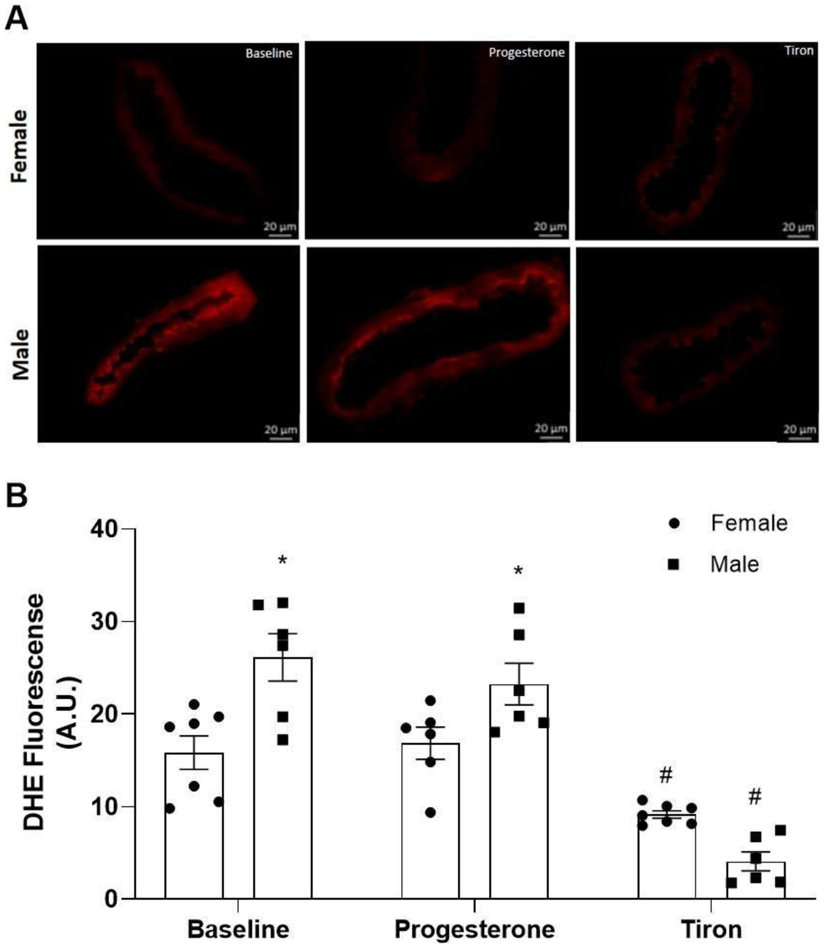
The protocol was performed according to Silva et al. (2016), with some modifications. Briefly, five slides per animal (female or male) were incubated for 30 minutes at 37 ºC and protected from light. The first slide was incubated with DHE (5 µM) to investigate basal O2●- production. The second slide was incubated with DHE + Tiron (10 µM), as a negative control for basal O2●- production. The third slide was incubated with DHE + progesterone (10 µM) to investigate whether incubation with progesterone induced changes in O2●- production.
Digital images were obtained at 40x magnification using a Leica DM 2500 inverted fluorescence photomicroscope. The images were analyzed using the Image J program (National Institutes of Health, USA). The mean fluorescence density was calculated from five images of the mesenteric segments of each animal. A sample number of 6-7 experimental animals per group was used.
2.5. Statistical analysis
Data were analyzed using the Graph-Pad Prism 8 statistical software and expressed as mean ± standard error of the mean (SEM). The Shapiro-Wilk test was used to confirm the normality of the data. For the analysis of the vasodilator response, two-way analysis of variance (two-way ANOVA) was used, followed by Sidak's post-hoc test. Rmax analysis was performed using one-way analysis of variance (one-way ANOVA) followed by Tukey's post-hoc test or Student t-test. The significance level was set at p < 0.05.
3. Results
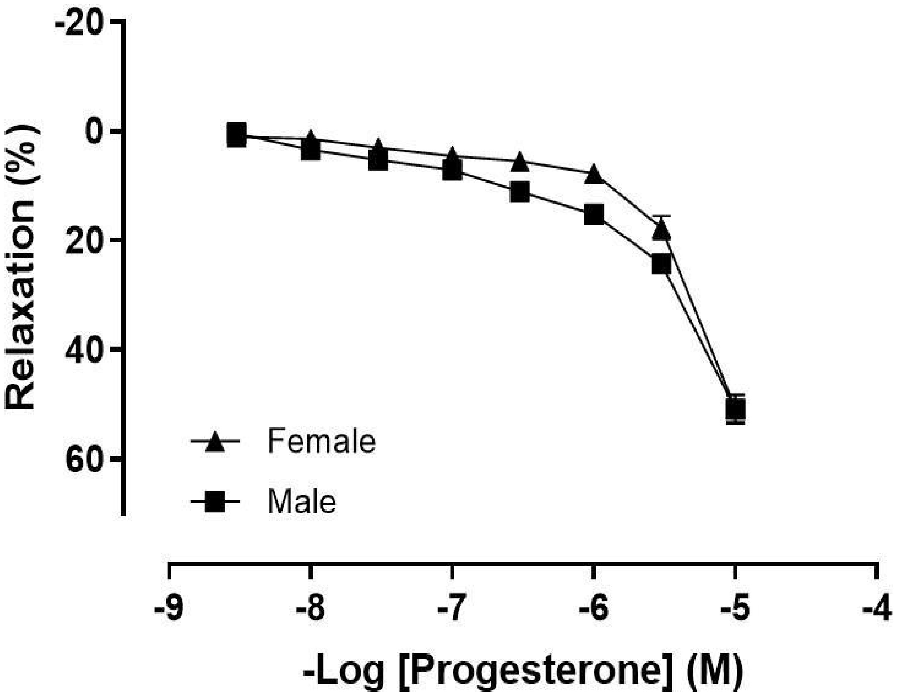
Increasing cumulative concentrations of progesterone similarly promoted relaxation in mesentery artery segments in both sexes, with females showing 50.6 ± 2.4% relaxation and males 50.8 ± 2.5% relaxation (Figure 1).
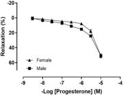
Concentration-response curve of progesterone (P4, 10 nM - 10 μM) in mesenteric resistance arteries in the presence of endothelium (Females, n = 35; Males, n = 37). Values were expressed as mean ± SEM. Two-way ANOVA was used for the analysis, followed by the Tukey’s post hoc test.
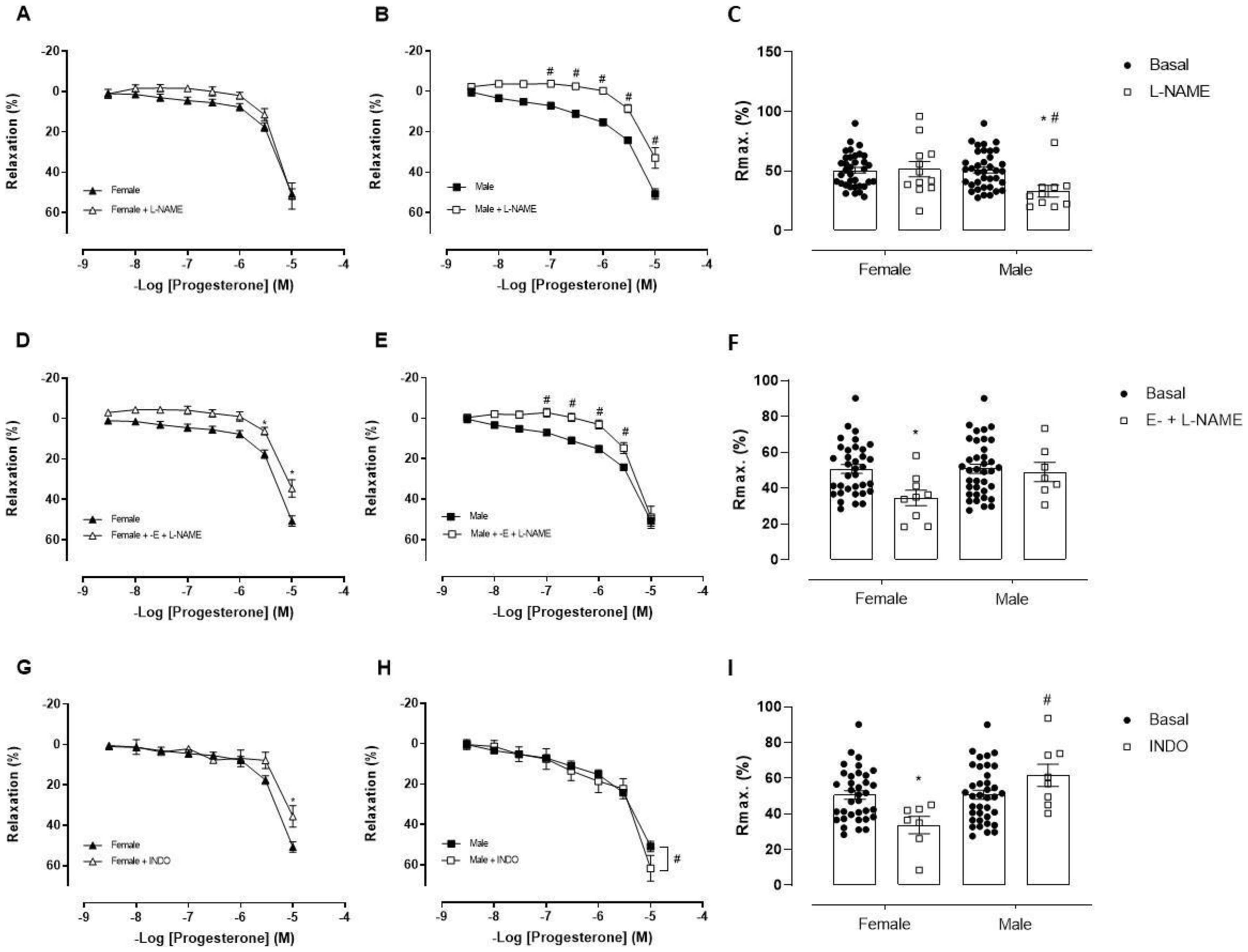
After confirming progesterone’s ability to dilate these arteries, the next step was to identify the endothelial mediators involved in this response. To do this, we first assessed the role of NO by incubating the arteries with L-NAME, a non-selective NOS inhibitor. In females, there was no significant change in the maximum relaxation response (L-NAME: without 50.6 ± 2.4% vs. 51.6 ± 6.4%) (Figure 22C). In contrast, males showed a reduction in the maximum relaxation response (L-NAME: without 50.8 ± 2.5% vs. 32.9 ± 5%) (Figure 22C), suggesting that NO may play a role in the progesterone-induced relaxation response in males.
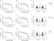
Vasodilator response to increasing concentrations of progesterone (10 nM - 10 μM) before and after inhibition with L-NAME [A: Female, n = 10; B: Male, n = 12; C: Rmax. (%)]; L-NAME in the absence of endothelium [D: Female, n = 9; E: Male, n = 7; F: Rmax. (%)] and indomethacin [G: Female, n = 7; H: Male, n = 8; I: Rmax. (%)]. Values were expressed as mean ± SEM. * P < 0.05 compared to the same dose in the basal control curve of females after incubations, # P < 0.05 compared to the same dose in the basal curve of males after incubations, + P < 0.05 compared to the female and male group after incubations. Curve analysis was carried out point-by-point through two-way ANOVA, followed by the Tukey’s post hoc test. Rmax. values were evaluated through one-way ANOVA followed by Tukey’s post hoc test.
Recognizing that NO production occurs not only in the endothelium but also in other vascular layers like the adventitia (Schwarz et al., 1999), we examined the role of extra-endothelial pathways in NO formation. To do this, we conducted incubations with L-NAME following endothelial removal. Our results revealed a significant decrease in the maximum relaxation response in females (L-NAME + endothelium removal: 50.6 ± 2.4% vs. 34.5 ± 4.3%) (Figure 22F). In males, however, only a slight reduction was observed at certain points on the curve, with no significant change in the maximum response (50.8 ± 2.5% vs. 48.9 ± 5.4%) (Figure 22F). These findings suggest that, in females, the endothelium does not appear to contribute to NO-mediated relaxation in response to progesterone, since the inhibitory effect of L-NAME was only evident after endothelial removal.
The second pathway evaluated was that of prostanoids (PNs). Using indomethacin, a non-selective COX inhibitor, we observed that only females experienced a reduction in the maximum relaxation response, suggesting the involvement of PNs in this group (INDO: without 50.6 ± 2.4 vs. 35.5 ± 5.2%) (Figure 22I). On the other hand, males showed an increase in the maximum relaxation response (INDO: without 50.8 ± 2.5% vs. 61 ± 6.2%) (Figure 22I), indicating that in males, this pathway might contribute to the production of vasoconstrictors or interfere with the action of vasodilators.
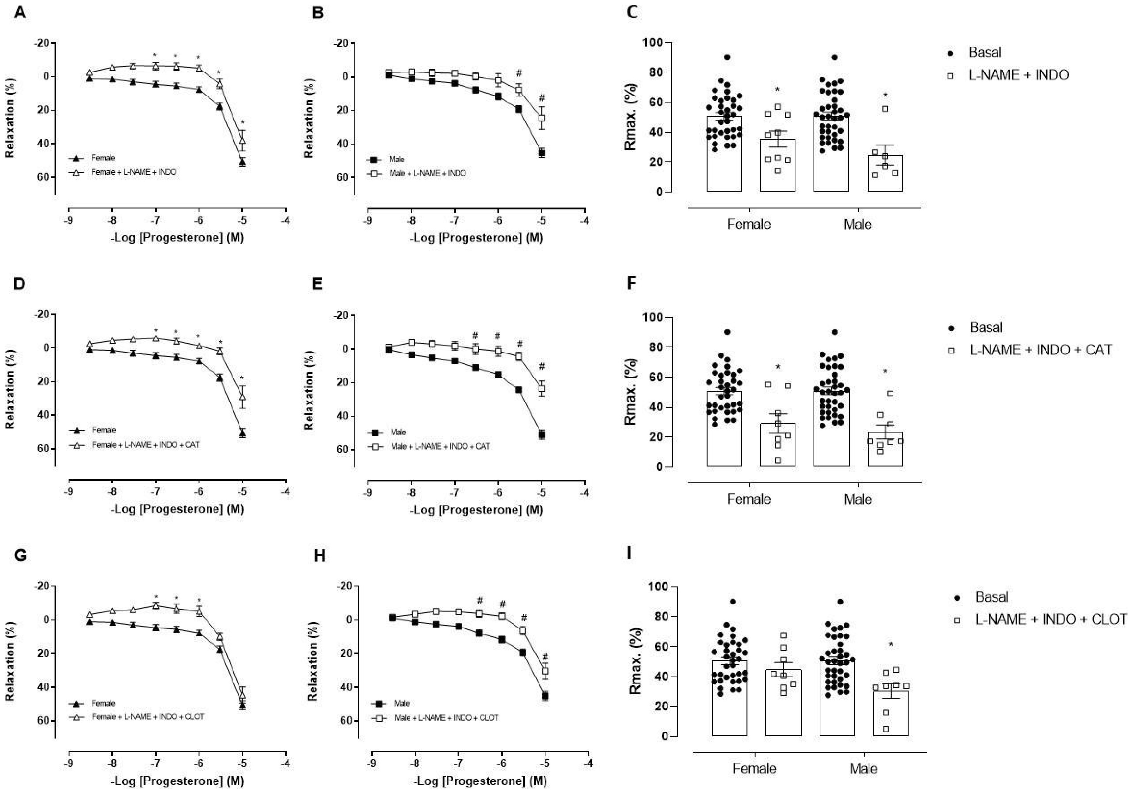
The role of the EDH pathway in the relaxation response of resistance arteries is well established (Matoba and Shimokawa, 2003; Shimokawa and Morikawa, 2005). To assess the involvement of this pathway, we conducted joint incubations with L-NAME and indomethacin and observed a reduction in the maximum relaxation response induced by progesterone in both sexes (L-NAME + INDO: without females: 50.6 ± 2.4% vs. 38 ± 6%; males: without 50.8 ± 2.5% vs. 24.6 ± 6.6%) (Figure 3A, 3B, 3C). These findings suggest that the residual relaxation after inhibition of NOS and COX pathways may be partially attributed to EDH. We next evaluated the role of H2O2 in the EDH-mediated vasodilatory response induced by progesterone. Therefore, catalase, an enzyme that degrades H2O2, was applied in combination with NOS and PNs pathway inhibitors. This approach did not lead to a further reduction in the relaxation response in either sex (L-NAME + INDO + Cat: females: 29.1 ± 6.4%; males: 23.5 ± 4.6%). Thus, in both females and males, H2O2 does not appear to contribute to the EDH response, as maximal relaxation remained similar between the two protocols.
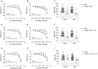
Vasodilator response to increasing concentrations of progesterone (10 nM - 10 μM) before and after inhibition with L-NAME + indomethacin (A: Female, n = 9; B: Male n = 7, C: Rmax. (%)); L-NAME + indomethacin + catalase (D: Female, n = 8, E: Male, n = 8; F: Rmax. (%)), and L-NAME + indomethacin + clotrimazole (G: Female, n = 8, H: Male, n = 8; I: Rmax. (%)). Values were expressed as mean ± SEM. * P < 0.05 compared to the same dose in the basal control curve of females after incubations, # P < 0.05 compared to the same dose in the basal curve of males after incubations, + P < 0.05 compared to the female and male group after incubations. Curve analysis was carried out point-by-point through two-way ANOVA, followed by the Tukey’s post hoc test. Rmax. values were evaluated through one-way ANOVA followed by Tukey’s post hoc test.
Furthermore, we evaluated the involvement of cytochrome P450 (CYP)-derived epoxyeicosatrienoic acids (EETs) in the EDH pathway. After inhibiting NO, PNs, and EETs, no significant reduction was observed in the maximal vasodilatory response to progesterone in either sex (L-NAME + INDO + CLOT: females: 44.6 ± 4.8%; males: 30.3 ± 4.7%). These findings indicate that EETs do not play a role in this response, suggesting that other EDH mechanisms may be involved.
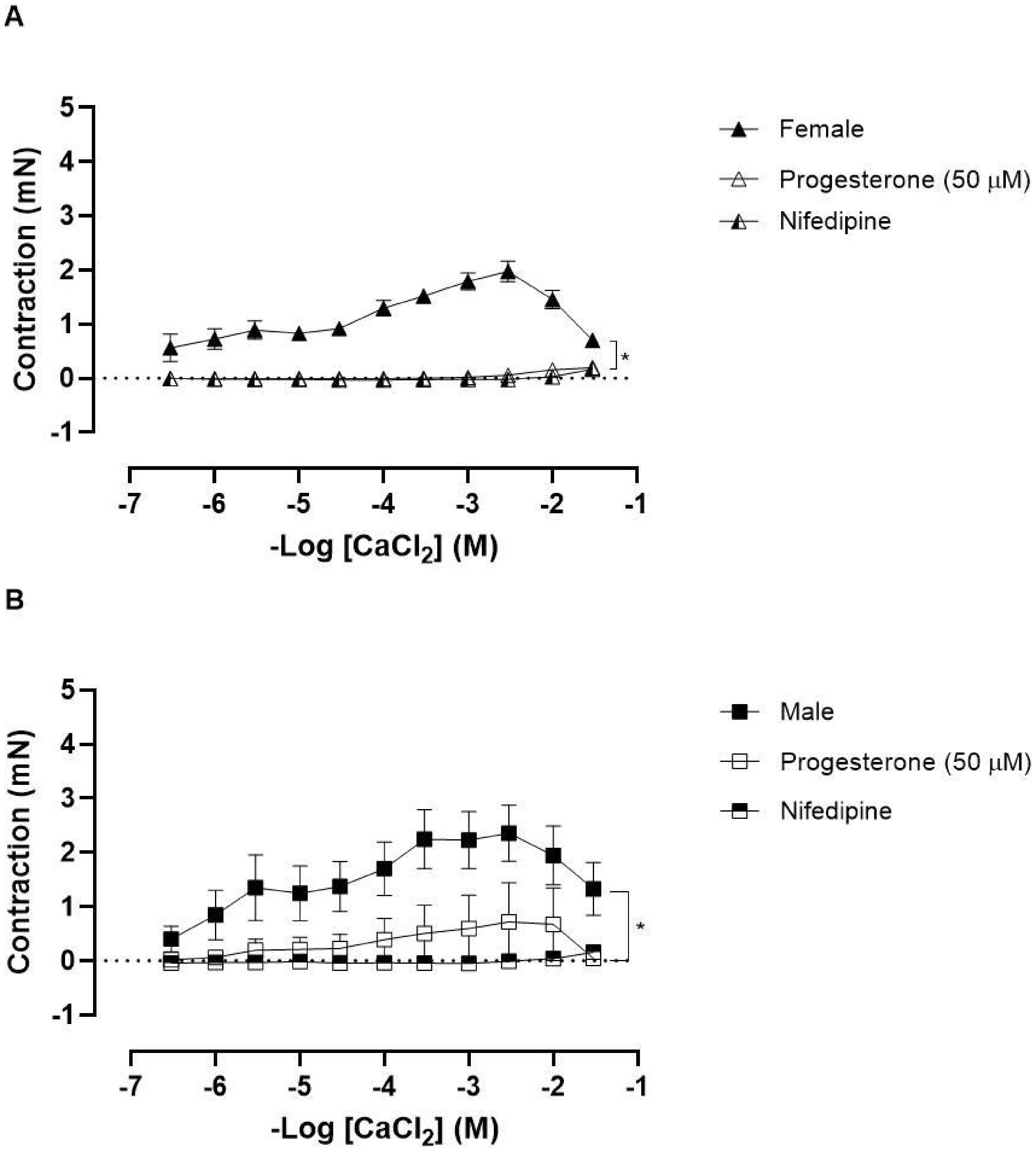
Even after inhibiting the primary endothelial relaxation pathways, the vasodilatory response was not completely abolished. Given that progesterone can directly affect VSM, we investigated whether Ca2+ might play a role in this response. To test this, mesenteric arteries from both sexes were exposed to cumulative CaCl2 concentrations in a Ca2+-free depolarizing solution, either in the presence of progesterone (50 μM) or nifedipine (1 μM) as a positive control. We observed a significant reduction in the maximum contractile response in the presence of both progesterone and nifedipine in both sexes (Figure 44B).
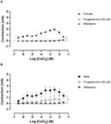
CaCl2 concentration-response curve (10 µM – 30 mM) in mesenteric resistance arteries maintained in Ca2+-free depolarizing solution, in the absence (female, n = 10; male, n = 8) and presence of progesterone (50 µM) (female n = 10 and male n = 6), and in the presence of nifedipine (female, n = 7; male, n = 5). Values are expressed as mean ± SEM. * P < 0.05 compared to the control curve. Statistical analysis was performed using two-way ANOVA followed by Tukey's post hoc test.
As shown in Figures 55B, fluorescence analysis for ROS revealed sex differences under both basal (females: 15.8 ± 1.8; males: 26.1 ± 2.5 A.U.) and stimulated with progesterone (females: 16.8 ± 1.7; males: 23.2 ± 2.2 A.U.), and Tiron (females: 9.1 ± 0.4; males: 4.0 ± 1 A.U.). Body weight and internal diameter of the mesenteric resistance arteries were assessed in both sexes. Female animals presented a mean body weight of 192 ± 3.3 g, while males presented 277 ± 8.7 g. Regarding vascular morphology, the internal diameter of the resistance arteries was 119 ± 6.4 µm in females and 220 ± 5.1 µm in males.
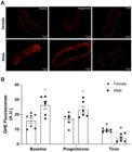
Fluorescence microscopy analysis emitted by DHE in mesenteric resistance arteries of females (n = 6) and males (n = 7) in the absence or presence of stimulation by progesterone (10 µM), with quantification of the fluorescence produced. Scale bar = 20 μm. Values were expressed as mean ± SEM. * P < 0.05 compared to the female group and # P < 0.05 compared to the baseline and progesterone incubations of the same group. Two-way ANOVA was used for the analysis, followed by the Sidak’s post hoc test.
4. Discussion
The main finding of this study is that progesterone promoted relaxation in mesenteric resistance arteries similarly in both females and males, but the underlying mechanisms differed between sexes. In females, progesterone-induced relaxation seems to rely more on PNs and extra-endothelial NO. In contrast, in males, this response appears to be primarily dependent on NO pathway. Despite these differences, progesterone seems to negatively modulate Ca2+ mobilization in both sexes.
We first observed that increasing concentrations of progesterone effectively promoted relaxation in mesenteric arterial segments from normotensive rats of both sexes. This aligns with previous studies showing progesterone-induced relaxation in various arterial segments across different species, such as aorta of male Sprague-Dawley rats (Barbagallo et al., 2001), porcine coronary arteries (Molinari et al., 2001), and female primate coronary arteries (Minshall et al., 2002). Our results further confirm that progesterone can induce relaxation in mesenteric arterial segments.
We then found that progesterone-induced relaxation in mesenteric arteries without differences between sexes. This finding is consistent with a previous study by our group, which reported similar results in the coronary vascular bed of normotensive rats (Giesen et al., 2020). With the confirmation that progesterone promotes relaxation in mesenteric arteries of both sexes equally, our next step was to investigate the specific relaxation pathways involved in each sex. It should be acknowledged that the maximum concentration of progesterone used in the experiments (10 µM) exceeds the physiological circulating plasma levels reported in female rats. This difference is inherent to the isolated organ in vitro model and is consistent with the well-recognized principle that acute membrane effects of steroid hormones frequently require concentrations above physiological circulating levels in this type of preparation (Nakano et al., 1998; Santos et al., 2004). It is plausible that chronic exposure of mesenteric resistance arteries to physiological concentrations of progesterone in vivo may produce functional effects equivalent to those observed with the supraphysiological concentrations employed in vitro. The results should therefore be interpreted within the context of an acute pharmacological ex vivo model, and caution is warranted when extrapolating directly to physiological conditions in intact animals.
We first examined the NO pathway by inhibiting its synthesis with L-NAME. We observed a reduction in the relaxation response only in males, suggesting that NO may mediate relaxation in mesenteric arteries specifically in males, as previously reported (Chan et al., 2001). However, when L-NAME was applied to vessels without endothelium, we found a reduction in relaxation in females as well, indicating the involvement of extra-endothelial NO sources in females. Indeed, NO can be produced by extra-endothelial sources such as the VSM (Mollace et al., 1991) or the adventitial layer (Schwarz et al., 1999). The variation in NO pathway response between sexes in the presence of endothelium may be linked to differences in ROS production. Although ROS production was higher in males under both basal and stimulated conditions, the relaxation response was similar between sexes. This suggests that the NO pathway might play a more significant role in males when the endothelium is present.
Another potential pathway for endothelial mediators involved in progesterone-induced vasodilation is the PNs pathway. To assess this, we used indomethacin to non-selectively inhibit COX activity. We found that this inhibition attenuated the relaxation response in females, suggesting that PNs play a role in this process. Previous studies have shown that progesterone can modulate COX activity, thereby increasing the bioavailability of PNs (Cutini et al., 2014). Additionally, progesterone has been shown to enhance the expression and activity of COX-1 and COX-2 enzymes in human endothelial cells (Hermenegildo et al., 2005). In contrast, in the coronary vascular bed, the PNs pathway contributes to progesterone-induced vasodilation only in males (Giesen et al., 2020). In the present study, males exhibited an enhanced vasodilatory response following inhibition of this pathway. This finding may be related to the higher formation of vasoconstrictor PNs, such as thromboxane A2 and prostaglandin F2α, in males (Ospina et al., 2003). Inhibiting PNs production in males likely reduces these vasoconstrictors, thereby enhancing the vasodilatory response to progesterone.
After inhibiting both the NO and PNs pathways, the progesterone-induced relaxation response was reduced but not eliminated. This suggests that the remaining relaxation might be mediated by a third endothelial relaxation pathway, a direct effect of progesterone on the VSM, or both. Given that H2O2 is known to play a role in the EDH pathway in mesenteric resistance arteries (Matoba and Shimokawa, 2003), we investigated its contribution to progesterone-induced vasodilation. However, in this study, we did not observe the participation of H2O2 as an EDH pathway in both sexes. In the coronary vascular bed of normotensive rats, H2O2 was previously shown to contribute to progesterone-induced vasodilation; this effect was observed only in females (Giesen et al., 2020). The criterion adopted to suggest the involvement of an EDH-like component, defined as the residual relaxation after combined NOS and COX inhibition, corresponds to a functional approach widely used in ex vivo vascular preparations and previously validated in the literature by the Shimokawa group in studies of rat mesenteric circulation (Shimokawa et al., 1996) and in the characterization of H2O2 as an EDH factor in animals and humans (Matoba and Shimokawa, 2003; Shimokawa and Morikawa, 2005). However, in the present study, this participation was inferred indirectly, as we did not perform a specific assessment of ion channels involved in hyperpolarization. Although the lack of contribution from H2O2 and EETs supports this interpretation, a definitive mechanistic attribution cannot be established. In addition, it should be considered that progesterone may act directly on VSM (Cairrão et al., 2012; Pang and Thomas, 2021) contributing to the observed relaxation. Thus, the findings are consistent with the presence of a residual hyperpolarizing component, whose direct characterization remains a perspective for future studies.
Next, we investigated another potential EDH pathway candidate: EETs, which are metabolites of the CYP enzyme. Our results indicated that EETs are not involved in the vasodilatory response to progesterone in either male or female rats. EETs are crucial for vascular reactivity, as they help regulate vascular tone by activating calcium-activated K+ channels in the endothelium and VSM, leading to hyperpolarization and relaxation (Campbell and Fleming, 2010). While this pathway has been well-documented in the relaxation responses of the coronary vascular bed in normotensive rats (Santos et al., 2004) and in hypertensive female rats (Santos et al., 2010), it has not been previously observed in mesenteric resistance vessels.
In certain arteries, such as coronary arteries, sex differences in relaxation responses to sex hormone receptor agonists are often linked to varying levels of ROS production between the sexes. This increased ROS production, particularly when stimulated by a selective agonist, can lead to a reduced vasodilatory response in the sex with higher ROS levels (Debortoli et al., 2017). Even though ROS generation in males was greater in both conditions (basal and stimulated by progesterone), vasodilation was similar in both sexes, one possible explanation is the greater participation of the NO pathway in the presence of the endothelium in males. It is important to acknowledge a methodological limitation related to the evaluation of ROS. Although DHE is widely used for the detection of O2•− in different biological systems, its specificity is limited due to interference from other oxidizing species, which compromises its accurate quantification (Kalyanaraman et al., 2012). Therefore, throughout the manuscript, we use the term "reactive oxygen species" to avoid misinterpretations regarding the results obtained with DHE.
Once the participation of endothelial mediators in progesterone-induced vasodilation was evaluated, our next step was to investigate the role of specific ions, particularly Ca2+, in this response. In addition to assessing the relaxation response induced by progesterone, we also examined its ability to modulate Ca2+ mobilization in the same arterial segments. To do so, we generated contraction curves induced by CaCl2 in the presence and absence of progesterone or nifedipine, a non-selective Ca2+- channel blocker. We observed that progesterone attenuated the contraction curve in both sexes, producing a response similar to that seen with nifedipine. The inhibitory effect of progesterone on Ca2+ influx in VSM cells has been demonstrated in the thoracic aorta, though this was studied only in Wistar males (Cairrão et al., 2012). In the caudal artery of male Sprague-Dawley rats, progesterone also inhibited vasoconstriction by reducing Ca2+ currents (Zhang et al., 2002). Consistently, progesterone induced a significant vasorelaxant effect in KCl-precontracted rings without endothelium from canine basilar and internal carotid arteries, suggesting a direct action on the VSM (Ramírez-Rosas et al., 2014). This supports the hypothesis that progesterone is capable of producing acute relaxation independently of endothelial factors, likely through modulation of Ca2+ dynamics in VSM. The VSM sarcolemma contains membrane receptors for progesterone (mPRα), and their activation leads to a reduction in cytosolic Ca2+ via the Gi protein (inhibitory) and mitogen-activated protein kinase (MAPK) signaling pathways. Additionally, progesterone enhances the expression of SERCA and the phosphorylation of phospholamban, which increases Ca2+ uptake from the cytosol to the sarcoplasmic reticulum (Pang and Thomas, 2021). Given this context, our findings related to the relaxation response may be linked to the actions of progesterone on VSM.
Taken together, these findings reinforce the concept that progesterone induces relaxation of mesenteric resistance arteries similarly in both sexes, though different relaxation pathways are involved. In females, the pathways include PNs and extra-endothelial NO, while in males, NO is the main mediator. Additionally, progesterone attenuates the Ca2+-dependent vasoconstrictor response in both sexes. These findings enhance our understanding of progesterone’s vascular effects and may contribute to the development of improved hormone replacement therapies for postmenopausal women.
Acknowledgements
The authors would like to thank Nathalie Tristão Banhos Delgado and Rakel Passos Simões for teaching a new technique and contributing to the development of the manuscript’s results. This study was supported by Conselho Nacional de Desenvolvimento Científico e Tecnológico (CNPq) – [grant number: #311868/2022-3] and Fundação de Amparo à Pesquisa e Inovação do Espírito Santo (FAPES) – [grant numbers: #959/2023 - P:2023-MW48T and #697/2022 - P: 2022-WKMH7].
Data Availability Statement
The data supporting the findings of this study are available from the corresponding author upon reasonable request.
-
Ethics and consent
The protocol for the research project was approved by the Animal Ethics Committee of the Federal University of Espirito Santo (No #18/2020). All procedures were conducted in accordance with the recommendations of the Brazilian Guidelines for the Care and Use of Animals for Scientific and Didactic Purposes and the Guidelines for the Practice of Euthanasia.
References
-
BARBAGALLO, M., DOMINGUEZ, L.J., LICATA, G., SHAN, J., BING, L., KARPINSKI, E., PANG, P.K. and RESNICK, L.M., 2001. Vascular effects of progesterone: role of cellular calcium regulation. Hypertension, vol. 37, no. 1, pp. 142-147. https://doi.org/10.1161/01.HYP.37.1.142 PMid:11208769.
» https://doi.org/10.1161/01.HYP.37.1.142 -
BOSCHITSCH, E., MAYERHOFER, S. and MAGOMETSCHNIGG, D., 2010. Hypertension in women: the role of progesterone and aldosterone. Climacteric : The Journal of the International Menopause Society, vol. 13, no. 4, pp. 307-313. https://doi.org/10.3109/13697131003624649 PMid:20443718.
» https://doi.org/10.3109/13697131003624649 - BRASIL. Ministério da Ciência, Tecnologia e Inovação - MCTI. Conselho Nacional de Controle de Experimentação Animal -CONCEA, 2023. Guia brasileiro de produção, manutenção ou utilização de animais em atividades de ensino ou pesquisa científica Brasília: CONCEA.
-
CAIRRÃO, E., ALVAREZ, E., CARVAS, J.M., SANTOS-SILVA, A.J. and VERDE, I., 2012. Non-genomic vasorelaxant effects of 17β-estradiol and progesterone on rat aorta are mediated by L-type Ca2+ current inhibition. Acta Pharmacologica Sinica, vol. 33, no. 5, pp. 615-624. https://doi.org/10.1038/aps.2012.4 PMid:22465948.
» https://doi.org/10.1038/aps.2012.4 -
CAMPBELL, W.B. and FLEMING, I., 2010. Epoxyeicosatrienoic acids and endothelium-dependent responses. Pflügers Archiv : European Journal of Physiology, vol. 459, no. 6, pp. 881-895. https://doi.org/10.1007/s00424-010-0804-6 PMid:20224870.
» https://doi.org/10.1007/s00424-010-0804-6 -
CHAN, H.Y., YAO, X., TSANG, S.Y., CHAN, F.L., LAU, C.W. and HUANG, Y., 2001. Different role of endothelium/nitric oxide in 17β-estradiol and progesterone-induced relaxation in rat arteries. Life Sciences, vol. 69, no. 14, pp. 1609-1617. https://doi.org/10.1016/S0024-3205(01)01235-8 PMid:11589501.
» https://doi.org/10.1016/S0024-3205(01)01235-8 -
CUNHA, T.R.D., GIESEN, J.A.S., ROUVER, W.N., COSTA, E.D., GRANDO, M.D., LEMOS, V.S., BENDHACK, L.M. and SANTOS, R.L.D., 2020. Effects of progesterone treatment on endothelium-dependent coronary relaxation in ovariectomized rats. Life Sciences, vol. 247, pp. 117391. https://doi.org/10.1016/j.lfs.2020.117391 PMid:32017871.
» https://doi.org/10.1016/j.lfs.2020.117391 -
CUTINI, P., SELLÉS, J. and MASSHEIMER, V., 2009. Cross-talk between rapid and long-term effects of progesterone on vascular tissue. The Journal of Steroid Biochemistry and Molecular Biology, vol. 115, no. 1-2, pp. 36-43. https://doi.org/10.1016/j.jsbmb.2009.02.014 PMid:19429459.
» https://doi.org/10.1016/j.jsbmb.2009.02.014 -
CUTINI, P.H., CAMPELO, A.E. and MASSHEIMER, V.L., 2014. Differential regulation of endothelium behavior by progesterone and medroxyprogesterone acetate. The Journal of Endocrinology, vol. 220, no. 3, pp. 179-193. https://doi.org/10.1530/JOE-13-0263 PMid:24301615.
» https://doi.org/10.1530/JOE-13-0263 -
DEBORTOLI, A.R., ROUVER, W.D.N., DELGADO, N.T.B., MENGAL, V., CLAUDIO, E.R.G., PERNOMIAN, L., BENDHACK, L.M., MOYSÉS, M.R. and SANTOS, R.L., 2017. GPER modulates tone and coronary vascular reactivity in male and female rats. Journal of Molecular Endocrinology, vol. 59, no. 2, pp. 171-180. https://doi.org/10.1530/JME-16-0117 PMid:28733475.
» https://doi.org/10.1530/JME-16-0117 -
DUBEY, R.K., IMTHURN, B., ZACHARIA, L.C. and JACKSON, E.K., 2004. Hormone replacement therapy and cardiovascular disease: what went wrong and where do we go from here? Hypertension, vol. 44, no. 6, pp. 789-795. https://doi.org/10.1161/01.HYP.0000145988.95551.28 PMid:15477384.
» https://doi.org/10.1161/01.HYP.0000145988.95551.28 -
FERNANDES, D.C., WOSNIAK JUNIOR, J., PESCATORE, L.A., BERTOLINE, M.A., LIBERMAN, M., LAURINDO, F.R. and SANTOS, C.X., 2007. Analysis of DHE-derived oxidation products by HPLC in the assessment of superoxide production and NADPH oxidase activity in vascular systems. American Journal of Physiology. Cell Physiology, vol. 292, no. 1, pp. C413-C422. https://doi.org/10.1152/ajpcell.00188.2006 PMid:16971501.
» https://doi.org/10.1152/ajpcell.00188.2006 -
GIESEN, J.A.S., ROUVER, W.D.N., COSTA, E.D., LEMOS, V.S. and DOS SANTOS, R.L., 2020. Sex differences in progesterone-induced relaxation in the coronary bed from normotensive rats. Journal of Molecular Endocrinology, vol. 64, no. 2, pp. 91-102. https://doi.org/10.1530/JME-19-0171 PMid:31834856.
» https://doi.org/10.1530/JME-19-0171 -
GRADY, D., RUBIN, S.M., PETITTI, D.B., FOX, C.S., BLACK, D., ETTINGER, B., ERNSTER, V.L. and CUMMINGS, S.R., 1992. Hormone therapy to prevent disease and prolong life in postmenopausal women. Annals of Internal Medicine, vol. 117, no. 12, pp. 1016-1037. https://doi.org/10.7326/0003-4819-117-12-1016 PMid:1443971.
» https://doi.org/10.7326/0003-4819-117-12-1016 -
GRIMES, D.A. and LOBO, R.A., 2002. Perspectives on the women’s health initiative trial of hormone replacement therapy. Obstetrics and Gynecology, vol. 100, no. 6, pp. 1344-1353. https://doi.org/10.1016/S0029-7844(02)02503-6 PMid:12468183.
» https://doi.org/10.1016/S0029-7844(02)02503-6 -
GRODSTEIN, F., STAMPFER, M.J., COLDITZ, G.A., WILLETT, W.C., MANSON, J.E., JOFFE, M., ROSNER, B., FUCHS, C., HANKINSON, S.E., HUNTER, D.J., HENNEKENS, C.H. and SPEIZER, F.E., 1997. Postmenopausal hormone therapy and mortality. The New England Journal of Medicine, vol. 336, no. 25, pp. 1769-1775. https://doi.org/10.1056/NEJM199706193362501 PMid:9187066.
» https://doi.org/10.1056/NEJM199706193362501 -
HATANO, Y., NAKAMURA, K., MORIYAMA, S., MORI, K. and TODA, N., 1989. The contractile responses of isolated dog cerebral and extracerebral arteries to oxybarbiturates and thiobarbiturates. Anesthesiology, vol. 71, no. 1, pp. 80-86. https://doi.org/10.1097/00000542-198907000-00015 PMid:2751144.
» https://doi.org/10.1097/00000542-198907000-00015 -
HENDERSON, B.E., PAGANINI-HILL, A. and ROSS, R.K., 1991. Decreased mortality in users of estrogen replacement therapy. Archives of Internal Medicine, vol. 151, no. 1, pp. 75-78. https://doi.org/10.1001/archinte.1991.00400010095012 PMid:1985611.
» https://doi.org/10.1001/archinte.1991.00400010095012 -
HERMENEGILDO, C., OVIEDO, P.J., GARCÍA-MARTÍNEZ, M.C., GARCÍA-PÉREZ, M.A., TARÍN, J.J. and CANO, A., 2005. Progestogens stimulate prostacyclin production by human endothelial cells. Human Reproduction, vol. 20, no. 6, pp. 1554-1561. https://doi.org/10.1093/humrep/deh803 PMid:15734756.
» https://doi.org/10.1093/humrep/deh803 -
HERMSMEYER, R.K., THOMPSON, T.L., POHOST, G.M. and KASKI, J.C., 2008. Cardiovascular effects of medroxyprogesterone acetate and progesterone: a case of mistaken identity? Nature Clinical Practice. Cardiovascular Medicine, vol. 5, no. 7, pp. 387-395. https://doi.org/10.1038/ncpcardio1234 PMid:18521110.
» https://doi.org/10.1038/ncpcardio1234 -
HULLEY, S., GRADY, D., BUSH, T., FURBERG, C., HERRINGTON, D., RIGGS, B. and VITTINGHOFF, E., 1998. Randomized trial of estrogen plus progestin for secondary prevention of coronary heart disease in postmenopausal women. Journal of the American Medical Association, vol. 280, no. 7, pp. 605-613. https://doi.org/10.1001/jama.280.7.605 PMid:9718051.
» https://doi.org/10.1001/jama.280.7.605 - INGEGNO, M.D., MONEY, S.R., THELMO, W., GREENE, G.L., DAVIDIAN, M., JAFFE, B.M. and PERTSCHUK, L.P., 1988. Progesterone receptors in the human heart and great vessels. Laboratory Investigation, vol. 59, no. 3, pp. 353-356. PMid:3411936.
-
JIANG, C.W., SARREL, P.M., LINDSAY, D.C., POOLE-WILSON, P.A. and COLLINS, P., 1992. Progesterone induces endothelium-independent relaxation of rabbit coronary artery in vitro. European Journal of Pharmacology, vol. 211, no. 2, pp. 163-167. https://doi.org/10.1016/0014-2999(92)90524-8 PMid:1319340.
» https://doi.org/10.1016/0014-2999(92)90524-8 -
KALYANARAMAN, B., DARLEY-USMAR, V., DAVIES, K.J., DENNERY, P.A., FORMAN, H.J., GRISHAM, M.B., MANN, G.E., MOORE, K., ROBERTS 2ND, L.J. and ISCHIROPOULOS, H., 2012. Measuring reactive oxygen and nitrogen species with fluorescent probes: challenges and limitations. Free Radical Biology & Medicine, vol. 52, no. 1, pp. 1-6. https://doi.org/10.1016/j.freeradbiomed.2011.09.030 PMid:22027063.
» https://doi.org/10.1016/j.freeradbiomed.2011.09.030 -
MARCONDES, F.K., BIANCHI, F.J. and TANNO, A.P., 2002. Determination of the estrous cycle phases of rats: some helpful considerations. Brazilian Journal of Biology, vol. 62, no. 4A, pp. 609-614. https://doi.org/10.1590/S1519-69842002000400008 PMid:12659010.
» https://doi.org/10.1590/S1519-69842002000400008 -
MATOBA, T. and SHIMOKAWA, H., 2003. Hydrogen peroxide is an endothelium-derived hyperpolarizing factor in animals and humans. Journal of Pharmacological Sciences, vol. 92, no. 1, pp. 1-6. https://doi.org/10.1254/jphs.92.1 PMid:12832848.
» https://doi.org/10.1254/jphs.92.1 -
MENDELSOHN, M.E. and KARAS, R.H., 2005. Molecular and cellular basis of cardiovascular gender differences. Science, vol. 308, no. 5728, pp. 1583-1587. https://doi.org/10.1126/science.1112062 PMid:15947175.
» https://doi.org/10.1126/science.1112062 -
MINSHALL, R.D., PAVCNIK, D., BROWNE, D.L. and HERMSMEYER, K., 2002. Nongenomic vasodilator action of progesterone on primate coronary arteries. Journal of Applied Physiology (Bethesda, Md.), vol. 92, no. 2, pp. 701-708. https://doi.org/10.1152/japplphysiol.00689.2001 PMid:11796684.
» https://doi.org/10.1152/japplphysiol.00689.2001 -
MOLINARI, C., BATTAGLIA, A., GROSSINI, E., MARY, D.A., STOKER, J.B., SURICO, N. and VACCA, G., 2001. The effect of progesterone on coronary blood flow in anaesthetized pigs. Experimental Physiology, vol. 86, no. 1, pp. 101-108. https://doi.org/10.1113/eph8602076 PMid:11434325.
» https://doi.org/10.1113/eph8602076 -
MOLLACE, V., SALVEMINI, D., ANGGARD, E. and VANE, J., 1991. Nitric oxide from vascular smooth muscle cells: regulation of platelet reactivity and smooth muscle cell guanylate cyclase. British Journal of Pharmacology, vol. 104, no. 3, pp. 633-638. https://doi.org/10.1111/j.1476-5381.1991.tb12481.x PMid:1724627.
» https://doi.org/10.1111/j.1476-5381.1991.tb12481.x -
MULVANY, M.J. and HALPERN, W., 1977. Contractile properties of small arterial resistance vessels in spontaneously hypertensive and normotensive rats. Circulation Research, vol. 41, no. 1, pp. 19-26. https://doi.org/10.1161/01.RES.41.1.19 PMid:862138.
» https://doi.org/10.1161/01.RES.41.1.19 -
MURPHY, J.G. and KHALIL, R.A., 1999. Decreased [Ca2+]i during inhibition of coronary smooth muscle contraction by 17β-estradiol, progesterone, and testosterone. The Journal of Pharmacology and Experimental Therapeutics, vol. 291, no. 1, pp. 44-52. https://doi.org/10.1016/S0022-3565(24)35069-4 PMid:10490885.
» https://doi.org/10.1016/S0022-3565(24)35069-4 -
NAKANO, Y., OSHIMA, T., MATSUURA, H., KAJIYAMA, G. and KAMBE, M., 1998. Effect of 17β-estradiol on inhibition of platelet aggregation in vitro is mediated by an increase in NO synthesis. Arteriosclerosis, Thrombosis, and Vascular Biology, vol. 18, no. 6, pp. 961-967. https://doi.org/10.1161/01.ATV.18.6.961 PMid:9633938.
» https://doi.org/10.1161/01.ATV.18.6.961 -
ORSHAL, J.M. and KHALIL, R.A., 2004. Gender, sex hormones, and vascular tone. American Journal of Physiology. Regulatory, Integrative and Comparative Physiology, vol. 286, no. 2, pp. R233-R249. https://doi.org/10.1152/ajpregu.00338.2003 PMid:14707008.
» https://doi.org/10.1152/ajpregu.00338.2003 -
OSPINA, J.A., DUCKLES, S.P. and KRAUSE, D.N., 2003. 17β-Estradiol decreases vascular tone in cerebral arteries by shifting COX-dependent vasoconstriction to vasodilation. American Journal of Physiology. Heart and Circulatory Physiology, vol. 285, no. 1, pp. H241-H250. https://doi.org/10.1152/ajpheart.00018.2003 PMid:12637362.
» https://doi.org/10.1152/ajpheart.00018.2003 -
PANG, Y. and THOMAS, P., 2021. Involvement of sarco/endoplasmic reticulum Ca2+-ATPase (SERCA) in mPRα (PAQR7)-mediated progesterone induction of vascular smooth muscle relaxation. American Journal of Physiology. Endocrinology and Metabolism, vol. 320, no. 3, pp. E453-E466. https://doi.org/10.1152/ajpendo.00359.2020 PMid:33427050.
» https://doi.org/10.1152/ajpendo.00359.2020 -
PANG, Y., DONG, J. and THOMAS, P., 2015. Progesterone increases nitric oxide synthesis in human vascular endothelial cells through activation of membrane progesterone receptor-alpha. American Journal of Physiology. Endocrinology and Metabolism, vol. 308, no. 10, pp. E899-E911. https://doi.org/10.1152/ajpendo.00527.2014 PMid:25805192.
» https://doi.org/10.1152/ajpendo.00527.2014 -
QUADROS, P.S., PFAU, J.L., GOLDSTEIN, A.Y., VRIES, G.J. and WAGNER, C.K., 2002. Sex differences in progesterone receptor expression: a potential mechanism for estradiol-mediated sexual differentiation. Endocrinology, vol. 143, no. 10, pp. 3727-3739. https://doi.org/10.1210/en.2002-211438 PMid:12239082.
» https://doi.org/10.1210/en.2002-211438 -
RAMÍREZ-ROSAS, M.B., COBOS-PUC, L.E., SÁNCHEZ-LÓPEZ, A., GUTIÉRREZ-LARA, E.J. and CENTURIÓN, D., 2014. Pharmacological characterization of the mechanisms involved in the vasorelaxation induced by progesterone and 17β-estradiol on isolated canine basilar and internal carotid arteries. Steroids, vol. 89, pp. 33-40. https://doi.org/10.1016/j.steroids.2014.07.010 PMid:25072792.
» https://doi.org/10.1016/j.steroids.2014.07.010 -
RENZO, G.C.D., TOSTO, V. and TSIBIZOVA, V., 2020. Progesterone: history, facts, and artifacts. Best Practice & Research. Clinical Obstetrics & Gynaecology, vol. 69, pp. 2-12. https://doi.org/10.1016/j.bpobgyn.2020.07.012 PMid:32943346.
» https://doi.org/10.1016/j.bpobgyn.2020.07.012 -
ROSSETTI, M.F., CAMBIASSO, M.J., HOLSCHBACH, M.A. and CABRERA, R., 2016. Oestrogens and progestagens: synthesis and action in the brain. Journal of Neuroendocrinology, vol. 28, no. 7, pp. 1-11. https://doi.org/10.1111/jne.12402 PMid:27306650.
» https://doi.org/10.1111/jne.12402 -
ROSSOUW, J.E., ANDERSON, G.L., PRENTICE, R.L., LACROIX, A.Z., KOOPERBERG, C., STEFANICK, M.L., JACKSON, R.D., BERESFORD, S.A., HOWARD, B.V., JOHNSON, K.C., KOTCHEN, J.M. , OCKENE, J., and WRITING GROUP FOR THE WOMEN’S HEALTH INITIATIVE INVESTIGATORS, 2002. Risks and benefits of estrogen plus progestin in healthy postmenopausal women: principal results from the Women’s Health Initiative randomized controlled trial. Journal of the American Medical Association, vol. 288, no. 3, pp. 321-333. https://doi.org/10.1001/jama.288.3.321 PMid:12117397.
» https://doi.org/10.1001/jama.288.3.321 -
SANTOS, R.L., ABREU, G.R., BISSOLI, N.S. and MOYSÉS, M.R., 2004. Endothelial mediators of 17β-estradiol-induced coronary vasodilation in the isolated rat heart. Brazilian Journal of Medical and Biological Research = Revista Brasileira de Pesquisas Médicas e Biológicas, vol. 37, no. 4, pp. 569-575. https://doi.org/10.1590/S0100-879X2004000400014 PMid:15064820.
» https://doi.org/10.1590/S0100-879X2004000400014 -
SANTOS, R.L., MARIN, E.B., GONÇALVES, W.L., BISSOLI, N.S., ABREU, G.R. and MOYSÉS, M.R., 2010. Sex differences in the coronary vasodilation induced by 17β-oestradiol in the isolated perfused heart from spontaneously hypertensive rats. Acta Physiologica (Oxford, England), vol. 200, no. 3, pp. 203-210. https://doi.org/10.1111/j.1748-1716.2010.02140.x PMid:20426771.
» https://doi.org/10.1111/j.1748-1716.2010.02140.x -
SCHNEIDER, M.A., DAVIES, M.C. and HONOUR, J.W., 1993. The timing of placental competence in pregnancy after oocyte donation. Fertility and Sterility, vol. 59, no. 5, pp. 1059-1064. https://doi.org/10.1016/S0015-0282(16)55928-7 PMid:8486174.
» https://doi.org/10.1016/S0015-0282(16)55928-7 -
SCHWARZ, P.M., KLEINERT, H. and FÖRSTERMANN, U., 1999. Potential functional significance of brain-type and muscle-type nitric oxide synthase I expressed in adventitia and media of rat aorta. Arteriosclerosis, Thrombosis, and Vascular Biology, vol. 19, no. 11, pp. 2584-2590. https://doi.org/10.1161/01.ATV.19.11.2584 PMid:10558999.
» https://doi.org/10.1161/01.ATV.19.11.2584 -
SHIMOKAWA, H. and MORIKAWA, K., 2005. Hydrogen peroxide is an endothelium-derived hyperpolarizing factor in animals and humans. Journal of Molecular and Cellular Cardiology, vol. 39, no. 5, pp. 725-732. https://doi.org/10.1016/j.yjmcc.2005.07.007 PMid:16122755.
» https://doi.org/10.1016/j.yjmcc.2005.07.007 -
SHIMOKAWA, H., YASUTAKE, H., FUJII, K., OWADA, M.K., NAKAIKE, R., FUKUMOTO, Y., TAKAYANAGI, T., NAGAO, T., EGASHIRA, K., FUJISHIMA, M. and TAKESHITA, A., 1996. The importance of the hyperpolarizing mechanism increases as the vessel size decreases in endothelium-dependent relaxations in rat mesenteric circulation. Journal of Cardiovascular Pharmacology, vol. 28, no. 5, pp. 703-711. https://doi.org/10.1097/00005344-199611000-00014 PMid:8945685.
» https://doi.org/10.1097/00005344-199611000-00014 -
SILVA, G.C., PEREIRA, A.C., REZENDE, B.A., DA SILVA, J.P., CRUZ, J.S., DE SOUZA, M., GOMES, R.A., TELES, Y.C., CORTES, S.F. and LEMOS, V.S., 2013. Mechanism of the antihypertensive and vasorelaxant effects of the flavonoid tiliroside in resistance arteries. Planta Medica, vol. 79, no. 12, pp. 1003-1008. https://doi.org/10.1055/s-0032-1328765 PMid:23877918.
» https://doi.org/10.1055/s-0032-1328765 -
SILVA, J.F., CAPETTINI, L.S., DA SILVA, J.F., SALES-JUNIOR, P., CRUZ, J.S., CORTES, S.F. and LEMOS, V.S., 2016. Mechanisms of vascular dysfunction in acute phase of Trypanosoma cruzi infection in mice. Vascular Pharmacology, vol. 82, pp. 73-81. https://doi.org/10.1016/j.vph.2016.03.002 PMid:26988253.
» https://doi.org/10.1016/j.vph.2016.03.002 -
SIMONCINI, T., MANNELLA, P., FORNARI, L., CARUSO, A., WILLIS, M.Y., GARIBALDI, S., BALDACCI, C. and GENAZZANI, A.R., 2004. Differential signal transduction of progesterone and medroxyprogesterone acetate in human endothelial cells. Endocrinology, vol. 145, no. 12, pp. 5745-5756. https://doi.org/10.1210/en.2004-0510 PMid:15358673.
» https://doi.org/10.1210/en.2004-0510 -
SMITH, J.L., KUPCHAK, B.R., GARITAONANDIA, I., HOANG, L.K., MAINA, A.S., REGALLA, L.M. and LYONS, T.J., 2008. Heterologous expression of human mPRα, mPRβ and mPRγ in yeast confirms their ability to function as membrane progesterone receptors. Steroids, vol. 73, no. 11, pp. 1160-1173. https://doi.org/10.1016/j.steroids.2008.05.003 PMid:18603275.
» https://doi.org/10.1016/j.steroids.2008.05.003 -
STAMPFER, M.J. and COLDITZ, G.A., 1991. Estrogen replacement therapy and coronary heart disease: a quantitative assessment of the epidemiologic evidence. Preventive Medicine, vol. 20, no. 1, pp. 47-63. https://doi.org/10.1016/0091-7435(91)90006-P PMid:1826173.
» https://doi.org/10.1016/0091-7435(91)90006-P -
STANCZYK, F.Z. and HENZL, M.R., 2001. Use of the name ‘progestin’. Contraception, vol. 64, no. 1, pp. 1-2. https://doi.org/10.1016/S0010-7824(01)00222-0 PMid:11535205.
» https://doi.org/10.1016/S0010-7824(01)00222-0 -
TANG, Y.T., HU, T., ARTERBURN, M., BOYLE, B., BRIGHT, J.M., EMTAGE, P.C. and FUNK, W.D., 2005. PAQR proteins: a novel membrane receptor family defined by an ancient 7-transmembrane pass motif. Journal of Molecular Evolution, vol. 61, no. 3, pp. 372-380. https://doi.org/10.1007/s00239-004-0375-2 PMid:16044242.
» https://doi.org/10.1007/s00239-004-0375-2 -
TARABORRELLI, S., 2015. Physiology, production and action of progesterone. Acta Obstetricia et Gynecologica Scandinavica, vol. 94, no. suppl. 161, pp. 8-16. https://doi.org/10.1111/aogs.12771 PMid:26358238.
» https://doi.org/10.1111/aogs.12771 -
THOMAS, P. and PANG, Y., 2013. Protective actions of progesterone in the cardiovascular system: potential role of membrane progesterone receptors (mPRs) in mediating rapid effects. Steroids, vol. 78, no. 6, pp. 583-588. https://doi.org/10.1016/j.steroids.2013.01.003 PMid:23357432.
» https://doi.org/10.1016/j.steroids.2013.01.003 -
THOMAS, P., PANG, Y., DONG, J., GROENEN, P., KELDER, J., DE VLIEG, J., ZHU, Y. and TUBBS, C., 2007. Steroid and G protein binding characteristics of the seatrout and human progestin membrane receptor α subtypes and their evolutionary origins. Endocrinology, vol. 148, no. 2, pp. 705-718. https://doi.org/10.1210/en.2006-0974 PMid:17082257.
» https://doi.org/10.1210/en.2006-0974 -
VÁZQUEZ, F., RODRÍGUEZ-MANZANEQUE, J.C., LYDON, J.P., EDWARDS, D.P., O’MALLEY, B.W. and IRUELA-ARISPE, M.L., 1999. Progesterone regulates proliferation of endothelial cells. The Journal of Biological Chemistry, vol. 274, no. 4, pp. 2185-2192. https://doi.org/10.1074/jbc.274.4.2185 PMid:9890981.
» https://doi.org/10.1074/jbc.274.4.2185 -
XIU, F., ANIPINDI, V.C., NGUYEN, P.V., BOUDREAU, J., LIANG, H., WAN, Y., SNIDER, D.P. and KAUSHIC, C., 2016. High physiological concentrations of progesterone reverse estradiol-mediated changes in differentiation and functions of bone marrow derived dendritic cells. PLoS One, vol. 11, pp. e0153304. https://doi.org/10.1371/journal.pone.0153304 PMid:27064901.
» https://doi.org/10.1371/journal.pone.0153304 -
YAFFE, K., SAWAYA, G., LIEBERBURG, I. and GRADY, D., 1998. Estrogen therapy in postmenopausal women: effects on cognitive function and dementia. Journal of the American Medical Association, vol. 279, no. 9, pp. 688-695. https://doi.org/10.1001/jama.279.9.688 PMid:9496988.
» https://doi.org/10.1001/jama.279.9.688 -
YOU, Y., TAN, W., GUO, Y., LUO, M., SHANG, F.F., XIA, Y. and LUO, S., 2020. Progesterone promotes endothelial nitric oxide synthase expression through enhancing nuclear progesterone receptor-SP-1 formation. American Journal of Physiology. Heart and Circulatory Physiology, vol. 319, no. 2, pp. H341-H348. https://doi.org/10.1152/ajpheart.00206.2020 PMid:32618512.
» https://doi.org/10.1152/ajpheart.00206.2020 -
ZHANG, M., BENISHIN, C.G. and PANG, P.K., 2002. Rapid inhibition of the contraction of rat tail artery by progesterone is mediated by inhibition of calcium currents. The Journal of Pharmacy and Pharmacology, vol. 54, no. 12, pp. 1667-1674. https://doi.org/10.1211/002235702405 PMid:12542897.
» https://doi.org/10.1211/002235702405
Edited by
-
Editor:
Takako Matsumura Tundisi