
 Inhibition of biofilm formation in Mycobacterium smegmatis by extracts of endophytic fungi from Poikilospermum suaveolens (Blume) Merr)
Abstract
Tuberculosis (TB) is the most common and lethal infectious illness on the planet, and it is still a global epidemic. Despite treatment options, this condition, caused by Mycobacterium tuberculosis. Surrogate organism Mycobacterium smegmatis is employed in antimycobacterial drug development tests. Mycobacteria, particularly Mycobacterium smegmatis, have been found to proliferate in biofilms. The goal of this study was to explore how a bioactive component from endophytic fungi affects M. smegmatis, specifically its growth and biofilm habits, to establish a scientific foundation for its medicinal use. After identification of endophytic fungi, the bioactive fraction were tested for antimycobacterial properties, MIC, IC50, and inhibition of biofilm. Screening for effective inhibitors of M. smegmatis growth and biofilm assembly was conducted using the micro-broth dilution technique. After measuring biofilm density with crystal violet stain and a spectrophotometer, the study examined the combined impact of the top-performing extract and rifampicin. DNA analysis identified the endophytic fungus 04BhSi1-1 as Fusarium oxysporum. Out of the three purified fractions, fraction 1 exhibited the most potent antimycobacterial properties with a minimum inhibitory concentration of 12.5 µg/mL and was uniquely effective at preventing biofilm growth. The synergy between this extract and rifampicin enhanced the inhibition, indicating that F. oxysporum derived from Poikilospermum suaveolens is a promising candidate for future drug discovery.
Keywords:
anti-mycobacterial; biofilm; endophytic; Poikilospermum suaveolens
Resumo
A tuberculose (TB) é a doença infecciosa mais comum e letal do planeta, constituindo uma epidemia global. Apesar das opções de tratamento, essa enfermidade, causada pelo Mycobacterium tuberculosis, persiste como um grave problema de saúde pública. O organismo substituto Mycobacterium smegmatis é empregado em testes de desenvolvimento de fármacos antimicobacterianos. Micobactérias, particularmente M. smegmatis, proliferam em biofilmes. O objetivo deste estudo foi investigar como um componente bioativo de fungos endofíticos afeta o M. smegmatis, especificamente seu crescimento e hábito de formação de biofilme, a fim de estabelecer uma base científica para seu uso medicinal. Após a identificação dos fungos endofíticos, a fração bioativa foi testada quanto às propriedades antimicobacterianas, CIM, CI50 e inibição de biofilme. A triagem de inibidores eficazes do crescimento de M. smegmatis e da montagem de biofilme foi conduzida utilizando a técnica de microdiluição em caldo. Após a mensuração da densidade do biofilme com coloração de cristal violeta e espectrofotômetro, o estudo examinou o efeito combinado do extrato com melhor desempenho e da rifampicina. A análise de DNA identificou o fungo endofítico 04BhSi1-1 como Fusarium oxysporum. Das três frações purificadas, a fração 1 exibiu as propriedades antimicobacterianas mais potentes, com concentração inibitória mínima de 12,5 µg/mL, e foi a única eficaz na prevenção do crescimento do biofilme. A sinergia entre esse extrato e a rifampicina potencializou a inibição, indicando que F. oxysporum derivado de Poikilospermum suaveolens é um candidato promissor para a descoberta de futuros medicamentos.
Palavras-chave:
antimicobacteriano; biofilme; endofítico; Poikilospermum suaveolens
1. Introduction
Mycobacterium tuberculosis serves as the causative pathogen for tuberculosis, which remains a high-risk infectious condition. It is often deadly because the germs are not limited to the respiratory tract and can invade other bodily organs. MOTT (Mycobacterium Other Than Tuberculosis) is a type of Mycobacterium that can cause respiratory issues and can occasionally interfere with the identification and treatment of pulmonary tuberculosis. Cough is not generally a characteristic TB sign in HIV positive individuals, hence cough symptoms may not typically persist 2 weeks or longer.
WHO projected that there were 8.6 million new cases of TB globally in 2012. Asia and Africa account for 58% and 27%, respectively, of all TB cases. Less instances are reported in the Eastern Mediterranean area (8%), Europe (4%), and America (3%) regions. TB came after South Africa, China, and India. Over 0.5 million TB infections were reported in Indonesia in 2012, according to estimates. Reports from the WHO in 2012 projected Indonesia's TB prevalence at 297 per 100,000, with incidence and death rates at 185 and 27 respectively. The disease persisted as a critical health issue in 2013, causing 1.5 million deaths among 9 million new cases, particularly in the developing world across the Asian and African continents (Zumla et al., 2015; Al-ebini et al., 2026).
Research by Al Abdallah et al. (2026) and Ojha et al. (2015) indicates that various mycobacteria can grow within biofilms. This formation serves as a key virulence factor, helping M. tuberculosis endure and survive inside a human host. Surface-attached bacterial populations encapsulated in an extracellular polymeric substance (EPS) (Barnes and Caskey, 2002; Mohammad et al., 2026a). EPS composition varies according to bacterial species and can include polysaccharides, proteins, nucleic acids, and lipids (Payne and Boles, 2016; Kambarov et al., 2024). Biofilm-borne bacteria have a different physiology than planktonic (free-floating) microorganisms. Biofilm cells may be more resistant to environmental challenges such as low pH, UV exposure, salt, and dehydration (Carvalho, 2018; Mohammad et al., 2026b). Resistance to disinfectants and antibiotics is conferred through physiological heterogeneity and lower growth rates within biofilms (Ryall et al., 2012; Al-Karmalawy et al., 2026a). Mycobacteria invade municipal water systems due to their propensity to cling to surfaces and their great resilience to chlorine-based disinfectants (Le Dantec et al., 2002; Ibragimova et al., 2025). This emphasizes the importance of alternate biofilm intervention techniques for EM. Nearly 99% of microorganisms in the biosphere can form multicellular colonies known as biofilms. These biofilms consist of stationary bacterial communities encased in an extracellular matrix they create themselves. Composed of DNA, proteins, and polysaccharides, this matrix functions as both a physical and physiological shield against antibiotic treatments and the host’s natural immune defenses (Ortiz-Perez et al., 2011; Oriquat et al., 2026; Ardah et al., 2026).
Documentation on how Mycobacterium tuberculosis develops biofilms remains limited; however, other mycobacterial strains, including M. marinum, M. avium, and M. smegmatis, are known to be successful at forming them (Chakraborty and Kumar, 2019). Identifying potent drugs and effective detection tools for biofilms in M. smegmatis models is crucial, as these findings help establish successful management tactics for various clinically important mycobacterial species. Moreover, an effective quantitative spectroscopic approach was used to assess the ability of different plant extracts to concurrently reduce M. smegmatis biofilm development and remove preexisting biofilms.
The aim of the research was to find bioactive chemicals with different inhibitory mechanisms, one of which might inhibit or impair M. smegmatis biofilm development. Anti-biofilm M. smegmatis bioactive substances are often derived from natural sources such as medicinal plant endophytic fungi. According to the research, the 70% ethanol extract of the Poikilospermum suaveolens (Blume) Merr.) plant showed flavonoids, saponins, tannins, and triterpenoids, whereas tannins and triterpenoids were detected in the ethyl acetate extract (Hapid et al., 2021; Al-Assi et al., 2026), so it is hoped that the endophytic molds will have the same compounds as their hosts.
2. Materials and Methods
2.1. Bacterial strains
The target bacterial strains used in this research was the Mycobacterium smegmatis (American Type Culture Collection, ATCC 11468) with potential clinical implications and are well-known to be resistance against antibiotics (rifampicin). The endophytic fungi used was the endophytic fungi strain 4BS1 isolated from fruit of Poikilospermum suaveolens (Blume) Merr.
2.2. Identification of the selected endophytic fungi
For this study, the endophytic fungus 4BS1 was harvested from fruit samples of Poikilospermum suaveolens in the Nangroe Aceh Darussalam Province of Indonesia. To determine its classification, the team performed both molecular and phylogenetic assessments.
The study identified the 5.8S rDNA by focusing on the ITS1 and ITS2 spacers within the DNA. Total DNA from the fungi was collected using PhytoPure extraction kits, followed by PCR amplification of the ITS rDNA region. The process involved 25 microliter reaction mixtures that combined DNA templates with GoTaq Green Master Mix, DMSO, and primers. Using the primer sets ITS4 and ITS5 as described by White et al. (1990), researchers amplified a 650-nucleotide segment covering the ITS1, ITS2, and 5.8S rDNA regions. The PCR process involved an initial 3-minute denaturation at 95° C, followed by 30 cycles of varying temperatures in a TaKaRa thermal cycler. After purifying and sequencing the resulting DNA, the researchers confirmed the reliability of the phylogenetic branches through 1000 bootstrap resamplings (Kumar et al., 2016).
2.3. Isolation of bioactive compounds
To refine the organic extract, 1600 mg was processed through a Merck Kieselgel 60 column using a 10:1 mixture of hexane and ethyl acetate. The resulting 5 mL eluent portions were captured in vials, and each sample was then evaluated through thin layer chromatography to check its composition. To visualize the plates, researchers used UV light at 254 and 366 nm or applied chemical sprays like cerium, vanillin, and phosphomolybdic acid. These treatments were followed by heating the plates on a hot plate at 100° C for 5 minutes. The corresponding fractions were combined in one vial, yielding nine homogenous fractions. Once concentrated via rotary evaporation and weighed, each fraction underwent antimicrobial testing. These tests were performed at 2.5 mg/mL in 96-well microtitre plates provided by Biologix Europe GmbH. The active fractions were repurified under the same conditions and eluted with Dichloromethane and methanol CH2Cl2 - i-MeOH (10:1) by analytical TLC, producing a pure and homogenous product.
2.4. The effect of endophytic fungi extracts on M. smegmatis growth
The effects of endophytic fungus extracts obtained through repeated exhaustive and total extraction on growth inhibition were examined using the microbroth dilution method. The extract was diluted in DMSO to produce a solution containing 50% DMSO and 2.5 mg/mL extract. The dissolved extract was serially diluted twice with media containing 50% DMSO until it reached a concentration of 2.5 mg/mL. For testing, extracts were added to 96-well cell culture plates. The M. smegmatis culture was standardized to a 0.5 McFarland level, resulting in a concentration of 2 × 10⁶ cfu/mL, before 48 microliters of the suspension were dispensed into the microplate wells. To find the MIC of the extracts and rifampicin, the study utilized a microplate reader at 590 nm and plotted the resulting optical density against concentration. This same spectrophotometric approach was used to calculate the overall inhibition percentage.
2.5. Bioautography
Mycobacterium smegmatis bacteria that have been grown in 7H9 broth media are poured in a sterile tray and diluted by adding 7H9 broth media. KLT plates that have been eluted and bottled samples are dipped in bacterial suspensions and stored in sterile petri dishes. Each side of the plate was placed with moist cotton and incubated for 18-24 hours at 37° C. After incubation, the cotton was removed and the plate was sprayed using resazurin indicator solution and reincubated for 60-120 minutes. Samples tat have anti-bacterial ability will be seen from the area around the spot or spot that is still blue, while for samples that do not have anti-bacterial ability, the plate is colorless because the blue color of resazurin has faded (Vishwas et al., 2025; Al-Karmalawy et al., 2026b).
2.6. Screening for an extract that effectively inhibits biofilm formation in M. smegmatis
The researchers adapted a method from Hawser and Douglas (1994) to examine biofilm development in Mycobacterium smegmatis. They prepared 1 cm2 PVC discs from piping and sanitized them using 70% alcohol. In each well of a 6-well Nunclon plate, three sterile discs were arranged at equal distances. A day-old culture of M. smegmatis was then centrifuged at 3500 rpm for 3 minutes. Following centrifugation, the liquid top layer was discarded, and the remaining cell pellet was rinsed with 10 mL of a 0.9% saline solution.
Researchers centrifuged the cell suspension for 3 minutes at 3500 rpm, subsequently gathering the cells and resuspending them in 5 mL of sterile medium. The final solution was standardized to 5x 10⁸ cfu/mL by matching its appearance to a 0.5 McFarland standard.
The researchers transferred 5 mL of the cell suspension into 6-well plates containing PVC discs. These plates were incubated for two hours at 37°C to facilitate cellular attachment to the disc surfaces. To remove any non-adherent cells, the discs were gently rinsed with 5 mL of 0.15 M PBS (pH 7.2). Subsequently, each disc was placed into an individual well of a 12-well tissue culture plate containing 3 mL of the bioactive substance at a concentration of 100 µg/mL. To ensure reliability, the bioactive samples, the positive control (rifampicin), and the negative control (DMSO) were each tested in triplicate.
Sterility was monitored using a cell-free well with PVC discs. Following a 72-hour incubation at 37°C and 30 rpm, the wells were emptied and washed five times. The remaining biofilms were colored with crystal violet for 45 minutes, rinsed, and dried. Quantification involved dissolving the dye in ethanol and measuring absorbance at 590 nm. Results were plotted to evaluate the plant extract. Given its superior performance, the ethanol extract underwent additional testing across various concentrations to find the best inhibition point.
2.7. The effect of combining the ethanol extract with rifampicin on biofilm formation
The researchers followed earlier protocols for biofilm formation but introduced minor variations in the chemical levels. They analyzed the results of lowering rifampicin from the initial 1.6 g/mL MIC to one-fifth of that amount while keeping the ethanol plant extract at 100 g/mL. Simultaneously, they tested the effect of reducing the ethanol plant extract concentration from 100 g/mL down to 12.5 g/mL while maintaining a fixed kanamycin level of 1.6 g/mL.
2.8. Scanning electron microscope
To process the bacterial cells after extract exposure, the suspension was centrifuged at 3500 rpm for 15 minutes. The pellet was then subjected to two rounds of washing with phosphate buffer and subsequent centrifugation. Once the filtrate was removed, the pellet was fixed using glutaraldehyde and cacodylate buffer for a 4-hour period. Next, the material was immersed in 2% tannic acid in cacodylate buffer for 12 hours, followed by another centrifugal separation to discard the supernatant. The pellet was then placed in 1% osmium tetroxide for 2 to 4 hours. To conclude the preparation, the pellet was cleaned with cacodylate buffer, centrifuged, and washed with cold 50% ethanol for a duration of 10 minutes. Next, it was centrifuged again and washed with 70%, 80%, 95%, and absolute ethanol for 10 min each. After centrifuging, the pellet was washed again with tert-butanol. Cell smears were applied to glass slides, and the slides were placed on stubs to be coated with gold for 1 hour under vacuum. The samples were observed using an electron microscope.
2.9. Statistical analyses
To identify significant differences between the control groups and experimental tests, the researchers employed a one-way ANOVA with Dunnett’s post-test. The software utilized for these calculations was GraphPad Prism 6 (Version 6.03, GraphPad Software Inc., San Diego, CA, USA).
3. Results and Discussion
3.1. Results
Identification and characterization of the selected endophytic are shown in Figure 1. Only bootstrap values above 80 are shown.

Neighbour-joining tree of 4BS1 based on ITS rDNA sequence and Alternaria allii as an outgroup.
Endophytes are bacteria that develop and colonize inside the host plant's live interior tissues without hurting it (Singh and Dubey, 2015). Endophytes are both fungi and bacteria, although the former are pervasive, adaptable, and widespread microorganisms that colonize plants in practically all geoclimatic settings. Fungi create bioactive secondary metabolites that help the host plant grow and develop, while the host plant serves as a home for these microorganisms. Ascomycetes fungi are the most researched endophytic fungi.
The macroscopic and microscopic view of potential endophytic fungi strain 4BS1 grown on potato dextrose agar (PDA), and 7 days incubation at 27 °C are illustrated in Figure 2. The classification of fungi has been revolutionized by the use of molecular tools like phylogenetic analysis, which allow for more precise identification of different fungal species (Sarwar et al., 2019; Al-Karmalawy et al., 2026b). For the purpose of identifying fungi, researchers frequently use the ITS region as a phylogenetic marker because it contains the highly conserved 5.8S rRNA and the variable ITS1 and ITS2 sequences (Xu, 2017). In addition, the variations found in the ITS2 sequence are more appropriate for distinguishing between species and reconstructing phylogenetic relationships within fungi and eukaryotes (Iwen et al., 2002). There is no question that ITS2 secondary structure is vital for creating functional 60S subunits and ensuring proper rRNA processing. Phylogenetic analysis and homology searches using ITS and ITS2 sequences identified the endophytic fungus 04BhSi1-1 as Fusarium oxysporum, as demonstrated by its clustering in the phylogenetic trees. Although the ITS2 secondary structures of pathogenic and endophytic F. oxysporum strains showed a close relationship, they exhibited notable differences in their motif structures and folding patterns. Similar observations from Padhi (2016) using the ITS2 structure as a phylogenetic marker is a viable way to compare different lifestyles of a single species, highlighting their shared traits and unique characteristics.

The macroscopic and microscopic view of potential endophytic fungi strain 4BS1: (a) were grown on potato dextrose agar (PDA), (b, c) 7 days incubation at 27 °C.
3.2. Fractionation of endophytic fungi extract
Fusarium oxysporum fungus is a type of pathogenic fungus in the soil which attacks the roots and tubers causing wilt disease plant until the plant dies. Because its activity in the roots is very makes it easier for this fungus to spread to other nearby plants through the media land. This fungus has several characteristics, namely forming a 1-celled micronidium, no colored, oval or oval in Semangun (Charirak et al., 2023). Endophytic fungi were cultured and then conducted for three weeks to allow the fungus to develop until it reached its stationary phase. After that, the endophytic fungi were extracted with ethyl acetate and allowed for two nights before being filtered. A rotating evaporator was used to extract the solvent under vacuum.
3.3. The effect of endophytic fungi extracts on M. smegmatis growth
The separation of mixture compounds in chromatography relies on their continuous distribution between a stationary phase and a moving phase. Silica gel chromatography is widely utilized for isolating and characterizing such compounds, which are differentiated and separated according to their chemical polarity. Based on chromatography results, three fraction were purified from endophytic fungi extract (Figure 3). The mycobacterial inhibitory from these fraction was 94.1%, 98.57%, and 97.67% for Mycobacterium smegmatis, respectively after extraction and fractionation. As shown in Figure 4, Fraction 1 exhibited strong antibacterial properties against M. smegmatis. The analysis revealed an MIC value of 12.5 µg/mL and an IC50 of 18.69.
3.4. Bioautography
The active fraction obtained from column chromatography showing double band on TLC (Figure 5). Therefore, the spots for fraction 1 that displayed the most promising antibacterial activity was utilised for further investigation by chromatography techniques. The antimycobacterial activities of the active fraction were also evaluated towards M. smegmatis by 96-well Micro Titer Plate technique in combination with TLC-bioautography. Based on TLC-bioautography, the spots most susceptible to M. smegmatis have Rf value 0.7-0.9 with eluent dichloromethane: methanol (10:1) (Figure 5). Compared to standard maceration, the granulated PDB extraction method proved more efficient by increasing yields and saving time. Fraction F1, derived from the F4 crude, showed a powerful 97.67% inhibitory effect on M. smegmatis. Testing through the microdilution method established an MIC of 12.5 µg/mL for the ethyl acetate fraction, and TLC-bioautography confirmed that F. oxysporum extract fractions with an Rf of 0.7 to 0.9 were the most effective against the bacteria.

TLC-bioautography of one bioactive fraction (F1), (a) TLC-bioautography of F1 against Mycobacterium smegmatis by three replications; (b) TLC results viewed with UV light 254 nm, (c) TLC results viewed with UV light 366 nm.
Antimicrobials have been proven to be resistant to biofilm formation by many pathogenic organisms at high doses (Wu et al., 2016). Comparable to biofilms, the structures in which Mycobacterium tuberculosis develops are referred to as pellicles, and these formations exhibit resistance to drugs (Ojha et al., 2015). The investigation revealed that when drug-sensitive M. tuberculosis mutants form pellicles, they gain resistance to drugs. Consequently, developing therapies that specifically target these M. tuberculosis biofilms might enhance the overall success of tuberculosis medical care.
The ability of Mycobacterium smegmatis to form biofilms has been identified as a factor in the development of venous catheter-associated bacteremia and peritonitis related to peritoneal dialysis (Jiang et al., 2011). This organism serves as a model for M. tuberculosis research because its biofilm development is significantly faster than that of other Mycobacterium species, such as M. kansasii and M. phlei (Bonkat et al., 2012).
3.5. Screening for an extract that effectively inhibits biofilm formation in M. smegmatis
This study suggests that fraction 1 inhibits the clumping of microbes instead of directly eliminating them. Since clumping is a precursor to biofilm formation, preventing this assembly ensures the microorganisms are left exposed to the immune system's broad intervention strategies (Ojha et al., 2015). At a concentration of 6.25 g/mL, the fraction failed to suppress Mycobacterium smegmatis biofilms and instead encouraged their formation, despite its overall anti-mycobacterial strength. This may be due to specific phytochemicals in the acetone extract that induced bacterial stress. Various studies have demonstrated that stress factors like antibiotics can turn on biofilm-related genes, causing bacteria to adopt a biofilm phenotype as a survival mechanism (Ackart et al., 2014). Rather than blocking biofilm development, this extract appears to have triggered a stress response that pushed the bacteria to adopt a biofilm lifestyle over a planktonic one. Findings illustrated in Figure 6 show that while the MIC was 6.25 µg/mL, the ethanol extract failed to hinder Mycobacterium smegmatis biofilms at 3.125 µg/mL. This implies that higher dosages of the sample are necessary to prevent the formation of biofilms than are required to inhibit standard bacterial multiplication.

Inhibition of biofilm for an extract that effectively inhibits biofilm formation in M. smegmatis.
The drug rifampicin is a semisynthetic form of ansamycin, which was first detected as an antibacterial agent in the medium where the soil-dwelling Nocardia mediterranei grew. These specific natural products were titled rifamycins (Chakraborty and Rhee, 2015). Researchers used a rifampicin-resistant version of Mycobacterium smegmatis, noting that the standard wild-type strain has a minimum inhibitory concentration of 0.39 µg/mL. By studying these resistant mutants, they proved that the drug works by binding to RNA polymerase, since nearly all resistance-linked mutations occur in a specific 81-nucleotide portion of the beta subunit gene (Levin and Hatfull, 1993).
3.6. The effect of combining the ethanol extract with rifampicin on biofilm formation in M. smegmatis
Rifampicin failed to suppress biofilm development when taken alone at the MIC level. Biofilm development was decreased when rifampicin was coupled with fraction 1. Since rifampicin alone at its minimum inhibitory concentration was unable to stop Mycobacterium smegmatis from forming biofilms, it was essential to determine if combining it with fraction 1 would produce a better result. To explore this, researchers analyzed how different concentrations of the extract affected biofilm development while keeping rifampicin at a constant level. It was discovered that 3.125 g/mL of portion 1 suppressed Mycobacterium smegmatis biofilm formation but not growth (Figure 7).

Inhibition of biofilm for an extract combining with rifampicin that effectively inhibits biofilm formation in M. smegmatis.
The results indicated a synergistic effect between fraction 1 and rifampicin. However, the study also found that a specific mixture of 12.5 µg/mL of fraction 1 and 12.5 µg/mL of rifampicin actually led to an increase in biofilm development. This suggests that at these particular low doses, the combination was ineffective and failed to inhibit the growth process. The ability of M. smegmatis to form colonies and biofilms suggests that the fraction 1/rifampicin mixture created an environment of antibiotic stress. This likely resulted in bacilli that were better adjusted to the specific chemical blend, enabling them to multiply effectively and organize into the observed biofilms (Paraušić et al., 2024).
Using the protocol established by Ojha et al. (2015), the researchers grew biofilms in polystyrene wells to gauge the pathogen's development capacity. M. smegmatis demonstrated a clear ability to adhere to PVC surfaces and establish biofilms. These results were validated by light and scanning electron microscopy (SEM), which provided detailed, high-resolution imaging of how the microbes adhered and organized themselves (Hannig and Hannig, 2009).
3.7. The effect of endophytic fungi extracts on M. smegmatis growth morphologically
The study employed light and SEM as tools for the qualitative analysis of Mycobacterium smegmatis biofilms. This approach enabled a close inspection of the microbial layers, offering insights into the density and patterns of the biofilm formation. The bacteria were found to establish a dense mat and exhibit strong cellular clumping, as illustrated in Figure 8. Similarly, SEM analysis at magnifications of 10,000x (Figure 9a) and 15,000x (Figure 9b) revealed the intricate details of this strong aggregation and the resulting thick biofilm structure. Mycobacterium smegmatis was identified as a dominant biofilm former, supporting previous research that Mycobacterium smegmatis has a strong proclivity to form biofilms (Shi et al., 2011). The biofilm reduction and removal potential of fraction 1 endophytic fungi was investigated in this study. Endophytic fungi fraction 1 had a significant biofilm reduction potential and significant biofilm removal potential.

Light microscopic analysis of biofilm formation by Mycobacterium smegmatis: (a) M. smegmatis cells treated with bioactive compound from F1 of endophytic fungi; (b) Untreated M. smegmatis cells.
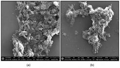
Scanning electron microscopic analysis of biofilm formation by Mycobacterium smegmatis: (a) M. smegmatis cells treated with bioactive compound from F1 of endophytic fungi; (b) Untreated M. smegmatis cells.
4. Conclusion
In summary, the study established that the endophyte found in Poikilospermum suaveolens (Blume) Merr. is Fusarium oxysporum. This fungus provides phytochemicals that act against M. smegmatis and interfere with its biofilm-building processes. A key finding is that growth inhibition and biofilm suppression are distinct mechanisms, as the former does not guarantee the latter. Furthermore, combining the active fraction with rifampicin improved overall effectiveness. Subsequent research should aim to purify the biofilm-inhibiting components from various solvent extracts and evaluate their performance against M. tuberculosis.
Acknowledgements
This work was supported by the National Research and Innovation Agency – BRIN through its post-doctoral programs. The authors also acknowledge the Research Center for Pharmaceutical Ingredient and Traditional Medicine and the Research Center for Biosystematics for granting access to the facilities necessary for this study. Part of the funding for this project came from a Rispro LPDP Indonesia grant (RIIM-B-803/II.7.5/FR/6/2022 and B1373/III.5/PR.03.08/6/2022) as part of the Reset Inovasi Untuk Indonesia Maju initiative. Support was also provided by the Science and Technology Research Partnership for Sustainable Development (SATREPS), a collaborative effort involving AMED and JICA from Japan.
Data Availability Statement
This research paper includes the complete set of data generated and examined during the investigation.
References
-
ACKART, D., HASCAL-DOVE, L., CERES, S.M., KIRK, N., PODELL, B., MALED, C., ORME, I.M., LEID, J.G., NICK, J.A., and BASARABA, R.S., 2014. Expression of antimicrobial tolerance by attached communities of Mycobacterium tuberculosis. Pathogens and Disease, vol. 70, no. 3, pp. 359-369. https://doi.org/10.1111/2049-632X.12144 PMid:24478060.
» https://doi.org/10.1111/2049-632X.12144 -
AL ABDALLAH, Q., ALANANBEH, K.M., AL-KARABLIEH, N. and ARAJ, S.-E., 2026. The chitinolytic enzymes from the entomopathogenic fungus metarhizium anisopliae: GH family distribution, domain architecture, and protein regulation. Frontiers in Fungal Biology, vol. 7, pp. 1732437. https://doi.org/10.3389/ffunb.2026.1732437 PMid:41783233.
» https://doi.org/10.3389/ffunb.2026.1732437 -
AL-ASSI, G., ABDULSAHIB, W.K., ABDAL-WAHAB, S.T., PADMA PRIYA, G., RAY, S., DIVAKARAN, S., MUKHERJEE, G., CHAUHAN, A.S. and RUZIYEVA, G., 2026. Non-invasive biological matrices on biosensor design. Clinica Chimica Acta; International Journal of Clinical Chemistry, vol. 582, pp. 120785. https://doi.org/10.1016/j.cca.2025.120785 PMid:41390074.
» https://doi.org/10.1016/j.cca.2025.120785 -
AL-EBINI, Y., CHANDRAMOULI, M., KOPPURAVURI, N.P., PASHA, T.Y., RAHAMATHULLA, M., ELTAWATY, S., THAJUDEEN, K.Y., AHMED, M.M. and SHIVANANDAPPA, T.B., 2026. Design, docking, synthesis, and biological evaluation of pyrazolone derivatives as potential dual-action antimicrobial and antiepileptic agents. Pharmaceuticals (Basel, Switzerland), vol. 19, no. 2, pp. 193. https://doi.org/10.3390/ph19020193 PMid:41754738.
» https://doi.org/10.3390/ph19020193 -
AL-KARMALAWY, A.A., ALABDALI, A.Y.M., EL-AMIER, Y.A., AL KHATIB, A.O., ALNAJJAR, R., KHALIFA, M.M., FAROUK, F., SAADELDIN, A.A., ALMUJRI, S.S., ABDULLAH ALZAHRANI, A.Y. and TAHER, R.F., 2026a. Exploring the anticancer potential of launaea mucronata: biological assessment, molecular docking, and molecular dynamics simulations. Current Medicinal Chemistry, vol. 33. Ahead of print. https://doi.org/10.2174/0109298673402314251104071536 PMid:42099141.
» https://doi.org/10.2174/0109298673402314251104071536 -
AL-KARMALAWY, A.A., EISSA, M.E., YOUSEF, T.A., SHARAKY, M., AL KHATIB, A.O. and HAWAS, S.S., 2026b. Targeting the menin– KMT2A interface: medicinal chemistry rules governing reversible, covalent, and degrader inhibitors. Chemical Biology & Drug Design, vol. 107, no. 4, pp. e70291. https://doi.org/10.1111/cbdd.70291 PMid:41934676.
» https://doi.org/10.1111/cbdd.70291 -
ARDAH, M.T., ABDULSAHIB, W.K., ABDAL-WAHAB, S.T., MALATHI, H., SENAPATI, J.N., SAMIKAN, K., MUKHERJEE, G., SINHA, A. and RIZAEV, J., 2026. Reprogramming the inflammatory response to promote neural STEM cell function after spinal cord injury. Molecular Neurobiology, vol. 63, no. 1, pp. 534. https://doi.org/10.1007/s12035-026-05819-7 PMid:41912935.
» https://doi.org/10.1007/s12035-026-05819-7 - BARNES, R.L. and CASKEY, D.K., 2002. Using ozone in the prevention of bacterial biofilm formation and scaling. Water Condıtıonıng and Purıfıcatıon Internatıonal., vol. 44, pp. 70-77.
- BONKAT, G., BACHMANN, A., SOLOKHINA, A., WIDMER, A.F., FREI, R., GASSER, T.C. and BRAISSANT, O., 2012. Growth of mycobacteria in urine determined by isothermal microcalorimetry: implications for urogenital tuberculosis and other mycobacterial infections.Urology, vol. 80, no. 5, pp. 1163.e9-1163.e12. https://doi.org/10.1016/j.urology.2012.04.050.
-
CARVALHO, C.C.C.R., 2018. Marine biofilms: a successful microbial strategy with economic implications. Frontiers in Marine Science, vol. 5, no. 126, pp. 1-11. https://doi.org/10.3389/fmars.2018.00126
» https://doi.org/10.3389/fmars.2018.00126 -
CHAKRABORTY, P. and KUMAR, A., 2019. The extracellular matrix of mycobacterial biofilms: could we shorten the treatment of mycobacterial infections? Microbial Cell, vol. 6, no. 2, pp. 105-122. https://doi.org/10.15698/mic2019.02.667 PMid:30740456.
» https://doi.org/10.15698/mic2019.02.667 -
CHAKRABORTY, S. and RHEE, K.Y., 2015. Tuberculosis drug development: history and evolution of the mechanism-based paradigm. Cold Spring Harbor Perspectives in Medicine, vol. 5, no. 8, pp. a021147. https://doi.org/10.1101/cshperspect.a021147 PMid:25877396.
» https://doi.org/10.1101/cshperspect.a021147 - CHARIRAK, P., PRAJANTASAN, R., PREMPRAYOON, K., SRIKACHA, N. and RATANANIKOM, K., 2023. In vitro antibacterial activity and mode of action of piper betle extracts against soft rot disease-causing bacteria. Scientifica, vol. 2023, pp. 5806841. https://doi.org/10.1155/2023/5806841. PMid:37766936.
-
HANNIG, C. and HANNIG, M., 2009. The oral cavity - a key system to understand substratum-dependent bioadhesion on solid surfaces in man. Clinical Oral Investigations, vol. 13, no. 2, pp. 123-139. https://doi.org/10.1007/s00784-008-0243-3 PMid:19137331.
» https://doi.org/10.1007/s00784-008-0243-3 -
HAPID, A., NAPITUPULU, M. and ZUBAIR, M.S., 2021. Phytochemical screening, GC-MS analysis, toxicity and antimicrobial properties of extracts outer shell Poikilospermum suaveolens (Blume) Merr. International Journal of Research and Innovation in Applied Science, vol. 6, no. 9, pp. 111-117. https://doi.org/10.51584/IJRIAS.2021.6903
» https://doi.org/10.51584/IJRIAS.2021.6903 -
HAWSER, S.P. and DOUGLAS, L.J., 1994. Biofilm formation by Candida species on the surface of catheter materials in vitro. Infection and Immunity, vol. 62, no. 3, pp. 915-921. https://doi.org/10.1128/iai.62.3.915-921.1994 PMid:8112864.
» https://doi.org/10.1128/iai.62.3.915-921.1994 -
IBRAGIMOVA, F., KALENDEROVA, G., KALMURATOVA, B., OKHUNOVA, M., KHOLMURADOVA, Z., IKRAMOVA, F. and AXMEDOV, N., 2025. The association between gut microbiota-derived metabolites and arterial stiffness in hypertensive patients. Revista Latinoamericana de Hipertensiòn, vol. 20, no. 11, pp. 818-824. https://doi.org/10.5281/zenodo.17925018
» https://doi.org/10.5281/zenodo.17925018 -
IWEN, P.C., HINRICHS, S.H. and RUPP, M.E., 2002. Utilization of the internal transcribed spacer regions as molecular targets to detect and identify human fungal pathogens. Medical Mycology, vol. 40, no. 1, pp. 87-109. https://doi.org/10.1080/mmy.40.1.87.109 PMid:11860017.
» https://doi.org/10.1080/mmy.40.1.87.109 -
JIANG, S.H., SENANAYAKE, S. and TALAULIKAR, G.S., 2011. Peritoneal dialysis-related peritonitis due to Mycobacterium smegmatis. Peritoneal Dialysis International : Journal of the International Society for Peritoneal Dialysis, vol. 31, no. 2, pp. 215-216. http://doi.org/10.3747/pdi.2010.00074 PMid:21427257.
» http://doi.org/10.3747/pdi.2010.00074 -
KAMBAROV, J., BOYNAZAROV, O., SHAROPOVA, N., KHUDAYBERGANOV, K., ABDUVALIYEVA, Z. and MATKARIMOV, I., 2024. Management and planning of population sustainability in rural settlements based on geographical-economic variables (tourism, agriculture and handicrafts). Economic Annals-XXI, vol. 211, no. 9-10, pp. 58-62. https://doi.org/10.21003/ea.V211-09
» https://doi.org/10.21003/ea.V211-09 -
KUMAR, S., STECHER, G. and TAMURA, K., 2016. MEGA7: molecular evolutionary genetics analysis version 7.0 for bigger datasets. Molecular Biology and Evolution, vol. 33, no. 7, pp. 1870-1874. https://doi.org/10.1093/molbev/msw054 PMid:27004904.
» https://doi.org/10.1093/molbev/msw054 -
LE DANTEC, C., DUGUET, J.P., MONTIEL, A., DUMOUTIER, N., DUBROU, S. and VINCENT, V., 2002. Chlorine disinfection of atypical mycobacteria isolated from a water distribution system. Applied and Environmental Microbiology, vol. 68, no. 3, pp. 1025-1032. https://doi.org/10.1128/AEM.68.3.1025-1032.2002 PMid:11872446.
» https://doi.org/10.1128/AEM.68.3.1025-1032.2002 -
LEVIN, M.E. and HATFULL, G.F., 1993. Mycobacterium smegmatis RNA polymerase: DNA supercoiling, action of rifampicin and mechanism of rifampicin resistance. Molecular Microbiology, vol. 8, no. 2, pp. 277-285. https://doi.org/10.1111/j.1365-2958.1993.tb01572.x PMid:8316080.
» https://doi.org/10.1111/j.1365-2958.1993.tb01572.x - MOHAMMAD, S., VASUDEVAN, A., ABED, S., NIGORA, O., ZAKHRO, A., XUDAYOROVA, S., FAZOLAT, K. and GATI, M., 2026a. Production of agricultural products to replace pharmaceutical and health materials: pharmaceutical and health industry. Procedia Environmental Science, Engineering and Management, vol. 13, no. 1, pp. 317-324.
- MOHAMMAD, S., VASUDEVAN, A., FURAIJL, H., KHIROMON, S., MUKHAMMADKHUJA, S., GULBAHOR, X., ILKHOMJON, R. and KHADDOUR, R., 2026b. Application of agricultural waste in the production of textile and dye products with an economic and environmental efficiency approach. Procedia environmental science Engineering and Management, vol. 13, no. 1, pp. 351-358.
-
OJHA, A., ANAND, M., JACOBS, W.R., BHATT, A., KREME, L. and HATFULL, G.F., 2015. GroEL1: a dedicated chaperone involved in mycolic acid biosynthesis during biofilm formation in mycobacteria. Cell, vol. 123, no. 5, pp. 861-873. https://doi.org/10.1016/j.cell.2005.09.012 PMid:16325580.
» https://doi.org/10.1016/j.cell.2005.09.012 -
ORIQUAT, G.H.M., MAHARANA, L., DHYANI, A., AL-HASNAAWEI, S., SINGH-CHAUHAN, A., ARORA, V., SHARMA, J. and SADEGHI-SAMARJAN, R., 2026. The gut microbiome in amyotrophic lateral sclerosis: emerging mechanisms and therapeutic potential. Molecular Neurobiology, vol. 63, no. 1, pp. 549. https://doi.org/10.1007/s12035-026-05826-8 PMid:41925964.
» https://doi.org/10.1007/s12035-026-05826-8 -
ORTIZ-PEREZ, A., MARTIN-DE-HIJAS, N., ALONSO-RODRIGUEZ, N., MOLINA-MANSO, D., FERNANDEZ-ROBLAS, R. and ESTEBAN, J., 2011. Importance of antibiotic penetration in the antimicrobial resistance of biofilm formed by non-pigmented rapidly growing mycobacteria against amikacin, ciprofloxacin and clarithromycin. Enfermedades Infecciosas y Microbiología Clínica, vol. 29, no. 2, pp. 79-84. https://doi.org/10.1016/j.eimc.2010.08.016 PMid:21333405.
» https://doi.org/10.1016/j.eimc.2010.08.016 -
PADHI, M.K., 2016. Importance of indigenous breeds of chicken for rural economy and their improvements for higher production performance. Scientifica, vol. 2016, pp. 2604685. https://doi.org/10.1155/2016/2604685 PMid:27144053.
» https://doi.org/10.1155/2016/2604685 -
PARAUŠIĆ, V., GRUJIĆ VUČKOVSKI, B., SUBIĆ, J., 2024. Value added agriculture in Serbia and its impact on agriculture export value: case study of GLOBALG. AP fruit and vegetables certification. International Journal for Quality Research, vol. 18, no. 3, pp. 779-792. https://doi.org/10.24874/IJQR18.03-09
» https://doi.org/10.24874/IJQR18.03-09 -
PAYNE, D.E. and BOLES, B.R., 2016. Emerging interactions between matrix components during biofilm development. Current Genetics, vol. 62, no. 1, pp. 137-141. https://doi.org/10.1007/s00294-015-0527-5 PMid:26515441.
» https://doi.org/10.1007/s00294-015-0527-5 -
RYALL, B., EYDALLIN, G. and FERENCI, T., 2012. Culture history and population heterogeneity as determinants of bacterial adaptation: the adaptomics of a single environmental transition. Microbiology and Molecular Biology Reviews : MMBR, vol. 76, no. 3, pp. 597-625. https://doi.org/10.1128/MMBR.05028-11 PMid:22933562.
» https://doi.org/10.1128/MMBR.05028-11 -
SARWAR, S., FIRDOUS, Q. and KHALID, A.N., 2019. Importance of molecular and phylogenetic analyses for identification of basidiomycetes. In: Z. YOUSAF, ed. Recent advances in phylogenetics London: IntechOpen. https://doi.org/10.5772/intechopen.80671
» https://doi.org/10.5772/intechopen.80671 -
SHI, T., FU, T. and XIE, J., 2011. Polyphosphate deficiency affects the sliding motility and biofilm formation of Mycobacterium smegmatis. Current Microbiology, vol. 63, no. 5, pp. 470-476. https://doi.org/10.1007/s00284-011-0004-4 PMid:21882007.
» https://doi.org/10.1007/s00284-011-0004-4 -
SINGH, R. and DUBEY, A.K., 2015. Endophytic actinomycetes as emerging source for therapeutic compounds. Indo Global Journal of Pharmaceutical Sciences, vol. 5, no. 2, pp. 106-116. https://doi.org/10.35652/IGJPS.2015.11
» https://doi.org/10.35652/IGJPS.2015.11 -
VISHWAS, S., NAIK, N., KOWSHIK, S., SURESH, S. 2025. An overview of integrating the internet of things and advanced automation technologies for sustainable crop growth and monitoring in precision agriculture. Proceedings on Engineering, vol. 7, no. 2, 1219-1230. https://doi.org/10.24874/PES07.02C.002
» https://doi.org/10.24874/PES07.02C.002 - WHITE, T.J., BRUNS, T.D., LEE, S.B. and TAYLOR, J.W., 1990, Amplification and direct sequencing of fungal ribosomal RNA genes for phylogenetics. In: M.A. INNIS, D.H. GELFAND, J.J. SNINSKY and T.J. WHITE, eds. PCR Protocols: a guide to methods and applications New York: Academic Press, Inc., pp. 315-322.
-
WU, S., LIU, G., JIN, W., XIU, P. and SUN, C., 2016. Antibiofilm and anti-infection of a marine bacterial exopolysaccharide against Pseudomonas aeruginosa. Frontiers in Microbiology, vol. 7, pp. 102. https://doi.org/10.3389/fmicb.2016.00102 PMid:26903981.
» https://doi.org/10.3389/fmicb.2016.00102 -
XU, J., 2017. Fungal DNA barcoding. Genome, vol. 59, no. 11, pp. 913-932. https://doi.org/10.1139/gen-2016-0046 PMid:27829306.
» https://doi.org/10.1139/gen-2016-0046 -
ZUMLA, A., GEORGE, A., SHARMA, V., HERBERT, R.H.N., BARONESS MASHAM OF ILTON, OXLEY, A. and OLIVER, M.,2015. The WHO 2014 global tuberculosis report further to go. The Lancet. Global Health, vol. 3, no. 1, pp. e10-e12. https://doi.org/10.1016/S2214-109X(14)70361-4 PMid:25539957.
» https://doi.org/10.1016/S2214-109X(14)70361-4
Edited by
-
Editor:
Takako Matsumura Tundisi


