Coronectomy: a long-term follow-up. Case series
Abstract
Aim Coronectomy is an alternative technique for the removal of lower third molars in close proximity to the inferior alveolar nerve (IAN), minimizing the risk of nerve damage by retaining the roots in situ. This study aimed to evaluate the long-term outcomes and complications of coronectomy in four clinical cases.
Methods Three healthy male patients (aged 28, 36, and 57 years) underwent four coronectomy procedures between 2010 and 2016. Preoperative panoramic radiographs and, in two cases, cone-beam computed tomography (CBCT) confirmed the proximity of the third molar roots to the IAN. Surgical protocols included standardized techniques, antibiotic prophylaxis, and postoperative follow-up with radiographic assessments at intervals ranging from 6 months to 10 years.
Results Root migration was observed primarily within the first postoperative year, stabilizing thereafter. No cases exhibited root exposure, infection, or neurosensory deficits. The 28-year-old patient demonstrated the highest migration rate (5.65 mm), consistent with age-related trends reported in the literature.
Conclusion Coronectomy is a viable and safe alternative for high-risk lower third molar extractions, significantly reducing the risk of IAN injury. Long-term follow-up confirmed minimal complications, though root migration remains a predictable occurrence, particularly in younger patients.
Keywords
Tooth extraction; Mandibular nerve; Molar, third; Tooth crown/surgery; Intraoperative complications
Introduction
Impacted lower third molar (I3M) is commonly associated with dental caries and pericoronitis1. Therefore, its removal is the most common surgical procedure of the oral cavity1,2.
Neurosensory deficits are unfortunate sequelae of treatment of I3M surgery and their occurrences are not uncommon, with an estimated 0.1%–22% for lingual nerve (LN) deficit and 0.26%– 8.4% for inferior alveolar nerve (IAN) deficit3. Injury to the inferior alveolar nerve (IAN) occurs due to the proximity between the IAN and the tooth roots4,5. The clinical signs of an IAN injury are anesthesia, paresthesia, or dysesthesia of the lower lip and gingiva5. For this reason, coronectomy has become a common procedure as an alternative to complete I3M removal, decreasing the risk of IAN injury6. The technique consists of removing only the crown of a lower third molar, leaving the remaining root within the dentoalveolar bone2,7,8. Coronectomy may also help to reduce the incidence of mandibular fracture in high-risk patients because less bone is removed, lower forces are required, and the root is retained to maintain the structure and integrity of the mandible9.
Radiographic signs showing a high risk of nerve involvement are deviation or/and narrowing of the IAN canal, root darkness, loss of lamina dura, deflection, and narrowing of the roots2. The presence of one of these signs indicates that a Cone-beam Computed Tomography (CBCT) can be of great value, even though it is not mandatory for the coronectomy procedure. This technique may be performed even when an ortho-pantographic image indicates the proximity of the roots of the I3M to the IAN or when the patient is anxious owing to the potential risk of IAN injury5,8.
A coronectomy is contraindicated when all the enamel of the tooth cannot be removed, if the roots are mobilized during the procedure, when the second molars are going to be distalized orthodontically, in the presence of mobility, apical disease, tumor, in association with cystic tissue that is unlikely to resolve if the root is left in situ, in immunocompromised patients or, whose received radiotherapy before10,11.
Many authors describe good results, and few difficulties associated with the coronectomy technique1,2,9,12-14. However, complications can occur during surgical procedures as bleeding, displacement of fragments of the root, and/or damage to adjacent structures. In the short-term postoperative, the damages are alveolar osteitis, infection, bleeding, and/or paresthesia. In the long-term postoperative, migration and eruption of roots can happen9. The technique has minimal morbidities such as infection, pain, dry socket, or development of pathologies, nevertheless, some roots migrate after surgery, which can be observed on radiographs as a periapical radiolucency, at least 3 months after the extraction1,10. Root exposure requires a second surgical procedure, in a few situations.1 Although root migration is common, it isn´t a potential complication. The roots can be easily removed without risk of IAN damage in the rare cases in which they become exposed to the oral cavity7,10,14. Age is a significant factor in root migration since the roots’ teeth following coronectomy of older patients seem to dislocate less than those of younger patients7.
The objective of this study is to report four cases of long-term follow-up coronectomy, analyzing root migration and other potential long-term issues associated with the technique.
Cases Series
Four I3M coronectomies were performed in three healthy males, with 28, 36, and 57 years old, from 2010 to 2016. All of them were referred to an oral and maxillofacial surgeon for I3M removal due to pain in the area. A panoramic radiograph evaluation revealed the great proximity of the I3M roots with the mandibular canal, indicating a coronectomy. A CBCT was performed in two patients. After the explanation about the risks of IAN injury and the coronectomy technique, all patients decided on the coronectomy procedure, and all of them signed an informed consent. In the 28-year-old patient, the coronectomy was bilateral, with a 15-day interval between each procedure. All surgeries were performed by the same surgeon using the same technique.
Two grams of amoxicillin and 8 mg of dexamethasone were prescribed 60 and 30 minutes before the surgery, respectively. Following effective local anesthesia of the IAN and buccal block, an envelope incision was performed from the mesial of the 1IM, with relaxing in the distal of the 2IM and a complete detachment of the mucoperiosteal flap. Buccal bone was removed with 702 fissure bur to expose the crown down to the cementoenamel junction (CEJ). Decoronation of the tooth was performed by cutting through the CEJ with the fissure bur taken approximately two-thirds of the way across the tooth and then the crown was fractured off with a straight elevator. The crown was then removed, and the root portion was smoothed using a #8 spherical diamond bur, under low rotation, taking care to leave the roots at least 3-4 mm below the alveolar crest and ensuring the removal of all remaining enamel. The wound was then irrigated with copious amounts of saline solution to remove tooth powder and debris, and the closure was performed with a 4.0 nylon suture. Chlorhexidine mouthwash was advised for post-operative care and the prescriptions included 500 mg amoxicillin and 4 mg dexamethasone every 8 hours, for 48 hours and 500 mg paracetamol with 30 mg codeine every 6 hours, in case of pain. There were no trans or postoperative intercurrences and the patients returned for suture removal one week later.
Follow-up with panoramic radiographs was requested soon after the surgical procedure: 7-14 days, 6 months, 1 year, and after that, an annual control. This schedule however was not fully respected by the patients and, although all of them had their immediate postoperative control at the right time, the second control was quite variable. (Table 1).
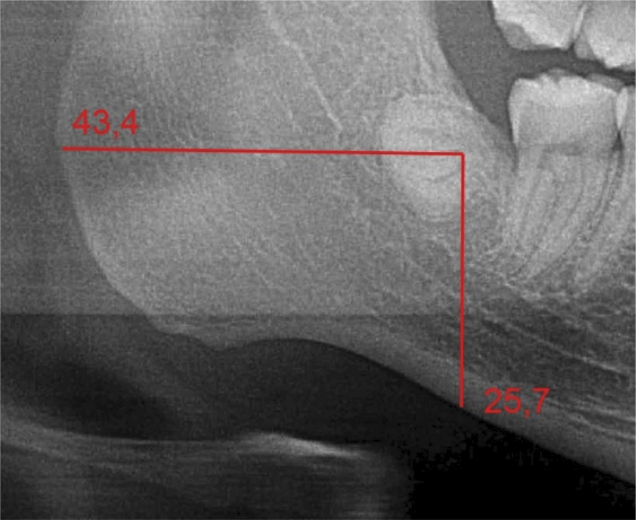
Measurements were performed to analyze the root migration throughout the years. Two straight lines, starting from the furcation region, were designed at a 90o angle: one horizontal towards the most posterior cortical bone of the ascending ramus of the mandible and another, vertical, towards the mandibular basal cortical bone. (Figure 1).
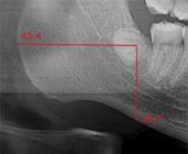
The difference between the horizontal and vertical lines, in millimeters, was calculated on each panoramic radiograph to establish the root migration.
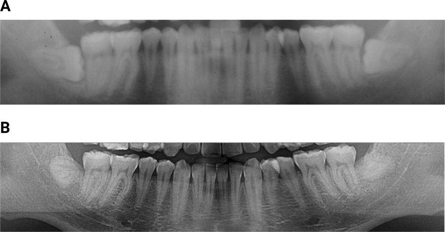
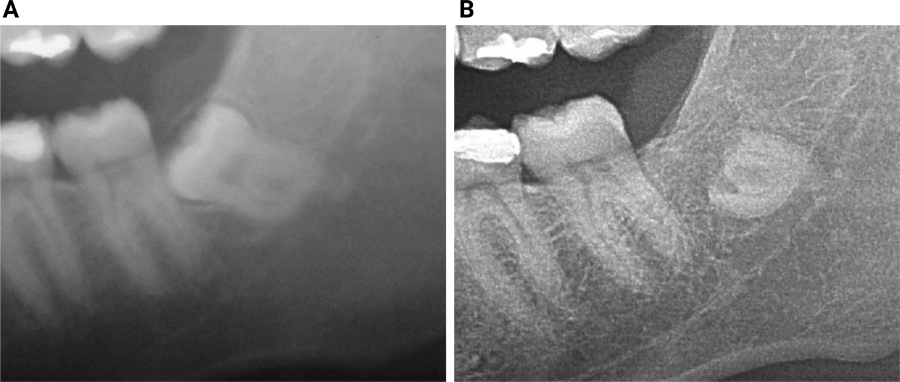
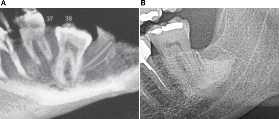
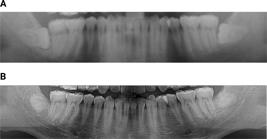
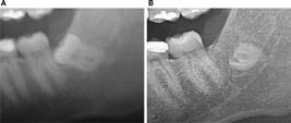
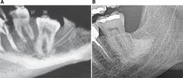
A total of three patients were selected, and 4 procedures were analyzed, being the coronectomy of 3 lower third molars on the left side and 1 lower third molar on the right, which resulted in an analysis of 12 panoramic radiographs, taken in the same panoramic radiographic device (Figures 2-4). Two blind-calibrated PhD dentists evaluated the images, with a very high concordance level. First, the root migration between the initial radiograph and the second follow-up was analyzed. Then, the migration that occurred between the second and the last follow-up was analyzed. It was observed that the migrations were significantly lower in the long term (Table 2). No root exposure in the oral cavity nor infection in any of the cases was observed.
Discussion
Coronectomy was first described in 1984 by Ecuyer and Debien and has become increasingly recommended as surgical treatment for mandibular third molars that need to be removed and are very close to the IAN. This technique is usually indicated for third molars, but it can also be performed on first and second molars to avoid IAN injuries15,16. Another important indication is when conventional third molar surgery presents a risk of mandibular fracture during the procedure.
Imaging exams such as panoramic radiographs are extremely important to assess the relationship between the IAN and the roots of the I3M. The panoramic image may contain some radiographic findings that indicate their proximity, such as darkening and/or sudden narrowing of the roots of the third molar, discontinuity and/or loss of cortical bone that surrounds the mandibular canal, and displacement of the canal caused by the roots2. Some studies state that panoramic radiography is enough in the decision-making on the coronectomy technique5,8. It is required to observe at least one of the above-mentioned radiographic signs such as narrowing and deviation of the mandibular canal5,6,10. For some authors, all roots that vertically overlap the outline of the canal or lie in prolonged contact with it should be considered for coronectomy16. However, CBCT is of great value before and during surgery to confirm the relationship of the I3M roots with the IAN, in addition to providing a 3-D assessment of the roots7. Actually, no guidelines for the preoperative radiological evaluation exist14. In the present study, the CBCT was not performed only in Case 1, due to the poor financial condition of the patient. However, its panoramic radiograph showed that the root apex of the I3M left was crossing the inferior cortex of the mandibular canal, one of the radiographic signs that indicates its proximity to the IAN.
It´s important to notice that horizontal impaction of I3M is not a contraindication for coronectomy. The higher risk occurs when the whole tooth, including its crown, is lying horizontally along the path of the IAN, and in this situation, coronectomy could present a higher risk of nerve involvement than just taking out the whole tooth10. Other authors emphasize that coronectomy can be done safely for all types of impacted third molars11. In our case series, both I3M of patient number 2 presented horizontal impaction, but their crowns weren´t in intimacy with the mandibular canal.
The root of the coronectomized tooth must be kept at least 3 mm below the bone crest, creating optimal conditions for bone deposition and healing around the remained roots10,13. Endodontic treatment is not necessary since it does not present any advantages for the procedure and even increases the risk of infections and intraoperative complications17,18. The endodontic treatment wasn´t performed in any of the cases of the present study.
Complications that can occur with coronectomy are the same as those associated with complete tooth removal, such as alveolar osteitis, bleeding, infection, hemorrhage, and paresthesia9. Postoperative pain is usually present in all patients, and the prescription of analgesics can solve the problem14. Hemorrhage rarely occurs, unless the patient has a hematological disorder. The treatment is the same as in routine tooth extractions, such as identifying the source of bleeding and using hemostatic agents9. The root fragment must be removed when it is the cause of a postoperative infection5,13.
The most reported long-term issues are root migration and eruption2,4,8,13. With a high prevalence, as in 90% of cases, they can be observed radiographically from three months to up to ten years after surgery2,4,8,14. Most cases happen in the first year after surgery, with a tendency to be stabilized4,8. This was also observed in our cases, where the root migrations were higher in the first follow-up.
The patient´s age is considered an important factor in root migration, with the highest rate observed in younger patients1,2,8. Our study agrees with these authors since a 5.65 mm migration occurred in the 28-year-old patient. Considering that the root apex of the inferior third molar completes its development around 18 to 25 years of age, the force of eruption must affect the root movement4. A higher migration rate is observed in women, supposedly due to its lower mandibular bone density1,4,10. This variable was not possible to assess, since all patients in the present study were male.
Root migration reduces over time and the stabilization occurs due to bone formation above the root, resulting in their immobilization4,8,16. However, the movement can be continuous and causes root eruption in the oral cavity. This is a rare occurrence, observed in 1.77% of reported cases, and may persist within 10 years5,14. Eruption is not a problem and can be considered beneficial, since the roots migrate to the upper surface, moving away from the IAN. A second surgery would not pose any risks, compared to the 3IM conventional surgery10. None of the 4 cases included in this study presented root eruption in the oral cavity.
Root removal is also indicated when late chronic infection and paresthesia occur5,8. In a rare case report, root migration resulted in the displacement of the IAN, which was attached to the apical portion of the root, due to a dilaceration, causing a late paresthesia of the lower lip6. Paresthesia was not reported in any of the present cases.
Studies compared the safety of coronectomy and conventional excision of I3M in which roots were close to the IAN, in terms of associated surgical complications and neurosensory disturbances19-22. In the first clinical trial comparing the incidence of injury to the IAN as a result of coronectomy and complete removal of I3M, 19% of nerves were damaged after extraction, and none after coronectomy; the authors concluded that patients at risk of iatrogenic injury to the IAN, coronectomy reduces the chance of injury with no adverse effect on morbidity19. In another randomized controlled trial, 231 patients underwent surgery for 349 lower I3M (171 coronectomies, 178 controls – complete removal); nine patients in the control group presented with IAN deficit, compared with 1 in the coronectomy group20. This study confirmed that coronectomy can significantly reduce the incidence of IAN deficit when compared with total excision of I3M with proximity to the inferior alveolar canal20.
One systematic review evaluated coronectomy effectiveness as an alternative for surgical extraction of I3M that have a high risk of trauma to the inferior alveolar canal and confirmed that coronectomy can significantly reduce the incidence of IAN deficit when compared with total extraction of I3M; the authors however emphasized that root migration requires an extended follow-up period21. On the other hand, in another systematic review, the authors concluded that, although evidence from two randomized controlled trials suggests that coronectomy can reduce the risk of IAN injury compared to total surgical removal of high-risk I3M, the quality of evidence is not enough to provide definitive conclusions regarding the preferred technique22.
In conclusion, the coronectomy technique is a good alternative for cases of I3M very close to the IAN, reducing the paresthesia risks. In the present study, despite the small number of cases, it was observed that the technique is viable, without complications. All patients, however, should receive instructions that root migration may occur and, despite rare, their late eruption can happen, leading to a second surgical procedure. Annual radiographic follow-up is essential.
Acknowledgments
The authors thank the patient and his parents for authorizing the report of this story.
References
-
1 Leung YY, Cheung LK. Long-term morbidities of coronectomy on lower third molar. Oral Surg Oral Med Oral Pathol Oral Radiol. 2016 Jan;121(1):5-11. doi: 10.1016/j.oooo.2015.07.012. Epub 2015 Jul 22.
» https://doi.org/10.1016/j.oooo.2015.07.012 -
2 Pitros P, Jackson I, O'Connor N. Coronectomy: a retrospective outcome study. Oral Maxillofac Surg. 2019 Dec;23(4):453-8. doi: 10.1007/s10006-019-00794-x.
» https://doi.org/10.1007/s10006-019-00794-x -
3 Cheung LK, Leung YY, Chow LK, Wong MC, Chan EK, Fok YH. Incidence of neurosensory deficits and recovery after lower third molar surgery: a prospective clinical study of 4338 cases. Int J Oral Maxillofac Surg. 2010 Apr;39(4):320-6. doi: 10.1016/j.ijom.2009.11.010.
» https://doi.org/10.1016/j.ijom.2009.11.010 -
4 Kohara K, Kurita K, Kuroiwa Y, Goto S, Umemura E. Usefulness of mandibular third molar coronectomy assessed through clinical evaluation over three years of follow-up. Int J Oral Maxillofac Surg. 2015 Feb;44(2):259-66. doi: 10.1016/j.ijom.2014.10.003. Epub 2014 Nov 8.
» https://doi.org/10.1016/j.ijom.2014.10.003 -
5 Frenkel B, Givol N, Shoshani Y. Coronectomy of the mandibular third molar: a retrospective study of 185 procedures and the decision to repeat the coronectomy in cases of failure. J Oral Maxillofac Surg. 2015 Apr;73(4):587-94. doi: 10.1016/j.joms.2014.10.011. Epub 2014 Oct 22.
» https://doi.org/10.1016/j.joms.2014.10.011 -
6 Steinberg MJ, Nick MJ. Displacement of the inferior alveolar bundle secondary to post coronectomy root migration: a case report. J Oral Maxillofac Surg. 2021 Sep;79(9):1837-41. doi: 10.1016/j.joms.2021.04.012.
» https://doi.org/10.1016/j.joms.2021.04.012 -
7 Yeung AWK, Wong NSM, Bornstein MM, Leung YY. Three-dimensional radiographic evaluation of root migration patterns 4-8.5 years after lower third molar coronectomy: a cone beam computed tomography study. Int J Oral Maxillofac Surg. 2018 Sep;47(9):1145-52. doi: 10.1016/j.ijom.2018.03.014.
» https://doi.org/10.1016/j.ijom.2018.03.014 -
8 Leung YY, Cheung KY. Root migration pattern after third molar coronectomy: a long-term analysis. Int J Oral Maxillofac Surg. 2018 Jun;47(6):802-8. doi: 10.1016/j.ijom.2018.01.015.
» https://doi.org/10.1016/j.ijom.2018.01.015 -
9 Patel V, Gleeson CF, Kwok J, Sproat C. Coronectomy practice. Paper 2: complications and long term management. Br J Oral Maxillofac Surg. 2013 Jun;51(4):347-52. doi: 10.1016/j.bjoms.2012.06.008. Epub 2012 Jul 12.
» https://doi.org/10.1016/j.bjoms.2012.06.008 -
10 Pogrel MA. Coronectomy: partial odontectomy or intentional root retention. Oral Maxillofac Surg Clin North Am. 2015 Aug;27(3):373-82. doi: 10.1016/j.coms.2015.04.003.
» https://doi.org/10.1016/j.coms.2015.04.003 -
11 Gleeson CF, Patel V, Kwok J, Sproat C. Coronectomy practice. Paper 1. Technique and trouble-shooting. Br J Oral Maxillofac Surg. 2012 Dec;50(8):739-44. doi: 10.1016/j.bjoms.2012.01.001.
» https://doi.org/10.1016/j.bjoms.2012.01.001 -
12 Leung YY. Management and prevention of third molar surgery-related trigeminal nerve injury: time for a rethink. J Korean Assoc Oral Maxillofac Surg. 2019 Oct;45(5):233-40. doi: 10.5125/jkaoms.2019.45.5.233.
» https://doi.org/10.5125/jkaoms.2019.45.5.233 -
13 Shokouhi B, Thavaraj S, Sproat C, Kwok J, Beneng K, Patel V. Coronectomy root retrievals: a review of 92 cases. Oral Surg Oral Med Oral Pathol Oral Radiol. 2019 Mar;127(3):200-9. doi: 10.1016/j.oooo.2018.09.001. Epub 2018 Sep 10.
» https://doi.org/10.1016/j.oooo.2018.09.001 -
14 Pedersen MH, Bak J, Matzen LH, Hartlev J, Bindslev J, Schou S, et al. Coronectomy of mandibular third molars: a clinical and radiological study of 231 cases with a mean follow-up period of 5.7 years. Int J Oral Maxillofac Surg. 2018 Dec;47(12):1596-603. doi: 10.1016/j.ijom.2018.06.006.
» https://doi.org/10.1016/j.ijom.2018.06.006 -
15 Elo JA, Sun HB, Dong F, Nguyen K, Zakhary K. Does bone grafting improve outcomes in coronectomy surgery? Long-term (5- to 9-year) clinical and radiographic follow-up of 78 adult patients. J Oral Maxillofac Surg. 2017 Jul;75(7):1330-7. doi: 10.1016/j.joms.2017.02.004.
» https://doi.org/10.1016/j.joms.2017.02.004 -
16 Leung YY, Yeung AWK, Ismail IN, Wong NSM. Bone regeneration at the distal aspect of the adjacent second molar after lower third molar coronectomy: a long-term analysis. Int J Oral Maxillofac Surg. 2020 Oct;49(10):1360-6. doi: 10.1016/j.ijom.2020.03.016.
» https://doi.org/10.1016/j.ijom.2020.03.016 -
17 Sencimen M, Ortakoglu K, Aydin C, Aydintug YS, Ozyigit A, Ozen T, et al. Is endodontic treatment necessary during coronectomy procedure? J Oral Maxillofac Surg. 2010 Oct;68(10):2385-90. doi: 10.1016/j.joms.2010.02.024. Erratum in: J Oral Maxillofac Surg. 2011 Jun;69(6):1847.
» https://doi.org/10.1016/j.joms.2010.02.024 -
18 Pogrel MA, Lee JS, Muff DF. Coronectomy: a technique to protect the inferior alveolar nerve. J Oral Maxillofac Surg. 2004 Dec;62(12):1447-52. doi: 10.1016/j.joms.2004.08.003.
» https://doi.org/10.1016/j.joms.2004.08.003 -
19 Renton T, Hankins M, Sproate C, McGurk M. A randomised controlled clinical trial to compare the incidence of injury to the inferior alveolar nerve as a result of coronectomy and removal of mandibular third molars. Br J Oral Maxillofac Surg. 2005 Feb;43(1):7-12. doi: 10.1016/j.bjoms.2004.09.002.
» https://doi.org/10.1016/j.bjoms.2004.09.002 -
20 Leung YY, Cheung LK. Safety of coronectomy versus excision of wisdom teeth: a randomized controlled trial. Oral Surg Oral Med Oral Pathol Oral Radiol Endod. 2009 Dec;108(6):821-7. doi: 10.1016/j.tripleo.2009.07.004.
» https://doi.org/10.1016/j.tripleo.2009.07.004 -
21 Abu-Mostafa N, AlRejaie LM, Almutairi FA, Alajaji RA, Alkodair MM, Alzahem NA. Evaluation of the outcomes of coronectomy procedure versus surgical extraction of lower third molars which have a high risk for inferior alveolar nerve injury: a systematic review. Int J Dent. 2021 Nov;2021:9161606. doi: 10.1155/2021/9161606.
» https://doi.org/10.1155/2021/9161606 -
22 Ali AS, Benton JA, Yates JM. Risk of inferior alveolar nerve injury with coronectomy vs surgical extraction of mandibular third molars-A comparison of two techniques and review of the literature. J Oral Rehabil. 2018 Mar;45(3):250-7. doi: 10.1111/joor.12589. Epub 2017 Dec 11.
» https://doi.org/10.1111/joor.12589
-
Consent for publication:
It was given by the patient.
-
Data Availability:
Datasets related to this article will be available upon request to the corresponding author.
-
Funding:
This research did not receive any specific grant from funding agencies in the public, commercial, or not-for-profit sectors.
Edited by
-
Editor:
Dr. Altair A. Del Bel Cury
Datasets related to this article will be available upon request to the corresponding author.