A retrospective analysis of maxillary sinus septa by cone beam computed tomography
Abstract
Aim To determine the prevalence, location, shape, size and base length of maxillary sinus septa using cone beam computed tomography, in order to provide critical anatomical information for safe and accurate surgical planning, particularly in procedures such as sinus lift surgeries.
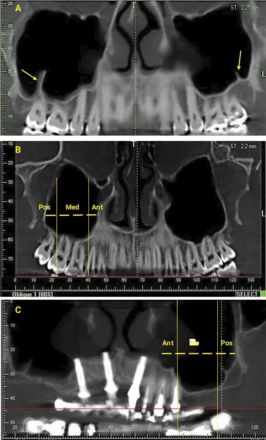
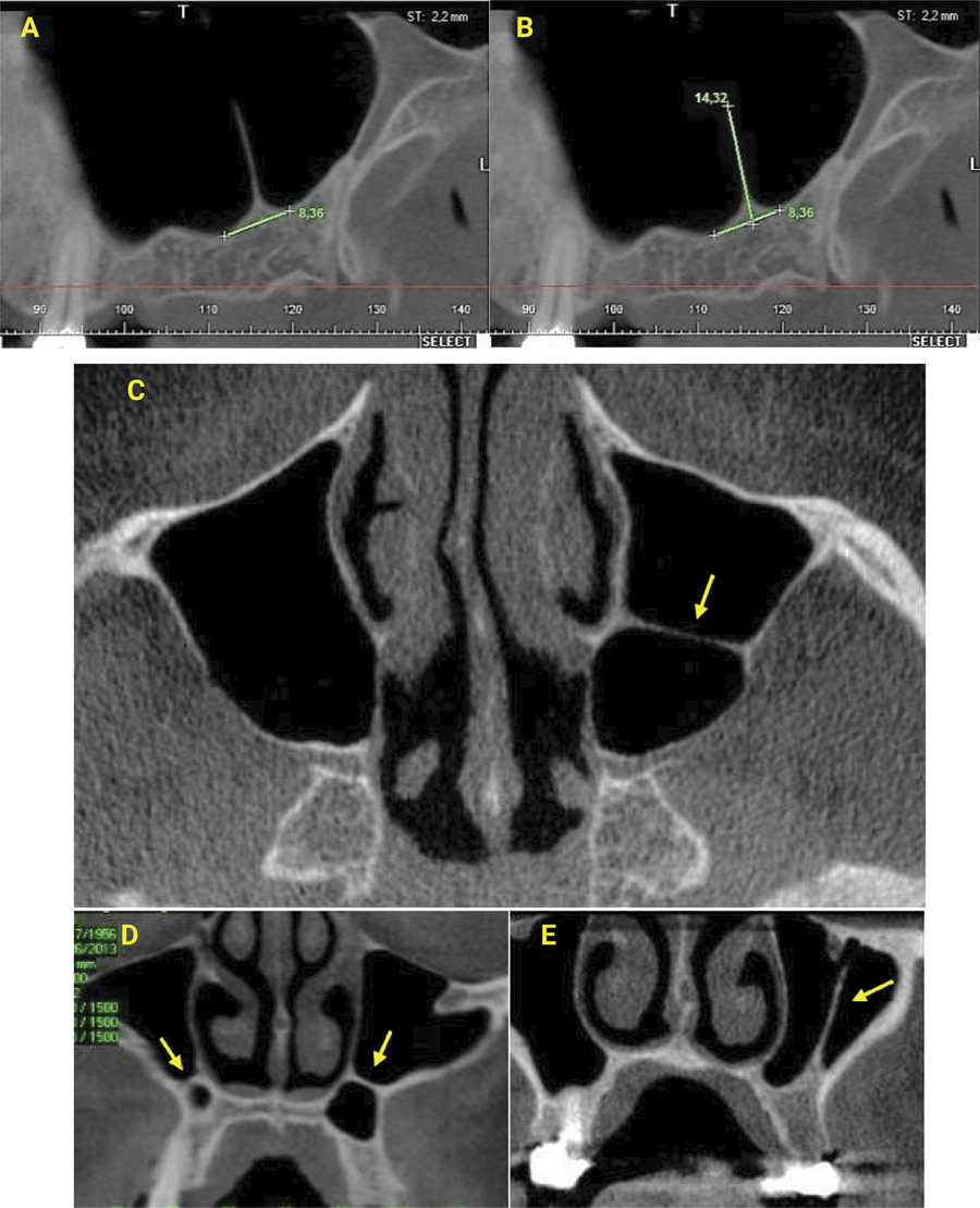
Methods CT images of 198 patients were selected. The images were analyzed using XoranCat® software. All patients were classified according to sex, age and type of dentition. The existence and number of septa were investigated. If septa were present, their locations were determined and the height and length of their base were measured. The septa were classified as buccopalatal, sagittal or transverse. Statistical analysis was performed to evaluate the prevalence, location and morphology of the septum within the maxillary sinus according to the different variables. The Pearson Chi-square, Mann-Whitney and Kruskal-Wallis statistical tests were used, adopting a significance level of 5% (p<0.05).
Results Septa were found in 99 patients studied, thus indicating a prevalence of 50%. No significant differences (p<0.05) were found in the frequency of septa according to dentition type, gender and different age groups. Most of the septa found were in the middle region, with a prevalence of 52.5%, and the buccopalatal orientation was the most frequent (75.5%). The mean height and base length found in this study were 6.12 ± 3.32 and 6.61 ± 2.38 mm, respectively, with the highest measurements found in the middle region.
Conclusion Making a detailed anatomical study by tomographic images essential before sinus floor elevation surgeries and other maxillary procedures, including diagnosis and treatment planning in implantology and sinus pathology.
Keywords
Maxillary sinus; Sinus floor augmentation; Cone-Beam Computed Tomography
 Thumbnail
 Thumbnail