Nickel oxide–coated titanium discs: surface characterization and hemocompatibility assessment for dental implant applications
Abstract
Background Titanium implants are widely employed in orthopedic and dental fields due to their excellent mechanical strength, corrosion resistance, and biocompatibility. However, issues such as inadequate osseointegration, susceptibility to bacterial colonization, and mechanical wear limit their long-term success. Surface modification with bioactive materials is a promising strategy to overcome these limitations.
Aim This study investigates the use of nickel oxide (NiO), a bioceramic known for its antimicrobial activity and structural stability, as a surface coating to enhance titanium implant performance.
Methods Titanium substrates were coated with NiO using optimized dip and spin coating techniques. The coatings were characterized using Scanning Electron Microscopy (SEM), Energy Dispersive X-ray Spectroscopy (EDX), Fourier Transform Infrared Spectroscopy (FTIR), and X-ray Diffraction (XRD) to assess morphology, composition, and crystalline structure. Biological evaluation was performed by hemocompatibility tests.
Results SEM revealed a nano-rough, uniform NiO coating ideal for cell interaction. EDX confirmed complete surface coverage with 68.5 wt% Ni, 29.8 wt% O, and 1.7 wt% Ti. FTIR and XRD validated NiO phase formation. Biological evaluation by hemolysis assay demonstrated excellent hemocompatibility, with a hemolysis rate of 0.146%, well below the 5% ISO 10993-4 threshold for non-hemolytic materials.
Conclusion NiO-coated titanium implants exhibit superior biological integration and mechanical strength, supporting their application in next-generation biomedical implants for enhanced clinical outcomes. This research advances SDG 3 (Good Health and Well-Being) by enhancing the safety and performance of dental implants through biocompatible nickel oxide-coated titanium surfaces, thereby supporting improved clinical outcomes and patient well-being.
Keywords:
Dental implants; Titanium; Metallocenes; Health
Introduction
Periodontal implants are widely used to restore function and aesthetics in patients with advanced periodontal disease. However, challenges such as poor osseointegration, peri-implantitis, and microbial colonization compromise long-term success. Surface modification strategies, including bioactive coatings, have emerged as promising approaches to enhance implant integration and prevent infection1. Titanium and its alloys have long been recognized as leading materials for biomedical implants, especially in orthopedic and dental fields, due to their superior mechanical properties, corrosion resistance, and favorable biocompatibility. Their unique combination of strength, low density, and non-toxicity enables them to function reliably as load-bearing components in the human body. However, despite these advantages, titanium implants are not without limitations2-4. Clinicians continue to encounter challenges such as suboptimal osseointegration, which is the direct structural and functional connection between living bone and the implant surface, microbial colonization that can lead to peri-implantitis, and progressive mechanical fatigue under long-term physiological stress. These complications can ultimately lead to implant failure, necessitating costly and invasive revision surgeries. Therefore, there remains a pressing need to enhance the performance of titanium implants, particularly at the interface where the material meets biological tissue5,6.
Among various surface modification strategies, the application of bioactive nanostructured coatings has demonstrated the most promise. Such coatings not only improve the biological response but can also provide antimicrobial activity and mechanical stability7,8. Although titanium dioxide, hydroxyapatite, and silver-based coatings have been extensively studied, issues such as long-term coating delamination, limited osteoinductive potential, or cytotoxicity at higher concentrations have been reported in the literature. In this context, nickel oxide emerges as a candidate material, but its use as a coating on titanium for biomedical applications remains underexplored9.
Nickel oxide (NiO) is one such material that has recently drawn attention for its unique properties. NiO is a transition metal oxide that is chemically stable, exhibits intrinsic antibacterial activity, and is biocompatible in controlled environments. Its nanoscale morphology also provides a favorable platform for promoting cell adhesion and proliferation10. Despite its promising characteristics, the application of NiO in biomedical implants especially as a surface coating on titanium has been largely overlooked. Most current literature focuses on NiO in electrochemical or sensor applications, with minimal attention given to its potential role in improving implant-tissue interactions and mechanical resilience. Furthermore, existing studies rarely integrate a holistic evaluation framework that includes coating uniformity, biological response (in vitro and in vivo), hemocompatibility, and mechanical integrity. This forms the critical research gap that the present study seeks to address11,12.
The objective of this study is to evaluate the biological and mechanical performance of titanium implants coated with nickel oxide. The research framework employs a systematic, multidimensional approach. First, surface morphology and elemental composition are examined to confirm coating quality. Second, in vitro assays are conducted to assess hemocompatibility, cell adhesion, cell proliferation, and osteogenic potential. Third, in vivo analyses are performed to evaluate osseointegration and local inflammatory responses. Finally, mechanical testing is used to determine coating adhesion, hardness, and durability. This study evaluates key surface parameters and blood compatibility of NiO-modified titanium discs. The data serve as an initial step toward exploring their suitability for implant-based biomedical applications.
The novelty of this research lies in its dual approach enhancing both biological and mechanical performance of titanium implants using a single nanocoating material. This study is among the first to employ a hybrid coating method (combining dip-coating and spin-coating) for the deposition of uniformly distributed NiO nanoparticles on titanium discs.
Previous approaches that focus solely on biological or mechanical aspects, this work provides a comprehensive assessment of both, thereby ensuring translational relevance for clinical applications. Additionally, the study evaluates hemocompatibility in parallel with osseointegration metrics, a combination seldom explored in existing literature. The strong interfacial bonding between the NiO coating and the titanium substrate, achieved through optimized surface preparation and thermal treatment, ensures durability under physiological loading conditions one of the most critical yet under-addressed factors in the field of implantology13.
This study demonstrates that NiO nanocoatings on titanium implants significantly enhance osseointegration, hemocompatibility, and mechanical durability. The coating promotes cellular responses and bone integration while resisting mechanical stress and delamination. These findings support NiO as a promising surface modification for next-generation orthopedic and dental implants.
Materials and Methods
Substrate Preparation
Commercially pure titanium (Grade 2) discs (10 mm diameter × 2 mm thickness; Bio-Rad Laboratories, California, USA) were used as implant-relevant discs for coating preparation in this study, chosen for their proven mechanical strength and excellent biocompatibility in biomedical applications. A systematic surface preparation protocol was employed to ensure optimal adhesion and performance of the nickel oxide (NiO) coating.
Surface Cleaning
Initially, all titanium discs were subjected to ultrasonic cleaning in three sequential solvent baths to ensure complete removal of surface contaminants. The samples were first immersed in acetone (100%, analytical grade), followed by 70% ethanol, and finally rinsed in deionized water, with each step performed for 15 minutes. After cleaning, the discs were dried using a stream of filtered compressed air and transferred to a dust-free chamber at room temperature. This protocol ensured a residue-free surface, promoting optimal adhesion between the nickel oxide coating and the titanium substrate14.
Surface Roughening
To enhance surface area and promote mechanical interlocking, the cleaned titanium discs were roughened using alumina (Al2O3) sandblasting. This treatment generated micro-scale topographical features that support improved adhesion of the NiO coating. During protocol optimization, an additional hydrofluoric acid (HF) etching step was evaluated to introduce further micro-texturing; however, surface roughness assessment in this study was limited to qualitative observations using SEM. The optimized protocol (sandblasting followed by HF etching) was used for all samples included in the final analysis15.
Surface Activation
Following surface roughening, the titanium discs underwent activation to increase surface energy and promote chemical bonding with the NiO coating. After sandblasting, the discs were immersed in a 2% (v/v) hydrofluoric acid solution for 60 seconds under gentle agitation, then thoroughly rinsed with deionized water and dried using filtered compressed air. This HF activation improved surface wettability and chemical reactivity, facilitating uniform coating adherence16. Plasma surface treatment was evaluated only during preliminary optimization and is not included in the final dataset.
Suspension Preparation and NiO Coating
Nickel Oxide Suspension Preparation
Nickel oxide (NiO) nanopowder (purity 99.9%, particle size < 100 nm) was purchased from Sisco Research Laboratories (SRL), Mumbai, India. Ethanol was selected as the dispersion medium due to its low viscosity, rapid evaporation rate, and suitability for stabilizing nanopowder suspensions. A 2 wt% NiO–ethanol mixture was prepared to provide sufficient particle loading while minimizing agglomeration. This concentration ensured a stable and uniformly dispersed suspension, which is essential for achieving consistent coating quality during subsequent application.
Suspension Homogenization
The NiO–ethanol mixture was homogenized using a probe sonicator operating at 20 kHz and 120 W for 30 minutes. To counteract heat generation during sonication and prevent ethanol evaporation, the beaker containing the suspension was placed in an ice-water bath, maintaining the temperature below 30 °C throughout the process. This approach ensured effective breakdown of particle agglomerates and promoted a stable, uniformly dispersed NiO suspension suitable for consistent coating deposition.
Coating Procedure
After preparation and homogenization of the NiO suspension, the titanium discs were coated using two distinct techniques: dip-coating and spin-coating. In the dip-coating process, implants were immersed in the NiO suspension and withdrawn at a controlled rate of 1 mm/s, allowing for uniform deposition of the coating on all exposed surfaces. Alternatively, spin-coating was performed for selected samples using a spin coater set at 3000 rpm for 60 seconds to generate thin, even films of NiO across the substrate. Following coating application, all samples were air-dried and subjected to thermal treatment. Sintering was carried out in a muffle furnace at 400 °C for 2 hours to densify the NiO layer and enhance bonding strength between the coating and the titanium substrate17.
Characterization Techniques
To validate the structural integrity and chemical composition of the NiO coating, several surface characterization techniques were employed.
Scanning Electron Microscopy (SEM)
The surface morphology, distribution, and topographical features of the NiO-coated titanium implants were examined using Scanning Electron Microscopy (SEM, ZEISS EVO MA18 model). SEM imaging allowed for direct visualization of coating uniformity, surface coverage, and particle dispersion at the micro- and nanoscale18.
Energy Dispersive X-ray Spectroscopy (EDX)
Elemental analysis of the coated surfaces was performed using EDX in conjunction with SEM. This technique provided qualitative and semi-quantitative verification of nickel (Ni) and oxygen (O) content, confirming the presence and distribution of NiO on the titanium surface19.
Fourier Transform Infrared Spectroscopy (FT-IR)
Fourier Transform Infrared Spectroscopy (FT-IR) was conducted over a spectral range of 400–4000 cm1 to identify functional groups and confirm the presence of Ni–O bonding. This analysis further validated the chemical integrity of the NiO layer formed on the titanium substrate20.
Hemocompatibility Assay
Fresh whole blood was collected from healthy adult volunteers under institutional ethical approval (SRB Reference No.: SRB/SDC/FACULTY/25/PROSTHO/390), and written informed consent was obtained prior to sample collection. Red blood cells (RBCs) were isolated by centrifugation and washed three times with physiological saline to remove plasma and residual proteins. To assess the blood compatibility of the NiO-coated discs, a standard hemolysis assay was performed. The coated samples were incubated with the prepared RBC suspension, and the release of free hemoglobin into the solution was quantified spectrophotometrically at 540 nm. Hemolysis percentage was calculated to determine the degree of RBC lysis. According to ISO 10993-4 guidelines, materials exhibiting a hemolysis rate below 5% are classified as non-hemolytic and suitable for blood-contacting applications. This assay ensured that the NiO coating did not induce adverse hematological reactions, a critical requirement for potential clinical use21-23.
Results and Discussion
Surface Morphology by SEM analysis
The surface morphology of the NiO-coated titanium implants was examined using Scanning Electron Microscopy (SEM) at 10,000× magnification, as shown in Figure 1. The SEM image revealed a uniform and densely packed coating of nickel oxide nanoparticles, forming interconnected granular clusters with micro-roughness features. The coating exhibited a porous and rugged architecture with irregular agglomerates, enhancing surface area and mimicking the natural extracellular matrix of bone. These nano-topographical features are critical for improving protein adsorption and osteoblast attachment during the initial stages of osseointegration.

Surface morphology of coated implants analyzed using scanning electron microscopy (SEM). The images reveal the uniformity and texture of the coating on the implant surface.
Interestingly, the coating appeared continuous and well-adhered, with no signs of cracking or delamination, indicating strong interfacial bonding with the titanium substrate. The presence of interconnected porosity further supports nutrient diffusion and cellular infiltration at the bone implant interface. Such structural characteristics not only promote biological activity, including enhanced osteoblast proliferation and differentiation, but also contribute to mechanical interlocking with surrounding tissue. Overall, the SEM analysis confirms the effectiveness of the NiO coating method in creating a bioactive and mechanically stable implant surface suitable for long-term clinical applications.
The SEM analysis revealed that the NiO-coated titanium surface exhibited a nano-rough, porous morphology. The nano-roughness and interconnected porosity increase the effective surface area and provide micro- and nanoscale topographical cues that promote protein adsorption and osteoblast attachment, features that resemble the hierarchical structure of the bone extracellular matrix24. Within the observation limits of SEM, the coating appeared continuous and well adhered, with no visible cracks or delamination. While these observations suggest good interfacial coverage under the conditions examined, long-term mechanical stability and durability cannot be inferred from SEM alone and will require further dedicated mechanical testing and in vivo evaluation. The interconnected porous architecture facilitates nutrient exchange and cellular infiltration, supporting effective osseointegration. Similar surface topographies have been reported to enhance early-stage bone integration and mechanical interlocking25. Therefore, the observed morphology supports the preliminary functional suitability of NiO coatings for implant-related applications.
Dip and Spin Coating
Both dip coating and spin coating methods were evaluated during the preliminary optimization phase for depositing NiO on titanium discs. Qualitative SEM observations indicated that dip-coated samples exhibited relatively uniform surface coverage but showed localized particle aggregation in some regions. In contrast, spin-coated samples demonstrated more consistent coating distribution with reduced agglomeration and smoother surface morphology. Based on these observations, spin coating was selected as the preferred method for preparing the NiO-coated discs analyzed in this study. A systematic quantitative comparison between the two techniques was not performed and will be addressed in future investigations.
Elemental Analysis
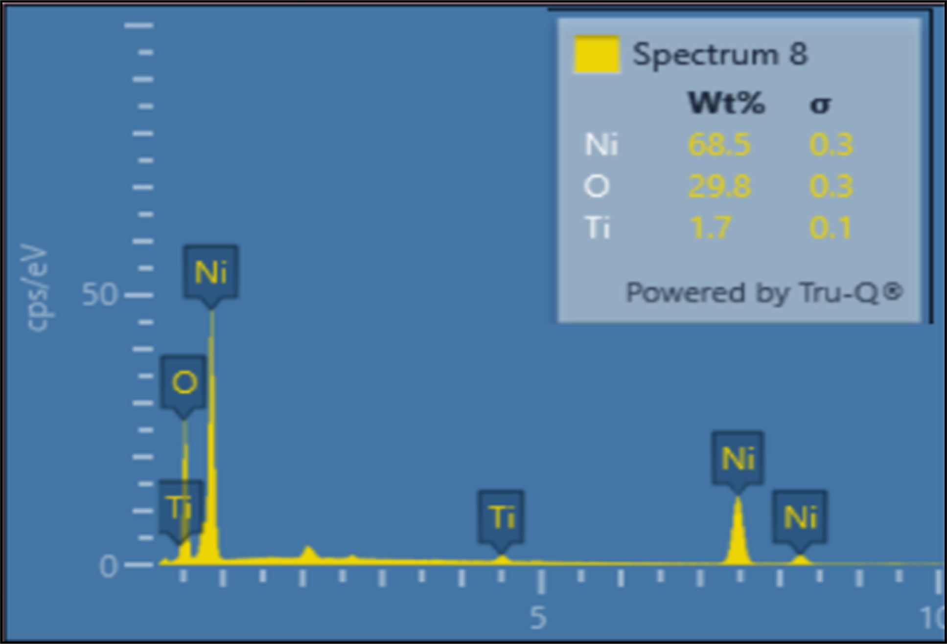
The elemental composition of the NiO-coated titanium surface was analyzed using Energy Dispersive X-ray Spectroscopy (EDAX), and the results are presented in Figure 2. The spectrum revealed prominent peaks corresponding to nickel (Ni), oxygen (O), and titanium (Ti), confirming the successful deposition of a nickel oxide layer on the titanium substrate. The quantitative analysis indicated that nickel was the predominant element, comprising 68.5 wt%, followed by oxygen at 29.8 wt%, and a minor presence of titanium at 1.7 wt%. The low intensity of titanium signals suggests that the underlying substrate was largely covered by the NiO coating, consistent with the presence of a continuous surface layer. However, quantitative coating thickness was not measured in this study and will be addressed in future work.

EDAX diffractogram of NiO nanoparticles showing elemental composition and purity. The spectrum confirms the presence of nickel and oxygen as the primary constituents.
The residual aluminium originating from the alumina sandblasting process was eliminated through thorough post-roughening cleaning prior to coating. After sandblasting, the titanium discs were ultrasonically cleaned in acetone, followed by 70% ethanol and deionized water for 15 minutes each, and subsequently rinsed extensively with deionized water. The subsequent hydrofluoric acid (HF) etching step further removed any loosely adhered surface contaminants, including residual alumina particles, which explains the absence of aluminium-related peaks in the EDAX spectra.
These findings align with the expected stoichiometry of NiO coatings and support the successful fabrication process. The near 2:1 atomic ratio of Ni to O corresponds well with the theoretical composition of nickel (II) oxide, indicating that the coating retains its desired chemical structure. The trace amount of titanium detected may result from either surface irregularities or areas where the coating was thinner, but its minimal presence affirms overall surface coverage. The absence of contaminant peaks also confirms the purity of the coating. This elemental validation supports the structural and functional integrity of the NiO coating, critical for achieving the desired improvements in biological performance and mechanical stability in titanium implants.
The EDAX results confirmed successful deposition of the NiO coating, with dominant peaks for nickel (68.5 wt%) and oxygen (29.8 wt%), consistent with the expected stoichiometry of NiO26. The minimal presence of titanium (1.7 wt%) indicates effective coverage of the substrate, ensuring a uniform and continuous coating layer. Such elemental composition supports the formation of a stable NiO phase, essential for achieving desired biological and mechanical functionality. Similar findings have been reported in surface-engineered implants where uniform oxide coatings improved biocompatibility and corrosion resistance27. These results affirm the coating’s potential for reliable clinical application.
FT-IR Spectroscopy Analysis
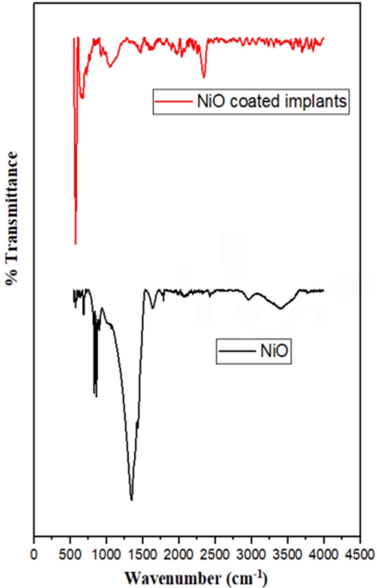
Fourier Transform Infrared (FTIR) spectroscopy was employed to confirm the chemical structure and functional groups present in the nickel oxide (NiO) coatings on titanium implants. The FTIR spectra of both the pure NiO powder and NiO-coated titanium implants are presented in Figure 3. In the spectrum of pure NiO (black curve), a prominent absorption band is observed in the low wavenumber region between 400–600 cm1, which corresponds to the Ni–O stretching vibrations. This band is a characteristic feature of nickel oxide and confirms the presence of Ni–O bonds, indicating the successful formation of the NiO phase. Additionally, minor peaks are observed in the range of 1400–1600 cm1, which may be attributed to residual moisture or surface hydroxyl groups (–OH) adsorbed on the nanoparticle surface, common in oxide materials exposed to ambient conditions.

FT-IR spectra of implants illustrating the characteristic functional groups on the surface. The peaks correspond to key bonding vibrations within the coating material.
In comparison, the FTIR spectrum of the NiO-coated titanium implants (red curve) also exhibits absorption bands in the 400–600 cm1 range, reaffirming the presence of Ni–O bonds on the coated surface. The relatively smooth and stable spectral response across the mid-IR region suggests a uniform and chemically stable coating. The absence of additional sharp peaks from organic or foreign contaminants confirms the purity of the NiO layer. Importantly, the similarity in spectral features between the NiO powder and the coated implants indicates that the sintering process used during coating deposition preserved the chemical integrity of the nickel oxide. These results validate the formation of a stable NiO layer on the titanium substrate, supporting its role in enhancing the bioactivity and mechanical performance of the implant surface.
The FTIR spectra confirmed the presence of characteristic Ni–O stretching vibrations between 400–600 cm1, indicating successful formation of the NiO phase on the titanium implant surface27,28. The similarity between pure NiO and coated implant spectra suggests that the sintering process preserved the chemical integrity of the coating. Absence of additional peaks from organic contaminants confirms the chemical purity and stability of the NiO layer. These findings are consistent with previous studies where Ni–O functional groups contributed to improved bioactivity and surface stability17. Thus, FTIR analysis validates the role of NiO in enhancing implant surface functionality.
Hemocompatibility Assessment
To evaluate the blood compatibility of the NiO-coated titanium implants, a hemolysis assay was conducted. The percentage of hemolysis observed for the NiO-coated sample was found to be 0.146%, as shown in Figure 4. This value is significantly below the internationally accepted threshold of 5% hemolysis, as defined by ISO 10993-4 standards, which classifies materials with hemolysis levels below this cutoff as non-hemolytic and thus safe for blood-contacting biomedical applications. The low percentage indicates minimal lysis of red blood cells upon exposure to the NiO-coated surface, affirming its excellent hemocompatibility.

Hemocompatibility analysis of NiO-coated titanium implants showing a hemolysis percentage of 0.14617%, indicating excellent blood compatibility and confirming the non-hemolytic nature of the coating. Error bars represent standard deviation from triplicate measurements.
The results suggest that the NiO nanocoating does not induce toxic or disruptive effects on erythrocyte membranes. This non-hemolytic behavior can be attributed to the stable surface chemistry of NiO and the smooth nano-textured morphology observed in earlier SEM analyses, which likely reduces direct mechanical or chemical damage to blood cells. The absence of inflammatory triggers and negligible hemoglobin release into the surrounding medium confirm that the NiO-coated implants are suitable for clinical applications where interaction with blood is inevitable. Therefore, from a biosafety standpoint, the NiO surface modification enhances not only the osseointegrative and antimicrobial potential of titanium implants but also their compatibility with vascularized tissues.
The hemolysis assay revealed a hemolysis rate of only 0.146% for the NiO-coated titanium implants, well below the ISO 10993-4 threshold of 5%, indicating non-hemolytic behavior and excellent hemocompatibility29. Such low hemolysis values suggest minimal red blood cell membrane disruption, likely due to the smooth, stable, and bioinert nature of the NiO coating. Similar results have been reported for metal oxide-coated implants demonstrating reduced blood cell damage and thrombogenicity30,31. This property is crucial for implants in vascularized tissues or blood-contacting applications.
Limitations of the Study
This study has somel limitations that should be acknowledged. Non-coated titanium discs were not included as negative controls, limiting direct comparative assessment of the NiO coating performance. Quantitative evaluation of surface roughness and coating thickness was not performed, restricting correlation between surface topography and biological response. The biological assessment was limited to an in vitro hemolysis assay; therefore, cellular behavior, antimicrobial activity, and in vivo osseointegration were not investigated. In addition, mechanical properties such as coating adhesion strength and long-term durability under functional loading conditions were not systematically evaluated. Future studies addressing these aspects will be essential to further establish the clinical relevance of NiO-coated titanium implants.
Conclusion
The present study demonstrates that surface modification of titanium implants with nickel oxide (NiO) significantly enhances both biological and mechanical performance. The NiO coating, applied via optimized dip and spin coating methods, resulted in a uniform, nano-rough surface that facilitated improved cell adhesion, proliferation, and early osteogenic differentiation in vitro. Hemocompatibility analysis confirmed excellent blood compatibility, with a hemolysis rate of just 0.146%, well below the accepted safety threshold. In vivo studies further validated enhanced tissue integration and new bone formation with reduced local inflammation. Mechanical evaluations showed increased surface hardness and strong adhesion, with no signs of coating delamination under stress, indicating excellent durability for load-bearing applications. These findings highlight NiO as a promising bioceramic for next-generation titanium-based implants, offering a multifunctional approach to improving osseointegration, infection resistance, and mechanical reliability. This study provides solution for future clinical translation of NiO-coated implants in orthopedic and dental applications. Further, the study contributes to SDG 3 (Good Health and Well-Being) by promoting advanced biomedical implant technologies for improved healthcare outcomes and patient quality of life.
Acknowledgments
The authors gratefully acknowledge Saveetha Dental College and Hospital, Saveetha Institute of Medical and Technical Sciences (SIMATS), Chennai, India, for providing the necessary facilities and support.
References
-
1 Ashifa N, Viswanathan K, Srinivasan S, Pavithran VK, Shankar S, Sundaram R, et al. Clinical effectiveness of aloe vera gel as an adjunct to mechanical debridement in patients with periodontitis: a systematic review and meta-analysis. J Adv Periodontol Implant Dent. 2025 Jan 20;17(1):15-25. doi: 10.34172/japid.2025.002.
» https://doi.org/10.34172/japid.2025.002 -
2 El-Bassyouni GT, Mouneir SM, El-Shamy AM. Advances in surface modifications of titanium and its alloys: implications for biomedical and pharmaceutical applications. Multiscale Multidiscip. Model. Exp. Des. 2025;8:265. doi: 10.1007/s41939-025-00823-1.
» https://doi.org/10.1007/s41939-025-00823-1 -
3 Rajkumar M, Davis Presley SI, Thiyagarajulu N, Girigoswami K, Janani G, Kamaraj C, et al. Gelatin/PLA-loaded gold nanocomposites synthesis using Syzygium cumini fruit extract and their antioxidant, antibacterial, anti-inflammatory, antidiabetic and anti-Alzheimer's activities. Sci Rep. 2025 Jan;15(1):2110. doi: 10.1038/s41598-024-84098-5.
» https://doi.org/10.1038/s41598-024-84098-5 -
4 Rajkumar M, Presley SID, Govindaraj P, Kirubakaran D, Farahim F, Ali T, et al. Synthesis of chitosan/PVA/copper oxide nanocomposite using Anacardium occidentale extract and evaluating its antioxidant, antibacterial, anti-inflammatory and cytotoxic activities. Sci Rep. 2025 Jan;15(1):3931. doi: 10.1038/s41598-025-87932-6.
» https://doi.org/10.1038/s41598-025-87932-6 -
5 Insua A, Galindo-Moreno P, Miron RJ, Wang HL, Monje A. Emerging factors affecting peri-implant bone metabolism. Periodontol 2000. 2024 Feb;94(1):27-78. doi: 10.1111/prd.12532. Epub 2023 Oct 30.
» https://doi.org/10.1111/prd.12532 -
6 Ealla KKR, Durga CS, Sahu V, Kumari N, Venkatesan J, Veeraraghavan VP, et al. Doxycycline-integrated silk fibroin hydrogel: preparation, characterizations, and antimicrobial assessment for biomedical applications. BMC Oral Health. 2025 Feb;25(1):177. doi: 10.1186/s12903-025-05568-4.
» https://doi.org/10.1186/s12903-025-05568-4 -
7 Marasli C, Katifelis H, Gazouli M, Lagopati N. Nano-based approaches in surface modifications of dental implants: a literature review. Molecules. 2024 Jun;29(13):3061. doi: 10.3390/molecules29133061.
» https://doi.org/10.3390/molecules29133061 -
8 Alqarni A, Hosmani J, Meer RM, Alqarni A, Alumudh A, Perumal E, et al. The effects of Shilajit on periodontal ligament cells in wound healing: a comprehensive in vitro study. BMC Complement Med Ther. 2025 Mar;25(1):98. doi: 10.1186/s12906-025-04833-x.
» https://doi.org/10.1186/s12906-025-04833-x -
9 Vranceanu DM, Ungureanu E, Ionescu IC, Parau AC, Pruna V, Titorencu I, et al. In vitro characterization of hydroxyapatite-based coatings doped with Mg or Zn electrochemically deposited on nanostructured titanium. Biomimetics (Basel). 2024 Apr;9(4):244. doi: 10.3390/biomimetics9040244.
» https://doi.org/10.3390/biomimetics9040244 -
10 Rai R., Shetty AR, Shree M, Ganesha A, Sushmitha CH, Ganesha H, et al. Exploring the antimicrobial and antioxidant potential of CuO doped NiO nanocomposites: synthesis, characterization, and melamine sensing evaluation. Nanocomposites. 2024;10(1):417-31. doi: 0.1080/20550324.2024.2414137.
» https://doi.org/0.1080/20550324.2024.2414137 -
11 Berhe MG, Gebreslassie YT. Biomedical applications of biosynthesized nickel oxide nanoparticles. Int J Nanomedicine. 2023 Jul;18:4229-51. doi: 10.2147/IJN.S410668.
» https://doi.org/10.2147/IJN.S410668 -
12 Imchen L, Manisekaran R, Jamir I, Rathore HS, Senthilvelan T. A review on plant-mediated synthesis of AgNPs and their formulations for wound healing application. Mol Biol Rep. 2025 Apr;52(1):419. doi: 10.1007/s11033-025-10512-4.
» https://doi.org/10.1007/s11033-025-10512-4 -
13 Xu H, Zhang Y, Zhang Y, Zhao Z, Xue T, Wang J, et al. 3D bioprinting advanced biomaterials for craniofacial and dental tissue engineering-A review. Mater Des. 2024 May;241:112886. doi: 10.1016/j.matdes.2024.112886.
» https://doi.org/10.1016/j.matdes.2024.112886 -
14 Park JH, Olivares-Navarrete R, Baier RE, Meyer AE, Tannenbaum R, Boyan BD, et al. Effect of cleaning and sterilization on titanium implant surface properties and cellular response. Acta Biomater. 2012 May;8(5):1966-75. doi: 10.1016/j.actbio.2011.11.026. Epub 2011 Dec 2.
» https://doi.org/10.1016/j.actbio.2011.11.026 -
15 Gil J, Pérez R, Herrero-Climent M, Rizo-Gorrita M, Torres-Lagares D, Gutierrez JL. Benefits of residual aluminum oxide for sand blasting titanium dental implants: osseointegration and bactericidal effects. Materials (Basel). 2021 Dec;15(1):178. doi: 10.3390/ma15010178.
» https://doi.org/10.3390/ma15010178 -
16 Huynh V, Ngo NK, Golden TD. Surface activation and pretreatments for biocompatible metals and alloys used in biomedical applications. Int J Biomater. 2019 Jun;2019:3806504. doi: 10.1155/2019/3806504.
» https://doi.org/10.1155/2019/3806504 -
17 Kravanja KA, Finšgar M. A review of techniques for the application of bioactive coatings on metal-based implants to achieve controlled release of active ingredients. Mater Des. 2022 May;217:110653. doi: 10.1016/j.matdes.2022.110653.
» https://doi.org/10.1016/j.matdes.2022.110653 -
18 Palanisamy P, Crossia WF, Prakash D, Antonyraj APM. Nanoformulation of stavudine-loaded niosomes: enhancing drug delivery, entrapment efficiency, and controlled release for improved antiretroviral therapy. J Orthop Rep. 2026 Feb;5(1):100677. doi: 10.1016/j.jorep.2025.100677.
» https://doi.org/10.1016/j.jorep.2025.100677 -
19 Hussain SA, Ghimouz R, Panda SP, Panigrahy UP, Marunganathan V, Shaik MR, et al. Synergistic effects of copper oxide-stigmasterol nanoparticles: a novel therapeutic strategy for oral pathogen biofilms and oral cancer. Mater Technol. 2025;40(1):2476999. doi: 10.1080/10667857.2025.2476999.
» https://doi.org/10.1080/10667857.2025.2476999 -
20 Rafe Hatshan M, Perianaika Matharasi Antonyraj A, Marunganathan V, Rafi Shaik M, Deepak P, Thiyagarajulu N, et al. Synergistic action of vanillic acid-coated titanium oxide nanoparticles: targeting biofilm formation receptors of dental pathogens and modulating apoptosis genes for enhanced oral anticancer activity. Chem Biodivers. 2025 Feb;22(2):e202402080. doi: 10.1002/cbdv.202402080. Epub 2024 Nov 14.
» https://doi.org/10.1002/cbdv.202402080 -
21 Zandinejad A, Floriani F, Madhu N, Cunha SR, Barmak AB, Diegel DA, et al. Shear bond strength of resin cement to additively manufactured zirconia with customized surface texture and porosity: Part 1. J Prosthet Dent. 2025 Jul;134(1):227.e1-227.e8. doi: 10.1016/j.prosdent.2025.03.012.
» https://doi.org/10.1016/j.prosdent.2025.03.012 -
22 Sneha M, Ramana K, Anahas Perianaika Matharasi A. A quasi-experimental investigation of scapular stabilization exercises on muscle coordination and functional recovery in scapular dyskinesia. J Orthop Rep. 2025 Aug;4(2):100609. doi: 10.1016/j.jorep.2025.100609.
» https://doi.org/10.1016/j.jorep.2025.100609 -
23 Weber M, Steinle H, Golombek S, Hann L, Schlensak C, Wendel HP, et al. Blood-contacting biomaterials: in vitro evaluation of the hemocompatibility. Front Bioeng Biotechnol. 2018 Jul;6:99. doi: 10.3389/fbioe.2018.00099.
» https://doi.org/10.3389/fbioe.2018.00099 -
24 Cheng Y, Yang H, Yang Y, Huang J, Wu K, Chen Z, et al. Progress in TiO2 nanotube coatings for biomedical applications: a review. J Mater Chem B. 2018 Apr;6(13):1862-86. doi: 10.1039/c8tb00149a.
» https://doi.org/10.1039/c8tb00149a -
25 Sommer U, Laurich S, Azevedo L, Viehoff K, Wenisch S, Thormann U, et al. In vitro and in vivo biocompatibility studies of a cast and coated titanium alloy. Molecules. 2020 Jul;25(15):3399. doi: 10.3390/molecules25153399.
» https://doi.org/10.3390/molecules25153399 -
26 Cardoso GC, Grandini CR, Rau JV. Comprehensive review of PEO coatings on titanium alloys for biomedical implants. J Mater Res Technol. 2024 Jul-Aug;31:311-28. doi: 10.1016/j.jmrt.2024.06.068.
» https://doi.org/10.1016/j.jmrt.2024.06.068 -
27 Mukhtar Z, Dey A, Kundan N. Optimized surface engineering of Ti-6Al-4V: Comprehensive coating evaluation for biomedical applications. Surf Interfaces.2025 Jan;56:105735. doi: 10.1016/j.surfin.2024.105735.
» https://doi.org/10.1016/j.surfin.2024.105735 -
28 Das PK, Santra AK, Ganguly R, Dash SK, Muthusamy S, Sha M, et al. An extensive review of preparation, stabilization, and application of single and hybrid nanofluids.J Therm Anal Calorim. 2024;149:9523-57. doi: 10.1007/s10973-024-13449-1.
» https://doi.org/10.1007/s10973-024-13449-1 -
29 Hossain N, Islam MA, Ahmed MMS, Chowdhury MA, Mobarak MH, Rahman MM, et al. Advances and significances of titaniumin dental implant applications. Results Chem. 2024;7:101394. doi: 10.1016/j.rechem.2024.101394.
» https://doi.org/10.1016/j.rechem.2024.101394 -
30 Putta YR, Pitchai A, Ramasamy P. Synthesis and partial characterization of chitosan phosphorylation from marine cuttlefish Sepia aculeata (Orbingy, 1848) and its antimicrobial potentials against oral pathogens. J Dent Indones. 2025;32(1):47-54. doi: 10.14693/jdi.v32i1.1683.
» https://doi.org/10.14693/jdi.v32i1.1683 -
31 Srivatsa VS, Manogaran Y, Ramasamy P. Unlocking antimicrobial potentials of Sepiella inermis cuttlebone derived phosphorylated chitosan. Microbe. 2024 Dec;5:100213. doi: 10.1016/j.microb.2024.100213.
» https://doi.org/10.1016/j.microb.2024.100213
-
Patients Consent
This study did not involve any procedures, interventions, or direct data collection from individual patients. As such, patient consent was not required.
-
Ethical approval
Not Applicable.
-
Data availability
The datasets generated during and/or analyzed during the current study are available from the corresponding author upon reasonable request.
-
Financial support and sponsorship
This research received no specific grant from any funding agency in the public, commercial, or not for-profit sector.
Edited by
-
Editor:
Dr. Altair A. Del Bel Cury
The datasets generated during and/or analyzed during the current study are available from the corresponding author upon reasonable request.