Effect of different endodontic sealer activation methods on bond strength and sealer filling quality in the root canal
Abstract
Aims To evaluate the influence of different endodontic sealer activation methods on the bond strength and sealer filling quality.
Methods Sixty bovine incisors with similar root anatomy were instrumented using a crown-down technique and randomly distributed into five groups (n = 12) according to the sealer activation protocol: No Activation; Activation using Easy Clean (EC) file attached to a handpiece – EC / Handpiece; EC in endodontic motor in rotary movement – EC / Rotatory; EC in endodontic motor in reciprocating movement – EC / Reciprocating; or Ultrasonic activation. The roots were filled using the lateral condensation technique with gutta-percha and AH Plus sealer, which was activated for 40 seconds, 2 mm short of the working length. Two slices were obtained from each root third: one for push-out and another to assess sealer filling quality using a stereomicroscope. Kruskal-Wallis with Dunn’s post hoc tests were used to compare the different sealer activation methods. Friedman tests were applied for repeated measurements within the same group, comparing root thirds (α = 0.05).
Results Significantly lower values of bond strength and a higher occurrence of voids in the middle and apical thirds were observed when the sealer was not activated (p < 0.05). No significant differences were observed among the activation methods (ultrasonic activation vs. EC activation in different kinematics) for bond strength and sealer filling quality (p > 0.05).
Conclusions Sealer activation, regardless of the method, improve the bond strength of the sealer for root dentin and a result in fewer voids on filling material.
Keywords
Root canal obturation; ultrasonics; endodontics
Introduction
Root canal filling is a crucial step in endodontic treatment, as its primary purpose is to seal the root canal with inert and antiseptic materials, eliminating the space previously occupied by the dental pulp, which was previously cleaned and shaped through chemical-mechanical preparation1. This step is essential for creating an airtight seal that prevents infection or reinfection within the root canal system2,3.
During root canal filling, it is desirable to achieve mechanical imbrication and adhesion between the endodontic sealer and dentin4. In recent research, the available evidence suggests that ultrasonic activation of endodontic sealer enhances the penetration of the sealer into the dentinal tubules, potentially promoting effective root sealing5,6. Furthermore, prior studies have also emphasized that ultrasonic activation of endodontic sealer holds the potential to enhance the outcomes of sealer adhesion to dentin7-9. However, studies evaluating bond strength after ultrasonic activation of the sealer are relatively scarce, and varying results are observed. This underscores the need for additional research to draw a definitive conclusion about its effectiveness.
In addition to ultrasonic activation, which was originally designed to agitate irrigating solutions within the root canal10,11 and subsequently found applicability in activating endodontic sealers5, other devices designed similarly may also hold potential for use in this purpose. In this context, compared to ultrasonic activation, the Easy Clean (EC) plastic file (Easy Equipamentos Odontológicos, Belo Horizonte, MG, Brazil) is a cost-effective device that has demonstrated promising results in activating irrigants, enhancing disinfection12,13, and improving root canal cleaning14,15. Additionally, the EC is a highly versatile instrument, expanding its usability for clinicians, as it can be operated in rotary and reciprocating modes with an endodontic motor14 or even in a handpiece16. However, the literature lacks studies evaluating this device effectiveness in activating endodontic sealer and its subsequent impact on bond strength between the sealer and dentin. Such studies are essential to confirm the EC suitability for this purpose. Finally, assessing various kinematic modes of device use is crucial for identifying the most effective approach and comparing it with ultrasonic activation, the most commonly studied method. In light of the above, this study was designed to address the gap in investigations comparing these devices and their effects on endodontic sealer activation during root canal filling, aiming to contribute to the growing body of evidence on this subject17.
Therefore, this study aimed to evaluate the influence of endodontic sealer activation on the bond strength between the sealer and root dentin, as well as its effect on the sealer filling quality in the root canal. For this purpose, the ultrasonic activation and activation methods with the EC plastic file were compared in different kinematics of use (rotary or reciprocating in an endodontic motor, or in a handpiece), with the no activation of the endodontic sealer during obturation. The study formulated the following null hypotheses: (I) there would be no statistically significant difference in the investigated outcomes between activating or not activating the endodontic sealer; and (II) the utilization of various devices and their respective application methods would not result in statistically significant differences among the methods.
Materials and Methods
Sample size and selection
Ethical approval for the study was obtained from the local ethics committee (no. 3956240423). The sample size was calculated (http://www.openepi.com/SampleSize/SSMean.htm) using previous parameters from a similar study7. The following values for AH Plus sealer were considered for the calculation: bond strength of 1.65 MPa (± 0.45) for no activated sealer and 2.25 Mpa (± 0.56) for ultrasonically activated sealer; 80% power; and a significance level of 5%. Based on these data, the estimated minimum size was 12 samples per group. Sixty bovine incisors from animals killed for commercial reasons were used. The teeth were included in the study considering the presence of complete root development and an apical opening smaller than or equal to a size #60 K-file (Dentsply Maillefer, Ballaigues, Switzerland). In addition, teeth with cracks/fractures or any other structural anomaly were excluded. The teeth were stored in distilled water at 4ºC until the initiation of the methodological procedures.
Sample preparation
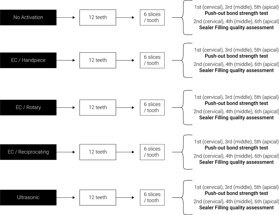
The crowns of the teeth were sectioned 18 mm from the apex with a diamond disc under constant irrigation in a cutting machine (Isomet 1000; Buehler Ltd, Lake Bluff, USA), to standardize the length of the roots. The working length was established 1 mm short of the apical foramen. The root canal was shaped using the crown-down technique, employing size #4 and #3 Gates-Glidden drills (Dentsply Maillefer) for the coronal and middle root thirds, respectively. The apical third was manually prepared with stainless steel instruments, up to size #80 K-file (Dentsply Maillefer). During root canal preparation, irrigation was carried out with 10 mL of 2.5% NaOCl. Following instrumentation, 5 mL of 17% EDTA was used for 5 minutes, followed by a final irrigation with 5 mL of distilled water. Then, the root canals were dried with compatible absorbent paper points (#80.02; Dentsply Maillefer). The AH Plus sealer was handled according to the manufacturer’s instructions and inserted into the root canals using a finger spreader #40.02 (MK Life Medical and Dental Products, Porto Alegre, RS, Brazil) until the root canal walls were completely covered by the sealer. The samples were then submitted to the type of endodontic sealer activation for root canal filling following prior random allocation (http://www.randomized.org) into five groups (n=12) (Figure 1), as follows:
One marker was missing, just like the other groups: no type of activation was performed on the sealer.
-
EC / Handpiece: The sealer was activated with the EC plastic file #25.04 attached to a handpiece at 20.000 rpm, 2 mm short of the working length. The sealer was activated for 20 seconds in the mesiodistal direction and another 20 seconds in the buccolingual direction.
-
EC / Rotary: The sealer was activated with an EC plastic file # 25.04 attached to an endodontic motor (X-Smart Plus; Dentsply Maillefer) at 1.000 rpm, 2 mm short of the working length. The sealer was activated for 20 seconds in the mesiodistal direction and another 20 seconds in the buccolingual direction.
-
EC / Reciprocating: The sealer was activated with an EC plastic file #25.04 attached to an endodontic motor (X-Smart Plus; Dentsply Maillefer) in “Wave One Gold” mode, 2 mm short of the working length. The sealer was activated for 20 seconds in the mesiodistal direction and another 20 seconds in the buccolingual direction.
-
Ultrasonic: The sealer was activated with the E1 Irrisonic tip (Helse Ultrasonic, Ribeirão Preto, SP, Brazil) attached to an ultrasonic device (Sonic Laxis BP LED, Schuster, Santa Maria, RS, Brazil) with a power level of 20%, 2 mm short of the working length. The sealer was activated for 20 seconds in the mesiodistal direction and another 20 seconds in the buccolingual direction.
Afterward, all root canals were filled with gutta-percha and AH Plus sealer using the lateral condensation technique (Dentsply Maillefer). Periapical radiographs were taken to verify the filling quality. The roots were sealed using interim restorative material (Coltosol; Coltene, Alstatten, Switzerland) and stored in distilled water at 37ºC for one week. After this period, the samples were sectioned using a precision cutting machine (Isomet 1000; Buehler Ltd). Six slices were obtained per sample, two corresponding to each root third, with a thickness of 1mm ± 0.1mm. The first slice (the most coronal) of each sample was subjected to the push-out bond strength test and subsequent failure mode analysis, while the second slice was used to evaluate the sealer filling quality in the root canal9.
Push-out bond strength test
The first sections obtained from each root third were positioned in a universal testing machine (Emic DL-2000; Emic, Pinhais, PR, Brazil) and subjected to the push-out test. The filling material was loaded in the apical-coronal direction using a stainless-steel plunger (Ø = 0.8 mm for the cervical and middle thirds, or Ø = 0.6 mm for the apical third) at a speed of 0.5 mm/min until failure. The data obtained, expressed in newtons (N), were converted to megapascals (MPa) using the following formula: σ = F / A, as described by a previous study18. F represented the force (N) recorded for specimen failure and A represented the bond area (mm2). To determine the area of the bond interface, the following formula was used:
In the formula, π = is the constant 3.14, R = coronal radius, r = apical radius, and h = slice thickness. A digital caliper was used to obtain measurements (CD-15C; Mitutoyo Co., Kawasaki, Japan).
Failure modes analysis
After failure, the samples were examined using a stereomicroscope (Discovery V20; Carl-Zeiss, Gottingen, Germany) at 25× magnification by an examiner blinded to the groups. Failures were categorized according to a previously established classification8. Consequently, failure patterns were divided into the following categories: adhesive, indicating failure at the sealer-dentin or sealer-core interface; cohesive, signifying failure within the sealer or dentin; or mixed, denoting a combination of adhesive and cohesive failures. The calibration process involved analyzing the fracture pattern of 20% of the sample, and this analysis was repeated after a two-week interval. Examiner reproducibility, calculated using the Kappa test, yielded a value of 0.886.
Sealer filling quality assessment
The second slices, obtained from each third of the root, were specifically employed to examine the effect of the different activation methods on the sealer filling quality in the root canal. The samples were evaluated by a blinded examiner, through a stereomicroscope (Discovery V20; Carl-Zeiss, Gottingen, Germany) at ×60 magnification. The number of voids was counted, and the presence of voids was assessed using the scoring system proposed by Kim et al.19: Score 1: well-condensed filling that showed only a few, minor air bubbles (less than 0.1 mm in diameter); Score 2: an imperfectly condensed filling that showed some minor air bubbles (more than 3 defects) or medium-sized air bubbles (0.1 mm to 0.2 mm in diameter); Score 3: inadequately condensed filling that showed many minor air bubbles (more than 5 defects) or large air bubbles (more than 0.2 mm in diameter); and Score 4: poorly condensed filling that showed many minor air bubbles (more than 7 defects) or empty. All analyses were conducted using ImageJ software (National Institutes of Health, Bethesda, USA) with a standardized zoom level set at 75%. The calibration process involved analyzing of 20% of the sample, and this analysis was repeated after a two-week interval. The intraclass correlation coefficient, used to assess the reproducibility of void counting by the examiner, yielded a value of 0.946. The Kappa test, used to assess the reproducibility of void scores by the examiner, yielded a value of 0.885.
Statistical analysis
A statistical software program (IBM SPSS Software; IBM, Armonk, NY, USA) was used with a significance level of 0.05 to perform the data analysis. The bond strength and void data were analyzed using the Shapiro-Wilk test, revealing a non-normal distribution. The different sealer activation methods were compared using Kruskal-Wallis and post hoc Dunn’s tests, while Friedman tests were applied for repeated measurements in the same group, enabling comparison among root thirds.
RESULTS
Push-out bond strength
The median and percentile of bond strength are shown in Table 1. The use of a sealer activation protocol had a significant effect on the bond strength compared to the No Activation group (p < 0.05). On the other hand, among the different types of activation investigated, there was no significant difference among the methods (p > 0.05). Regarding the comparison among the root thirds for the same group, a significant difference was only observed for the group in which the sealer was not activated, with the apical third having lower bond strength than the cervical third (p < 0.05).
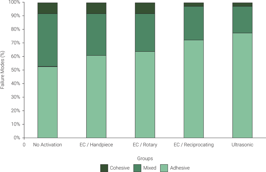
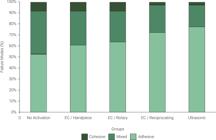
The failure mode results are presented in Figure 2. According to the grouped data, in all groups, the most common failure was adhesive failure, followed by mixed failure, and finally, cohesive failure. The relative frequency was 65.56%, 28.33%, and 6.11%, respectively.
Sealer filling quality
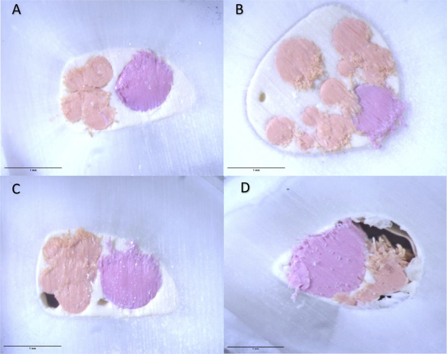
The median and percentile of the number of voids and void scores are shown in Tables 2 and 3, respectively. Regarding the number of voids, no significant differences were identified among the different types of activation (p > 0.05). When comparing the root thirds for the same group, a greater number of voids were observed in the middle and apical thirds when the sealer was not activated compared to the cervical third (p < 0.05). As for the data obtained for void scores, no significant differences were identified regardless of the type of sealer activation and the root third (p > 0.05). Representative images of void scores analysis are presented in Figure 3.
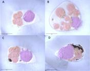
Representative stereomicroscope images of void scores at ×60 magnification. (A) = score 1; (B) = score 2; (C) = score 3; and (D) = score 4.
DISCUSSION
This study evaluates the influence of endodontic sealer activation on the bond strength between sealer and dentin, as well as its subsequent influence on the sealer filling quality in the root canal. Additionally, it stands out as one of the few studies that examine the effectiveness of ultrasonic activation for sealer activation, and the first to investigate the application of the Easy Clean (EC) plastic file in different kinematics of use for this purpose. The study findings revealed significantly lower bond strength values and a higher occurrence of voids in the middle and apical thirds when the sealer was not activated. Thus, the first null hypothesis formulated was rejected. However, no significant differences were observed among the activation methods (ultrasonic activation vs. EC activation in different kinematics), which confirms the acceptance of the second hypothesis as initially formulated.
The decrease in bond strength observed when the sealer was not activated is consistent with the findings in the existing, albeit limited, body of research on this topic7,8. During the activation process, the heat produced has the potential to decrease the sealer viscosity, facilitating improved flow within the root canal system20. Consequently, the activation of endodontic sealer results in enhanced material penetration into the dentinal tubules, along with the creation of more extensive and denser tags5,7. It is plausible that this improved mechanical interaction at the tubular level contributes to the observed higher bond strength. However, it is important to note that this connection is not yet firmly established in the literature and warrants further investigation21.
Although our study did not reveal significant differences in sealer filling quality scores between activating or not activating sealer (regardless of activation method), we did observe a higher occurrence of voids in the middle and apical thirds when the sealer was not activated, as opposed to the cervical third. Consequently, these results led to a reduction in bond strength in the apical third. This finding is associated with the fact that the greater pressure and projection of the sealer against the canal walls generated during the activation of the sealer favors a more homogeneous filling with fewer voids5. This explains the voids when the sealer activation step is omitted. Furthermore, the number of dentinal tubules per mm2 decreases from the coronal portion of the root canal to the apical region22. This underscores the significance of activating the sealer in the final millimeters of the root canal, enhancing the interaction of the sealer with the tubular structure, and improving the uniformity and sealing of the filling.
When comparing sealer activation using ultrasonic and EC techniques, encompassing rotary, reciprocating, and handpiece modes, with the same application time, we did not observe variations in bond strength or sealer filling quality, as indicated by the number of voids and void scores. Notably, ultrasonic activation operates at the highest frequency, typically between 25.000 and 30.000 Hz, which promotes acoustic transmission and cavitation10, favoring the diffusion of sealer into irregularities23. However, the frequency did not appear to be a decisive factor, as EC in different kinematics also yielded similar results. In this context, the act of activating the sealer in some manner seems to carry greater significance than the specific parameters used for activation. Nonetheless, it is crucial to underscore that the factors related to equipment frequency, usage kinematics, and application time should be subjected to further investigation to establish comprehensive protocols. Additionally, it is worth highlighting that the EC plastic file Clean is readily available to clinicians for a low investment, does not necessitate the acquisition of additional equipment, and has shown effective results when used to agitate irrigants12,24, making it an interesting alternative for use in endodontic clinical practice.
The push-out test is a method that accurately estimates bond strength because it results in failure parallel to the bonding interface, simulating a true shear load similar to the clinical condition25. Ideally, adhesion test results should primarily involve adhesive failures, a characteristic observed in the present study in all groups26. In addition, as recommended by Pane et al.25, the stainless-steel plunger diameter should range from 70 to 90% of the root canal size. This is why we selected a Ø = 0.8 stainless-steel plunger for the cervical and middle thirds and a Ø = 0.6 stainless-steel plunger for the apical third. One of the most advanced methods for assessing the sealer filling quality in the root canal is through microcomputed tomography (micro-CT)27. However, recent research has suggested that micro-CT analysis might be less sensitive than the sectioning method followed by stereomicroscope analysis when it comes to detecting voids18. This is because sealers are highly radiopaque, which can make it challenging to identify small voids within the filling volume using micro-CT28. Given this context, in our study, we opted to assess the sealer filling quality visually using a stereomicroscope. This approach has been adopted previously and shown to be a suitable option9.
The use of bovine teeth can be identified as a limitation of the current study. While bovine teeth are commonly regarded as suitable substitutes for human teeth in dental research, it is important to acknowledge that the anatomical complexity of human teeth might present a more challenging scenario and should be explored in future studies29. Nonetheless, we emphasize that, for the purposes of this study, the use of bovine teeth adequately fulfilled the outlined objectives. Additionally, we evaluated the activation process in only one type of endodontic sealer. Sealers with higher fluidity may have an increased capacity to enhance the activation of the sealer, as they are more likely to efficiently fill irregularities30,31. In this sense, it is imperative to consider assessing sealers with different compositions because each type of sealer may exhibit a distinct behavior. Finally, it is imperative to conduct future clinical investigations to assess the influence of sealer activation on the outcomes of long-term endodontic treatment.
In conclusion, regardless of the activation method employed, activating the endodontic sealer led to higher bond strength and a filling without differences in voids across the root thirds compared to no activation of the sealer. Furthermore, the ultrasonic activation and EC activation methods in different kinematics of use demonstrated similar findings in terms of bond strength and sealer filling quality.
Acknowledgements
We would like to thank the Coordenação de Aperfeiçoamento de Pessoal de Nível Superior (CAPES) for their support.
References
-
1 Tomson RM, Polycarpou N, Tomson PL. Contemporary obturation of the root canal system. Br Dent J. 2014 Mar;216(6):315-22. doi: 10.1038/sj.bdj.2014.205.
» https://doi.org/10.1038/sj.bdj.2014.205 -
2 Pinheiro ET, Gomes BP, Ferraz CC, Sousa EL, Teixeira FB, Souza-Filho FJ. Microorganisms from canals of root-filled teeth with periapical lesions. Int Endod J. 2003 Jan;36(1):1-11. doi: 10.1046/j.1365-2591.2003.00603.x.
» https://doi.org/10.1046/j.1365-2591.2003.00603.x -
3 Song M, Park M, Lee CY, Kim E. Periapical status related to the quality of coronal restorations and root fillings in a Korean population. J Endod. 2014 Feb;40(2):182-6. doi: 10.1016/j.joen.2013.10.017. Epub 2013 Nov 25.
» https://doi.org/10.1016/j.joen.2013.10.017 -
4 Souza MA, Padilha Rauber MG, Zuchi N, Bonacina LV, Ricci R, Dias CT, et al. Influence of final irrigation protocols and endodontic sealer on bond strength of root filling material with root dentin previously treated with photodynamic therapy. Photodiagnosis Photodyn Ther. 2019 Jun;26:137-41. doi: 10.1016/j.pdpdt.2019.03.013.
» https://doi.org/10.1016/j.pdpdt.2019.03.013 -
5 Guimarães BM, Amoroso-Silva PA, Alcalde MP, Marciano MA, de Andrade FB, Duarte MA. Influence of ultrasonic activation of 4 root canal sealers on the filling quality. J Endod. 2014 Jul;40(7):964-8. doi: 10.1016/j.joen.2013.11.016.
» https://doi.org/10.1016/j.joen.2013.11.016 -
6 Keles A, Askerbeyli Ors S, Purali N, Kucukkaya Eren S. Effect of different sealer activation techniques on dentinal tubule penetration. Aust Endod J. 2023 Dec;49(3):470-5. doi: 10.1111/aej.12738.
» https://doi.org/10.1111/aej.12738 -
7 Wiesse PEB, Silva-Sousa YT, Pereira RD, Estrela C, Domingues LM, Pécora JD, et al. Effect of ultrasonic and sonic activation of root canal sealers on the push-out bond strength and interfacial adaptation to root canal dentine. Int Endod J. 2018 Jan;51(1):102-11. doi: 10.1111/iej.12794. Epub 2017 Jun 19.
» https://doi.org/10.1111/iej.12794 -
8 De Bem IA, de Oliveira RA, Weissheimer T, Bier CAS, Só MVR, Rosa RAD. Effect of ultrasonic activation of endodontic sealers on intratubular penetration and bond strength to root dentin. J Endod. 2020 Sep;46(9):1302-8. doi: 10.1016/j.joen.2020.06.014.
» https://doi.org/10.1016/j.joen.2020.06.014 -
9 Padoin K, Bohrer TC, Ceolin LG, Bier CAS, Rosa RAD, Morgental RD. Does ultrasonic activation improve the bond strength and root canal filling quality of endodontic sealers? Braz Dent J. 2022 May-Jun;33(3):28-37. doi: 10.1590/0103-6440202204728.
» https://doi.org/10.1590/0103-6440202204728 -
10 van der Sluis LW, Versluis M, Wu MK, Wesselink PR. Passive ultrasonic irrigation of the root canal: a review of the literature. Int Endod J. 2007 Jun;40(6):415-26. doi: 10.1111/j.1365-2591.2007.01243.x.
» https://doi.org/10.1111/j.1365-2591.2007.01243.x -
11 Boutsioukis C, Arias-Moliz MT. Present status and future directions - irrigants and irrigation methods. Int Endod J. 2022 May;55 Suppl 3(Suppl 3):588-612. doi: 10.1111/iej.13739.
» https://doi.org/10.1111/iej.13739 -
12 Nogueira L, Amaral G, Silva E, Tinoco J, Alves F, Sassone LM. Bacterial reduction in oval-shaped root canals after different irrigant agitation methods. Eur Endod J. 2021 Apr;6(1):110-6. doi: 10.14744/eej.2020.42204.
» https://doi.org/10.14744/eej.2020.42204 -
13 Matoso FB, Montagner F, Jardine AP, Quintana RM, Grecca FS, Kopper PMP. Effect of different disinfection protocols in bacterial viability of an intraradicular biofilm formed in situ. Braz Dent J. 2023 May-Jun;34(3):42-9. doi: 10.1590/0103-6440202305244.
» https://doi.org/10.1590/0103-6440202305244 -
14 Cesario F, Hungaro Duarte MA, Duque JA, Alcalde MP, de Andrade FB, Reis So MV, et al. Comparisons by microcomputed tomography of the efficiency of different irrigation techniques for removing dentinal debris from artificial grooves. J Conserv Dent. 2018 Jul-Aug;21(4):383-7. doi: 10.4103/JCD.JCD_286_16.
» https://doi.org/10.4103/JCD.JCD_286_16 -
15 Monteiro LPB, de Sousa SEM, de Castro RF, da Silva EJNL, da Silva Brandão JM. Mechanical activation with Easy Clean device enhanced organic tissue removal from simulated internal root resorption in a laboratory evaluation. BMC Oral Health. 2023 Jun;23(1):385. doi: 10.1186/s12903-023-03122-8.
» https://doi.org/10.1186/s12903-023-03122-8 -
16 Oliveira KV, Silva BMD, Leonardi DP, Crozeta BM, Sousa-Neto MD, Baratto-Filho F, et al. Effectiveness of different final irrigation techniques and placement of endodontic sealer into dentinal tubules. Braz Oral Res. 2017 Dec;31:e114. doi: 10.1590/1807-3107BOR-2017.vol31.0114.
» https://doi.org/10.1590/1807-3107BOR-2017.vol31.0114 -
17 Pauletto G, Padoin Y, de Pellegrin SF, Bier CAS. The use of sonic and ultrasonic activation of endodontic sealer to improve sealer filling quality in the root canal: a scoping review. Odontology. 2024 Jul;112(3):657-71. doi: 10.1007/s10266-024-00901-9.
» https://doi.org/10.1007/s10266-024-00901-9 -
18 Pauletto G, Carlotto IB, Rosa LSD, Pereira GKR, Bier CAS. Effect of sodium/calcium hypochlorite on adhesion and adaptation of fiber posts luted with a dual resin cement. Braz Dent J. 2023 May-Jun;34(3):111-8. doi: 10.1590/0103-6440202305396.
» https://doi.org/10.1590/0103-6440202305396 -
19 Kim JA, Hwang YC, Rosa V, Yu MK, Lee KW, Min KS. Root canal filling quality of a premixed calcium silicate endodontic sealer applied using gutta-percha cone-mediated ultrasonic activation. J Endod. 2018 Jan;44(1):133-8. doi: 10.1016/j.joen.2017.07.023. Epub 2017 Nov 6.
» https://doi.org/10.1016/j.joen.2017.07.023 - 20 Bittmann B, Haupert F, Schlarb AK. Ultrasonic dispersion of inorganic nanoparticles in epoxy resin. Ultrason Sonochem. 2009 Jun;16(5):622-8. doi: 10.1016/j.ultsonch.2009.01.006.
-
21 Machado R, Silva Neto UX, Carneiro E, Fariniuk LF, Westphalen VP, Cunha RS. Lack of correlation between tubular dentine cement penetration, adhesiveness and leakage in roots filled with gutta percha and an endodontic cement based on epoxy amine resin. J Appl Oral Sci. 2014 Jan-Feb;22(1):22-8. doi: 10.1590/1678-775720130247.
» https://doi.org/10.1590/1678-775720130247 -
22 Mjör IA, Smith MR, Ferrari M, Mannocci F. The structure of dentine in the apical region of human teeth. Int Endod J. 2001 Jul;34(5):346-53. doi: 10.1046/j.1365-2591.2001.00393.x.
» https://doi.org/10.1046/j.1365-2591.2001.00393.x -
23 Arslan H, Abbas A, Karatas E. Influence of ultrasonic and sonic activation of epoxy-amine resin-based sealer on penetration of sealer into lateral canals. Clin Oral Investig. 2016 Nov;20(8):2161-4. doi: 10.1007/s00784-016-1737-z.
» https://doi.org/10.1007/s00784-016-1737-z -
24 Aveiro E, Chiarelli-Neto VM, de-Jesus-Soares A, Zaia AA, Ferraz CCR, Almeida JFA, et al. Efficacy of reciprocating and ultrasonic activation of 6% sodium hypochlorite in the reduction of microbial content and virulence factors in teeth with primary endodontic infection. Int Endod J. 2020 May;53(5):604-18. doi: 10.1111/iej.13261.
» https://doi.org/10.1111/iej.13261 -
25 Pane ES, Palamara JE, Messer HH. Critical evaluation of the push-out test for root canal filling materials. J Endod. 2013 May;39(5):669-73. doi: 10.1016/j.joen.2012.12.032.
» https://doi.org/10.1016/j.joen.2012.12.032 - 26 Drummond JL, Sakaguchi RL, Racean DC, Wozny J, Steinberg AD. Testing mode and surface treatment effects on dentin bonding. J Biomed Mater Res. 1996 Dec;32(4):533-41. doi: 10.1002/(SICI)1097-4636(199612)32:4<533::AID-JBM6>3.0.CO;2-S.
-
27 Zaslansky P, Fratzl P, Rack A, Wu MK, Wesselink PR, Shemesh H. Identification of root filling interfaces by microscopy and tomography methods. Int Endod J. 2011 May;44(5):395-401. doi: 10.1111/j.1365-2591.2010.01830.x.
» https://doi.org/10.1111/j.1365-2591.2010.01830.x -
28 Jung M, Lommel D, Klimek J. The imaging of root canal obturation using micro-CT. Int Endod J. 2005 Sep;38(9):617-26. doi: 10.1111/j.1365-2591.2005.00990.x.
» https://doi.org/10.1111/j.1365-2591.2005.00990.x -
29 Yassen GH, Platt JA, Hara AT. Bovine teeth as substitute for human teeth in dental research: a review of literature. J Oral Sci. 2011 Sep;53(3):273-82. doi: 10.2334/josnusd.53.273.
» https://doi.org/10.2334/josnusd.53.273 -
30 Amoroso-Silva PA, Guimarães BM, Marciano MA, Duarte MA, Cavenago BC, Ordinola-Zapata R, et al. Microscopic analysis of the quality of obturation and physical properties of MTA Fillapex. Microsc Res Tech. 2014 Dec;77(12):1031-6. doi: 10.1002/jemt.22432.
» https://doi.org/10.1002/jemt.22432 -
31 Piai GG, Duarte MAH, Nascimento ALD, Rosa RAD, Só MVR, Vivan RR. Penetrability of a new endodontic sealer: A confocal laser scanning microscopy evaluation. Microsc Res Tech. 2018 Nov;81(11):1246-9. doi: 10.1002/jemt.23129.
» https://doi.org/10.1002/jemt.23129
-
Data Availability:
Datasets related to this article will be available to the corresponding author upon request.
Edited by
-
Editor:
Dr. Altair A. Del Bel Cury
Datasets related to this article will be available to the corresponding author upon request.