
 Effect of aging on surface roughness and color stability of resin composites with color adjustment potential
Abstract
Aim This study aimed to evaluate the effect of six-month water aging on surface roughness and color stability of resin composites with color adjustment potential.
Methods Disc specimens from Omnichroma (OMNI), Vittra Unique (UNI), Harmonize (HAR), Filtek Universal (FIL), and Filtek Z350XT (Z350) (n=5) were prepared and immersed in water. Color was assessed at baseline, 7 days, 1, 2, 3, and 6 months. CIEDE2000 color differences were calculated between baseline and each time point. Surface roughness was measured at baseline and 6 months. One-way ANOVA with Tukey’s post-hoc test analyzed differences among the composites, while repeated measures ANOVA with Bonferroni’s post-hoc test evaluated differences over time. Color differences were interpreted based on perceptibility and acceptability thresholds, and roughness based on the plaque retention threshold. Pearson correlation assessed the relationship between ΔE00 and surface roughness.
Results Water aging significantly affected CIELab color coordinates for all composites except HAR. All composites exhibited color differences within the acceptable threshold for three months. After 6 months, surface roughness was different among the resins (p = 0.043), only the FIL maintained mean values below Ra=0.2μm. Over the 6-month evaluation period, no correlation between color difference and surface roughness (R=0.057, P=0.788) was demonstrated.
Conclusions All resin composites presented color stability for three months. However, after six months, color changes for Filtek Universal and Vittra Unique were above the acceptability threshold, demonstrating moderately unacceptable discoloration. Surface roughness was affected by six months of water aging, exceeding the plaque retention threshold for all resin composites except Filtek Universal.
Keywords
Dental materials; Composite resins; Surface properties; Polymers; Color perception
Introduction
Resin composites with pronounced blending effect achieve superior color matching when integrated with the surrounding structures, compared to when they are observed separately from the tooth1. This effect is mostly attributed to the translucent nature of the resin composites that facilitates its interaction with the enamel and dentin tissues2. The concept of color adjustment refers to the ability of materials to assimilate and reflect the colors of the of surrounding structures3. The terms blending, color shifting and color assimilation also refer to this property and are considered more appropriate than the dental jargon “chameleon effect”1,4.
Group shaded resin composites present a limited number of shades designed match a determined set of shade tabs from the VITA Classical shade guide4,5. However, more recently, new resin composites known as universal, one-shade or single-shade composites have been developed3. These materials were formulated with a broader concept of color matching, and were designed to match all tooth colors within the VITA Classical shade guide5,6.
Recently, a new classification has been introduced for resin composites, taking into account their shade matching ability7. Based on color adjustment potential, the number of shades available, and the shade selection process, the resin composites may be classified into three categories: polyshades, simplyshades or monoshades. Polyshade composites comprise systems that have more than five shades, including enamel, body, and/or dentin opacities. Simplyshade composites, include kits that have different opacities, and a few body shades, generally up to 3-5, with enhanced color adjustment potential. While, monoshades have only one shade option that matches all 16 Vita classical shade tabs, with Omnichroma being the first developed in this category7.
Color adjustment is a highly desired property as it facilitates accurate shade matching, simplifies the color selection process, optimizes time, and reduces the complexity of clinical protocols2,3. Moreover, it enhances color matching in restorations, thereby increasing the overall efficiency6.
The longevity of resin composite restorations is primarily related to their esthetic appearance, particularly the preservation of long-term surface properties and color stability8. Ideally, resin composites should exhibit certain color stability over time9. However, color mismatches between the resin composite restoration and the surrounding structures are considered one of the main reasons for restoration replacement in the anterior region10.
Among the primary factors contributing to the extrinsic discoloration of restorations is the consumption of foods and beverages containing pigments, particularly tea, wine, and coffee11. Intrinsic discolorations, on the other hand, results from the degradation of resin composite components due to inherent challenges within the oral environment12. Both types of discoloration significantly impact the overall esthetic appearance of the restorations, leading to perceptible changes in their optical properties12.
Effective finishing and polishing play a crucial role in enhancing and maintaining both the longevity and esthetics of resin composite restorations. The lack of proper polishing or an increase in surface roughness due to deterioration can contribute to staining, plaque accumulation, recurrent cavities, and gingival issues over time13. Moreover, inadequate surface properties can cause discomfort to patients and making plaque removal difficult14. Additionally, increased surface roughness reduces light reflection and shine, directly impacting the esthetics, texture and color of the restorations12.
Most studies compare the color adjustment potential of different resin composites. However, alongside comparative studies, a thorough investigation of the color stability and surface roughness of these composites is also crucial, especially for understanding how these properties change over time and for predicting restoration longevity. However, there is a lack of studies assessing the impact of aging on the optical and surface properties of resin composites. In this context, the purpose of this study was to investigate the effect of six-month water aging on the surface roughness and color stability of resin composites with color adjustment potential, and to compare the different tested materials. The hypotheses being tested are as follows: 1) Water aging will result in differences in CIEL*a*b* color coordinates of the resin composites, leading to color difference values exceeding the 50:50% perceptibility threshold for color discrimination; 2) Water aging will induce changes in surface roughness among the tested resins, resulting in values surpassing the surface roughness threshold for bacterial plaque retention.
Materials and Methods
Study design
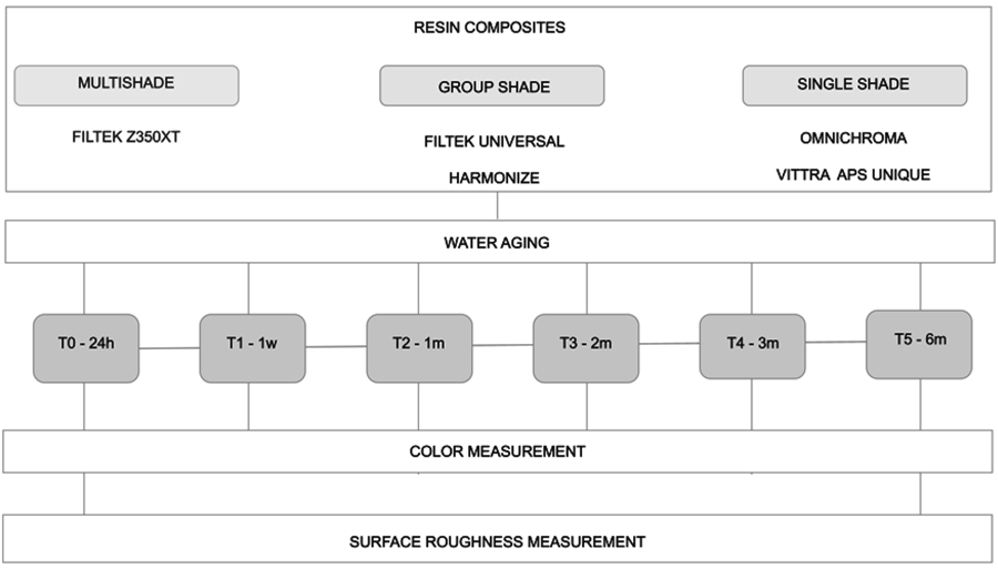
This in-vitro study evaluated the impact of water aging on color stability and surface roughness of different resin composites. Two single-shaded resin composites - Omnichroma (OMNI), Tokuyama Dental, Tokyo, Japan and Vittra APS Unique (UNI), FGM, Joinville, SC, Brazil, and two group-shaded resin composites - Harmonize (HAR), Kerr Dental, Orange, CA, USA; FiltekTM Universal (FIL), 3M ESPE, St. Paul, MN, USA, were tested. A conventional nanhybrid multi-shaded resin composite was considered as a control group FiltekTM Z350XT (Z350), 3M ESPE, St. Paul; MN, USA. The resin composites, manufacturer, composition, shades, and batch number are presented in Table 1. The experimental design, group distribution and different water aging time points are shown in Figure 1.
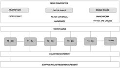
The number of specimens for color difference and surface roughness evaluation was estimated by using the Open Source Epidemiologic Statistics for Public Health, version 3.01 (free open source software). The confidence interval of 95% and power of 80% were used, resulting in a minimum sample size of n=4 for each group for both the surface roughness and color stability evaluation. The mean values of the CIEDE2000 color difference and standard deviations after aging of Omnichroma and Vittra Unique (ΔE00 1.5 (0.3) and ΔE00 0.9 (0.3)) and mean values of surface roughness of Omnichroma before and after water aging (0.12 (0.06) and 0.303 (0.10)) reported previously15,16 were used for sample calculation.
Specimen preparation
Specimens were prepared by one trained operator using a cylindrical metallic device with 8.0-mm diameter and 2.0-mm thickness (n=5). The resin composite was placed in one increment and the upper and lower surfaces were covered with polyester strips, and a glass plate was applied before light activation in order to standardize the top surface of the specimens, to ensure that the excess of resin composite was extruded, and to obtain a flat and smooth surface. Each specimen was light-cured for 40 seconds using a light-emitting-diode (LED; Bluephase, Ivoclar Vivadent, Schaan, Liechtenstein) with 1000 mW/cm2irradiance.
Then, finishing was executed by the same operator, using coarse, medium and fine polishing discs and a low-speed hand piece with mild hand pressure. Each specimen was held and polished for 10 seconds, turned 90 degrees, then polished for another 10 seconds for each sequential polishing disc (coarse, medium and fine). The polishing discs were replaced after every 5 specimens. The specimens were stored in distilled water for 6 months at 37°C. During this period, the water was replaced weekly.
Color measurement
The color of each resin composite specimen was measured using a calibrated reflectance spectrophotometer (SP60, EX-Rite/Grand Rapid, MI, USA), over a white background (L* = 91.85, a* = -1.01, b* = 6.71). CIE 2o Standard Observer and CIE D65 Standard Illuminant were used. The illuminating/measuring geometry corresponded to CIE diffuse/8o. CIE L*a*b*. Color coordinates were measured at six time points: 24 hours of water aging (T0-Baseline), 7 days of water aging (T1), 1 month of water aging (T2), 2 months of water aging (T3), 3 months of water aging (T4), 6 months of water aging (T5). The color of each specimen was measured in triplicate by the same operator, with similar conditions of illumination and room temperature.
The CIEDE2000 color difference (ΔE00) was calculated between the CIEL*a*b* coordinates of the baseline measurement (T0) and each time-point (T1, T2, T3, T4 and T5), according to the following formula17:
Surface Roughness
Surface roughness (Ra) was measured using a rugosimeter (Suftest SJ – 201P, Mitutoyo, Tokyo, Japan) with a diamond tip of 5 μm in diameter, angled at 90o. A total of twelve measurements (Ra, μm) per specimen were performed: six in the x-axis and six in the y-axis. The cut-off value and measurement speed were 0.8 mm and 0.5 mm/s, respectively. The surface roughness values were considered from the arithmetic means between the peaks and valleys. These values were obtained in 2 stages, after 24 h of immersion in water (T0) and after 6 months (T5) of immersion.
Statistical analyses
The differences of CIEL*a*b* color coordinates among the resin composites was analyzed using one-way ANOVA followed by Tukey’s post-hoc test (level of significance p < 0.05). The difference among the water exposure time points (T1-T0, T0-T2, T0-T3, T0-T4, and T0-T5) for each resin composite was analyzed using ANOVA with repeated measures (ANOVA-RM) and subsequently Bonferroni’s post-hoc test (level of significance p < 0.05). Color differences were interpreted based on the 50:50% perceptibility (PT00 = 0.8) and 50:50% acceptability (AT00 = 1.8) thresholds18. In addition, AT00 ratings were used for interpreting color differences as follows: Excellent match - EM (∆E ≤ 0.8), Acceptable match - AM) (0.8 < ∆E ≤ 1.8), Moderately unacceptable mismatch - MU ( 1.8 < ∆E ≤ 3.6), Clearly Unacceptable mismatch - CU (3.6 < ∆E ≤ 5.4) and Extremely unacceptable mismatch - EU ( ∆E > 5.4)19.
Surface roughness differences among the resin composites were analyzed using ANOVA, followed by Tukey’s post hoc test. The difference between time-points within each resin composite was examined through repeated measures ANOVA. Surface roughness was additionally interpreted using the roughness parameter for plaque retention (Ra=0.2μm)13. The correlation between ΔE00 and roughness was analyzed using the Pearson correlation test.
Results
Table 2 presents the differences in CIEL*a*b* color coordinates among the resin composites and evaluation time periods. Significant differences among the resin composites were shown for all color coordinates, except FIL and HAR at T0 for coordinates a* (p=0.53) and b* (p=0.195), and Z350 and FIL at T4 for color coordinate a* (p=0.536). Notably, HAR was the only resin that presented similar mean L*, a* and b* values across all evaluation periods: L* - p=0.706 (T0), p=0.271 (T1), p= 0.516 (T2), p=0.501 (T3), p=0.405 (T4), p=0.324 (T5); a* - p=0.366 (T0), p=0.291 (T1), p=0.139 (T2), p=0.312 (T3), p=0.256 (T4), p=0.2654 (T5), and b* - p=1.312 (T0), p=0.805 (T1), p=0.609 (T2), p=0.695 (T3), p=0.517 (T4), p=0.789 (T5).
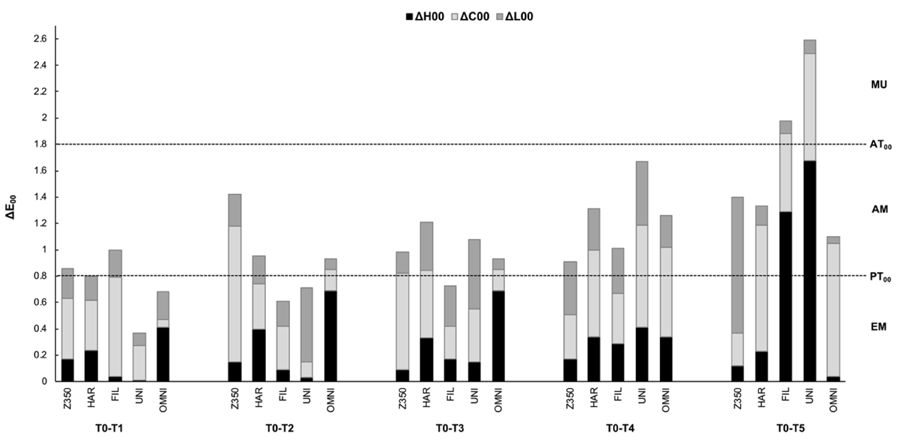
The mean color difference and the 50:50% PT00 and AT00 visual color thresholds and their respective ratings are shown in figure 2. The color difference between the baseline (T0) and the respective time points remained within acceptable limits for all resin composites until time point T4 (3 months). However, at time point T5 (6 months), FIL and UNI presented mean ∆E00 values above the visual acceptability threshold.
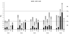
The total color differences were predominantly influenced by ΔC00, at T4 (3 months), for all resin composites. However, at T5 (6 months), ΔC00 had a predominant impact on the total color difference only for HAR and OMNI. For FIL and UNI, total color difference was more influenced by ΔH00, and for Z350, by ΔL00 at T5.
Table 3 presents the comparison of surface roughness among the resin composites across different time-points. The resin composites showed similar mean surface roughness values at T0 (p=0.20). However, after 6 months (T5), significant differences were observed among the tested resins (p=0.008), FIL presented significantly lower surface roughness compared to HAR (p=0.022). At T0, all resin composites, except HAR, presented mean surface roughness values below Ra=0.2μm. However, after 6 months, only the FIL maintained mean values below Ra=0.2μm. Over the 6-month evaluation period, no correlation was observed between color difference and surface roughness (R=0.057, P=0.788).
Discussion
Color stability is essential for maintaining the longevity of esthetic restorations, as color changes can lead to patient dissatisfaction, premature restoration replacement and clinical failure10. In addition to color discrepancies between the tooth and the resin composite restoration, alterations in surface roughness also affect the appearance of restorations. Smoother surfaces tend to reflect more light, while rougher surfaces may appear more opaque, being more susceptible to extrinsic staining12.
Resin composites with color adjustment potential were developed to achieve proper color matching with adjacent tooth structures using the structural color concept, differing from traditional resin composites that rely on the addition of pigments to provide multiple shade options. These composites offer versatility by simplifying the shade selection process, improving color matching, and reducing the number of resin composites required in dental practice to cover all 16 VITA shade colors plus bleach shades5,6.
Composites with color adjustment potential are available in two categories: single-shade and group-shade. Single-shade resin composites use structural color technology without the addition of any pigments 5,6, while group-shade resins, such as HAR and FIL, offer a limited range of shades, each designed to cover a specific group of shade tabs of the VITA Classic shade guide5. The color adjustment potential of the tested composites (OMNI, UNI, FIL, HAR) has been evaluated in previous studies, which have reported successful outcomes in terms of color matching with surrounding dental structures for both single-shade and group-shade composites3,5,20,21.
Given the recent introduction of resin composites with color adjustment potential, there is currently limited research on their long-term performance6,16,22. Longitudinal clinical trials and laboratory studies that simulate the oral environment are crucial for assessing these factors over time. Various methodologies are used to simulate the challenging conditions that composite materials may experience in the oral environment. These include water aging, artificial accelerated aging22, staining, immersion in different media16, simulated tooth brushing, and thermal and mechanical cycling23.
The present study was designed to evaluate the influence of prolonged water aging on the color stability and surface roughness of resins with color adjustment potential, rather than directly assessing the impact of aging on the color adjustment potential itself. Although further studies specifically focused on the direct impact of aging on the color adjustment potential of these materials would be valuable, analyzing color stability over time, as conducted in the present study, indirectly provides insights into the long-term color compatibility of these materials. This approach assumes that any visually perceived alterations in the initial color after aging could potentially affect the materials’ ability to maintain a satisfactory color match with dental structures in a clinical scenario. Therefore, our findings contribute to understanding the durability of color stability under aging conditions, which is relevant for predicting the longevity and esthetic outcomes of restorations.
In general, the tested resin composites showed differences in the CIEL*a*b* color coordinates after water exposure. The CIEDE2000 color difference between baseline and after three months of water aging (T4) exceeded the visual perceptibility threshold in all tested resins, thus confirming the first hypothesis of the study. Concerning surface roughness, there were no statistically significant differences observed between the time periods for most resin composites. However, when considering the bacterial plaque retention threshold (0.2µm)13, after six months of water exposure, the majority of resins demonstrated surface roughness values surpassing the established limit, therefore the second hypothesis of the study was partially accepted.
For precise color measurements, instrumental methods are preferred over visual assessments due to their objectivity and accuracy, thus eliminating the subjectivity inherent of visual assessments12. In this study, color was assessed using a spectrophotometer, an instrument that provides precise and repeatable measurements24. Furthermore, interpreting mean color difference results based on perceptibility and acceptability thresholds18 is crucial for determining the clinical relevance of numerical values19.
In this study, the color difference was evaluated based on the 50:50% perceptibility (PT00 = 0.8) and acceptability (AT00 = 1.8) thresholds. At three months of water aging (T4), all resin composites exhibited mean color difference values above 0.8 ∆E00 units, yet still remaining within the visual acceptability threshold (0.8 < ∆E00 ≤ 1.8). However, after 6 months of water aging (T5), two resin composites (FIL and UNI) showed mean color difference values above 1.8 units, surpassing the acceptability threshold, with mean color difference values classified as moderately unacceptable (1.8 < ∆E00 ≤ 3.6). These findings are consistent with a prior study on prolonged water aging of resin composites25, which also demonstrated that color stability was impacted by water exposure, resulting in unacceptable color mismatches after 6 months25.
Chroma plays an important role in color perception, significantly affecting how color differences are perceived. Samples with higher chroma levels present greater challenges for color matching acceptability compared to those with medium or low chroma26. In this study, after three months, chromatic changes became more pronounced, primarily due to variations in chroma for all tested resin composites. This finding contrasts with a previous study that identified luminosity as the primary component responsible for total color difference after prolonged water aging25. The discrepancy between the findings may be attributed to differences in research methodologies, the composition of the tested resin composites, and the resin composite placement techniques, as the layering concept was assessed in the previous study.
In addition to considering the perceptibility threshold for interpreting color difference outcomes, it is important to provide clinical relevance to the surface roughness findings of this study by also considering the surface roughness thresholds for bacterial plaque retention13 and roughness detection by patients27. The 0.2 μm threshold for Ra, supported by the theory of bacterial adhesion and retention13, indicates that Ra values exceeding this threshold are associated to increased biofilm accumulation. Moreover, a roughness value of 0.28 μm can be perceived by the tip of the tongue27. In the present study, all tested composite resins, except for FIL, presented surface roughness values above the bacterial retention threshold after six months of water aging. In addition to promoting bacterial plaque accumulation, rough surfaces may also be detected by patients, leading to discomfort and favoring extrinsic staining12,27.
Based on the results of the present study, water aging influenced the color stability of the resin composites. Differences in the CIELab color coordinates were observed among the resin composites and at different time points. After six months, both FIL and UNI exhibited a moderately unacceptable color change, while the other tested composites remained within the acceptability threshold throughout all time points.
The intrinsic discoloration of polymer-based materials may be influenced by their monomer composition, filler content and composition, resin-matrix volume, resin-matrix debonding and alterations of the resin matrix interface28,29. Analyzing the monomer composition of the tested composites revealed a similar matrix composition among most materials, which predominantly consist of a mixture of Bis-GMA, TEGDMA, and UDMA. Notably, only FIL presents a distinct composition, described by the manufacturer as a novel formulation containing AUDMA, AFM, Diurethane-DMA, and 1,12-dodecane-DMA. However, the substitution of traditional monomers by this novel formulation did not result in improved color stability for FIL.
It is well established that monomers such as Bis-GMA and TEGDMA are more susceptible to water sorption12,30, while UDMA exhibits greater resistance to water sorption29. Nevertheless, despite the presence of UDMA in its composition, UNI, also demonstrated a higher degree of color change compared to the other tested composites after six months.
In addition to matrix composition, filler content and size may also influence water sorption. A higher percentage of particles and smaller filler sizes are associated with lower water sorption and solubility, contributing to better color stability and surface roughness28. Among the tested resins, FIL and UNI presented the lowest weight/volume ratio, which may have influenced their reduced long-term color stability.
The FIL group presented the lowest surface roughness mean values among all tested materials. However, it was the only group that exhibited a significant increase in surface roughness after six months of water aging. Nonetheless, FIL remained the only group that maintained surface roughness below the plaque retention threshold after six months. This outcome may be attributed to the size and distribution of the fillers. Regarding filler size, both FIL and Z350 contain nanoparticles of 20 nm silica and 4–11 nm zirconia, aggregated into silica/zirconia clusters. In contrast, HAR exhibits filler sizes ranging from 5 to 400 nm, while OMNI contains fillers of approximately 260 nm. The manufacturer does not specify the filler size for UNI.
While the correlation between color difference and surface roughness has been established previously16, this study was unable to establish a correlation between these properties. The discrepancies in the results may be attributed to methodological differences, such as water exposure duration16, types of immersion solutions, pH of the immersion solutions, staining protocols, the application of artificial accelerated aging (AAA)15 and thermal cycling6. Additionally, a rigorous polishing protocol was applied, resulting in low mean surface roughness values at baseline. Moreover, surfaces lacking proper polishing reflect less light and gloss, leading to a greater impact on the color of the restorations and accelerating surface degradation12.
Limitations of this study are particularly associated to in vitro models. In the oral environment color and surface roughness can be influenced by many factors. The aging simulation relied exclusively on water exposure; other factors that could have potentially impact the study outcomes, such as artificial saliva, pH cycling, simulated tooth brushing, and thermal and mechanical cycling, were not assessed. Additionally, accelerated artificial ageing (AAA) application and visual assessments were not conducted. AAA has the potential to simulate many years of clinical service22, while visual assessment could offer relevant information into the perception of color outcomes. Future studies should address these limitations to provide more comprehensive information on both properties.
Nevertheless, despite its limitations, this study contributes to addressing gaps within this category of composites. The evaluation of two critical properties - color stability and surface roughness - while also assessing several commercially available composites with color adjustment potential are strengths of the study. Furthermore, the prolonged water immersion period of six months, which exceeds the average immersion time used in previous studies (typically 12 to 14 days)6,16,22, explores the impact of long-term water exposure. Clinicians should be attentive to the color stability and surface characteristics of composite resins with color adjustment potential, as these properties may change over time, potentially leading to esthetic alterations and restoration failure10.
All resin composites presented color differences within the limits of visual acceptability for up to three months of water aging, indicating color stability until this time point. However, after six months of water aging, surface roughness exceeded the plaque retention threshold for all resin composites except FIL. The color difference between the baseline and the respective time points was above visual acceptability threshold for FIL and UNI. No correlation between surface roughness and color differences was demonstrated.
References
-
1 Trifkovic B, Powers JM, Paravina RD. Color adjustment potential of resin composites. Clin Oral Investig. 2018 Apr;22(3):1601-7. doi: 10.1007/s00784-017-2260-6. Epub 2017 Oct 25.
» https://doi.org/10.1007/s00784-017-2260-6 -
2 Abdelraouf RM, Habib NA. Color-matching and blending-effect of universal shade bulk-fill-resin-composite in resin-composite-models and natural teeth. Biomed Res Int. 2016;2016:4183432. doi: 10.1155/2016/4183432.
» https://doi.org/10.1155/2016/4183432 -
3 Iyer RS, Babani VR, Yaman P, Dennison J. Color match using instrumental and visual methods for single, group, and multi-shade composite resins. J Esthet Restor Dent. 2021 Mar;33(2):394-400. doi: 10.1111/jerd.12621. Epub 2020 Aug 25.
» https://doi.org/10.1111/jerd.12621 -
4 Ismail EH, Paravina RD. Color adjustment potential of resin composites: optical illusion or physical reality, a comprehensive overview. J Esthet Restor Dent. 2022 Jan;34(1):42-54. doi: 10.1111/jerd.12843. Epub 2021 Nov 29.
» https://doi.org/10.1111/jerd.12843 -
5 Lucena C, Ruiz-López J, Pulgar R, Della Bona A, Pérez MM. Optical behavior of one-shaded resin-based composites. Dent Mater. 2021 May;37(5):840-8. doi: 10.1016/j.dental.2021.02.011.
» https://doi.org/10.1016/j.dental.2021.02.011 -
6 El-Rashidy AA, Abdelraouf RM, Habib NA. Effect of two artificial aging protocols on color and gloss of single-shade versus multi-shade resin composites. BMC Oral Health. 2022 Aug;22(1):321. doi: 10.1186/s12903-022-02351-7.
» https://doi.org/10.1186/s12903-022-02351-7 -
7 Korkut B, Tarçin B, Atali PY, Özcan M. Introduction of a new classification for resin composites with enhanced color adjustment potential. Curr Oral Heal Reports 2023;10:223-32. doi: 10.1007/s40496-023-00351-2.
» https://doi.org/10.1007/s40496-023-00351-2 -
8 Mizutani K, Takamizawa T, Ishii R, Shibasaki S, Kurokawa H, Suzuki M, et al. Flexural properties and polished surface characteristics of a structural colored resin composite. Oper Dent. 2021 May;46(3):E117-E131. doi: 10.2341/20-154-L.
» https://doi.org/10.2341/20-154-L -
9 Sedrez-Porto JA, Münchow EA, Brondani LP, Cenci MS, Pereira-Cenci T. Effects of modeling liquid/resin and polishing on the color change of resin composite. Braz Oral Res. 2016 Aug;30(1):S1806-83242016000100275. doi: 10.1590/1807-3107BOR-2016.vol30.0088.
» https://doi.org/10.1590/1807-3107BOR-2016.vol30.0088 -
10 Demarco FF, Collares K, Coelho-de-Souza FH, Correa MB, Cenci MS, Moraes RR, et al. Anterior composite restorations: a systematic review on long-term survival and reasons for failure. Dent Mater. 2015 Oct;31(10):1214-24. doi: 10.1016/j.dental.2015.07.005.
» https://doi.org/10.1016/j.dental.2015.07.005 -
11 Prodan DA, Gasparik C, Mada DC, Miclaus V, Baciut M, Dudea D. Influence of opacity on the color stability of a nanocomposite. Clin Oral Investig. 2015 May;19(4):867-75. doi: 10.1007/s00784-014-1315-1. Epub 2014 Sep 13.
» https://doi.org/10.1007/s00784-014-1315-1 - 12 Alkhadim YK, Hulbah MJ, Nassar HM. Color shift, color stability, and post-polishing surface roughness of esthetic resin composites. Materials (Basel). 2020 Mar;13(6):1376. doi: 10.3390/ma13061376.
-
13 Bollen CM, Lambrechts P, Quirynen M. Comparison of surface roughness of oral hard materials to the threshold surface roughness for bacterial plaque retention: a review of the literature. Dent Mater. 1997 Jul;13(4):258-69. doi: 10.1016/s0109-5641(97)80038-3.
» https://doi.org/10.1016/s0109-5641(97)80038-3 -
14 Rodrigues CS, Nora BD, Mallmann A, May LG, Jacques LB. Repolishing resin composites after bleaching treatments: effects on color stability and smoothness. Oper Dent. 2019 Jan-Feb;44(1):54-64. doi: 10.2341/17-107-L. Epub 2018 Jun 1.
» https://doi.org/10.2341/17-107-L -
15 Fidan M, Yagci Ö. Effect of aging and fiber-reinforcement on color stability, translucency, and microhardness of single-shade resin composites versus multi-shade resin composite. J Esthet Restor Dent. 2024 Apr;36(4):632-42. doi: 10.1111/jerd.13125. Epub 2023 Aug 25.
» https://doi.org/10.1111/jerd.13125 -
16 Rohym S, Tawfeek HEM, Kamh R. Effect of coffee on color stability and surface roughness of newly introduced single shade resin composite materials. BMC Oral Health. 2023 Apr;23(1):236. doi: 10.1186/s12903-023-02942-y.
» https://doi.org/10.1186/s12903-023-02942-y - 17 Carter EC, Schanda JD, Hirschler R, Jost S, Luo MR, Melgosa M, et al. Technical report: colorimetry 4 th edition. Vienna: Commission Internationale de l'eclairage, 2018. 113 p. CIE 015:2018.
-
18 Paravina RD, Ghinea R, Herrera LJ, Bona AD, Igiel C, Linninger M, et al. Color difference thresholds in dentistry. J Esthet Restor Dent. 2015 Mar-Apr;27 Suppl 1:S1-9. doi: 10.1111/jerd.12149.
» https://doi.org/10.1111/jerd.12149 -
19 Paravina RD, Pérez MM, Ghinea R. Acceptability and perceptibility thresholds in dentistry: a comprehensive review of clinical and research applications. J Esthet Restor Dent. 2019 Mar;31(2):103-12. doi: 10.1111/jerd.12465.
» https://doi.org/10.1111/jerd.12465 -
20 Pereira Sanchez N, Powers JM, Paravina RD. Instrumental and visual evaluation of the color adjustment potential of resin composites. J Esthet Restor Dent. 2019 Sep;31(5):465-70. doi: 10.1111/jerd.12488.
» https://doi.org/10.1111/jerd.12488 -
21 Durand LB, Ruiz-López J, Perez BG, Ionescu AM, Carrillo-Pérez F, Ghinea R, et al. Color, lightness, chroma, hue, and translucency adjustment potential of resin composites using CIEDE2000 color difference formula. J Esthet Restor Dent. 2021 Sep;33(6):836-43. doi: 10.1111/jerd.12689. Epub 2020 Dec 7.
» https://doi.org/10.1111/jerd.12689 -
22 Sensi L, Winkler C, Geraldeli S. Accelerated aging effects on color stability of potentially color adjusting resin-based composites. Oper Dent. 2021 Mar;46(2):188-96. doi: 10.2341/20-099-L.
» https://doi.org/10.2341/20-099-L -
23 El-Rashidy AA, Shaalan O, Abdelraouf RM, Habib NA. Effect of immersion and thermocycling in different beverages on the surface roughness of single- and multi-shade resin composites. BMC Oral Health. 2023 Jun;23(1):367. doi: 10.1186/s12903-023-03069-w.
» https://doi.org/10.1186/s12903-023-03069-w -
24 Yang H, Ding S, Jiang P. Precision analysis of color measurement by different spectrophotometers. Adv. Eng. Res. 2015 Aug27:983-6. doi: 10.2991/ic3me-15.2015.190.
» https://doi.org/10.2991/ic3me-15.2015.190 -
25 Perez BG, Pérez MM, Ruiz-López J, Gaidarji B, Durand LB. Effect of layering strategy and prolonged water aging on masking ability of composite resins. J Prosthet Dent. 2023 Nov;130(5):745.e1-745.e8. doi: 10.1016/j.prosdent.2023.08.031.
» https://doi.org/10.1016/j.prosdent.2023.08.031 -
26 Tejada-Casado M, Pérez MM, Della Bona A, Lübbe H, Ghinea R, Herrera LJ. Chroma-dependence of CIEDE2000 acceptability thresholds for dentistry. J Esthet Restor Dent. 2024 Mar;36(3):469-76. doi: 10.1111/jerd.13153. Epub 2023 Oct 20.
» https://doi.org/10.1111/jerd.13153 -
27 Jones CS, Billington RW, Pearson GJ. The in vivo perception of roughness of restorations. Br Dent J. 2004 Jan;196(1):42-5; discussion 31. doi: 10.1038/sj.bdj.4810881.
» https://doi.org/10.1038/sj.bdj.4810881 -
28 Mazzitelli C, Paolone G, Sabbagh J, Scotti N, Vichi A. Color stability of resin cements after water aging. Polymers (Basel). 2023 Jan;15(3):655. doi: 10.3390/polym15030655.
» https://doi.org/10.3390/polym15030655 -
29 Barutcigil Ç, Yildiz M. Intrinsic and extrinsic discoloration of dimethacrylate and silorane based composites. J Dent. 2012 Jul;40 Suppl 1:e57-63. doi: 10.1016/j.jdent.2011.12.017.
» https://doi.org/10.1016/j.jdent.2011.12.017 -
30 Ardu S, Duc O, Di Bella E, Krejci I. Color stability of recent composite resins. Odontology. 2017 Jan;105(1):29-35. doi: 10.1007/s10266-016-0234-9. Epub 2016 Feb 18.
» https://doi.org/10.1007/s10266-016-0234-9
-
Data Availability:
Datasets related to this article will be available upon request to the corresponding author.
Edited by
-
Editor:
Dr. Altair A. Del Bel Cury
Datasets related to this article will be available upon request to the corresponding author.

 **
Abbreviations: PT
00
= 0.8; AT
00
= 1.8; EM - Excellent match ΔE
00
≤ 0.8; AM – Acceptable match 0.8 < ΔE
00
≤ 1.8; MU – Moderately Unacceptable mismatch 1.8 < ΔE
00
≤ 3.6.
**
Abbreviations: Z330 – Filtek Z350 XT; HAR – Harmonize; Fil – Filtek Universal; UNI – Vittra APS Unique; OMNI – Ominichroma; T0 – baseline; T1 – 7 days; T2 – 1 month; T3 – 2 months; T4 – 3 months and T5 – 6 months.
**
Abbreviations: PT
00
= 0.8; AT
00
= 1.8; EM - Excellent match ΔE
00
≤ 0.8; AM – Acceptable match 0.8 < ΔE
00
≤ 1.8; MU – Moderately Unacceptable mismatch 1.8 < ΔE
00
≤ 3.6.
**
Abbreviations: Z330 – Filtek Z350 XT; HAR – Harmonize; Fil – Filtek Universal; UNI – Vittra APS Unique; OMNI – Ominichroma; T0 – baseline; T1 – 7 days; T2 – 1 month; T3 – 2 months; T4 – 3 months and T5 – 6 months.