Immunoneurogenic pulp tissue response: a narrative review of the molecular and cellular mechanisms related to pulp tissue regeneration
Abstract
Aim to evaluate advances in dental pulp regeneration and challenges for regenerative endodontic therapies, particularly nervous tissues and their components.
Methodology for the analysis of this narrative review, a search for relevant articles was conducted across various scientific databases, including PubMed, Scopus, Web of Science, and Embase, covering the period from 2000 to 2023. The inclusion criteria were set to primarily encompass original research articles, systematic reviews, and meta-analyses that demonstrated relevance to the topics of interest.
Results neurogenesis occurs during pulp repair but is impaired by ongoing lesions due to reduced vascular supply. The results suggest that while various therapeutic approaches using biomaterials are being explored, efficient neural regeneration though achieved in some studies remains a significant challenge.
Conclusion Several studies have demonstrated the return of pulp sensitivity and the formation of neural tissue. However, despite growing interest and promising results, challenges remain to be addressed, the variability in individual responses and the complexity of tissue revitalization mechanisms, which are not yet fully understood.
Keywords
Inflammation; Dental pulp; Immune system; Tissue scaffolds
Introduction
The dental pulp is a loose connective tissue located within the root canals, surrounded by dentin walls1,2. It is highly vascularized and contains various cells such as undifferentiated mesenchymal cells, fibroblasts, odontoblasts, vascular and defense cells, blood vessels, and sensory neurons. Among these cells, fibroblasts play a crucial role in pulp regeneration by inducing pluripotent stem cell reprogramming and regulating cellular hypoxia through factors like VEGF and PDGF2,3. Additionally, odontoblasts, located at the dentin interface, are responsible for the formation of primary and secondary dentin, contributing to the structural integrity of the pulp4,5.
Pulp tissue also has a well-organized sensory system. Most of its nerve fibers are unmyelinated C fibers originating from the trigeminal nerve, while the remaining fibers come from myelinated sympathetic ganglia1. These neurons release neuropeptides such as substance P (SP), neurokinin A (NKA), and calcitonin gene related peptide (CGRP), which act as neurotransmitters and play roles in the defense system and tissue regeneration3,6. Odontogenic pain arises when the physical barriers of the tooth are compromised, leading to the release of inflammatory mediators such as histamine and prostaglandins, causing vasodilation and initial pain, often diagnosed as hyperemia7,8.
When the infectious process persists, pulp inflammation can progress to pulpitis and eventually pulp necrosis9. This occurs when defense cells can no longer control the infection, leading to the destruction of odontoblasts and further stimulation of afferent sensory neurons10. The resulting release of neuropeptides and activation of receptors like TRP channels contribute to the pain and tissue degeneration associated with pulp disease. These processes highlight the complex mechanisms involved in pulp defense and regeneration11,12.
A key aspect of the neurogenic pain mechanism is the activation of ion channels such as ASIC and PIEZO channels, which contribute to the perception of touch and temperature13,14. TRP channels, specifically, play a significant role in the acute temperature sensitivity of teeth. Understanding the activation and modulation of these receptors is essential for developing new regenerative therapies aimed at improving sensory function and overall dental tissue health1,12.
Then, this review highlights advances in neurogenesis and tissue engineering technologies that are paving the way for innovative methods to regenerate dental tissues15,16. Emerging techniques, such as the controlled assembly of cells and biomaterials, show great potential for clinical endodontics17,18. The integration of artificial intelligence, tissue engineering, and gene therapy could lead to personalized treatments, revolutionizing dental practices and improving patient quality of life19.
Methodology
This narrative review included 114 papers using PubMed, Scopus, Web of Science, and Embase databases, covering the period from 2000 to 2023. The search strategy was designed to capture a wide range of studies at the intersection of advanced biomedical engineering and dental science20. Keywords such as “inflammation”, “dental nerve”, “immune system”, and “scaffolds” were used in various combinations to ensure a comprehensive retrieval of articles.
The inclusion criteria were set to primarily encompass original research articles, systematic reviews, and meta-analyses that demonstrated relevance to the topics of interest20. The selected articles were further assessed for quality and relevance, particularly in terms of their findings’ significance for advancing the understanding of topics such as neurogenesis, tissue engineering, and clinical applications in endodontics. Studies from various geographic regions and research contexts were included to provide a global perspective on advancements in these areas. As an exclusion criterion, articles that were not available in English were disregarded, ensuring consistency in data analysis.
Results
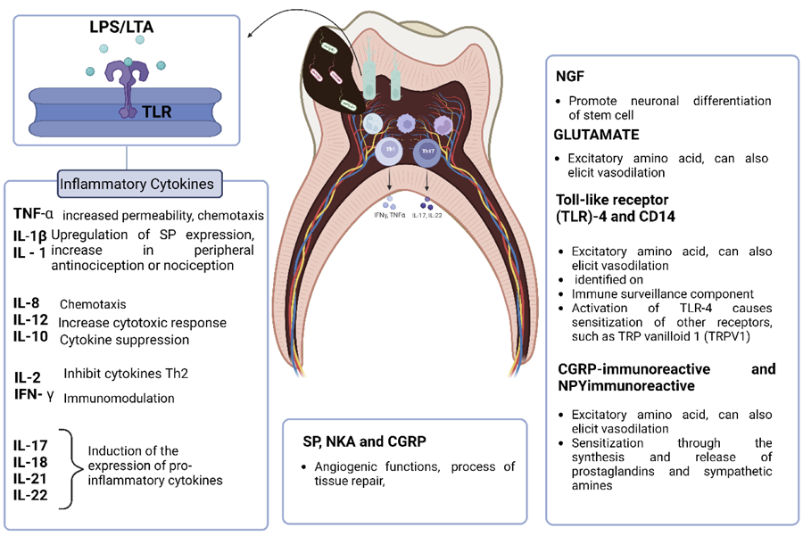
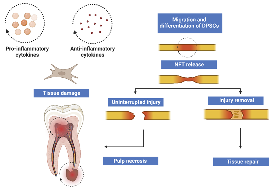
A study was conducted to evaluate the interaction between the immune and nervous systems of the dental pulp during pulp pathologies, demonstrating the response of the dentin-pulp complex to microbial, traumatic, or iatrogenic insults. This study revealed that such insults involve complex defense mechanisms and directly affect the nervous tissue (Figure 1). Neurogenesis was observed during pulp repair; however, its occurrence was significantly reduced in cases of continuous lesion progression due to diminished vascular supply, modification of cellular niches, and obliteration of the pulp chamber, which compromises regenerative capacity and leads to pulp necrosis (Figure 2).
Illustrative scheme of pulp tissue after bacterial invasion and detection by odontoblasts and immune cells. Subsequently, inflammatory cytokines, neuropeptides, proteins, and growth factors are released and induce tissue repair or damage. Inflammation, tissue damage and elimination of infectious bacteria are even more events. TLR: toll like receptor; Th: T helper; LPS: lipopolysaccharides; LTA: lipoteichoic acid; NKA: neurokinin A; CGRP: peptide related to the calcitonin gene; NPY: neuropeptide Y; SP: substance P; IL: interleukins; NGF: nerve growth factor; TNF: tumor necrosis factor. Created by BioRender.com.
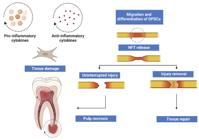
Tissue drainage and induction of inflammatory cytokines by fibroblasts to the affected region. After the injurious stimulus, pulpal stern cells migrate and act in the nervous tissue repair process. Neurotrophic factor (NTF) has the unique potential to support neuronal survival and enhance neuronal function in the injured and diseased nervous system. Removing the stimulus and action of NFTs, together with immune cells, promotes nerve tissue repair. Created by BioRender.com.
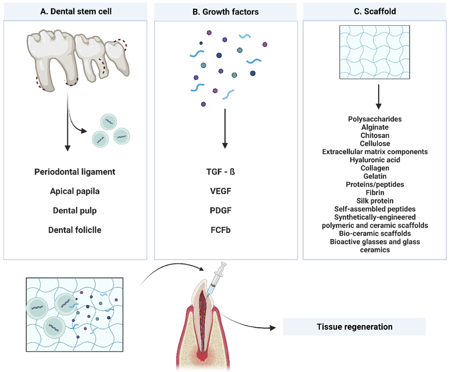
Various studies have explored several therapeutic approaches using cells and natural, biological, and synthetic biomaterials. In regenerative dentistry, scaffolds are developed with biomaterials that enhance the stability and reproducibility of tissue engineering therapies, providing a controlled environment designed for specific applications (Figure 3). Despite advancements in regeneration with the formation of tissues and blood vessels, neural regeneration remains limited and under-documented. Efficient neural regeneration in regenerative treatments continues to be a significant challenge, as evidenced by the reviewed studies (Table 1).
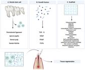
Regenerative potential of stem cells and the use of biomaterials in tissue regeneration. (A) Dental stem cells. (B) Growth factors that regulate cellular events and are important in tissue repair, including transforming growth factor beta (TGF-β), vascular endothelial growth factor (VEGF), platelet-derived growth factor (PDGF), and basic fibroblast growth factor (bFGF). (C) Scaffolds that support cell growth. Biomaterials associated with dental stem cells and growth factors present regenerative potential, enabling tissue formation similar to the original tissue. Created with BioRender.com.
Discussion
Relationship between the dental pulp immune and nervous systems during dental pulp pathologies
The odontoblasts present at the dentin-pulp interface act as an initial protective barrier through the activation of Pattern Recognition Receptors (PRRs) present on the cell surface, by recognizing Pathogen Associated Molecular Patterns (PAMPs)35. The membrane proteins of the PRRs present in odontoblast cells belong to the Toll-like receptor (TLR) family36. These membrane proteins recognize peptidoglycans and lipoteichoic acid (LTA) from Gram-positive bacteria and lipopolysaccharides (LPS) mainly from Gram-negative bacteria. This recognition leads to the activation of a pro-inflammatory response, including the production of cytokines and chemokines, nitric oxide, and eicosanoids37-39.
After the stimulus is detected by odontoblasts, a repair process begins in the dentin, which promotes the formation of tertiary dentin to block the entry of irritating agents into the pulp tissue. As the intensity of aggression increases, TGF-β1 and TGF-β3 growth factors are released by the odontoblasts, promoting the formation of reparative dentin and, consequently, calcifications of the pulp chamber40,41. The initial confrontation of the invasion of these pathogens also occurs through the production of nitric oxide (NO) and beta-defensins, in addition to the synthesis of pro-inflammatory chemokines such as CCL2, CXCL2, CXCL8, and CXCL1037,42.
Bacterial colonization during the progression of pulpal inflammation establishes a biofilm composed mostly of Gram-positive and Gram-negative bacteria, as well as fungi, archaea, and viruses12,43. A recent study evaluated different patterns of inflammatory cytokines induced by LPS from E. coli and LPS from P. gingivalis. These bacteria virulence factors significantly increased the expression of pro-inflammatory cytokines after 24h, including interleukins IL-1β, IL-6, tumor necrosis factor (TNF-α), and IL-8. The increase in the production of chemokines, cytokines, and defensins promotes cellular recruitment of leukocytes, neutrophils, macrophages, and dendritic cells, which initiate the innate immune response with the secretion of cytokines (ILs) and influence the adaptive immune response12,44,45.
This process occurs from the presentation of antigens by dendritic cells that activate B lymphocytes and CD4+ T lymphocytes or T helper 0 (Th0) cells, which differentiate into Th1, Th2, and Th17 cells, important regulators in the production of cytokines. Depending on the intensity of the stimulus, these cytokines can act both in the repair and the pathological process of pulp degradation and necrosis, together with matrix metalloproteinases (MMPs), which in uncontrolled production act in synergy with (ILs) in excessive tissue destruction9,44,46.
The inflammatory reaction when subodontoblastic levels are reached promotes a decrease in local blood flow, capable of suppressing the activity of nerve fibers that are located along the pulp tissue, promoting changes such as compression of nerve fibers and consequently induction of pain. These fibers are classified as type A fibers located in the most coronal region of the pulp and which present higher oxygen consumption and lower survival capacity when there are exacerbated inflammatory stimuli. On the other hand, type C fibers that are more resistant to hypoxia are located in the central portion of the pulp tissue27,42,47-49.
The type A fibers detect early stimuli in cases of reversible pulpitis with localized pain. Upon receiving stimuli, these fibers induce the production of neurotrophins and tissue neuropeptides, such as NGF (neuronal growth factor), CGRP (genetically related peptide to calcitonin), substance P, and neurokinin A, which help in inducing tissue repair, leading to angiogenesis, and production of tertiary dentin by odontoblasts. Type C fibers receive stimulation in the transition from innate to adaptive immune response, mediated by B and T lymphocytes, which becomes an irreversible symptomatic lymphocyte state. It is characterized by severe, intermittent pain; in addition, the ability to recover pulp tissue is lost because the robust stimulus is not removed. Consequently, there is an increase in blood flow permeability and a transient increase in interstitial tissue pressure that compromises pulp vitality. Studies indicate that the production of TNF, responsible for the process of dilation and increased permeability of blood vessels, decreases dramatically as the process of tissue necrosis occurs. This is because infected necrotic pulp tissue is not accessible to both innate and adaptive immune defense mechanisms49-51.
This neural network, together with fibroblasts, acts in tissue protection by recognizing stimuli that impair its functioning, acting in the secretion and recruitment of resident endothelial cells and stem cells involved in repair. Therefore, relatively low stimuli can contain the destructive advance of the inflammatory immune reaction and promote repair, which is possible by directly interfering with the cause of the problem. Chronic levels of infection can induce an irreversible destructive effect on the pulp tissue, due to the exacerbated recruitment of resident cells and the intense action of the immune response27,47,48.
Cells involved with the neurogenesis process after its destruction
Fibroblasts, which are the main cells of pulp tissue, secrete into the inflamed pulp important factors for the recruitment of stem cells. These cell types recruited during the inflammatory process are directly involved in repair. Various populations of progenitor cells, including dental pulp stem cells (DPSCs), migrate to injured pulp sites and differentiate into odontoblast-like cells during dentinogenesis and reparative neurogenesis. Neurogenesis acts on the formation of new neurons and involves stages of proliferation, migration, and differentiation of neuronal networks. For the neurogenic process to be initiated, the presence of neural stem cells (NSC) in the surrounding microenvironment is important16,52.
The process of neurogenesis occurs during the pulp repair process and is reduced when the injurious progress of pulp tissue is not stopped. This is because the decrease in vascular supply, modification of the niches and obliteration of the pulp chamber alter the regenerative capacity of stem cells. In this way, the ability of pulp tissue to recover if lost, promoting a decrease in local oxygen supply and necrosis of the pulp tissue53,54
In the later stage, necrosis extends beyond the apical foramen and into the periapical tissue, resulting in acute/chronic periodontitis. This lesion occurs because of exacerbated pulp inflammation. Consequently, functional interactions occur between the immune and nervous systems that play important roles in the progression of apical periodontitis. Sympathetic nerves inhibit the production of pro-inflammatory bone-resorbing cytokine IL-1α, which can increase the number of osteoclasts by slowing the progression of local bone resorption. In this process, there are changes in the level of innate immune cells and in neural changes which modulate the inflammatory response. As the density of sensory nerves peaks, tissue repair begins to occur55-58.
DPSCs have proved to be interesting for dental and non-dental therapeutic applications, at the level of neuronal repair, through the secretion of anti-inflammatory and pro-growth molecular signals, in addition to neurotrophic factors (NTFs). Thus, DPSCs promote a fundamental action in pulp regeneration, since the NTFs released by these cells also regulate the processes of dentinogenesis and repair. This repair process begins with the formation of the vascular network with the local circulatory system and the supply of oxygen and essential nutrients for the elimination of waste, which occurs mainly with the regulatory capacity of DPSCs in inducing platelet-derived growth factor β (PDGFRβ), α-smooth muscle actin (α-SMA), vascular endothelial growth factor (VEGF), platelet-derived growth factor (PDGF), monocyte chemotactic protein-1 (MCP-1), fibroblast growth factor base (bFGF) and endothelin 1 (EDN1)38,48.
DPSCs are characterized by the presence of surface markers that determine their properties. Among them are the markers STRO-1, CD29, CD44, CD73, CD90, CD105, CD146, CD166, CD271, CD34, CD117, OCT-3/4 and NANOG. DPSCs can differentiate into neural cells and glial cells, in addition to specific neuronal markers such as neuronal nuclei (NeuNq) that promote neuronal differentiation under neuronal induction conditions. DPSCs grown in neuronal inductive media containing growth factors such as glial cell line-derived neurotrophic factor (GDNF) and brain-derived neurotrophic factor (BDNF) are known to differentiate into neuron-like cells59,60.
Biotechnological opportunities to regenerate the nervous dental pulp tissue
The regeneration of damaged/lost biological tissues represents a significant challenge for tissue engineering and involves their restoration and return of biological functions. However, for this goal, three objectives must be achieved: elimination of symptoms and evidence of bone healing, increase in root wall thickness and in root length, and a positive response to vitality tests61-63. Scientific advances in tissue engineering enable the use of protocols that stimulate tissue regeneration, aiming to imitate the complexity of structures and restore their main functions. The incorporation of synthetic and biological scaffolds with signaling molecules and stem cells are models produced in laboratories that allow in vitro tissue reconstruction. This model, although much studied, still does not allow complete tissue mimicry in line with its specificity. The biggest challenge has been to create an ideal model that enables neural regeneration64-66.
Given this, more advanced technologies are emerging to reproduce an environment that develops the physiological functions of tissues. This is how bioprinting technologies appear, structures designed to mimic replacement tissues, cell assembly, and biomaterials in a controlled manner67-68.
Stem cells
Previous studies have provided strong evidence that human dental pulp is an accessible “niche” of stromal stem cells with potential differentiation into adipogenic, osteogenic, chondrogenic, myogenic, and neurogenic lineages without any stimulus for differentiation under basal conditions. It is also important to point out that stem cells from the pulp of permanent teeth represent less than 1% of the cells in the dental pulp. They can produce morphological tissue with functional characteristics like dental pulp, increasing clinical success in conservative endodontic therapies69,70.
Cells from the pulp tissue remaining from exfoliated primary teeth, SHEDs, were identified as a population of highly proliferative clonogenic cells capable of differentiating into a variety of cell types, including neural cells, adipocytes, and odontoblasts. After in vivo transplantation, SHEDs were found to be able to induce bone formation, generate dentin, and survive in the mouse brain along with the expression of neural markers71.
The apical papilla is known to be a source of odontoblasts for the pulp tissue, and these odontoblasts are responsible for dentinogenesis, playing an essential role in the root formation process. Studies have shown that apical papilla stem cells (SCAPs) have more significant potential for dentin regeneration than dental pulp stem cells (DPSCs), demonstrating higher levels of essential molecules that act in mediating cell proliferation and differentiation, in addition to expressing a wide variety of neurogenic markers, such as nestin and neurofilament M, after stimulation with a neurogenic medium. For this reason, undifferentiated cells from this tissue have been the focus of several studies in endodontics related to regenerative treatments. Therefore, it was concluded that SCAPs are like DPSCs, but a distinct source of potent dental stem/progenitor cells72,73. Another dental component is the periodontal ligament, which plays a vital role in physically supporting teeth and contributes to the nutrition, homeostasis, and regeneration of adjacent tissues. In addition, it is a niche of stem cells, and, given their specificity, these cells were named periodontal ligament stem cells (PDLSCs). According to the study, which proved the existence of stem cells in the periodontal ligament, they can be obtained by scraping the dental root and then expanded in vitro for future clinical application74,75.
These cells showed the ability to regenerate rudimentary periodontal ligament and cementum, but subsequent studies revealed that these cells also exhibit osteogenic, adipogenic, chondrogenic, and immunoregulatory/immunosuppressive properties. Given this, several analyses have already demonstrated that stem cells from the periodontal ligament are capable of regenerating periodontal tissues, both the ligament component (connective), as well as bone and cementum, including on implants – as revealed in a study carried out in rats – with possible applicability in humans; and diabetes treatment76-82.
Thus, stem cells derived from dental tissues have been studied in the treatment of neural injuries as well as neurodegenerative diseases due to their ability to regenerate neural tissue. In 2008, a group of researchers used stem cells from the pulp of human deciduous teeth injected locally into the muscles of puppies with muscular dystrophy (MD). After that, a significant clinical improvement in the dogs was observed; it was suggested, therefore, that this type of cell therapy could delay the progression of the dystrophic process by expressing immunoregulatory paracrine factors82,83.
For degenerative diseases such as Parkinson’s disease, one of the most common neurodegenerative pathologies caused by the loss of dopaminergic neurons, stem cells from the pulp of deciduous teeth, previously induced to neurogenic differentiation in vitro, reduced behavioral deficits after transplantation in Parkinsonian rats. Alzheimer’s disease is another disorder of interest in this area of research. Alzheimer’s is a progressive neurodegenerative disease characterized by a decline in cognitive ability and the formation of myeloid plaques in the brain. Pulp stem cells from deciduous teeth injected intranasally in vivo substantially improved cognitive function in mice with Alzheimer’s. Furthermore, the performance of SHEDs was superior to that of bone marrow stem cells in these animal models14,83,84.
A recent study has compared the neural protection capacity of stem cells from the pulp of deciduous teeth and stem cells from the pulp of permanent teeth with other postnatal stem cells. In adult rats with the spinal cord wholly transected, the cells resulted in the marked recovery of hindlimb locomotor functions. It should also be noted that human nerve pulp stem cells exhibit three central neuro regenerative activities – namely: the cells enhanced apoptosis induced by spinal cord secretion of neurons, astrocytes, and oligodendrocytes, which improved the preservation of neuronal filaments and myelin sheaths; promoted the inhibition of transected axons by directly inhibiting several axon growth inhibitors, including proteoglycans, chondroitin sulfate, and myelin-associated glycoprotein, through paracrine disruption; and replaced the lost cells by differentiating into mature oligodendrocytes under the conditions of spinal cord injury85
In this context, the neurogenic potential is already well established, mainly due to the ability of these cells to secrete multiple neurotrophins and which, at least in part, were responsible for stimulating neuroprotection, neurogenesis, and axonogenesis, both in vitro and in vivo for treating optic nerve injuries, and other studies with neural involvement13. DPSCs also performed better than bone marrow stem cells restoring neural function after intravitreal transplantation. It is believed, therefore, that these reported facts were due to the origin of these cells being the neural crest and, therefore, presenting neural markers. It is also possible that they are different from other already known types of stem cells, such as those from the bone marrow86.
Biomaterials and biomolecules
The ideal materials for dentine-pulp complex regeneration should be biocompatible and have clinical applicability. They should therefore allow adhesion to the cell surface and promote migration, differentiation, and cell function19,61,87,88. The number of nerve fibers in the regenerated pulp determines the degree of sensory function. Given this, neural regeneration is indispensable for maintaining the structure and function of the pulp tissue.
Recent animal studies have reported pulpal regeneration with vascularization and neural innervation; however, there was no significant response to clinical sensitivity test. Similar results were verified for some human patients, as only a few patients with post-treatment pulp sensitivity were reported. Another study, evaluating two dental elements submitted to a revascularization treatment, observed the presence of peripheral afferent neurons in the space of the necrotic canal, suggesting a solid indication of regeneration/repair. Furthermore, these elements responded positively to the sensitivity test, which indicates a possible regenerative success18,89-92.
Currently, the biggest challenge has been to find compatible biomaterials associated with stem cells and growth factors that promote regenerative success based on the formation of nerve fibers, complete root formation, absence of periapical lesions, and asymptomatic dental elements after treatment. Endodontics has evolved considerably, with a wide range of concepts and treatment options in this practice. The homing cell technique, for example, has been applied more frequently, followed by methods with natural scaffolds of hydrogels/collagen, PRF, and PRP. These scaffolds, in addition to the characteristics already mentioned, must be degradable after implantation and replaced by newly formed tissues. Degradation of the scaffold too quickly can compromise its cellular support function, while a degradation rate that is too slow can prevent the formation of new tissues93-97.
Natural materials such as fibrin, collagen, gelatin, and decellularized matrix offer a favorable environment for differentiation and biodegradability control. In in vitro studies, the results prove that these scaffolds allow significantly greater cell viability, proliferation, and migration. Recent studies have demonstrated that human pulp tissue can regenerate itself using decellularization methods, making use of the dental pulp as a scaffold. It is possible to identify a variety of cells present in the tissue, including stem cells that have the potential to induce differentiation into several different cell classes, including osteoblasts, odontoblasts, neural cells, and adipocytes93,98,99.
New tissue regeneration strategies are being combined with the aforementioned scaffolds, because there are still major challenges regarding the risk of infection and non-vascular formation. In the search for possibilities for neural inheritance, nanometric materials present physical-chemical and biological properties, because their size favors the ability to interact with cells that promote characteristics of adhesion, migration, and differentiation of fibroblasts, neural and vascular cells. These nanomaterials can be applied alone or accompanied by medications that favor antibacterial activity, or they can also be associated with stem cells and growth factors along with scaffolds, increasing the efficiency of their functionality.
Regenerative endodontic therapy must be associated with a previous decontamination protocol before inducing apical bleeding. The most used protocol includes the use of sodium hypochlorite (NaOCl) due to its antimicrobial potential and ability to dissolve organic matter, associated with ethylenediaminetetraacetic acid (EDTA) due to its ability to accommodate the dentin100. Recent studies have evaluated the influence of EDTA irrigation on regenerative endodontic processes, and it is possible to observe improved results when compared with groups using only NaOCl100,101.
In addition, recent studies have evaluated the influence of irrigation with EDTA on regenerative endodontic processes, and better results are observed when compared with groups that used only sodium hypochlorite (NaOCl)102. These studies reported total closure of the apex while the groups with only NaOCl adopted partial closure, in addition to the release of important cytokines in the endodontic regenerative process, such as IL-1, TGF-β and IGF, favoring cell recruitment and differentiation103-105. Although it has a high capacity to induce cell proliferation and migration, other studies have observed that the use of EDTA after irrigation with NaOCl and without association with saline solution to remove residues can compromise cell differentiation. Another study proved that the activation of ultrasound irrigation can also positively favor cell recruitment. These characteristics may induce the neurogenic potential of DPSCs recruited in the root canal106,107.
Pulp regeneration and new perspectives
All recent results are important for tissue regeneration, although they do not present enough clinical data for the clinical applicability of many studied models. These difficulties in translating to human models are due to experiences regarding the use of correct protocols for instrumentation and interference. Human studies, in addition to clinical case studies, should be further explored to obtain accurate results. There are still few studies where there was complete pulp revitalization, although some studies are related to the recovery of partial sensitivity, mainly in electrical tests. In human studies, few recovered sensitivities after regeneration; however, they developed pulp tissue similar to living tissue, increased length/thickness of the root, and remission of the inflammatory/infectious process, with no symptoms. These findings indicate that although neural formation is an indicator of expected success, the results obtained so far indicate a great clinical advance for regenerative endodontic therapy97,98.
New approaches are being tested in order to enhance the neurogenic potential in regenerative therapies and improve clinical outcomes. Synthetic biology has contributed to the construction of genetic circuits that allow the predictable programming of new cellular functions. These synthetic gene schedules can control the location, timing, and dosage of therapeutic activities in response to disease-specific biomarkers. The construction of culture models and 3D and 4D scaffolds presents conditions for improving the techniques already performed today108-112.
Synthetic biology has also developed clustered regulatory interspaced short palindromic experiments technology (CRISPR) – Cas9, for example, allows a new genetic configuration in most of the cells present in the organism. DNA is fragmented by genetic processing that stimulates cellular DNA repair change and results in changes to the original DNA sequence113,114. Tests carried out in vitro and in vivo influence neural influence and the influence of other important tissues. This is mainly due to the genetic ability of this technique to reprogram a type of cell that is present in greater quantity in the body, by another type of reduced cell to restore compromised tissue areas. This strategy may possibly facilitate neural recovery, which remains a research challenge115,116.
A recently conducted study obtained strong results in promoting neural cells, neurite outgrowth in vivo, axons and remyelination by inducing transdifferentiation. The study developed a baculovirus (BV) hybrid vector to target the CRISPRa system in activating BDNF, GDNF and NGDF: these neurotrophic factors were activated for 21 days and promoted a positive result for the sciatic nerve immunity of mice117. These techniques use a viral vector to target the CRISPRa system to the desired location, while techniques with non-viral vectors offer reduced efficiency as they use microparticle or nanoparticle delivery methods. In addition to CRISPRa, genetically modified spheroids also showed controlled regenerative potential in preclinical trials118.
New perspectives are addressed with the aim of reproducing an environment that has all the ideal characteristics and regulation of natural biological processes. The construction of a scaffold/synthetic cell that self-regenerates and self-replicates and that reproduces a natural biological environment has evolved, and despite seeming a distant goal, it represents a great advance for technical engineering.
Despite the advances in dental pulp tissue engineering, challenges remain, such as variability in individual responses and the lack of standardized protocols. The future of this field points to more precise and personalized approaches, using technologies like gene therapy and artificial intelligence to optimize the creation of biomaterials tailored to each patient’s specific needs. Interdisciplinary collaboration and the development of new technologies will be essential for ensuring more effective and predictable regenerative treatments.
Acknowledgments
This study was supported by Conselho Nacional de Desenvolvimento Científico e Tecnológico (CNPq) (305242/2022-9), Coordenação de Aperfeiçoamento de Pessoal de Nível Superior (CAPES) (200085891), Fundação de Apoio à Pesquisa do Distrito Federal (FAPDF) (00193– 00000782/2021-63 and 00193-00001118/2021-31).
References
-
1 Manuja N, Nagpal R, Pandit IK, Chaudhary S. Dental pulp neuropathophysiology. J Clin Pediatr Dent. 2010 Winter;35(2):121-7. doi: 10.17796/jcpd.35.2.t13t4834j3567rp5.
» https://doi.org/10.17796/jcpd.35.2.t13t4834j3567rp5 -
2 Baker A, Karpagaselvi K, Kumaraswamy J, Ranjini MR, Gowher J. Role of dental pulp in age estimation: a quantitative and morphometric study. J Forensic Dent Sci. 2019 May-Aug;11(2):95-102. doi: 10.4103/jfo.jfds_57_19. Epub 2020 Jan 24.
» https://doi.org/10.4103/jfo.jfds_57_19 - 3 Hossain MZ, Bakri MM, Yahya F, Ando H, Unno S, Kitagawa J. The role of transient receptor potential (TRP) channels in the transduction of dental pain. Int J Mol Sci. 2019 Jan;20(3):526. doi: 10.3390/ijms20030526.
-
4 Ravindran S, George A. Biomimetic extracellular matrix mediated somatic stem cell differentiation: applications in dental pulp tissue regeneration. Front Physiol. 2015 Apr;6:118. doi: 10.3389/fphys.2015.00118.
» https://doi.org/10.3389/fphys.2015.00118 -
5 Lee K, Lee BM, Park CK, Kim YH, Chung G. Ion channels involved in tooth pain. Int J Mol Sci. 2019 May;20(9):2266. doi: 10.3390/ijms20092266.
» https://doi.org/10.3390/ijms20092266 -
6 Matoug-Elwerfelli M, Duggal MS, Nazzal H, Esteves F, Raïf E. A biocompatible decellularized pulp scaffold for regenerative endodontics. Int Endod J. 2018 Jun;51(6):663-73. doi: 10.1111/iej.12882. Epub 2017 Dec 16.
» https://doi.org/10.1111/iej.12882 -
7 Mitsiadis TA, Woloszyk A. Odyssey of human dental pulp stem cells and their remarkable ability to survive in extremely adverse conditions. Front Physiol. 2015 Mar;6:99. doi: 10.3389/fphys.2015.00099.
» https://doi.org/10.3389/fphys.2015.00099 -
8 Bae YC, Yoshida A. Morphological foundations of pain processing in dental pulp. J Oral Sci. 2020;62(2):126-30. doi: 10.2334/josnusd.19-0451.
» https://doi.org/10.2334/josnusd.19-0451 - 9 Sasaki H, Balto K, Kawashima N, Eastcott J, Hoshino K, Akira S, et al. Gamma interferon (IFN-gamma) and IFN-gamma-inducing cytokines interleukin-12 (IL-12) and IL-18 do not augment infection-stimulated bone resorption in vivo. Clin Diagn Lab Immunol. 2004 Jan;11(1):106-10. doi: 10.1128/cdli.11.1.106-110.2004.
-
10 Diogenes A. Trigeminal sensory neurons and pulp regeneration. J Endod. 2020 Sep;46(9S):S71-S80. doi: 10.1016/j.joen.2020.06.038.
» https://doi.org/10.1016/j.joen.2020.06.038 -
11 Mehboob R, Hassan S, Gilani SA, Hassan A, Tanvir I, Waseem H, et al. Enhanced neurokinin-1 receptor expression is associated with human dental pulp inflammation and pain severity. Biomed Res Int. 2021 May;2021:5593520. doi: 10.1155/2021/5593520.
» https://doi.org/10.1155/2021/5593520 -
12 Liu J, Que K, Liu Y, Zang C, Wen J. Tumor necrosis factor-a regulates the TRPA1 expression in human odontoblast-like cells. J Pain Res. 2020 Jul;13:1655-64. doi: 10.2147/JPR.S255288.
» https://doi.org/10.2147/JPR.S255288 -
13 Gokoffski KK, Peng M, Alas B, Lam P. Neuro-protection and neuro-regeneration of the optic nerve: recent advances and future directions. Curr Opin Neurol. 2020 Feb;33(1):93-105. doi: 10.1097/WCO.0000000000000777.
» https://doi.org/10.1097/WCO.0000000000000777 -
14 Wang J, Wang X, Sun Z, Wang X, Yang H, Shi S, et al. Stem cells from human-exfoliated deciduous teeth can differentiate into dopaminergic neuron-like cells. Stem Cells Dev. 2010 Sep;19(9):1375-83. doi: 10.1089/scd.2009.0258.
» https://doi.org/10.1089/scd.2009.0258 -
15 Goldberg M, Njeh A, Uzunoglu E. Is pulp inflammation a prerequisite for pulp healing and regeneration? Mediators Inflamm. 2015;2015:347649. doi: 10.1155/2015/347649.
» https://doi.org/10.1155/2015/347649 -
16 Liu K, Yu S, Ye L, Gao B. The regenerative potential of bFGF in dental pulp repair and regeneration. Front Pharmacol. 2021 Jul ;12:680209. doi: 10.3389/fphar.2021.680209.
» https://doi.org/10.3389/fphar.2021.680209 -
17 Seker S, Elçin AE, Elçin YM. Advances in regenerative medicine and biomaterials. Methods Mol Biol. 2023;2575:127-52. doi: 10.1007/978-1-0716-2716-7_7.
» https://doi.org/10.1007/978-1-0716-2716-7_7 -
18 Brizuela C, Huang GT, Diogenes A, Botero T, Khoury M. The four pillars for successful regenerative therapy in endodontics: stem cells, biomaterials, growth factors, and their synergistic interactions. Stem Cells Int. 2022 Sep;2022:1580842. doi: 10.1155/2022/1580842.
» https://doi.org/10.1155/2022/1580842 -
19 Haugen HJ, Basu P, Sukul M, Mano JF, Reseland JE. Injectable biomaterials for dental tissue regeneration. Int J Mol Sci. 2020 May;21(10):3442. doi: 10.3390/ijms21103442.
» https://doi.org/10.3390/ijms21103442 -
20 Green BN, Johnson CD, Adams A. Writing narrative literature reviews for peer-reviewed journals: secrets of the trade. J Chiropr Med. 2006 Autumn;5(3):101-17. doi: 10.1016/S0899-3467(07)60142-6.
» https://doi.org/10.1016/S0899-3467(07)60142-6 -
21 Sousa MGC, Almeida GC, Martins Mota DC, Costa RA, Dias SC, Limberger SN, et al. Antibiofilm and immunomodulatory resorbable nanofibrous filing for dental pulp regenerative procedures. Bioact Mater. 2022 Feb;16:173-86. doi: 10.1016/j.bioactmat.2022.01.027.
» https://doi.org/10.1016/j.bioactmat.2022.01.027 -
22 Meng H, Hu L, Zhou Y, Ge Z, Wang H, Wu CT, et al. A sandwich structure of human dental pulp stem cell sheet, treated dentin matrix, and matrigel for tooth root regeneration. Stem Cells Dev. 2020 Apr;29(8):521-32. doi: 10.1089/scd.2019.0162.
» https://doi.org/10.1089/scd.2019.0162 -
23 Iohara K, Zayed M, Takei Y, Watanabe H, Nakashima M. Treatment of pulpectomized teeth with trypsin prior to transplantation of mobilized dental pulp stem cells enhances pulp regeneration in aged dogs. Front Bioeng Biotechnol. 2020 Aug;8:983. doi: 10.3389/fbioe.2020.00983.
» https://doi.org/10.3389/fbioe.2020.00983 -
24 Murakami M, Hayashi Y, Iohara K, Osako Y, Hirose Y, Nakashima M. Trophic effects and regenerative potential of mobilized mesenchymal stem cells from bone marrow and adipose tissue as alternative cell sources for pulp/dentin regeneration. Cell Transplant. 2015;24(9):1753-65. doi: 10.3727/096368914X683502. Epub 2014 Jul 30.
» https://doi.org/10.3727/096368914X683502 -
25 Palma PJ, Ramos JC, Martins JB, Diogenes A, Figueiredo MH, Ferreira P, et al. Histologic evaluation of regenerative endodontic procedures with the use of chitosan scaffolds in immature dog teeth with apical periodontitis. J Endod. 2017 Aug;43(8):1279-87. doi: 10.1016/j.joen.2017.03.005.
» https://doi.org/10.1016/j.joen.2017.03.005 -
26 Nakashima M, Iohara K. Regeneration of dental pulp by stem cells. Adv Dent Res. 2011 Jul;23(3):313-9. doi: 10.1177/0022034511405323.
» https://doi.org/10.1177/0022034511405323 -
27 Mounir MMF, Rashed FM, Bukhary SM. Regeneration of neural networks in immature teeth with non-vital pulp following a novel regenerative procedure. Int J Stem Cells. 2019 Nov 30;12(3):410-8. doi: 10.15283/ijsc19026.
» https://doi.org/10.15283/ijsc19026 -
28 Abu Zeid ST, Alamoudi RA, Alothmani OS, Mokeem Saleh AA, Siddiqui AY. A prospective study of long-term regenerative endodontics outcomes of necrotic immature permanent teeth: an 8-year follow-up. Healthcare (Basel). 2021 Dec;9(12):1670. doi: 10.3390/healthcare9121670.
» https://doi.org/10.3390/healthcare9121670 -
29 Chrepa V, Joon R, Austah O, Diogenes A, Hargreaves KM, Ezeldeen M, et al. Clinical outcomes of immature teeth treated with regenerative endodontic procedures-a San Antonio study. J Endod. 2020 Aug;46(8):1074-84. doi: 10.1016/j.joen.2020.04.008.
» https://doi.org/10.1016/j.joen.2020.04.008 - 30 Ahmed E, Saleh T, Xu M. Recellularization of native tissue derived acellular scaffolds with mesenchymal stem cells. Cells. 2021 Jul;10(7):1787. doi: 10.3390/cells10071787.
-
31 Nazzal H, Ainscough S, Kang J, Duggal MS. Revitalisation endodontic treatment of traumatised immature teeth: a prospective long-term clinical study. Eur Arch Paediatr Dent. 2020 Oct;21(5):587-96. doi: 10.1007/s40368-019-00501-0. Epub 2019 Dec 6.
» https://doi.org/10.1007/s40368-019-00501-0 -
32 Mittmann CW, Kostka E, Ballout H, Preus M, Preissner R, Karaman M, et al. Outcome of revascularization therapy in traumatized immature incisors. BMC Oral Health. 2020 Jul;20(1):207. doi: 10.1186/s12903-020-01193-5.
» https://doi.org/10.1186/s12903-020-01193-5 -
33 Bezgin T, Yilmaz AD, Celik BN, Kolsuz ME, Sonmez H. Efficacy of platelet-rich plasma as a scaffold in regenerative endodontic treatment. J Endod. 2015 Jan;41(1):36-44. doi: 10.1016/j.joen.2014.10.004. Epub 2014 Nov 12.
» https://doi.org/10.1016/j.joen.2014.10.004 -
34 Mittal N, Baranwal HC, Kumar P, Gupta S. Assessment of pulp sensibility in the mature necrotic teeth using regenerative endodontic therapy with various scaffolds - Randomised clinical trial. Indian J Dent Res. 2021 Apr-Jun;32(2):216-20. doi: 10.4103/ijdr.ijdr_253_21.
» https://doi.org/10.4103/ijdr.ijdr_253_21 -
35 Staquet MJ, Carrouel F, Keller JF, Baudouin C, Msika P, Bleicher F, et al. Pattern-recognition receptors in pulp defense. Adv Dent Res. 2011 Jul;23(3):296-301. doi: 10.1177/0022034511405390.
» https://doi.org/10.1177/0022034511405390 -
36 Kanno K, Shimizu K, Shinoda M, Hayashi M, Takeichi O, Iwata K. Role of macrophage-mediated Toll-like receptor 4-interleukin-1R signaling in ectopic tongue pain associated with tooth pulp inflammation. J Neuroinflammation. 2020 Oct;17(1):312. doi: 10.1186/s12974-020-01995-y.
» https://doi.org/10.1186/s12974-020-01995-y -
37 Geraldeli S, Li Y, Hogan MM, Tjaderhane LS, Pashley DH, Morgan TA, et al. Inflammatory mediators in fluid extracted from the coronal occlusal dentine of trimmed teeth. Arch Oral Biol. 2012 Mar;57(3):264-70. doi: 10.1016/j.archoralbio.2011.08.012. Epub 2011 Sep 18.
» https://doi.org/10.1016/j.archoralbio.2011.08.012 -
38 Muñoz-Carrillo JL, Vázquez-Alcaraz SJ, Vargas-Barbosa JM, Ramos-Gracia LG, Alvarez-Barreto I, Medina-Quiroz A, et al. The role of microRNAs in pulp inflammation. Cells. 2021 Aug;10(8):2142. doi: 10.3390/cells10082142.
» https://doi.org/10.3390/cells10082142 -
39 Rechenberg DK, Galicia JC, Peters OA. Biological markers for pulpal inflammation: a systematic review. PLoS One. 2016 Nov;11(11):e0167289. doi: 10.1371/journal.pone.0167289.
» https://doi.org/10.1371/journal.pone.0167289 -
40 Kritikou K, Greabu M, Imre M, Miricescu D, Totan AR, Burcea M, et al. ILs and MMPs levels in inflamed human dental pulp: a systematic review. Molecules. 2021 Jul;26(14):4129. doi: 10.3390/molecules26144129.
» https://doi.org/10.3390/molecules26144129 - 41 Kokkas A, Goulas A, Stavrianos C, Anogianakis G. The role of cytokines in pulp inflammation. J Biol Regul Homeost Agents. 2011 Jul-Sep;25(3):303-11.
-
42 Hu Z, Chen Y, He J, Liu H, Hui T. EZH2 might affect macrophage chemotaxis and anti-inflammatory factors by regulating CCL2 in dental pulp inflammation. Stem Cells Int. 2021 Dec;2021:3060480. doi: 10.1155/2021/3060480.
» https://doi.org/10.1155/2021/3060480 -
43 Siqueira Jr JF, Rôças IN. Present status and future directions: microbiology of endodontic infections. Int Endod J. 2022 May;55 Suppl 3:512-30. doi: 10.1111/iej.13677.
» https://doi.org/10.1111/iej.13677 -
44 Harms RZ, Creer AJ, Lorenzo-Arteaga KM, Ostlund KR, Sarvetnick NE. Interleukin (IL)-18 binding protein deficiency disrupts natural killer cell maturation and diminishes circulating IL-18. Front Immunol. 2017 Aug;8:1020. doi: 10.3389/fimmu.2017.01020.
» https://doi.org/10.3389/fimmu.2017.01020 -
45 Omidi S, Bagheri M, Fazli M, Ahmadiankia N. The effect of different pulp-capping materials on proliferation, migration and cytokine secretion of human dental pulp stem cells. Iran J Basic Med Sci. 2020 Jun;23(6):768-75. doi: 10.22038/ijbms.2020.41511.9814.
» https://doi.org/10.22038/ijbms.2020.41511.9814 -
46 Lan C, Chen S, Jiang S, Lei H, Cai Z, Huang X. Different expression patterns of inflammatory cytokines induced by lipopolysaccharides from Escherichia coli or Porphyromonas gingivalis in human dental pulp stem cells. BMC Oral Health. 2022 Apr;22(1):121. doi: 10.1186/s12903-022-02161-x.
» https://doi.org/10.1186/s12903-022-02161-x -
47 Farges JC, Alliot-Licht B, Renard E, Ducret M, Gaudin A, Smith AJ, et al. Dental pulp defence and repair mechanisms in dental caries. Mediators Inflamm. 2015;2015:230251. doi: 10.1155/2015/230251.
» https://doi.org/10.1155/2015/230251 -
48 Kwack KH, Lee HW. Clinical potential of dental pulp stem cells in pulp regeneration: current endodontic progress and future perspectives. Front Cell Dev Biol. 2022 Apr;10:857066. doi: 10.3389/fcell.2022.857066.
» https://doi.org/10.3389/fcell.2022.857066 - 49 Xin B, Lin Y, Tian H, Song J, Zhang L, Lv J. Identification of pulpitis-related potential biomarkers using bioinformatics approach. Comput Math Methods Med. 2021 Sep;2021:1808361. doi: 10.1155/2021/1808361.
-
50 Jain N, Gupta A, N M. An insight into neurophysiology of pulpal pain: facts and hypotheses. Korean J Pain. 2013 Oct;26(4):347-55. doi: 10.3344/kjp.2013.26.4.347.
» https://doi.org/10.3344/kjp.2013.26.4.347 - 51 Galler KM, Weber M, Korkmaz Y, Widbiller M, Feuerer M. Inflammatory response mechanisms of the dentine-pulp complex and the periapical tissues. Int J Mol Sci. 2021 Feb;22(3):1480. doi: 10.3390/ijms22031480.
-
52 Huang X, Li Z, Liu A, Liu X, Guo H, Wu M, et al. Microenvironment influences odontogenic mesenchymal stem cells mediated dental pulp regeneration. Front Physiol. 2021 Apr;12:656588. doi: 10.3389/fphys.2021.656588.
» https://doi.org/10.3389/fphys.2021.656588 -
53 Al Madhoun A, Sindhu S, Haddad D, Atari M, Ahmad R, Al-Mulla F. Dental pulp stem cells derived from adult human third molar tooth: a brief review. Front Cell Dev Biol. 2021 Oct;9:717624. doi: 10.3389/fcell.2021.717624.
» https://doi.org/10.3389/fcell.2021.717624 -
54 Yoshida S, Tomokiyo A, Hasegawa D, Hamano S, Sugii H, Maeda H. Insight into the role of dental pulp stem cells in regenerative therapy. Biology (Basel). 2020 Jul;9(7):160. doi: 10.3390/biology9070160.
» https://doi.org/10.3390/biology9070160 -
55 Burns LE, Ramsey AA, Emrick JJ, Janal MN, Gibbs JL. Variability in capsaicin-stimulated calcitonin gene-related peptide release from human dental pulp. J Endod. 2016 Apr;42(4):542-6. doi: 10.1016/j.joen.2015.12.010.
» https://doi.org/10.1016/j.joen.2015.12.010 -
56 Baudry A, Schneider B, Launay JM, Kellermann O. Serotonin in stem cell based-dental repair and bone formation: a review. Biochimie. 2019 Jun;161:65-72. doi: 10.1016/j.biochi.2018.07.030. Epub 2018 Aug 2.
» https://doi.org/10.1016/j.biochi.2018.07.030 -
57 Austah ON, Ruparel NB, Henry MA, Fajardo RJ, Schmitz JE, Diogenes A. Capsaicin-sensitive innervation modulates the development of apical periodontitis. J Endod. 2016 Oct;42(10):1496-502. doi: 10.1016/j.joen.2016.06.009.
» https://doi.org/10.1016/j.joen.2016.06.009 -
58 Zhan C, Huang M, Yang X, Hou J. Dental nerves: a neglected mediator of pulpitis. Int Endod J. 2021 Jan;54(1):85-99. doi: 10.1111/iej.13400. Epub 2020 Sep 26.
» https://doi.org/10.1111/iej.13400 -
59 Barglow P, Sadow L. Visual perception: its development and maturation from birth to adulthood. J Am Psychoanal Assoc. 1971 Jul;19(3):433-50. doi: 10.1177/000306517101900303.
» https://doi.org/10.1177/000306517101900303 -
60 Liu Y, Gan L, Cui DX, Yu SH, Pan Y, Zheng LW, et al. Epigenetic regulation of dental pulp stem cells and its potential in regenerative endodontics. World J Stem Cells. 2021 Nov;13(11):1647-66. doi: 10.4252/wjsc.v13.i11.1647.
» https://doi.org/10.4252/wjsc.v13.i11.1647 -
61 Soares DG, Bordini EAF, Swanson WB, Costa CAS, Bottino MC. Platform technologies for regenerative endodontics from multifunctional biomaterials to tooth-on-a-chip strategies. Clin Oral Investig. 2021 Aug;25(8):4749-79. doi: 10.1007/s00784-021-04013-4.
» https://doi.org/10.1007/s00784-021-04013-4 -
62 Siddiqui Z, Acevedo-Jake AM, Griffith A, Kadincesme N, Dabek K, Hindi D, et al. Cells and material-based strategies for regenerative endodontics. Bioact Mater. 2021 Nov;14:234-49. doi: 10.1016/j.bioactmat.2021.11.015.
» https://doi.org/10.1016/j.bioactmat.2021.11.015 -
63 Xie Z, Shen Z, Zhan P, Yang J, Huang Q, Huang S, et al. Functional dental pulp regeneration: basic research and clinical translation. Int J Mol Sci. 2021 Aug;22(16):8991. doi: 10.3390/ijms22168991.
» https://doi.org/10.3390/ijms22168991 -
64 Sharma N, Cao S, Msallem B, Kunz C, Brantner P, Honigmann P, et al. Effects of steam sterilization on 3D printed biocompatible resin materials for surgical guides-an accuracy assessment study. J Clin Med. 2020 May;9(5):1506. doi: 10.3390/jcm9051506.
» https://doi.org/10.3390/jcm9051506 -
65 Ressler A. Chitosan-based biomaterials for bone tissue engineering applications: a short review. Polymers (Basel). 2022 Aug;14(16):3430. doi: 10.3390/polym14163430.
» https://doi.org/10.3390/polym14163430 -
66 Loukelis K, Helal ZA, Mikos AG, Chatzinikolaidou M. Nanocomposite bioprinting for tissue engineering applications. Gels. 2023 Jan;9(2):103. doi: 10.3390/gels9020103.
» https://doi.org/10.3390/gels9020103 -
67 Persaud A, Maus A, Strait L, Zhu D. 3D bioprinting with live cells. Eng Regen. 2022 Sept;3(3):292-309. doi: https://doi.org/10.1016/j.engreg.2022.07.002
» https://doi.org/10.1016/j.engreg.2022.07.002 -
68 Chen EP, Toksoy Z, Davis BA, Geibel JP. 3D bioprinting of vascularized tissues for in vitro and in vivo applications. Front Bioeng Biotechnol. 2021 May;9:664188. doi: 10.3389/fbioe.2021.664188.
» https://doi.org/10.3389/fbioe.2021.664188 -
69 Xuan K, Li B, Guo H, Sun W, Kou X, He X, et al. Deciduous autologous tooth stem cells regenerate dental pulp after implantation into injured teeth. Sci Transl Med. 2018 Aug;10(455):eaaf3227. doi: 10.1126/scitranslmed.aaf3227.
» https://doi.org/10.1126/scitranslmed.aaf3227 -
70 Karaöz E, Dogan BN, Aksoy A, Gacar G, Akyüz S, Ayhan S, et al. Isolation and in vitro characterisation of dental pulp stem cells from natal teeth. Histochem Cell Biol. 2010 Jan;133(1):95-112. doi: 10.1007/s00418-009-0646-5. Epub 2009 Oct 9.
» https://doi.org/10.1007/s00418-009-0646-5 -
71 Miura M, Gronthos S, Zhao M, Lu B, Fisher LW, Robey PG, et al. SHED: stem cells from human exfoliated deciduous teeth. Proc Natl Acad Sci U S A. 2003 May;100(10):5807-12. doi: 10.1073/pnas.0937635100.
» https://doi.org/10.1073/pnas.0937635100 -
72 Zhang W, Zhang X, Ling J, Liu W, Zhang X, Ma J, et al. Proliferation and odontogenic differentiation of BMP2 gene-transfected stem cells from human tooth apical papilla: an in vitro study. Int J Mol Med. 2014 Oct;34(4):1004-12. doi: 10.3892/ijmm.2014.1862.
» https://doi.org/10.3892/ijmm.2014.1862 -
73 Sonoyama W, Liu Y, Fang D, Yamaza T, Seo BM, Zhang C, et al. Mesenchymal stem cell-mediated functional tooth regeneration in swine. PLoS One. 2006 Dec 20;1(1):e79. doi: 10.1371/journal.pone.0000079.
» https://doi.org/10.1371/journal.pone.0000079 -
74 Seo BM, Miura M, Gronthos S, Bartold PM, Batouli S, Brahim J, et al. Investigation of multipotent postnatal stem cells from human periodontal ligament. Lancet. 2004 Jul;364(9429):149-55. doi: 10.1016/S0140-6736(04)16627-0.
» https://doi.org/10.1016/S0140-6736(04)16627-0 -
75 Bartold PM, McCulloch CA, Narayanan AS, Pitaru S. Tissue engineering: a new paradigm for periodontal regeneration based on molecular and cell biology. Periodontol 2000. 2000 Oct;24:253-69. doi: 10.1034/j.1600-0757.2000.2240113.x.
» https://doi.org/10.1034/j.1600-0757.2000.2240113.x -
76 Lee JS, An SY, Kwon IK, Heo JS. Transdifferentiation of human periodontal ligament stem cells into pancreatic cell lineage. Cell Biochem Funct. 2014 Oct;32(7):605-11. doi: 10.1002/cbf.3057.
» https://doi.org/10.1002/cbf.3057 -
77 Lin Y, Gallucci GO, Buser D, Bosshardt D, Belser UC, Yelick PC. Bioengineered periodontal tissue formed on titanium dental implants. J Dent Res. 2011 Feb;90(2):251-6. doi: 10.1177/0022034510384872. Epub 2010 Dec 13.
» https://doi.org/10.1177/0022034510384872 -
78 Tsumanuma Y, Iwata T, Washio K, Yoshida T, Yamada A, Takagi R, et al. Comparison of different tissue-derived stem cell sheets for periodontal regeneration in a canine 1-wall defect model. Biomaterials. 2011 Sep;32(25):5819-25. doi: 10.1016/j.biomaterials.2011.04.071.
» https://doi.org/10.1016/j.biomaterials.2011.04.071 -
79 Lin NH, Gronthos S, Bartold PM. Stem cells and periodontal regeneration. Aust Dent J. 2008 Jun;53(2):108-21. doi: 10.1111/j.1834-7819.2008.00019.x.
» https://doi.org/10.1111/j.1834-7819.2008.00019.x -
80 Hasegawa M, Yamato M, Kikuchi A, Okano T, Ishikawa I. Human periodontal ligament cell sheets can regenerate periodontal ligament tissue in an athymic rat model. Tissue Eng. 2005 Mar-Apr;11(3-4):469-78. doi: 10.1089/ten.2005.11.469.
» https://doi.org/10.1089/ten.2005.11.469 -
81 Wada N, Menicanin D, Shi S, Bartold PM, Gronthos S. Immunomodulatory properties of human periodontal ligament stem cells. J Cell Physiol. 2009 Jun;219(3):667-76. doi: 10.1002/jcp.21710.
» https://doi.org/10.1002/jcp.21710 -
82 Kerkis I, Ambrosio CE, Kerkis A, Martins DS, Zucconi E, Fonseca SA, et al. Early transplantation of human immature dental pulp stem cells from baby teeth to golden retriever muscular dystrophy (GRMD) dogs: local or systemic? J Transl Med. 2008 Jul 3;6:35. doi: 10.1186/1479-5876-6-35.
» https://doi.org/10.1186/1479-5876-6-35 -
83 Bertram L, Lill CM, Tanzi RE. The genetics of Alzheimer disease: back to the future. Neuron. 2010 Oct 21;68(2):270-81. doi: 10.1016/j.neuron.2010.10.013.
» https://doi.org/10.1016/j.neuron.2010.10.013 -
84 Mita T, Furukawa-Hibi Y, Takeuchi H, Hattori H, Yamada K, Hibi H, et al. Conditioned medium from the stem cells of human dental pulp improves cognitive function in a mouse model of Alzheimer's disease. Behav Brain Res. 2015 Oct;293:189-97. doi: 10.1016/j.bbr.2015.07.043.
» https://doi.org/10.1016/j.bbr.2015.07.043 -
85 Sakai K, Yamamoto A, Matsubara K, Nakamura S, Naruse M, Yamagata M, et al. Human dental pulp-derived stem cells promote locomotor recovery after complete transection of the rat spinal cord by multiple neuro-regenerative mechanisms. J Clin Invest. 2012 Jan;122(1):80-90. doi: 10.1172/JCI59251. Epub 2011 Dec 1.
» https://doi.org/10.1172/JCI59251 -
86 Mead B, Logan A, Berry M, Leadbeater W, Scheven BA. Intravitreally transplanted dental pulp stem cells promote neuroprotection and axon regeneration of retinal ganglion cells after optic nerve injury. Invest Ophthalmol Vis Sci. 2013 Nov;54(12):7544-56. doi: 10.1167/iovs.13-13045.
» https://doi.org/10.1167/iovs.13-13045 -
87 Luzuriaga J, Polo Y, Pastor-Alonso O, Pardo-Rodríguez B, Larrañaga A, Unda F, et al. Advances and perspectives in dental pulp stem cell based neuroregeneration therapies. Int J Mol Sci. 2021 Mar;22(7):3546. doi: 10.3390/ijms22073546.
» https://doi.org/10.3390/ijms22073546 -
88 Moreira MS, Sarra G, Carvalho GL, Gonçalves F, Caballero-Flores HV, Pedroni ACF, et al. Physical and biological properties of a chitosan hydrogel scaffold associated to photobiomodulation therapy for dental pulp regeneration: an in vitro and in vivo study. Biomed Res Int. 2021 Jan;2021:6684667. doi: 10.1155/2021/6684667.
» https://doi.org/10.1155/2021/6684667 -
89 Fried K, Nosrat C, Lillesaar C, Hildebrand C. Molecular signaling and pulpal nerve development. Crit Rev Oral Biol Med. 2000;11(3):318-32. doi: 10.1177/10454411000110030301.
» https://doi.org/10.1177/10454411000110030301 -
90 Wei Y, Lyu P, Bi R, Chen X, Yu Y, Li Z, et al. Neural regeneration in regenerative endodontic treatment: an overview and current trends. Int J Mol Sci. 2022 Dec;23(24):15492. doi: 10.3390/ijms232415492.
» https://doi.org/10.3390/ijms232415492 -
91 Eramo S, Natali A, Pinna R, Milia E. Dental pulp regeneration via cell homing. Int Endod J. 2018 Apr;51(4):405-19. doi: 10.1111/iej.12868. Epub 2017 Nov 7.
» https://doi.org/10.1111/iej.12868 -
92 Kim JY, Xin X, Moioli EK, Chung J, Lee CH, Chen M, et al. Regeneration of dental-pulp-like tissue by chemotaxis-induced cell homing. Tissue Eng Part A. 2010 Oct;16(10):3023-31. doi: 10.1089/ten.TEA.2010.0181.
» https://doi.org/10.1089/ten.TEA.2010.0181 -
93 Alexander A, Torabinejad M, Vahdati SA, Nosrat A, Verma P, Grandhi A, et al. Regenerative endodontic treatment in immature noninfected ferret teeth using blood clot or synoss putty as scaffolds. J Endod. 2020 Feb;46(2):209-15. doi: 10.1016/j.joen.2019.10.029. Epub 2019 Dec 16.
» https://doi.org/10.1016/j.joen.2019.10.029 -
94 He L, Zhong J, Gong Q, Cheng B, Kim SG, Ling J, et al. Regenerative endodontics by cell homing. Dent Clin North Am. 2017 Jan;61(1):143-59. doi: 10.1016/j.cden.2016.08.010.
» https://doi.org/10.1016/j.cden.2016.08.010 -
95 Sallam NM, El Kalla IH, Wahba AH, Salama NM. Clinical and radiographic evaluation of platelet-rich fibrin for revascularization of necrotic immature permanent teeth: A controlled clinical trial. Pediat Dent J. 2000;30(3):182-90. doi: 10.1016/j.pdj.2020.09.001.
» https://doi.org/10.1016/j.pdj.2020.09.001 -
96 Duggal M, Tong HJ, Al-Ansary M, Twati W, Day PF, Nazzal H. Interventions for the endodontic management of non-vital traumatised immature permanent anterior teeth in children and adolescents: a systematic review of the evidence and guidelines of the European Academy of Paediatric Dentistry. Eur Arch Paediatr Dent. 2017 Jun;18(3):139-51. doi: 10.1007/s40368-017-0289-5. Erratum in: Eur Arch Paediatr Dent. 2017 Jun;18(3):153. doi: 10.1007/s40368-017-0292-x.
» https://doi.org/10.1007/s40368-017-0289-5 -
97 Minic S, Vital S, Chaussain C, Boukpessi T, Mangione F. Tissue characteristics in endodontic regeneration: a systematic review. Int J Mol Sci. 2022 Sep;23(18):10534. doi: 10.3390/ijms231810534.
» https://doi.org/10.3390/ijms231810534 -
98 El Halaby HM, Abu-Seida AM, Fawzy MI, Farid MH, Bastawy HA. Evaluation of the regenerative potential of dentin conditioning and naturally derived scaffold for necrotic immature permanent teeth in a dog model. Int J Exp Pathol. 2020 Dec;101(6):264-76. doi: 10.1111/iep.12372.
» https://doi.org/10.1111/iep.12372 -
99 Alqahtani Q, Zaky SH, Patil A, Beniash E, Ray H, Sfeir C. Decellularized swine dental pulp tissue for regenerative root canal therapy. J Dent Res. 2018 Dec;97(13):1460-7. doi: 10.1177/0022034518785124.
» https://doi.org/10.1177/0022034518785124 -
100 Martin DE, Almeida JF, Henry MA, Khaing ZZ, Schmidt CE, Teixeira FB, et al. Concentration-dependent effect of sodium hypochlorite on stem cells of apical papilla survival and differentiation. J Endod. 2014 Jan;40(1):51-5. doi: 10.1016/j.joen.2013.07.026. Epub 2013 Oct 25.
» https://doi.org/10.1016/j.joen.2013.07.026 -
101 Galler KM, Widbiller M, Buchalla W, Eidt A, Hiller KA, Hoffer PC, et al. EDTA conditioning of dentine promotes adhesion, migration and differentiation of dental pulp stem cells. Int Endod J. 2016 Jun;49(6):581-90. doi: 10.1111/iej.12492. Epub 2015 Jul 22.
» https://doi.org/10.1111/iej.12492 -
102 Hancerliogullari D, Erdemir A, Kisa U. The effect of different irrigation solutions and activation techniques on the expression of growth factors from dentine of extracted premolar teeth. Int Endod J. 2021 Oct;54(10):1915-24. doi: 10.1111/iej.13589.
» https://doi.org/10.1111/iej.13589 -
103 Reis-Prado AH, Abreu LG, Fagundes RR, Oliveira SC, Bottino MC, Ribeiro-Sobrinho AP, et al. Influence of ethylenediaminetetraacetic acid on regenerative endodontics: A systematic review. Int Endod J. 2022 Jun;55(6):579-612. doi: 10.1111/iej.13728.
» https://doi.org/10.1111/iej.13728 -
104 Bracks IV, Espaladori MC, Barros P, Brito LCN, Vieira LQ, Ribeiro Sobrinho AP. Effect of ethylenediaminetetraacetic acid irrigation on immune-inflammatory response in teeth submitted to regenerative endodontic therapy. Int Endod J. 2019 Oct;52(10):1457-65. doi: 10.1111/iej.13136.
» https://doi.org/10.1111/iej.13136 -
105 Zeng Q, Nguyen S, Zhang H, Chebrolu HP, Alzebdeh D, Badi MA, et al. Release of growth factors into root canal by irrigations in regenerative endodontics. J Endod. 2016 Dec;42(12):1760-6. doi: 10.1016/j.joen.2016.04.029.
» https://doi.org/10.1016/j.joen.2016.04.029 -
106 Widbiller M, Eidt A, Hiller KA, Buchalla W, Schmalz G, Galler KM. Ultrasonic activation of irrigants increases growth factor release from human dentine. Clin Oral Investig. 2017 Apr;21(3):879-88. doi: 10.1007/s00784-016-1824-1. Epub 2016 Apr 25.
» https://doi.org/10.1007/s00784-016-1824-1 -
107 Aksel H, Albanyan H, Bosaid F, Azim AA. Dentin conditioning protocol for regenerative endodontic procedures. J Endod. 2020 Aug;46(8):1099-104. doi: 10.1016/j.joen.2020.05.010.
» https://doi.org/10.1016/j.joen.2020.05.010 -
108 Yang C, Luo J, Polunas M, Bosnjak N, Chueng SD, Chadwick M, et al. 4D-printed transformable tube array for high-throughput 3D cell culture and histology. Adv Mater. 2020 Oct;32(40):e2004285. doi: 10.1002/adma.202004285.
» https://doi.org/10.1002/adma.202004285 - 109 Ong LJY, Islam A, DasGupta R, Iyer NG, Leo HL, Toh YC. A 3D printed microfluidic perfusion device for multicellular spheroid cultures. Biofabrication. 2017 Sep;9(4):045005. doi: 10.1088/1758-5090/aa8858.
-
110 Wan Z, Zhang P, Liu Y, Lv L, Zhou Y. Four-dimensional bioprinting: Current developments and applications in bone tissue engineering. Acta Biomater. 2020 Jan;101:26-42. doi: 10.1016/j.actbio.2019.10.038. Epub 2019 Oct 28.
» https://doi.org/10.1016/j.actbio.2019.10.038 -
111 Limongi T, Guzzi F, Parrotta E, Candeloro P, Scalise S, Lucchino V, et al. Microfluidics for 3D cell and tissue cultures: microfabricative and ethical aspects updates. Cells. 2022 May;11(10):1699. doi: 10.3390/cells11101699.
» https://doi.org/10.3390/cells11101699 -
112 Nies C, Rubner T, Lorig H, Colditz V, Seelmann H, Müller A, et al. A microcavity array-based 4D cell culture platform. Bioengineering (Basel). 2019 May;6(2):50. doi: 10.3390/bioengineering6020050.
» https://doi.org/10.3390/bioengineering6020050 -
113 Raikwar SP, Kikkeri NS, Sakuru R, Saeed D, Zahoor H, Premkumar K, et al. Next generation precision medicine: CRISPR-mediated genome editing for the treatment of neurodegenerative disorders. J Neuroimmune Pharmacol. 2019 Dec;14(4):608-41. doi: 10.1007/s11481-019-09849-y.
» https://doi.org/10.1007/s11481-019-09849-y -
114 Kolli N, Lu M, Maiti P, Rossignol J, Dunbar GL. Application of the gene editing tool, CRISPR-Cas9, for treating neurodegenerative diseases. Neurochem Int. 2018 Jan;112:187-96. doi: 10.1016/j.neuint.2017.07.007.
» https://doi.org/10.1016/j.neuint.2017.07.007 -
115 Ma Y, Zhang L, Huang X. Genome modification by CRISPR/Cas9. FEBS J. 2014 Dec;281(23):5186-93. doi: 10.1111/febs.13110.
» https://doi.org/10.1111/febs.13110 -
116 Sharma G, Sharma AR, Bhattacharya M, Lee SS, Chakraborty C. CRISPR-Cas9: a preclinical and clinical perspective for the treatment of human diseases. Mol Ther. 2021 Feb;29(2):571-86. doi: 10.1016/j.ymthe.2020.09.028. Epub 2020 Sep 20.
» https://doi.org/10.1016/j.ymthe.2020.09.028 -
117 Hsu MN, Liao HT, Truong VA, Huang KL, Yu FJ, Chen HH, et al. CRISPR-based activation of endogenous neurotrophic genes in adipose stem cell sheets to stimulate peripheral nerve regeneration. Theranostics. 2019 Aug;9(21):6099-6111. doi: 10.7150/thno.36790.
» https://doi.org/10.7150/thno.36790 -
118 Guan L, Han Y, Yang C, Lu S, Du J, Li H, et al. CRISPR-Cas9-Mediated gene therapy in neurological disorders. Mol Neurobiol. 2022 Feb;59(2):968-82. doi: 10.1007/s12035-021-02638-w. Epub 2021 Nov 23.
» https://doi.org/10.1007/s12035-021-02638-w
-
Data Availability:
Datasets related to this article will be available upon request to the corresponding author.
Edited by
-
Editor:
Dr. Altair A. Del Bel Cury
Datasets related to this article will be available upon request to the corresponding author.