 Comparative Evaluation of Polymers for Fabrication of Mucoadhesive Tablets: Impact on Mucoadhesion Properties and In-vitro Drug Release
Abstract
This study explores the effect of various polymers on drug release and mucoadhesion, using miconazole as a model drug. Tablets were prepared with polymers such as Eudragit RS, hydroxypropyl methylcellulose (HPMC), carbomer 940, cress seed gum (CSG), basil seed gum (BSG), carrageenan (CAR), and guar gum (GG). All formulations were assessed for physicochemical properties, with additional tests carried out on selected formulations. In group A, formulations containing BSG and hydrophilic polymers such as HPMC or carbomer revealed limited drug release but favorable mucoadhesion. However, formulation F5, with Eudragit RS (a hydrophobic polymer) and BSG, demonstrated optimal performance. In group B, CAR-based tablets presented poor adhesion and release, while GG-containing formulations had adequate mucoadhesion but insufficient drug release. Thus, tablets containing BSG and CSG were subjected to further tests thanks to their acceptable mucoadhesion and release. Finally, formulation F5 was found as the most promising, releasing over 96% of the drug over 12 hours, following Higuchi kinetics and non-Fickian release. It also indicated a mucoadhesive strength of
16.3±0.6 grams and remained attached for over 12 hours. In conclusion, Eudragit RS combined with mucoadhesive polymers offers potential for prolonged drug release. Further, BSG, with improved mucoadhesion properties, serves as a promising candidate for mucoadhesive drug delivery systems.
Keywords:
Mucoadhesive tablets; Drug release; Mucoadhesion properties; Polymers.
INTRODUCTION
The mucosal surface presents a promising avenue for drug delivery with the capacity to influence both localized and systemic sites. Nevertheless, natural clearance mechanisms constraint the use of these membranes, causing shortened drug residence time and thus restricting drug absorption. Fortunately, the development of mucoadhesive systems has contributed to overcoming these challenges whereby medications prepared using this system, thanks to enhanced adherence, show better absorption and effectiveness (Golshani, Vatanara, Amin, 2022; Jawadi et al., 2022).
Among the different accessible and comfortable routes, the buccal mucoadhesive system stands out. Different dosage forms, including tablets, gels, films and more, can be formulated for this system. Buccal tablets are chosen based on their inherent advantages. For instance, in comparison to gels, tablets offer more precise dosing. Additionally, buccal films exhibit a shorter duration of drug release compared to tablets (Arabi et al., 2020; El-Say, Ahmed, 2022).
Mucoadhesive polymers can be classified into three groups based on their sources: natural polymers, semi synthetic polymers, and synthetic polymers (Bandi, Bhatnagar, Venuganti, 2021). The adhesive properties of these polymers, promoting prolonged adherence to biological surfaces, stem from interfacial molecular attractive forces (Jawadi et al., 2022). Carbomers, belonging to the synthetic polyacrylic acid derivatives, and hydroxypropyl methylcellulose (HPMC), a semisynthetic cellulose derivative, are widely utilized in mucoadhesive drug delivery system (da Silva et al., 2021; Russo et al., 2016; Singla, Chawla, Singh, 2000). Currently, natural polymers have gained popularity given their eco-friendly, sustainable, and cost-effective properties. The terms gum and mucilage, which are sometimes used interchangeably, refer to substances with water-absorptive capacities, rendering them adhesive and gel-like in the presence of water (Behrouzian, Razavi, Phillips, 2014; Tosif et al., 2021). Both cress seed gum (CSG) and basil seed gum (BSG), which are generated from Lepidium sativum seeds of the Cruciferae family and Ocimum basilicum seeds of the Labiatae family, respectively, present a notable mucilaginous content, qualifying them for use as mucoadhesive polymers (Behrouzian, Razavi, Phillips, 2014; Embafrash Berhe et al., 2023; Johnselvaraj, Balasubramanian, Ramalingam, 2019; Razavi, Naji-Tabasi, 2017). Furthermore, guar gum (GG) and carrageenan (CAR), polysaccharide derivatives, can also be used as mucoadhesive polymers across various dosage form (Amjed et al., 2024; Volod’ko et al., 2024; Zhang et al., 2023).
Several studies have indicated that water-absorptive hydrophilic polymers, despite their ability to adhere to mucosa, function poorly at controlling drug release, which is one of the challenges preventing their widespread use (Derakhshandeh, Soleymani, 2010; Reza, Quadir, Haider, 2003; Shaikh et al., 2011). Thus, hydrophobic polymers, such as Eudragit RS and RL from polymethacrylate family, can be employed to address this issue. These hydrophobic polymers form insoluble skeleton matrices that decelerate the penetration of liquid into them, thus prolonging drug release. These characteristics make them a favorable choice for extended release. For example, water insolubility, pH-independent swelling, and low permeability of Eudragit RS make it a good option for retarding drug release (Aguilar-de-Leyva, et al., 2019; Derakhshandeh, Soleymani, 2010; Mehrgan, Mortazavi, 2005; Rowe, Sheskey, Owen, 2006).
The objective of the present study was to assess the impact of hydrophobic polymer as well as mucoadhesive polymer, characterized by hydrophilic properties, on drug release, mucoadhesion characteristics, and kinetics of release. Miconazole, an imidazole antifungal agent, was selected as the model drug given its FDA approval as a buccal tablet (Lalla, Bensadoun, 2011; Russo et al., 2016).
MATERIAL AND METHODS
Material
Miconazole nitrate was procured from FDC Limited, India. HPMC 4000 cps was supplied by Shin Etsu, Japan. Carbomer 940 was provided by Corel Pharma chem, India, and Eudragit RS powder was received as a gift sample from Evonik, Germany. CSG and BSG were purchased from Reyhan Gum Parsian, Iran. CAR was sourced from Adonis drug, Iran. GG was purchased from Rama gum Industries Ltd, India, and povidone (PVP) K30 was obtained from Rahavard Tamin Pharmaceutical Co, Iran. In addition, magnesium stearate was purchased from Behansar, Iran, with ethanol acquired from Kimia Alcohol Zanjan, Iran.
Methods
Preparation of Miconazole Mucoadhesive Tablets
Miconazole mucoadhesive tablets were prepared through wet granulation. The target weight for each tablet was 100 mg. Each tablet contained 50 mg of miconazole nitrate which was mixed with different concentrations of polymers, including HPMC 4000, carbomer 940, Eudragit RS (powder), BSG, CSG, CAR, and GG. Mannitol was employed as a filler, with the granulation process carried out using PVP dissolved at a 1:1 ratio of purified water and ethanol 96%. The mixture was sieved through a 14-mesh sieve and then dried before passing through a 16-mesh sieve. Finally, magnesium stearate was added as a lubricant, where the final mixture was compressed employing a single punch tablet press machine to achieve a hardness of 4 to 7 KgF with a 7 mm diameter flat punch. The formulations are listed below, Table I.
Composition of 100 mg mucoadhesive tablet formulations with fixed masses of 50 mg miconazole, 5 mg PVP, and 2 mg magnesium stearate
Due to the absence of a monograph for miconazole buccal tablets, all assessments were obtained from various studies as specified in the references.
Physical Characterization Studies
Following compression, tablets were evaluated for uniformity of mass, hardness, thickness, and friability. For evaluating uniformity of mass, 20 tablets were randomly chosen and individually weighed. The mean weight of these tablets was calculated. Next, a total of 10 tablets were selected for each test of hardness (Tablet hardness tester, ERWEKA TBH 425, Germany) and thickness, with the following measurements recorded. For friability again 10 tablets were accurately weighed and placed in a friability tester (ERWEKA TAR II, Germany), rotating at a speed of 25 rpm. After undergoing 100 rotations, the tablets were re-weighed, where the percentage of friability was calculated using Equation 1:
Drug Assay
To evaluate the percentage of drug assay, three tablets of different formulations were randomly selected and completely powdered. A powder content of 50 milligrams of miconazole nitrate was added to a 100 mL volumetric flask, after which volume was adjusted by adding a mixture of 1:1 ethanol 96% and water. The final mixture was stirred for 30 minutes and sonicated. Thereafter, 10 mL of the solution was filtered using filter paper, with the UV absorbance measured at 272 nm. Before determining drug release and drug assay, a calibration curve was plotted using 1:1 ethanol 96% and water, whereby absorbance values were measured using a spectrophotometer (UV-VIS, Shimadzu, Japan) at 272 nm (y = 0.0013x-0.0024, R2 = 0.9996) (Ahmed et al., 2012; Mohammed, Khedr, 2003).
Determination of In Vitro Drug Release
For ascertaining in vitro drug release, a dissolution tester (ERWEKA Dt800, Germany) equipped with a paddle rotating at 50 rpm was utilized. Specifically, 500 milliliters of 1:1 ethanol 96% and water were utilized as the medium for each tablet. Three random tablets were examined, with 5 mL samples taken at 0.5, 1, 2, 3, 4, 5, 6, 7, and 8 hours. To maintain sink conditions, after each sampling, an equal volume of medium was pipetted into vessels. The in vitro drug release study continued for up to 12 hours for selected formulations, with additional samples collected at 9, 10, 11, and 12 hours. The released miconazole content was determined by measuring absorbance values through UV spectroscopy at 272 nm (Madgulkar, Kadam, Pokharkar, 2009; Mohammed, Khedr, 2003).
Determination of Mucoadhesive Strength
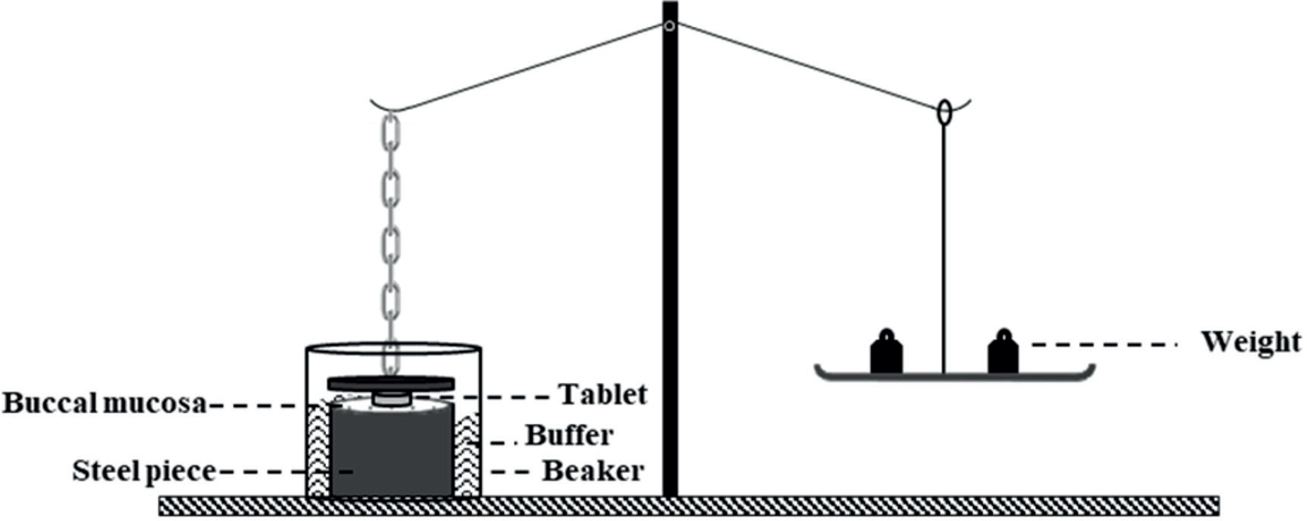
To assess the mucoadhesive strength of the formulations, a modified pan balance was applied. This test was performed on sheep buccal mucosa which was frozen in phosphate buffer pH 6.8 for one day and defrosted at room temperature prior to the experiment. An appropriately sized sheep mucosa was washed with phosphate buffer and attached to the lower platform of the pan balance by an elastic band. The mucoadhesive tablet (n=3) was affixed to the upper platform using cyanoacrylate glue (Figure 1). The tablet and mucosa were brought into contact, whereby a constant force was applied for 2 minutes to facilitate attachment. The weight on the other pan was elevated at a constant speed of 1 g until the two platforms detached. The total weight required for separation was recorded (Arabi et al., 2020; Koirala et al., 2021).

Complementary Evaluations
Infrared spectroscopic studies, determination of uniformity of dosage units, assessment of mucoadhesion duration, and drug release kinetics analysis were performed on selected formulations (F5 and F6).
Infrared Spectroscopic Studies
To ascertain any potential incompatibilities between the active pharmaceutical ingredient (API) and excipients, the Fourier-transform infrared (FT-IR) spectra of the physical mixtures of the selected formulations and pure miconazole nitrate were assessed using an FT-IR spectrometer (Nicolet Avatar 380, USA). Approximately 1-2 mg of each sample, blended with dry potassium bromide, was analyzed in transmission mode over a wavenumber range of 4000 to 400 cm-1.
Determination of Uniformity of Dosage Units
This test was employed to ensure the uniformity of dosage units in the selected formulations. As per the British Pharmacopeia, when the API equals or exceeds 25 mg and constitutes 25% or more of the total tablet weight, weight variation analysis is applied. Ten tablets underwent precise weighing, with their average mass calculated. The API percentage for each tablet was then determined using pharmacopeial methods. Next, the acceptance value for each selected formulation was reported (British Pharmacopoeia, 2022).
Assessment of Mucoadhesion Duration
This test aimed to assess mucoadhesion duration. The preparation of tablets, mucosa, and their assembly, was carried out as described in the determination of mucoadhesive strength. Nevertheless, to assess the duration of mucoadhesion, a 5 g weight was applied to the pan of the balance, with the chronometer initiating to measure the time until detachment occurred. The recorded time represented the duration of mucoadhesion (Arabi et al., 2020; Mortazavi, Smart, 1994).
Drug Kinetics Analysis
For evaluating kinetics of release of selected formulations, data collected from in vitro drug release studies were fitted into different kinetic mathematical models. These models include zero order, first order, Higuchi, and Korsmeyer-Peppas, with their corresponding equations listed in Table II (Dash, et al., 2010; Mortazavi, Mortazavi, 2020).
Statistical Analysis
Statistical analyses were performed using independent sample t-tests, ANOVA, and Tukey post-hoc tests to determine the existence of statistical significance among the dataset. The SPSS Statistics software package version 16.0 was employed for these analyses. Significance levels were established with a threshold of p < 0.05 for comparing data.
RESULTS
Evaluation of tablet properties
Physical Characterization
The results of mass uniformity, hardness, thickness, and friability for all formulations are reported in Table III. In the case of mass uniformity, tablets lied within a range between 98.3 mg and 103.7 mg, complying with the British pharmacopeia acceptance criteria, which stipulates a ±7.5% deviation for tablets within the 80-250 mg range. The thickness of tablets, which is a function of the type of polymers, exhibited a range between 1.9 mm to 2.1 mm. Friability assessments for all formulations offered favorable outcomes, registering at less than 1.0%. Further, hardness values were observed within 4.0-7.0 KgF. Both friability and hardness values collectively indicated desirable compactness and adequate resistance against mechanical shock as well as abrasion across all formulations (British Pharmacopoeia, 2022).
Quality control evaluation parameters of mucoadhesive tablet formulations (data are expressed as mean ±SD)
Drug Assay
As delineated in Table III, the drug assay was assessed for each formulation. The observed variability in assay findings spanned from 94.0% to 108.7%, falling within the prescribed range of 90.0-110.0%, complying with the common pharmacopeial limit (Rockville, 2024).
Determination of In Vitro Drug Release
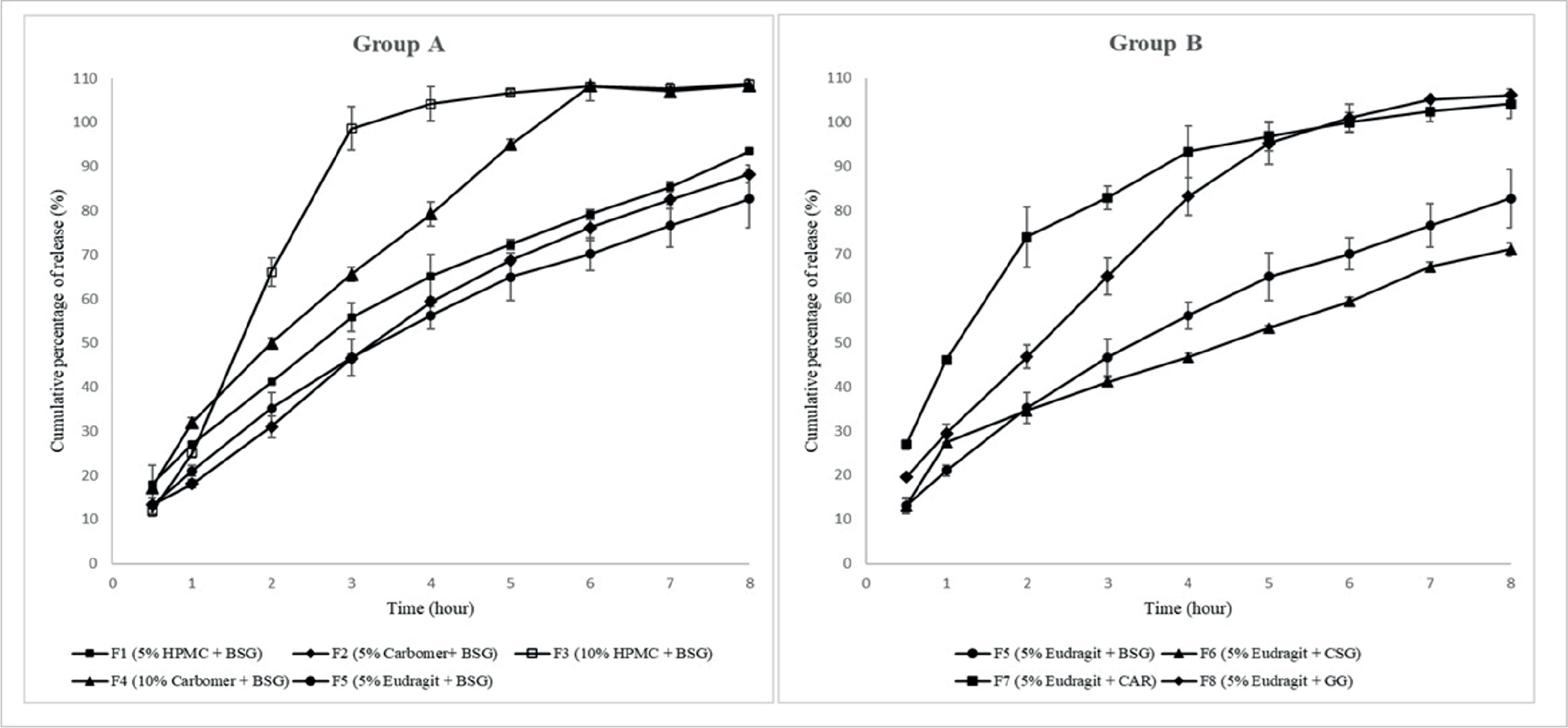
Figure 2 displays the cumulative release profiles of group Aand group B formulations. In group A, examining the effects of common hydrophilic mucoadhesive polymers and Eudragit RS, a hydrophobic polymer, in conjunction with BSG on drug release, Formulations F1 and F2 presented drug release percentages of 93.48± 0.47 and 88.28± 2.02 after 8 hours, respectively, while F3 and F4 achieved complete drug release within 4 and 6 hours, respectively. Further, F5 released 82.96 ± 6.67% after 8 hours. In group B, formulations F5-F9 were compared based on their natural mucoadhesive polymers employed in them. Formulations F6, F7, and F8 demonstrated drug release percentages of 71.16±1.53, 104.14± 3.35, and 106.12±0.31 after 8 hours, and the drug release percentage for formulation F5 had been previously noted.

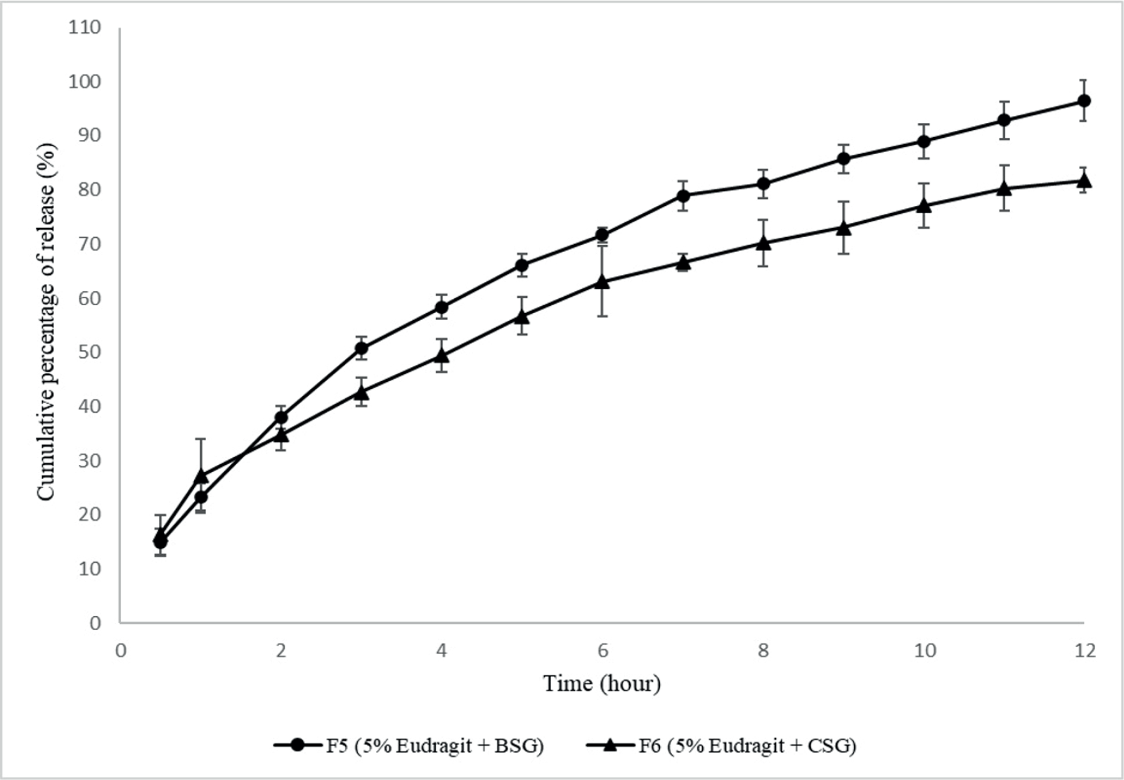
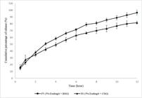
Based on the in vitro 8-hour drug release data from both experimental groups, formulations F5 and F6 were selected for a 12-hour drug release experiment, during which they released 96.43± 3.76 and 81.72± 2.21% of the drug, respectively, after 12 hours (Figure 3).
Determination of Mucoadhesive Strength
The findings from mucoadhesive strength assessment, as depicted in Figure 4, indicate that in group A formulations, formulation F4 presented the highest mucoadhesive strength with 24.0±1.0 grams (p <0.05), while formulation F5 displayed the lowest strength, approximately 16.3±0.6 grams (p <0.05).

In group B, the highest observed mucoadhesive strength was 35.0±1.0 grams (p <0.05) as evidenced by formulation F8, which contained GG, while formulation F7 (containing CAR) demonstrated the lowest mucoadhesive strength with 11.0±1.0 grams (p <0.05).
Complementary evaluations
Infrared Spectroscopic Studies
Even though previous studies reported no interaction between the API and excipients (Hariharaputhraayyanar et al., 2025; Janhavi, Anuradha, 2023; Madgulkar et al., 2009), FT-IR analysis was performed to evaluate the compatibility of the API with excipients in the selected formulations. As presented in Figure 5, characteristic peaks of miconazole nitrate were identified, including C=C and C=N stretching vibrations between 1500-1600 cm-1 (with two peaks), and an aromatic C-H stretch at 3107 cm-1. Additional peaks corresponding to C-Cl (812 cm-1), C-O (1085 cm-1), and C-N (1330 cm-1) stretches were also observed. These distinctive peaks remained in formulations F5 and F6 spectra without any notable shifts or disappearance, demonstrating the absence of significant interactions between the drug and formulation components (Bhalekar et al., 2009; Kumar Jha, Nama, Jain, 2024).

Determination of Uniformity of Dosage Units
The outcomes of the assessment for uniformity of dosage units revealed that the chosen formulations (F5 and F6), maintained consistency in their API contents. According to the British Pharmacopeia (2022), the acceptance value for the first 10 tablets should be less than or equal to L1, where L1 is defined as 15.0. The acceptance values for F5 and F6 were 6.8 and 4.2, respectively, and both were below 15.0, resulting in their acceptance.
Assessment of Mucoadhesion Duration
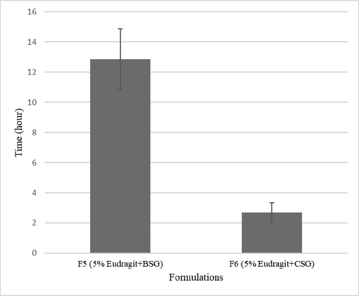
The test was conducted for two selected formulations, F5 and F6, with the results presented in Figure 6. Formulation F5 exhibited sustained adhesion to sheep buccal mucosa for a duration of 12.0 ±2.0 hours, while detachment occurred for formulation F6 after 2.5±0.7 hours.

Analysis of Drug Kinetics
Regarding in vitro drug release kinetics, data derived from drug release studies for selected formulations were subjected to fitting with different mathematical models. Table IV details the R2 values of zero order, first order, Higuchi, and Korsmeyer-Peppas model. Higher R2 values for Higuchi and first order kinetics indicated their suitability for F5 and F6, respectively. To understand the release mechanism from chosen mucoadhesive tablets, the initial 60% drug release data were fitted to the Korsmeyer-Peppas model. The resultant n values for F5 and F6 were 0.6867 and 0.5179, respectively.
Regression values of in-vitro release kinetic study of selected mucoadhesive tablet formulations
DISCUSSION
An ideal mucoadhesive tablet is characterized by prolonged pharmacological effects and sufficient residence time in the buccal cavity (Koirala et al., 2021). To fulfill these criteria, various parameters of the miconazole tablets were evaluated. The findings of physicochemical evaluations and drug assay revealed that all formulations fulfilled the required standards, making them eligible for further investigation, such as determining in vitro drug release, mucoadhesive strength, and additional tests.
The results of in vitro drug release of group A were unexpected. Formulations F3 and F4, containing higher concentrations of mucoadhesive polymers (carbomer/ HPMC+ BSG), presented faster drug release compared to F1 and F2, respectively. This is contrary to previous findings, suggesting that elevated concentrations of polymers, such as carbomer or HPMC, typically contribute to slower drug release (Arshad et al., 2022; Tavakoli et al., 2012). Considering our findings, the higher polymer concentrations seems to have led to excessive water absorption. This heightened water uptake in turn resulted in the loss of structural integrity of the formulations, ultimately causing their disintegration. In the research by Shanker et al. (2009), on developing tizanidine HCl buccal tablets, similar findings were noted. Formulations with high concentrations of HPMC disintegrated after 2 hours owing to a significant degree of swelling. Further, in line with previous findings, carbomer-containing formulations revealed lower release rates compared to HPMC (Samani, Montaseri, Kazemi, 2003). Among these, formulation F5 demonstrated a release of approximately 83% in 8 hours. Its release profile indicates the possibility of sustained drug release for up to 12 hours. This prolonged release may be attributed to the hydrophobic nature of Eudragit RS, delaying the penetration of the dissolution medium and lowers drug dissolution. Boyapally et al. (2009), reported that higher levels of Eudragit RS in combination with HPMC 6 cps led to diminished drug release from matrices. In group B, the findings suggested that the drug release characteristics were heavily tied to the composition of the mucoadhesive polymer utilized in each formulation. However, CAR demonstrated characteristics in accordance with studies demonstrating its inadequacy in prolonging drug release when used alone, thereby resulting in accelerated release in formulated products (Bartoníková, et al., 2024; Pacheco-Quito, et al., 2020).
Furthermore, in developingpropranolol vaginoadhesive tablets, Tasdighi et al. (2012), observed that formulations containing GG were insufficient in prolonging drug release to the acceptable duration. On the other hand, Embafrash Berhe et al. (2023), employed cress seed mucilage to develop mucoadhesive ibuprofen microspheres, and compared them to HPMC. The findings showed superior drug release extension of formulations including this mucilage compared to HPMC. Likewise, Lodhi et al. (2019), concluded in their study that BSG serves as a sustained release agent given its capability to effectively prolong drug release. According to these findings, formulations F5 and F6 were chosen as preferred candidates, and sampling of in vitro drug release continued up to 12 hours. The analysis of drug release profiles for selected formulations indicated that CSG could more effectively control the release rate of drug molecules. In addition, when assessing kinetics of release, based on the n values achieved from the Korsmeyer-Peppas model, the drug release mechanisms for both formulations F5 and F6 predominantly involve anomalous/non-Fickian diffusion. This suggests that swelling, diffusion, and gradual erosion contribute to drug release from these formulations.
Mucoadhesive strength is a measure of the adherence between polymers embedded into formulations and the mucus and/or epithelial surface. This adhesive strength can vary based on factors such as residence time within the mucus, polymer concentration, composition, and other relevant variables (Koirala et al., 2021). In this test, as previously indicated, formulation F5 presented a lower strength compared to formulations F1-F4, possibly because of the absence of mucoadhesive polymers such as carbomer or HPMC. In addition, in formulation F1-F4, formulations F3 and F4 revealed a better mucoadhesive strength in comparison to F1 and F2, respectively (p <0.05). These results highlight the remarkable impact of mucoadhesive polymer concentrations on the adhesion strength, aligning with previous studies that indicate elevated polymer concentrations would result in longer penetrating chain lengths and enhanced adhesion (Boddupalli et al., 2010; Phan, Tran, Tran, 2020). According to the results of this experiment, despite a twofold rise in the concentrations of HPMC or carbomer in formulations F3 and F4 compared to F1 and F2 respectively, there was no considerable enhancement observed in the mucoadhesive strength. This can arise from overhydration induced by the high polymer concentration. The overhydration led to loss of integrity and subsequently adhesive joint failure in these formulations (Boyapally, Nukala, Douroumis, 2009; Mortazavi, Mortazavi, 2020). Additionally, in group B, it was noted that the formulation containing GG presented the highest mucoadhesive strength, while the CAR-containing formulation exhibited the lowest strength. Meanwhile, it is widely acknowledged that different factors, including molecular weight, hydrophilicity, and the presence of functional groups (e.g., hydroxyl, carboxyl, sulfate, and amide), can significantly influence mucoadhesive behavior. In accordance with these findings, Bartoníková et al. (2024), in developing metronidazole vaginal mucoadhesive tablets observed that GG exhibited a superior mucoadhesive strength compared to CAR. This discrepancy may be attributed to the wide range of molecular masses of CAR gums, stemming from the diversity of species from which these gums are derived, thereby affecting their mucoadhesive properties (Shaikh et al., 2011). Statistical analyses comparing the adhesion strengths of buccal tablets across groups A and B indicated that formulations comprising GG presented superior adhesive properties in comparison to all other formulations (p <0.05). The outcomes of the mucoadhesion duration test demonstrated that formulation F5 offered extended adherence to the mucosal surface. This concords with the previous research conducted by Sakhare and Sayyad (2019), who employed BSG to develop mucoadhesive carvedilol-containing gastrospheres. Optimization procedures indicated that gastrospheres incorporating an optimal percentage of basil seed mucilage could retain adhesion to the mucosal surface for up to 8 hours, underscoring the potential for formulations containing BSG to achieve prolonged mucosal adhesion.
CONCLUSION
Mucoadhesive drug delivery systems have been extensively studied, with certain polymers recognized for their application in these systems. A comparative analysis of hydrophilic and hydrophobic polymers, including carbomer, HPMC, and Eudragit RS (group A), indicated that formulation F5 (Eudragit + BSG) could extend drug release for 12 hours, which may be attributed to the intrinsic properties of Eudragit RS. Furthermore, formulation F5 exhibited a satisfactory mucoadhesive strength and prolonged duration of adhesion. Comparison of natural polymers (group B) such as BSG, CSG, CAR, and GG consistently favored formulation F5 (Eudragit + BSG) for its desirable mucoadhesive characteristics as well as favorable in vitro drug release profiles characterized by Higuchi release kinetics and a non-Fickian release mechanism, demonstrating a release mechanism controlled by diffusion and erosion. In conclusion, Eudragit RS, with its hydrophobic characteristics, offers potential for synergistic use with mucoadhesive polymers to boost their retardation abilities. Further, BSG, a lesser-known mucoadhesive polymer, exhibited desirable mucoadhesive attributes, including acceptable adhesion strength and prolonged mucosal adherence, making it a promising candidate for incorporation into mucoadhesive drug delivery systems.
ACKNOWLEDGMENT
This paper is a part of Pharm. D. thesis of Sadaf Eshraghi at School of Pharmacy, Islamic Azad University of Medical Sciences, Tehran, Iran.
DECLARATION OF GENERATIVE AI AND AI ASSISTED TECHNOLOGIES IN THE WRITING PROCESS
The authors declare that they did not use AI tools during the writing process.
DATA AVAILABILITY STATEMENT
Use of data not disclosed.
REFERENCES
- Aguilar-de-Leyva Á, Campiñez MD, Jost F, Gavira M, Caraballo I. Study of the critical points in combined matrix tablets containing both inert and swelling excipients. J Drug Deliv Sci Technol. 2019;52:885-894.
- Ahmed TA, El-Say KM, Mahmoud MF, Samy AM, Badawi AA. Miconazole nitrate oral disintegrating tablets: in vivo performance and stability study. AAPS PharmSciTech. 2012;13(3):760-771.
- Amjed N, Zeshan M, Farooq A, Naz S. Applications of guar gum polysaccharide for pharmaceutical drug delivery: A review. Int J Biol Macromol. 2024;257:128390.
- Arabi M, Mortazavi SA, Jafariazar Z, Farhadnejad H, Alipour Harisa G, Fatahi Y. Fabrication and in-vitro evaluation of buccal mucoadhesive tablet of Meloxicam. Iran J Pharm Res. 2020;19(3):63-76.
- Arshad MS, Kiran M, Mudassir J, Farhan M, Hussain A, Abbas N. Formulation, Optimization, in vitro and in-vivo evaluation of levofloxacin hemihydrate Floating Tablets. Braz J Pharm Sci. 2022;58.
- Bandi SP, Bhatnagar S, Venuganti VVK. Advanced materials for drug delivery across mucosal barriers. Acta Biomater. 2021;119:13-29.
- Bartoníková K, Špaglová M, Papadakos M, Hanko M, Macho O. Mucoadhesive vaginal tablets containing metronidazole: screening of optimal natural polymer in the composition. Sci Pharm. 2024;92(1):10.
- Behrouzian F, Razavi SMA, Phillips GO. Cress seed (Lepidium sativum) mucilage, an overview. Bioact Carbohydr Diet Fibre. 2014;3(1):17-28.
- Bhalekar MR, Pokharkar V, Madgulkar A, Patil N, Patil N. Preparation and evaluation of miconazole nitrate-loaded solid lipid nanoparticles for topical delivery. AAPS PharmSciTech. 2009;10(1):289-296.
- Boddupalli BM, Mohammed ZN, Nath RA, Banji D. Mucoadhesive drug delivery system: An overview. J Adv Pharm Technol Res. 2010;1(4):381-387.
- Boyapally H, Nukala RK, Douroumis D. Development and release mechanism of diltiazem HCl prolonged release matrix tablets. Drug Deliv. 2009;16(2):67-74.
- British Pharmacopoeia 2022. ed. Commission BP. London: Stationery Office Books; 2022 p.
- da Silva JB, Dos Santos RS, da Silva MB, Braga G, Cook MT, Bruschi ML. Interaction between mucoadhesive cellulose derivatives and Pluronic F127: Investigation on the micelle structure and mucoadhesive performance. Mater Sci Eng C Mater Biol Appl. 2021;119:111643.
- Dash S, Murthy PN, Nath L, Chowdhury P. Kinetic modeling on drug release from controlled drug delivery systems. Acta Pol Pharm. 2010;67(3):217-223.
- Derakhshandeh K, Soleymani M. Formulation and in vitro evaluation of nifedipine-controlled release tablet: Influence of combination of hydrophylic and hydrophobic matrix forms. Asian J. Pharm. 2010;4(4).
- El-Say KM, Ahmed TA. Buccal Route of Drug Delivery. In: Talevi A, editor. The ADME Encyclopedia: A comprehensive guide on biopharmacy and pharmacokinetics. 1st ed. Switzerland: Springer Cham; 2022. p. 222-231.
- Embafrash Berhe H, Tesfay Mezgebo D, Abrha S, Gebremeskel Haile T, Molla F. Extraction, characterization, and evaluation of Lepidium sativum Linn. Mucilage as a Mucoadhesive polymer. Adv Pharmacol Pharm Sci. 2023;2023:5535344.
- Golshani S, Vatanara A, Amin M. Recent advances in oral mucoadhesive drug delivery. J Pharm Pharm Sci. 2022;25:201-217.
- Hariharaputhraayyanar G, Arivazhagan C, Bhuvaneshwaran A, Dhanush M, Jayabharath J, Keerthan T, et al. Formulation, characterization and evaluation of topical gel containing miconazole nitrate for fungal infection. Int J Recent Sci Res. 2025;16(02):126-132.
- Janhavi AV, Anuradha KS. Formulation and evaluation of antifungal topical gel by applying full factorial design. Bull Env Pharmacol Life Sci. 2023;12(3):26-31.
- Jawadi Z, Yang C, Haidar ZS, Santa Maria PL, Massa S. Bio-Inspired Muco-Adhesive Polymers for Drug Delivery Applications. Polymers. 2022;14(24):5459.
- Johnselvaraj G, Balasubramanian A, Ramalingam K. Application of novel natural mucoadhesive polymer in the development of pentoxifylline mucoadhesive tablets. Int J Appl Pharm. 2019;11(6):37-41.
- Koirala S, Nepal P, Ghimire G, Basnet R, Rawat I, Dahal A, et al. Formulation and evaluation of mucoadhesive buccal tablets of aceclofenac. Heliyon. 2021;7(3):e06439.
- Kumar Jha A, Nama N, Jain H. Preparation and evaluation of drug miconazole nitrate (gel form) for topical delivery with the help of deep eutectic mixture. Afr J Biol Sci. 2024;6(Si4):4871-4891.
- Lalla RV, Bensadoun RJ. Miconazole mucoadhesive tablet for oropharyngeal candidiasis. Expert Rev Anti Infect Ther. 2011;9(1):13-17.
- Lodhi BA, Hussain MA, Sher M, Haseeb MT,Ashraf MU, Hussain SZ, et al. Polysaccharide-based superporous, superabsorbent, and stimuli responsive hydrogel from Sweet Basil: A novel material for sustained drug release. Adv Polym Technol. 2019;2019:9583516.
- Madgulkar A, Kadam S, Pokharkar V. Development of buccal adhesive tablet with prolonged antifungal activity: Optimization and ex vivo Deposition Studies. Indian J Pharm Sci. 2009;71(3):290-294.
- Mehrgan H, Mortazavi S. The release behavior and kinetic evaluation of diltiazem HCl from various hydrophilic and plastic based matrices. Iran J Pharm Res. 2005;4(3):137-146.
- Mohammed FA, Khedr H. Preparation and in vitro/in vivo evaluation of the buccal bioadhesive properties of slow-release tablets containing miconazole nitrate. Drug Dev Ind Pharm. 2003;29(3):321-337.
- Mortazavi SA, Smart JD. An in-vitro method for assessing the duration of mucoadhesion. J Control Release. 1994;31(2):207-212.
- Mortazavi SM, Mortazavi SA. Propranolol Hydrochloride Buccoadhesive Tablet: Development and In-vitro Evaluation. Iran J Pharm Res. 2020;19(2):22-33.
- Pacheco-Quito EM, Ruiz-Caro R, Rubio J, Tamayo A, Veiga MD. Carrageenan-Based Acyclovir Mucoadhesive Vaginal Tablets for Prevention of Genital Herpes. Mar Drugs. 2020;18(5).
- Phan TQ, Tran PHL, Tran TTD. The relationship between mucoadhesive polymers and surface coating in tablets for the controlled colonic delivery of a poorly water-soluble drug. DARU J Pharm Sci. 2020;28(2):545-553.
- Razavi SMA, Naji-Tabasi S. Chapter 16 - Rheology and Texture of Basil Seed Gum: A New Hydrocolloid Source. In: Ahmed J, et al., editors. Advances in Food Rheology and Its Applications. 1st ed: Woodhead Publishing; 2017. p. 405-435.
- Reza MS, Quadir MA, Haider SS. Comparative evaluation of plastic, hydrophobic and hydrophilic polymers as matrices for controlled-release drug delivery. J Pharm Pharm Sci. 2003;6(2):282-291.
- Rockville M. United States Pharmacopeia. ed. United States: U.S. Pharmacopeial Convention; 2024 p.
- Rowe RC, Sheskey PJ, Owen SC. Handbook of Pharmaceutical Excipients. 5th ed. London: Pharmaceutical Press; 2006 p.
- Russo E, Selmin F, Baldassari S, Gennari CGM, Caviglioli G, Cilurzo F, et al. A focus on mucoadhesive polymers and their application in buccal dosage forms. J Drug Deliv Sci Technol. 2016;32:113-125.
- Sakhare S, Sayyad F. Design, Development, and Characterization of Ocimum basilicum mucilage-based, modified relaese mucoadhesive gastrospheres of Carvedilol. Asian J Pharm Clin Res. 2019;12(10):218-225.
- Samani SM, Montaseri H, Kazemi A. The effect of polymer blends on release profiles of diclofenac sodium from matrices. Eur J Pharm Biopharm. 2003;55(3):351-355.
- Shaikh R, Raj Singh TR, Garland MJ, Woolfson AD, Donnelly RF. Mucoadhesive drug delivery systems. J Pharm Bioallied Sci. 2011;3(1):89-100.
- Shanker G, Kumar CK, Gonugunta CS, Kumar BV, Veerareddy PR. Formulation and evaluation of bioadhesive buccal drug delivery of tizanidine hydrochloride tablets. AAPS PharmSciTech. 2009;10(2):530-539.
- Singla AK, Chawla M, Singh A. Potential applications of carbomer in oral mucoadhesive controlled drug delivery system: a review. Drug Dev Ind Pharm. 2000;26(9):913-924.
- Tasdighi E, Jafari Azar Z, Mortazavi SA. Development and In-vitro Evaluation of a Contraceptive Vagino-Adhesive Propranolol Hydrochloride Gel. Iran J Pharm Res. 2012;11(1):13-26.
- Tavakoli N, Varshosaz J, Dorkoosh F, Motaghi S, Tamaddon L. Development and evaluation of a monolithic floating drug delivery system for acyclovir. Chem Pharm Bull (Tokyo). 2012;60(2):172-177.
- Tosif MM, Najda A, Bains A, Kaushik R, Dhull SB, Chawla P, et al. A comprehensive review on plant-derived mucilage: Characterization, functional properties, applications, and its utilization for nanocarrier fabrication. Polymers (Basel). 2021;13(7).
- Volod’ko AV, Son EY, Glazunov VP, Davydova VN, Alexander-Sinkler EI, Aleksandrova SA, et al. Carrageenan films as promising mucoadhesive ocular drug delivery systems. Colloids Surf B Biointerfaces. 2024;237:113854.
- Zhang M, Qu Y, Li J, Yin L, Huang Q, Jia X. Charge characteristics of guar gums on the three-stage interaction mechanism with mucin to improve the mucoadhesion ability. Food Hydrocolloids. 2023;145:109107.