Dental anomalies and medical history: prematurity as a risk factor for agenesis
Abstract
The aims of this retrospective radiographic study were to assess the occurrence of dental anomalies in children and patients with disabilities and to investigate the association of these anomalies with pre-, peri-, and post-natal medical history. A total of 289 panoramic radiographs of children (0–14 years) and patients with disabilities (including all age groups) were used to identify developmental dental anomalies, and information was collected from medical records for statistical analysis. Data were expressed as relative frequency (percentage). The chi-square test, Fisher’s exact test, relative risk calculation, and the Mann-Whitney test were used for statistical comparisons with a significance level of 5%. Occurrence of dental alterations was 50.34% in children and 37.50% in patients with disabilities. Agenesis was the most common alteration (24.14% in children and 30.55% in patients with disabilities). The study found a relative risk of 2.6 for agenesis in children with a history of prematurity (p = 0.02). The variable “performing medical treatment” was a risk factor for the presence of dental alterations in children. The age and the medical conditions of the patients with disabilities varied greatly, with neurological diseases being the most prevalent disability. However, the health history of mothers during pregnancy was not associated with dental alterations in patients with disabilities (p > 0.05). Developmental dental anomalies were frequently observed in both groups, with agenesis being the most common condition. Prematurity was identified as a predictive factor for agenesis.
Descriptors
Anamnesis; Tooth Abnormalities; Radiography, Panoramic
Introduction
Dental anomalies can arise at different stages of tooth development and have a complex and multifactorial etiology that is not fully understood. Genetic and epigenetic defects along the stages of morpho- or histodifferentiation of tooth development have been considered as the main etiologic factors.1 At the same time, environmental factors such as medical conditions during the pre- and postnatal period of development may also be involved in the etiology of dental alterations. Depending on the stage at which the disorder occurs, malformation disorders can manifest as anomalies in the eruption, number, size, shape, position, color, and structure of the teeth,2,3 in addition to root malformations.
These dental alterations can cause local complications such as root resorption of adjacent teeth or cyst formation,3 impair the quality of life,4 and cause psychosocial problems in the child. Dental abnormalities can significantly affect a patient’s quality of life for several reasons, including: a) esthetics and self-esteem: abnormalities such as crowded, missing, or malformed teeth can affect the patient’s self-image, resulting in a loss of confidence and self-esteem; b) chewing function: agenesis, poorly positioned, or misaligned teeth can interfere with proper chewing; c) speech: the position and condition of the teeth can affect speech and some abnormalities can cause problems in the pronunciation of certain sounds; d) pain and discomfort: dental abnormalities can cause pain due to abnormal pressure to the jaw and facial muscles, or lead to infections associated with compromised teeth; and e) psychosocial impact: dental problems can be embarrassing and cause the patient to avoid social or professional interactions. It should be noted that developmental dental anomalies can result in the clinician having difficulties in behavioral management, diagnosis, and preparation of a treatment plan, and knowledge and understanding of this topic is essential for therapeutic success.5
To date, there is no consensus on the prevalence of dental anomalies with high variability (4.7 to 74.8%).3,6-10 Changes vary according to location, population, ethnicity, and geographic conditions, highlighting the environmental and genetic factors that may be associated.7 In addition, medical conditions such as childhood cancer,11,12 renal problems,13-16 premature birth, and low birth weight17 may affect dental development and contribute to the appearance of anomalies. Patients with certain syndromes, such as Apert syndrome, cleidocranial dysplasia, familial adenomatous polyposis and others often have dental agenesis, supernumerary teeth, and malformations.18,19
Recently, pre-, peri-, and postnatal conditions have been associated with defects in enamel development.20 The use of antibiotics, diseases/infections, alcohol consumption and smoking, complications during pregnancy and/or childbirth, high fever episodes, and neonatal illness among others have been shown to affect enamel development.20
Currently, there are no retrospective studies evaluating the incidence of radiographic dental anomalies in pediatric patients and patients with special needs treated at the Ribeirão Preto School of Dentistry of University of São Paulo (FORP-USP). The possible causal relationship between pre-, peri-, and postnatal medical history and incidence of dental anomalies has not been fully elucidated.
Based on the above, the present study aims to describe the occurrence of all types of dental anomalies that can be diagnosed on panoramic radiographs in a large sample of healthy children and patients with disabilities treated at the Pediatric Dentistry Clinic of the FORP-USP over a period of 10 years (2008–2018). At the same time, we sought to investigate the association between pre-, peri- and postnatal medical history and the development of these dental anomalies.
Methods
The present study was approved by the Research Ethics Committee of the Ribeirão Preto School of Dentistry (Protocol number 3,361,089).
The inclusion criteria of the present study were patients registered at the Pediatric Dentistry Clinic (code 541) and the Clinic for Patients with Disabilities (code 247) at FORP-USP, who had a panoramic radiograph in their medical record during the period 2008-2018. Then, the study population was determined by convenience. A list of all patients who underwent panoramic radiography (code 182) during this period was generated in the Romeu System - Computerization System of Dental Clinics at USP, and the number of corresponding records was recorded for retrospective evaluation. Exclusion criteria were cases with radiographic exams that were poorly stored or/and of no diagnostic value.
Next, the selected panoramic radiographs were evaluated on a computer for the diagnosis of dental anomalies by three calibrated, experienced examiners and specialists in pediatric dentistry.21 The radiographic findings of the developmental dental anomalies found in the study were analyzed using a concordance test among the three examiners.
The presence or absence, quantity, and type of dental changes found on each panoramic radiograph were recorded. The following anomalies were identified through radiographic analysis: agenesis, supernumerary tooth, mesiodens, rotation, microdontia, macrodontia, taurodontia, claw-shaped cusp, Hutchinson’s incisor, fusion, twinning, mulberry-shaped molar, tooth invagination, impacted tooth (except third molars), ectopic eruption, delayed eruption, transposition, root dilaceration, severe crowding, incisor hypomineralization, and dentigerous cyst.
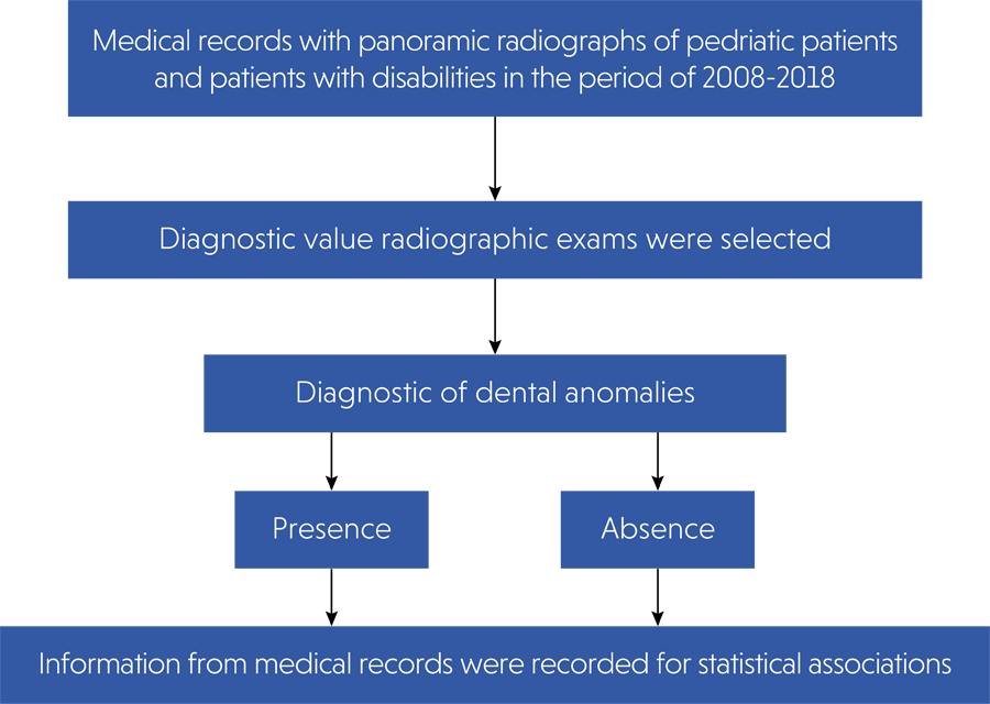
Subsequently, all the medical records included were accessed and analyzed by the main examiner. Information such as age, gender, race, and pre-, peri-, and postnatal dental and medical history were recorded for analysis of possible statistical associations. The patients from the pediatric dentistry clinic were aged between 0 and14 years, while the group of patients with disabilities included all ages. The flow diagram of the study is presented in Figure.

Because patients with disabilities have a variety of medical conditions, diseases were grouped into categories according to the type of systemic impairment to facilitate data analysis and interpretation. The categories were: infectious diseases, genetic, neurological impairment, cardiac, gastric, hepatic and renal, respiratory, endocrine, motor, auditory and visual, blood and dermatological and bone.
All data were categorized, recorded in an Excel spreadsheet, and statistically analyzed. Information that was not adequately filled out in the medical record was noted in the spreadsheet and was not considered for statistical associations.
Frequency distribution tables were created using the lower case letter “n” to indicate the absolute frequency and the symbol “%” to indicate the relative frequency (percentage).
Associations of nominal variables were performed using the chi-square test, Fisher’s exact test, and relative risk. For the ordinal variable (age group), the Mann Whitney test was used. The computational program Past version 4 22 was used for all analyses with a significance level of 5% (p ≤ 0.05).
Results
The Kappa value for the reliability between the three examiners for radiographic findings (referring to the diagnosis of dental anomalies) of pediatric patients was 0.822 and of patients with disabilities was 0.92, showing an almost perfect agreement.
Children from the Pediatric Dentistry Clinic
The records of 172 children were assessed, of which 145 had panoramic radiographs, 72 of whom were of patients without dental alterations or anomalies and 73 with dental alterations. Table 1 shows the variables race, gender, age at the time of the X-ray, illnesses and medications during pregnancy, prematurity, type of delivery, type of breastfeeding, medical treatment at the time of the X-ray and the child’s history of illnesses.
The item “Undergoing medical treatment at the moment” was a risk factor for the presence of dental alterations (p = 0.02). There was a higher occurrence of anomalies in the upper arch (56.16%). The most frequent alterations in permanent teeth according to the type of tooth affected are listed in Table 2.
Most frequent alterations in permanent teeth in pediatric dental patients and disabled patients.
Agenesis was the most prevalent type of anomaly, so the relative risk of this occurrence was calculated in relation to prematurity and type of delivery. Prematurity increased the risk of agenesis by 2.6 times (Table 3) (p = 0.02).
Developmental dental anomalies were grouped into the main types (shape, agenesis, and position) and further analysis was performed to detect possible associations with the most relevant medical conditions (prematurity and infectious diseases). The relative risk was calculated and the results confirmed that only prematurity increased the risk of agenesis (p = 0.03). For history of infectious diseases, no significant association was observed for any type of anomaly (p > 0.05).
Disabled Patients
The records of 214 disabled patients were assessed, of which 144 had quality panoramic radiographs, of which 90 were from patients without dental alterations or anomalies and 54 were from patients with dental alterations. Table 4 presents the variables race, gender, age at the time of the radiograph, illnesses and medications during pregnancy, prematurity, type of delivery, type of breastfeeding, medical treatment at the time of the radiograph and history of the child’s illnesses in relation to the presence or not of dental anomalies, with no significant association being observed for any parameter (p > 0.05).
Patients with one type of alteration predominated (72.22%), with no significant difference concerning the arch. The most frequent dental alterations or anomalies are shown in Table 2.
As in healthy children, in the group of patients with disabilities, agenesis was the most common alteration. However, the relative risk of this anomaly with prematurity and type of delivery was not significant (p > 0.05).
The diseases were grouped according to their systemic involvement (Table 5) to facilitate statistical analysis. According to these groups, neurological diseases were the most prevalent both in the group of patients without dental anomalies (42.31%) and in the group of patients with any type of anomaly (35.95%), but no statistically significant association was found (chi-square test, p = 0.099).
As with the group of pediatric patients, the developmental dental anomalies were grouped into their main types in order to make statistical associations. Statistical tests were then performed on the groups of genetic disorders and neurological disorders, which were predominantly recorded in the medical records. However, after statistical analysis, it was not possible to identify any risk (or protection) for the variables tested (p > 0.05).
Child Patients X Patients with Disabilities
The prevalence of dental anomalies between pediatric patients and patients with disabilities was compared and a statistically significant difference was observed. The number of healthy children with dental anomalies was higher compared to patients with disabilities.
Although the percentage of participants with disabilities and dental alterations was lower, the number of teeth affected was higher, with the 54 patients having 150 teeth with alterations (ratio between number of altered teeth and number of patients with special needs of 2.78). Among the children, 73 had 152 altered teeth with a ratio between the number of altered teeth and the number of children of 2.08.
Both groups were also compared with regard to the occurrence of the main types of anomalies, with no significant difference being observed (p > 0.05).
Discussion
The occurrence of radiographically identifiable dental alterations in the present study was 50.34% for children seen at the Pediatric Dentistry Clinic and 37.50% for the group of patients with disabilities, results that fall within the prevalence range of 4.7 to 74.8% reported in the literature 3, 6, 8-10. This variation is justified by the fact that studies are carried out in different populations and geographic regions,3,8,23, with varied samples,24-26 age groups27-29 and methodologies.23,26,30,31 This diversity of methodological factors makes it difficult to standardize this type of study with an established experimental design that would allow comparisons between prevalence surveys.
Some frequencies found in the present study reinforce the existing data3,25,29,31 indicating that dental anomalies occur more frequently in the upper arch, the most affected teeth are the incisors, and most patients have some type of dental alteration, both in the group of healthy children and in patients with disabilities. Additionally, more anomalies were observed in the permanent dentition than in the primary dentition, similarly to the study by Temiola et al.29
Similar to the present study, in which 24.14% of children and 30.55% of patients with disabilities had agenesis, Hummel et al.10 and Pallikaraki et al.,3 reported that agenesis was the most common type of anomaly, with a frequency of 12.0% and 6.4%, respectively, in each study. In the study by Vorwaller et al.,9 this alteration was the second most prevalent developmental dental defect after hyperdontia (8.1%), affecting 7.9% of children, with numerical anomalies predominating, which were also the most prevalent in the group of patients with disabilities in the present study. At the same time, according to Bilge et al.,8 when analyzed by type, position (60.8%) and shape (27.8%) anomalies were the most frequent, which is partly consistent with the present study in which these two types were among the most common and the three most frequent in both groups of patients studied.
The lack of significant associations between pre-, peri-, and postnatal medical factors and the presence of dental changes is common in the literature for a variety of reasons. Dental changes may be multifactorial, involving a complex combination of genetic predisposition, environmental exposures, and interactions between these factors. Identifying a single specific medical cause can be difficult because of this complexity. In addition, many studies may be limited by small sample sizes, inadequate control of variables, and different methods of diagnosing and classifying dental changes. This can lead to inconsistent or nonsignificant results. Genetic and phenotypic variability between individuals may result in different responses to the same pre-, peri-, and postnatal medical factors. Therefore, this variability can make it difficult to establish universal associations. Some studies may not adequately account for all potential confounding factors, such as lifestyle, diet, environmental exposures, and oral health care, which may also influence the presence of dental changes. Establishing a clear temporal relationship between pre-, peri-, and postnatal medical factors and the development of dental changes can be challenging, especially when the effects of these factors occur at different times in a patient’s life. Dental changes include a wide range of conditions, from structural malformations to problems with tooth eruption and changes in enamel formation. The heterogeneity of these conditions can make it difficult to identify consistent associations with specific medical factors.
In the present study, statistical analysis showed a significant association between agenesis and prematurity in the sample of healthy children, indicating that prematurity is a risk factor for agenesis. This finding corroborates a previous study that demonstrated that prematurity can be a predictive factor for an abnormal number of teeth, both in the primary and permanent dentition.17
Premature birth is associated with complications and postnatal medical treatments as well as impaired growth and development.32 Any stressful event during childbirth can lead to metabolic disorders during tooth formation. These defects may result from injuries arising from dysfunctions of the main organs affected.17 Thus, these mechanisms can support and explain the association between preterm delivery and agenesis, evidenced in the present study.
Although there was a significant association between dental alteration and postnatal health history in pediatric dental patients (with regard to the item “Undergoing medical treatment at the moment”), it was not possible to establish a causal relationship with any chronic systemic disease that could affect odontogenesis and cause a developmental anomaly in the deciduous or mixed dentition of the present study population. This absence of statistical association may be due to the limited information available in the medical records, with incomplete data on pre-, peri- and postnatal medical history.
Chickenpox or primary varicella, caused by the varicella-zoster virus (VZV), is a benign infectious disease of mild severity and is relatively common in children. However, it can manifest itself in a severe form in pregnant women and susceptible adults.33-35 In the present study, although there was no evidence of significant statistical association between chickenpox and dental anomalies, chickenpox was the most frequent disease (17 and 21 cases in children with and without dental alteration, respectively).
In the group of patients with disabilities, the probable association between dental agenesis and some systemic condition, including prematurity, must be interpreted with caution. In this sample group, prematurity did not significantly increase the risk of agenesis. The main limitation was the heterogeneous sample, including the age range of patients treated, of which 57.41% were adults with disabilities and some type of dental anomaly. In the analysis of agenesis, bias may have affected the findings, since it was difficult to correctly distinguish between missing teeth due to malformation or tooth extraction.
Supernumerary teeth are associated with various Mendelian syndromes.18 Since the sample was of patients with disabilities, a significant association between diseases of genetic etiology and the occurrence of supernumerary teeth was expected, as found in the literature.18,36,37 However, the occurrence of supernumerary teeth was low in the population studied. Although the heterogeneity of the sample, mainly because it included adults, was also a limiting factor for the study since patients may have already have had their supernumerary tooth extracted before to the time of the radiographic examination.
The assessment of statistical associations between pre-, peri-, and postnatal medical history and the occurrence of developmental dental anomalies in patients with disabilities proved to be a limiting and challenging task. Although diseases and medications were grouped into global categories, the analyses performed did not show significant associations. Some patients in this group had combinations of pathologies and complementary medications that prevented more relevant associations. In addition to the diversity of medical conditions presented by patients with disabilities, making it difficult to obtain a sufficiently large number for statistical significance, the lack of associations may have been due to incorrectly filled records.
Future studies can provide a better and more in-depth understanding of the prevalence and factors associated with dental anomalies, taking important issues into account. The sample studied must be representative of the target population in terms of age, gender, ethnicity, and socioeconomic characteristics. This will help with the generalizability of the results to the general population. Consistent and standardized diagnostic methods should be used to identify and classify dental abnormalities. This includes the use of digital radiographs, detailed clinical examinations, and, when appropriate, advanced technologies such as computed tomography (CT) or 3D imaging. Clear definitions of dental abnormalities to be assessed and precise criteria for inclusion and exclusion of participants must be established. This will help avoid bias and ensure the consistency of results. The prevalence of dental abnormalities must be placed in the context of broader epidemiologic considerations, including time trends, geographic variations, and demographic changes.
Despite the limitations, the findings from this study are clinically and scientifically relevant, as they shed light on etiological factors of dental anomalies and may improve the clinical management of these patients.
Conclusion
Based on the methodology used and the results obtained, it can be concluded that the occurrence of developmental dental anomalies was high and varied, with agenesia being the most prevalent alteration, both in healthy children and in patients with disabilities. In the group of children, prematurity proved to be a predictive factor for agenesia. In the group of disabled patients, no significant association could be observed between pre-, peri- and post-natal medical factors and the presence of dental alterations.
Acknowledgements
This work was supported by Coordenação de Aperfeiçoamento de Pessoal de Nível Superior—Brazil (Capes)—Finance Code 001. The authors thank Dr. Solange de Oliveira Braga Franzolin for the statistical support of this study.
References
-
1 Wagner VP, Arrué T, Hilgert E, Arús NA, Silveira HL, Martins MD, et al. Prevalence and distribution of dental anomalies in a paediatric population based on panoramic radiographs analysis. Eur J Paediatr Dent. 2020 Dec;21(4):292-8. https://doi.org/10.23804/ejpd.2020.21.04.7
» https://doi.org/10.23804/ejpd.2020.21.04.7 -
2 Brook AH, Jernvall J, Smith RN, Hughes TE, Townsend GC. The dentition: the outcomes of morphogenesis leading to variations of tooth number, size and shape. Aust Dent J. 2014 Jun;59(s1 Suppl 1):131-42. https://doi.org/10.1111/adj.12160
» https://doi.org/10.1111/adj.12160 -
3 Pallikaraki G, Sifakakis I, Gizani S, Makou M, Mitsea A. Developmental dental anomalies assessed by panoramic radiographs in a Greek orthodontic population sample. Eur Arch Paediatr Dent. 2020 Apr;21(2):223-8. https://doi.org/10.1007/s40368-019-00476-y
» https://doi.org/10.1007/s40368-019-00476-y -
4 Coffield KD, Phillips C, Brady M, Roberts MW, Strauss RP, Wright JT. The psychosocial impact of developmental dental defects in people with hereditary amelogenesis imperfecta. J Am Dent Assoc. 2005 May;136(5):620-30. https://doi.org/10.14219/jada.archive.2005.0233
» https://doi.org/10.14219/jada.archive.2005.0233 - 5 Cameron A, Widmer R, editors. Handbook of pediatric dentistry. Elsevier; 2013
- 6 Ezoddini AF, Sheikhha MH, Ahmadi H. Prevalence of dental developmental anomalies: a radiographic study. Community Dent Health. 2007 Sep;24(3):140-4.
-
7 Goncalves-Filho AJ, Moda LB, Oliveira RP, Ribeiro AL, Pinheiro JJ, Alver-Junior SR. Prevalence of dental anomalies on panoramic radiographs in a population of the state of Pará, Brazil. Indian J Dent Res. 2014;25(5):648-52. https://doi.org/10.4103/0970-9290.147115
» https://doi.org/10.4103/0970-9290.147115 -
8 Bilge NH, Yesiltepe S, Törenek Agirman K, Çaglayan F, Bilge OM. Investigation of prevalence of dental anomalies by using digital panoramic radiographs. Folia Morphol (Warsz). 2018;77(2):323-8. https://doi.org/10.5603/FM.a2017.0087
» https://doi.org/10.5603/FM.a2017.0087 - 9 Vorwaller R, Kratunova E, da Fonseca MA, Alapati SB, Hill B, Stanford C. Prevalence of radiographically identifiable dental anomalies in children and association with health status. Pediatr Dent. 2021 Nov;43(6):451-6.
- 10 Hummel B, Yu Q, Frazier J, Ballard RW, Johnson JT, Armbruster PC. The prevalence of developmental dental and eruption anomalies assessed using panoramic radiographs: a retrospective study. Gen Dent. 2023 Jan-Feb;71(1):19-23.
-
11 Sonis AL, Tarbell N, Valachovic RW, Gelber R, Schwenn M, Sallan S. Dentofacial development in long-term survivors of acute lymphoblastic leukemia. A comparison of three treatment modalities. Cancer. 1990 Dec;66(12):2645-52. https://doi.org/10.1002/1097-0142(19901215)66:12<2645::AID-CNCR2820661230>3.0.CO;2-S
» https://doi.org/10.1002/1097-0142(19901215)66:12<2645::AID-CNCR2820661230>3.0.CO;2-S -
12 Minicucci EM, Lopes LF, Crocci AJ. Dental abnormalities in children after chemotherapy treatment for acute lymphoid leukemia. Leuk Res. 2003 Jan;27(1):45-50. https://doi.org/10.1016/S0145-2126 (02)00080-2
» https://doi.org/10.1016/S0145-2126 (02)00080-2 -
13 Nunn JH, Sharp J, Lambert HJ, Plant ND, Coulthard MG. Oral health in children with renal disease. Pediatr Nephrol. 2000 Sep;14(10-11):997-1001. https://doi.org/10.1007/s004670050061
» https://doi.org/10.1007/s004670050061 -
14 Al-Nowaiser A, Roberts GJ, Trompeter RS, Wilson M, Lucas VS. Oral health in children with chronic renal failure. Pediatr Nephrol. 2003 Jan;18(1):39-45. https://doi.org/10.1007/s00467-002-0999-7
» https://doi.org/10.1007/s00467-002-0999-7 -
15 Davidovich E, Schwarz Z, Davidovitch M, Eidelman E, Bimstein E. Oral findings and periodontal status in children, adolescents and young adults suffering from renal failure. J Clin Periodontol. 2005 Oct;32(10):1076-82. https://doi.org/10.1111/j.1600-051X.2005.00812.x
» https://doi.org/10.1111/j.1600-051X.2005.00812.x -
16 Silva TM, Alves LA, Garrido D, Watanabe A, Mendes FM, Ciamponi AL. Health and oral health-related quality of life of children and adolescents with chronic kidney disease: a cross-sectional study. Qual Life Res. 2019 Sep;28(9):2481-9. https://doi.org/10.1007/s11136-019-02196-8
» https://doi.org/10.1007/s11136-019-02196-8 -
17 Prokocimer T, Amir E, Blumer S, Peretz B. Birth-Weight, Pregnancy Term, Pre-Natal and Natal Complications Related to Child's Dental Anomalies. J Clin Pediatr Dent. 2015;39(4):371-6. https://doi.org/10.17796/1053-4628-39.4.371
» https://doi.org/10.17796/1053-4628-39.4.371 -
18 Lubinsky M, Kantaputra PN. Syndromes with supernumerary teeth. Am J Med Genet A. 2016 Oct;170(10):2611-6. https://doi.org/10.1002/ajmg.a.37763
» https://doi.org/10.1002/ajmg.a.37763 -
19 López-Estudillo AS, Rosales-Bérber MA, Ruiz-Rodríguez S, Pozos-Guillén A, Noyola-Frías MÁ, Garrocho-Rangel A. Dental approach for Apert syndrome in children: a systematic review. Med Oral Patol Oral Cir Bucal. 2017 Nov;22(6):e660-8. https://doi.org/10.4317/medoral.21628
» https://doi.org/10.4317/medoral.21628 -
20 Butera A, Maiorani C, Morandini A, Simonini M, Morittu S, Barbieri S, et al. Assessment of genetical, pre, peri and post natal risk factors of Deciduous Molar Hypomineralization (DMH), Hypomineralized Second Primary Molar (HSPM) and Molar Incisor Hypomineralization (MIH): a narrative review. Children (Basel). 2021 May;8(6):432. https://doi.org/10.3390/children8060432
» https://doi.org/10.3390/children8060432 -
21 Kolokitha OE, Balli D, Zarkadi AE, Gizani S. Association between maxillary canine impaction and other dental anomalies: radiological study of a mixed dentition children's cohort from an orthodontic clinic. Eur Arch Paediatr Dent. 2023 Jun;24(3):401-7. https://doi.org/10.1007/s40368-023-00798-y
» https://doi.org/10.1007/s40368-023-00798-y - 22 Hammer O, Haper DAT, Ryan PD (2001). Past: Palaeontological statistics software package for education Palaeontologia Eletronica. 2001;4:9.
-
23 Vinjolli F, Zeqaj M, Dragusha E, Malara A, Danesi C, Laganà G. Dental anomalies in an Albanian orthodontic sample: a retrospective study. BMC Oral Health. 2023 Jan;23(1):47. https://doi.org/10.1186/s12903-023-02711-x
» https://doi.org/10.1186/s12903-023-02711-x -
24 Septer S, Bohaty B, Onikul R, Kumar V, Williams KB, Attard TM, et al. Dental anomalies in pediatric patients with familial adenomatous polyposis. Fam Cancer. 2018 Apr;17(2):229-34. https://doi.org/10.1007/s10689-017-0035-5
» https://doi.org/10.1007/s10689-017-0035-5 -
25 Bagci N, Pamukçu U, Altunkaynak B, Peker I. Dental anomalies in consanguineous marriage: a clinical-radiological study. Int Dent J. 2022 Feb;72(1):133-40. https://doi.org/10.1016/j.identj.2021.02.003
» https://doi.org/10.1016/j.identj.2021.02.003 -
26 Halperson E, Matalon V, Goldstein G, Saieg Spilberg S, Herzog K, Fux-Noy A, et al. The prevalence of dental developmental anomalies among childhood cancer survivors according to types of anticancer treatment. Sci Rep. 2022 Mar;12(1):4485. https://doi.org/10.1038/s41598-022-08266-1
» https://doi.org/10.1038/s41598-022-08266-1 -
27 Bardellini E, Amadori F, Pasini S, Majorana A. Dental Anomalies in Permanent Teeth after Trauma in Primary Dentition. J Clin Pediatr Dent. 2017;41(1):5-9. https://doi.org/10.17796/1053-4628-41.1.5
» https://doi.org/10.17796/1053-4628-41.1.5 -
28 Fekonja A. Prevalence of dental developmental anomalies of permanent teeth in children and their influence on esthetics. J Esthet Restor Dent. 2017 Jul;29(4):276-83. https://doi.org/10.1111/jerd.12302
» https://doi.org/10.1111/jerd.12302 -
29 Temilola DO, Folayan MO, Fatusi O, Chukwumah NM, Onyejaka N, Oziegbe E, et al. The prevalence, pattern and clinical presentation of developmental dental hard-tissue anomalies in children with primary and mix dentition from Ile-Ife, Nigeria. BMC Oral Health. 2014 Oct;14(1):125. https://doi.org/10.1186/1472-6831-14-125
» https://doi.org/10.1186/1472-6831-14-125 -
30 Wuollet E, Laisi S, Salmela E, Ess A, Alaluusua S. Molar-incisor hypomineralization and the association with childhood illnesses and antibiotics in a group of Finnish children. Acta Odontol Scand. 2016 Jul;74(5):416-22. https://doi.org/10.3109/00016357.2016.1172342
» https://doi.org/10.3109/00016357.2016.1172342 -
31 Udoye CI, Jafarzadeh H. Dilaceration among Nigerians: prevalence, distribution, and its relationship with trauma. Dent Traumatol. 2009 Aug;25(4):439-41. https://doi.org/10.1111/j.1600-9657.2009.00796.x
» https://doi.org/10.1111/j.1600-9657.2009.00796.x - 32 Rythén M. Preterm infants: odontological aspects. Swed Dent J Suppl. 2012;(S224):2p-106.
-
33 Field N, Amirthalingam G, Waight P, Andrews N, Ladhani SN, Hoek AJ, et al. Validity of a reported history of chickenpox in targeting varicella vaccination at susceptible adolescents in England. Vaccine. 2014 Feb;32(10):1213-7. https://doi.org/10.1016/j.vaccine.2013.06.098
» https://doi.org/10.1016/j.vaccine.2013.06.098 -
34 Andrade AL, da Silva Vieira MA, Minamisava R, Toscano CM, Souza MBL, Fiaccadori F, et al. Single-dose varicella vaccine effectiveness in Brazil: a case-control study. Vaccine. 2018 Jan;36(4):479-83. https://doi.org/10.1016/j.vaccine.2017.12.011
» https://doi.org/10.1016/j.vaccine.2017.12.011 -
35 Lo Presti C, Curti C, Montana M, Bornet C, Vanelle P. Chickenpox: an update. Med Mal Infect. 2019 Feb;49(1):1-8. https://doi.org/10.1016/j.medmal.2018.04.395
» https://doi.org/10.1016/j.medmal.2018.04.395 -
36 Wijn MA, Keller JJ, Giardiello FM, Brand HS. Oral and maxillofacial manifestations of familial adenomatous polyposis. Oral Dis. 2007 Jul;13(4):360-5. https://doi.org/10.1111/j.1601-0825.2006.01293.x
» https://doi.org/10.1111/j.1601-0825.2006.01293.x -
37 Cammarata-Scalisi F, Avendaño A, Callea M. Main genetic entities associated with supernumerary teeth. Arch Argent Pediatr. 2018 Dec;116(6):437-44. https://doi.org/10.5546/aap.2018.eng.437
» https://doi.org/10.5546/aap.2018.eng.437
-
Data availability:
The authors declare that all data generated or analyzed during this study are included in this published article.
Edited by
-
Editor-in-Chief:
Saul Paiva
The authors declare that all data generated or analyzed during this study are included in this published article.