
 Management of Cervical Eosinophilic Granuloma
ABSTRACT
To report the clinical presentation, radiological findings, management, and outcomes of three cases of eosinophilic granuloma (EG) of the cervical spine, in addition to reviewing the current literature on the topic. A retrospective study was conducted, including all cases of histopathologically confirmed cervical EG treated between 2003 and 2024 at a quaternary-level orthopedic hospital within a public healthcare system. A narrative literature review was subsequently performed, focusing on epidemiological, clinical, diagnostic, and therapeutic aspects. Three cases were treated during the study period, all involving patients aged three to four years with persistent cervical pain. Two cases were unifocal, and one was multifocal. One patient presented with atlantoaxial instability and underwent surgical treatment. No complications occurred, and all patients experienced symptom resolution. Cervical spine EG is rare but should be considered in patients with persistent neck pain and radiographic evidence of lytic lesions or instability. Treatment should be individualized and range from conservative management to surgical arthrodesis, depending on disease extent, spinal instability, or neurological deficit. Level of Evidence IV; Case Series.
Keywords:
Spinal Neoplasms; Neck Pain; Eosinophilic Granuloma.
RESUMO
Relatar a apresentação clínica, os achados radiológicos, o manejo e os desfechos de três casos de granuloma eosinófilo (GE) da coluna cervical, além de revisar a literatura atual sobre o tema. Foi realizado estudo retrospectivo com inclusão de todos os casos de GE cervical, com confirmação histopatológica, tratados entre 2003 e 2024 em um hospital ortopédico de nível quaternário de um sistema público de saúde. Em seguida, foi realizada uma revisão narrativa da literatura, com ênfase em aspectos epidemiológicos, clínicos, diagnósticos e terapêuticos. Foram tratados três casos no período do estudo, todos pacientes entre 3 e 4 anos de idade com dor cervical persistente, sendo dois com lesão unifocal e um multifocal. Um dos casos apresentava instabilidade atlantoaxial e foi tratado cirurgicamente. Nenhum dos pacientes apresentou complicações e todos tiveram resolução dos sintomas. O GE da coluna cervical é raro mas deve ser considerado em pacientes com cervicalgia persistente na presença de lesão lítica ou instabilidade aos exames de imagem. O tratamento deve ser individualizado e varia de conservador a cirúrgico com artrodese, conforme extensão da doença, instabilidade ou presença de déficit neurológico. Nível de Evidência IV; Série de Casos.
Descritores:
Neoplasias da Coluna Vertebral; Cervicalgia; Granuloma Eosinófilo.
RESUMEN
Reportar la presentación clínica, hallazgos radiológicos, manejo y evolución de tres casos de granuloma eosinófílo (GE) de la columna cervical, además de revisar la literatura actual sobre el tema. Se realizó un estudio retrospectivo que incluyó todos los casos de GE cervical con confirmación histopatológica, tratados entre 2003 y 2024 en un hospital ortopédico de nivel cuaternario del sistema público de salud. Posteriormente, se llevó a cabo una revisión narrativa de la literatura, con énfasis en los aspectos epidemiológicos, clínicos, diagnósticos y terapéuticos. Se trataron tres casos durante el período del estudio, todos en pacientes entre 3 y 4 años con cervicalgia persistente. Dos casos eran unifocales y uno multifocal. Un paciente presentó inestabilidad atlantoaxoidea y fue tratado quirúrgicamente. No hubo complicaciones y todos los pacientes evolucionaron con resolución de los síntomas. El GE cervical es raro, pero debe considerarse en pacientes con cervicalgia persistente y lesiones líticas o inestabilidad en estudios de imagen. El tratamiento debe individualizarse y puede variar desde el manejo conservador hasta la artrodesis quirúrgica, según la extensión de la enfermedad, la inestabilidad o la presencia de déficit neurológico. Nivel de Evidencia IV; Serie de Casos.
Descriptores:
Neoplasias de la Columna Vertebral; Dolor de Cuello; Granuloma Eosinófilo.
INTRODUCTION
Eosinophilic granuloma (EG) is a benign but locally destructive osteolytic bone lesion that may occur in isolation or as part of the spectrum of disorders known as Langerhans cell histiocytosis (LCH). Its etiology remains unknown, with proposed hypotheses ranging from autoimmune and inflammatory processes to infectious agents, adverse immune reactions, and viral infections.1
LCH is a rare condition, with an estimated incidence of approximately 1 in 1.5 million individuals. About 6.5% of LCH cases involve the spine, and among these, 11% are located in the cervical spine.1 As of 2002, only 54 cases of cervical spine involvement had been reported in the literature, contributing to the lack of consensus regarding optimal management.1 While it is considered a benign lesion, EGs can behave aggressively at the local level, potentially compromising adjacent neural structures.2
The objective of this study was to present the clinical and radiological features, management strategies, and outcomes of cervical spine eosinophilic granuloma cases treated by the authors, in addition to providing a comprehensive review of the current literature on the topic.
METHODS
This is a retrospective, descriptive case series involving patients with histopathologically confirmed cervical eosinophilic granuloma (EG) treated at a quaternary-level public orthopedic hospital in Brazil between January 2003 and December 2024. The study was conducted by the ethical standards of the institutional research committee and the 1964 Helsinki Declaration and its later amendments. Approval was granted by the institution’s research ethics board (number 56429322.7.0000.5273), and the requirement for informed consent was waived due to the retrospective nature of the study.
Patient identification was performed through a review of the hospital’s surgical records, using diagnostic codes and histopathological confirmation. For each case, the following data were extracted from medical records: age at diagnosis, sex, presenting symptoms, neurological status at presentation, affected vertebral level(s), imaging findings (including plain radiographs, computed tomography [CT], and magnetic resonance imaging [MRI]), treatment approach, postoperative outcomes, and follow-up duration.
The collected cases were then compared with data from the existing literature. A narrative literature review was conducted, and relevant findings were synthesized and organized into the following thematic categories: Epidemiology, Clinical Presentation, Imaging, Differential Diagnosis, Diagnosis, and Treatment.
RESULTS
We reviewed the three cases of cervical EG treated at our institution during the study period.
Case number 1
A three-year-old girl was admitted to our institution with a six-month history of cervical and lumbar pain, without any associated neurological symptoms. There was no family history of spinal lesions or neoplastic conditions. She had been previously treated with a cervical collar for one month, but her symptoms persisted.
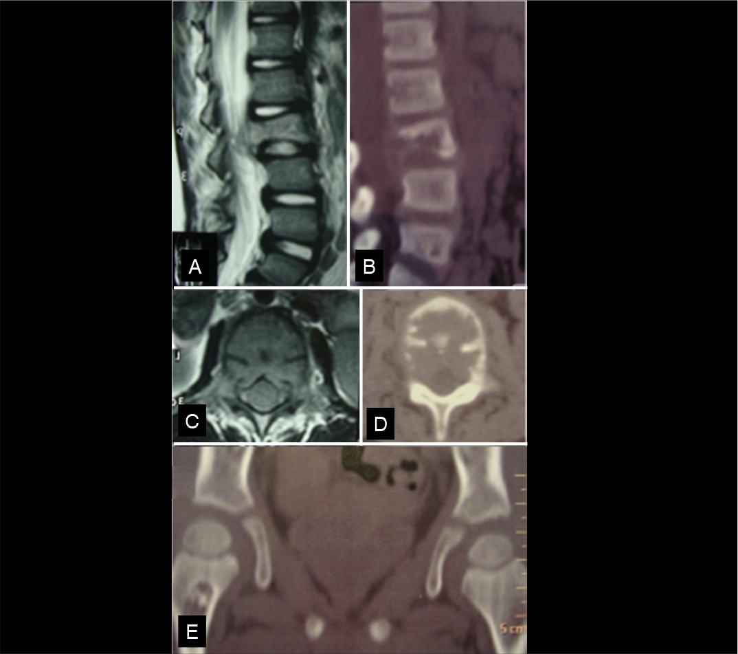
Imaging studies revealed osteolytic lesions involving the C4 vertebral body, L2 vertebra, and the proximal right femur. The femoral lesion was asymptomatic. Inflammatory markers, including C-reactive protein (CRP) and erythrocyte sedimentation rate (ESR), were within normal limits, and no other significant laboratory abnormalities were observed. (Figure 1)

A and B. Sagittal T2-weighted MRI and Sagittal CT show a L2 hyperintense and lytic lesion. C and D. Axial T2-weighted MRI and Axial CT show mild stenosis and destruction of the L2 vertebral body. E. Coronal CT of the hips presenting with a metaphyseal lytic lesion in the right femur, site chosen for biopsy.
A CT-guided needle biopsy of the femoral lesion was performed, and histopathological analysis confirmed the diagnosis of eosinophilic granuloma.
Given the presence of multifocal skeletal involvement, systemic treatment was initiated, consisting of chemotherapy in combination with systemic corticosteroids, while maintaining continuous use of a cervical collar. The patient demonstrated complete resolution of symptoms after three months of systemic therapy and was able to return to daily activities.
Case number 2
A four-year-old boy presented to our hospital with a six-month history of cervical pain. A CT scan performed at an outside institution revealed a lytic lesion involving the C2 vertebral body. At the time of presentation, the patient was using a Philadelphia cervical collar and reported neck pain but exhibited no neurological symptoms or signs. (Figure 2 A-F)

A and B. Tn-99 PET Scan shows an isolated hypermetabolic lesion in C2. C, D, and E. Cervical CT scan presents a lytic lesion in C2. F. Sagittal T2-weighted cervical MRI shows destruction of C2 body. G, H. 3 years post-C2 biopsy show no instability in the cervical region. I, J, K. CT and MRI scans, 3 years post C2 biopsy, presenting with complete reossification of the lesion and recovery of C2 height.
A right transpedicular biopsy was performed, and histopathological examination confirmed the diagnosis of eosinophilic granuloma. Following the procedure, the patient continued to use the cervical collar and experienced rapid symptom resolution within one month. At three-year follow-up, the patient remained asymptomatic, with complete radiographic resolution of the C2 lesion and no evidence of cervical instability or recurrence. (Figure 2 G-K)
Case number 3
A four-year-old girl presented to our hospital with a 10-month history of cervical pain, torticollis, and intermittent fever. She exhibited no neurological deficits, no loss of muscle strength, and had no family history of spinal tumors. Before admission, she had been treated empirically with antibiotics for 14 days at another institution, with no clinical improvement.
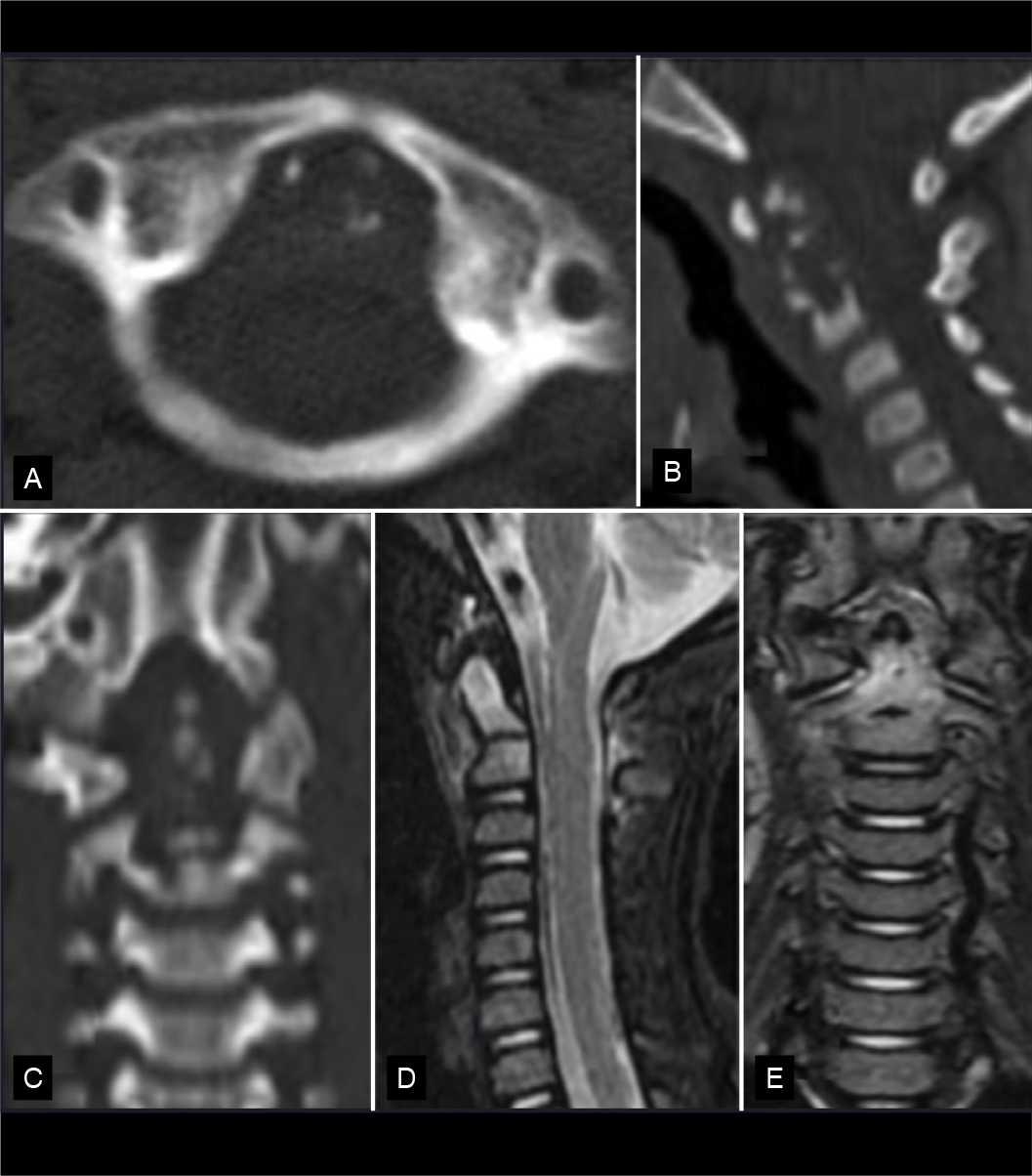
Imaging studies revealed cervical instability, with destruction of the odontoid process, associated with atlantoaxial dislocation by a lytic lesion and mild prevertebral soft tissue edema. Inflammatory markers, including CRP and ESR, were within normal ranges, and no additional laboratory abnormalities were identified. (Figure 3)

A, B, and C. Axial, Sagittal, and Coronal CT scans show destruction of the odontoid process and part of the body of C2. D and E. Sagittal and coronal T2-weighted MRI show hyperintensity in the odontoid process and part of the C2 body.
To confirm the diagnosis, a transoral biopsy was performed using a Jamshidi needle (Figure 4). Histopathological analysis revealed dissociated trabeculae, connective tissue, edema, and histiocytic cells. Immunohistochemical staining showed positivity for CD1a, S100, and Cyclin D1, confirming the diagnosis of eosinophilic granuloma.

Details of the needle biopsy. A. Patient positioning, with the orotracheal tube pushed to the right side. B. Alan Crockand transoral retractor and Jamshidi needle positioning. C. Lateral fluoroscopy shows needle biopsy in the body of C2. D. Biopsy fragment.
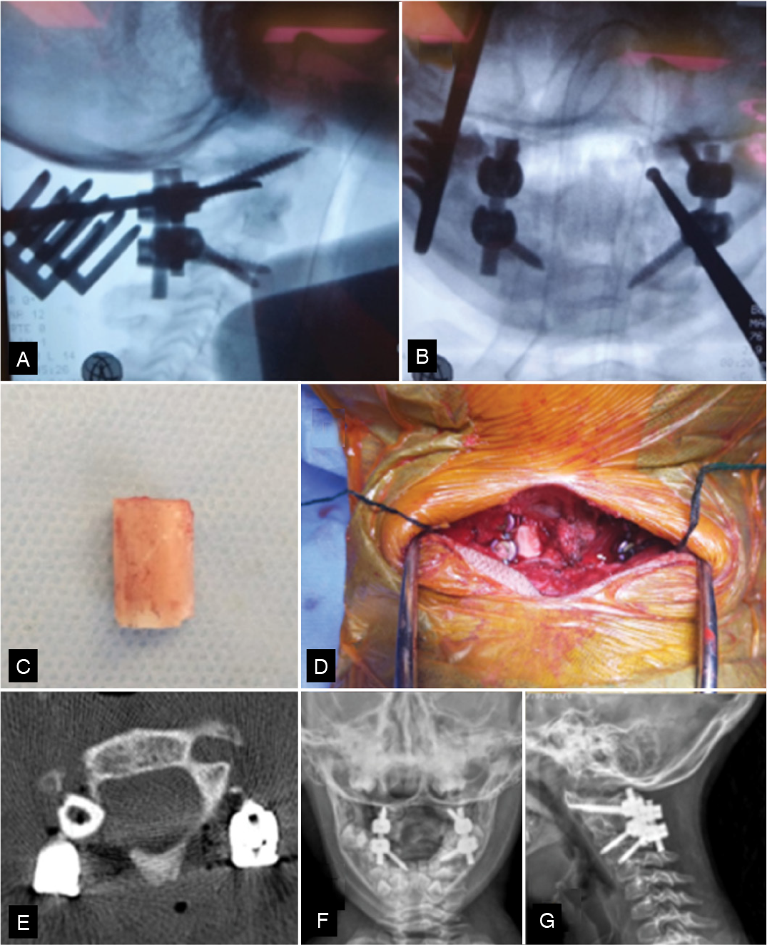
Definitive surgical treatment was performed one month after the biopsy via a posterior approach. The procedure involved intralesional resection by transpedicular approach of the right pedicle of C2 associated with C1 lateral mass screw and C3 pedicular instrumentation. As the lesion completely compromised the left lateral mass of C2 and was unsuitable for fixation, a fibular strut graft from the institution’s bone tissue bank was used to reconstruct the defect (Figure 5). Successful arthrodesis was confirmed by imaging at three months postoperatively, and at one-year follow-up, the patient remained asymptomatic, with complete clinical and radiographic resolution of the lesion.

Surgical Procedure. A and B. Lateral and AP fluoroscopy showing curettage of the lesion by transpedicular approach. C. Structural homologous fibular graft used for arthrodesis. D. Structural bone used for stability and fusion from the inferior lateral mass of C1 to the Superior facet of C3. E. Axial CT scan shows a Strut graft substituting the lateral mass. F and G. Postoperative AP and Lateral x-rays showing C1 lateral mass screws and C3 CPS.
DISCUSSION
Epidemiology
EG is a benign, locally aggressive osteolytic bone lesion that belongs to the spectrum of disorders known as LCH, which also includes more aggressive and multifocal forms such as Letterer-Siwe disease and Hand-Schuller-Christian disease. Its etiology remains unclear, with proposed mechanisms including autoimmune, inflammatory, infectious, and reactive processes.1
The estimated incidence of LCH is approximately 1 in 1.5 million individuals, with 79% of cases presenting as solitary lesions, 7% as multifocal, and 14% as part of syndromic forms. The spine is affected in only 6.5% of cases, while the skull (27%) and femur (15%) are more frequently involved while the skull (27%) and femur (15%) are more frequently involved.1 Within spinal lesions, the cervical spine accounts for 11%, the thoracic spine for 54%, and the lumbar spine for 35%.1 In our series, two cases (66.6%) were solitary and one (33.3%) was multifocal, findings consistent with the literature.
In a 2002 meta-analysis by Bertram et al., which reviewed the 53 published cases of cervical EG reported up to that time, the average age was 13 years, 7.4 years among pediatric patients, and 35 years among adults. In children, there was a male predominance with a ratio of 1.7:1, while in adults, a female predominance was observed with a ratio of 1.5:1. Among pediatric cases, 60% were located between C3 and C5. In contrast, in adults, 54% occurred at C2-C3.1 The majority of cases affected the vertebral body (86% in children and 80% in adults), and involvement of other bones was reported in 24% of cases. 1 In our series, two cases involved C2, and one case involved C4.
A systematic review focused exclusively on adult cervical EG by Prasad and Divya (2019) identified 62 cases, reporting a male-to-female ratio of 3.1:1 and a mean age of 32.8 years (range: 18 - 71). The average time from symptom onset to diagnosis was 13.7 weeks. Similarly, in our cohort, we observed a male predominance (2:1).
Clinical Presentation
The clinical presentation of EG in the cervical spine exhibits certain specific features when compared to lesions in other spinal regions. Pain is the most common symptom, reported in 76% of pediatric cases. Limitation of range of motion is also frequent, occurring in 76% of children and 60% of adults. However, neurological symptoms are present in only 14% of cervical cases, compared to 64% in the thoracic spine and 75% in the lumbar spine. One hypothesis proposed by the authors is that the greater mobility of the cervical spine may result in earlier onset of pain and, consequently, earlier diagnosis, before significant progression of the disease. 1
In adults, 94% present with neck pain, 58% with limb weakness, 19% with radiculopathy, 10% with kyphotic deformity, and 10% with torticollis. Extravertebral lesions are found in 20% of cases.3
In our series, all patients presented with pain (100%) and had been initially managed with a cervical collar at another institution. Notably, none of the patients exhibited neurological deficits at the time of presentation.
Imaging
Imaging typically reveals lytic lesions, although the classic finding of vertebra plana is relatively uncommon in cervical EG, observed in only 18% of pediatric cases and just one adult case. This contrasts with thoracic involvement, where vertebra plana occurs in 84% of cases, and lumbar involvement, where it is seen in 40%. There are no disease-specific laboratory abnormalities, and bone scintigraphy using technetium-99m (Tc-99m) is generally not recommended due to its low sensitivity.1
In adults, 82% presented with single-level involvement, while 18% have multilevel vertebral lesions. The most commonly affected vertebra was C2 (36%), followed by C1 (8%), and then the subaxial cervical spine (43%). The vertebral body was involved in 82% of cases, isolated posterior element involvement occurred in 8%, and combined anterior and posterior involvement was seen in 10% of cases.3
In adult cases of cervical EG, 96% present with lytic lesions, while vertebra plana is observed in only 4%, and endplate destruction in 2%. Atlantoaxial instability is reported in 18% of cases, and a paravertebral soft tissue component is present in 47%.3
In our series, all patients exhibited lytic lesions in the vertebral body with simultaneous involvement of the posterior elements.
Differential Diagnosis
Pediatric torticollis can result from a variety of pathological processes and may occur with or without associated cervical pain. Differential diagnoses include muscular contracture, Grisel’s syndrome, eosinophilic granuloma, spinal cord tumors, posterior fossa tumors, congenital scoliosis, and atlantoaxial instability, among other conditions.4
From a radiological perspective, the presence of vertebra plana should prompt consideration of alternative diagnoses, including Ewing sarcoma, leukemia, lymphoma, multiple myeloma, and bone metastases. Additionally, other local and systemic conditions such as vertebral fractures, Kümmell’s disease, and chronic recurrent multifocal osteomyelitis (CRMO) must also be considered in the differential.1
Diagnosis
Eosinophilic granuloma involving the spine is one of the few conditions in which a clinical and radiological presentation may allow diagnosis without the need for tissue biopsy. In young patients presenting with spinal pain, localized tenderness on palpation, and an isolated vertebra plana confirmed by CT and MRI, the diagnosis can often be established. However, because other conditions such as Ewing sarcoma, tuberculosis, osteogenesis imperfecta, and other tumors can also present with vertebra plana, and given that vertebra plana is uncommon in cervical EG, histopathological confirmation is generally required.5
A solitary vertebra plana in the pediatric spine, with preserved adjacent discs, disc spaces one-third wider than adjacent levels, and homogeneous density of the collapsed vertebral body, has been described as pathognomonic for EG.1 In our study, biopsy was performed in two cases to confirm the diagnosis, as the radiological features were not sufficiently characteristic to establish EG without tissue confirmation.
Treatment
Treatment is variable and should be individualized for each patient. Key factors influencing therapeutic decisions include symptom severity, spinal stability, extent of disease, and patient age. Whenever feasible, less invasive strategies are preferred, in line with the benign and often self-limiting nature of the disease.5
In cases of LCH, spontaneous progressive recovery may occasionally occur, with or without corticosteroid injection. However, in the presence of neurological compromise due to vertebral collapse, surgical intervention is indicated.6
Immobilization
In cases without neurological deficits or spinal instability, immobilization with a Minerva brace or cervical collar, combined or not with analgesics or anti-inflammatory medications, can be an effective treatment option, often resulting in favorable clinical outcomes.5 In our series, all patients were initially managed with cervical immobilization, either prescribed at the referring institution or initiated upon presentation.
Biopsy
In most cases, a biopsy is required to confirm the diagnosis of EG. Interestingly, manipulation of the lesion and surrounding bone tissue during the procedure may itself stimulate local healing. In cases of solitary cervical lesions reported by Jain et al., Ngu et al., and Greenlee et al., complete symptom resolution and radiographic evidence of lesion remodeling and fusion were observed following biopsy alone, indicating remission of the eosinophilic granuloma.5,7,8
In patients with multifocal disease, it is generally recommended that the most accessible extraspinal lesion be biopsied first, before considering spinal biopsy. Once the diagnosis is confirmed histologically, nonoperative management may be considered in asymptomatic, neurologically intact patients with stable and nonprogressive lesions.3
In our cohort, the biopsy in the multifocal case was performed in the femur, as it was the most accessible site. In another patient (Case 2), the biopsy itself appeared to promote lesion healing, with subsequent resolution of symptoms and radiographic improvement.
Corticosteroid infiltration
Rimondi et al. performed CT-guided injections of 40 to 80 mg of methylprednisolone into solitary eosinophilic granulomas in 19 patients. With a mean follow-up of six years, complete clinical and radiological resolution was observed in 17 patients. In the two cases with persistent symptoms, subsequent evaluation revealed multifocal disease requiring systemic treatment.9
Adjuvant treatment
Adjuvant radiotherapy is indicated for persistently painful lesions, neurological deficits, and recurrent, progressive, or refractory cases.10 However, it should be avoided in pediatric patients due to risks including impaired growth of endochondral plates, secondary malignancies, radiation-induced myelitis, and fibrosis of the esophagus and trachea.1
Chemotherapy is typically reserved for systemic or multifocal disease, but may be considered as first-line treatment in pediatric patients when the lesion’s location precludes safe and complete surgical resection.10 Nevertheless, the literature provides no clear consensus regarding the efficacy of agents such as vinblastine.3
Vertebroplasty, Kyphoplasty, and Resection
Tan et al. described the use of percutaneous vertebroplasty in the treatment of a C4 eosinophilic granuloma in a 10-year-old girl. Following biopsy confirmation, vertebroplasty was performed via an anterolateral approach under fluoroscopic guidance. A total of 3 mL of polymethylmethacrylate (PMMA) cement was injected into the C4 vertebral body, resulting in pain relief within two days, resolution of cervical stiffness within one week, and preservation of vertebral height at six-month follow-up.11
Zheng et al. evaluated the use of kyphoplasty combined with temporary instrumentation in nine pediatric patients with thoracic or lumbar EG. A Jamshidi needle biopsy with intraoperative frozen section analysis was performed. Upon confirmation of the diagnosis, a posterior midline approach was used, followed by curettage through one pedicle, unilateral kyphoplasty, and temporary instrumentation of the adjacent levels. One year later, after vertebral height preservation was confirmed, instrumentation was removed through the same approach. No complications were reported, and all patients achieved the expected outcomes.12
Arthrodesis
Surgery is mandatory in cases involving the upper cervical spine, spinal instability, neurologic deficits (regardless of severity), or when biopsy results are inconclusive. Radical procedures such as en bloc resection or total spondylectomy are usually unnecessary, and the addition of spinal fusion is considered optional depending on the case.3
Zhong et al. retrospectively analyzed 19 pediatric patients with cervical EG presenting with either neurological deficits or spinal instability. Lesions were located at C1-C2 in 7 patients and at C3-C7 in 12 patients. Surgical approach was individualized based on the Weinstein-Boriani-Biagini staging system, with all procedures performed in a single stage. The most common symptoms included loss of range of motion, neck pain, neurological symptoms, and torticollis. For atlantoaxial tumors, anterior tumor resection was combined with posterior instrumentation.13
In subaxial cases with anterior column involvement, an anterior approach was used for excision and instrumentation. For posterior element tumors, resection was followed by pedicle screw instrumentation. All patients with neurological deficits showed significant improvement in Frankel grade, and only one case experienced recurrence in the thoracic spine.13 In cases with anterior column compromise, corpectomy with reconstruction using autologous bone graft, plate, and screws may be performed, as described by Hüseyin Per et al. in a 5-year-old patient with a C6 EG and radiculopathy.4
In the surgical treatment of EG of the pediatric cervical spine, it is important to consider the instrumentation technique in cases requiring posterior arthrodesis. Although pediatric patients have smaller and less dense cervical bone structures compared to adults, cervical instrumentation has proven to be safe in this population.14 Additionally, cervical pedicle screws, due to their superior biomechanical stability and higher resistance to pullout compared to lateral mass screws, allow for a shorter construct and better preservation of cervical mobility, thus allowing better preservation of the physiological growth of the cervical spine in skeletally immature patients.15
In a 2019 systematic review by Prasad et al. involving adult patients, 74.5% underwent surgical intervention, with or without fusion. Conservative treatment was applied in 29% of cases, including 6 with immobilization and analgesia, 4 with radiotherapy, 1 with chemotherapy, and 1 with combined radiotherapy and chemotherapy. Among these, 7 later required fusion due to failure of conservative management.3 In the 2003 review by Bertram et al., which included pediatric and adult patients, 13 underwent conservative treatment with immobilization, of whom six also received radiotherapy. Three were treated with radiotherapy alone, while two received combined radiotherapy and chemotherapy. Surgical management included curettage in 2 patients, curettage with bone grafting in 3, and anterior fusion in 8 patients.1
Reported outcomes are generally favorable regardless of the treatment method used. Long-term follow-up is recommended due to the rare possibility of recurrence. Serial MRI is essential to monitor therapeutic response.3
CONCLUSION
Eosinophilic granuloma of the cervical spine, though rare, should always be considered in the differential diagnosis of persistent neck pain in children and adults, particularly when associated with torticollis and lytic radiographic changes. Surgical treatment should be reserved for cases presenting with spinal instability or neurological deficits.
-
Reviewed by: Helton Delfino
-
Study conducted by the Instituto Nacional de Traumatologia e Ortopedia Jamil Haddad, Av. Brasil, 500, Caju, Rio de Janeiro, RJ, Brasil. 20940-070.
REFERENCES
-
1 Bertram C, Madert J, Eggers C. Eosinophilic granuloma of the cervical spine. Spine (Phila Pa 1976). 2002;27(13):1408-13. doi: 10.1097/00007632-200207010-00007.
» https://doi.org/10.1097/00007632-200207010-00007. -
2 Denaro L, Longo UG, Papalia R, Di Martino A, Maffulli N, Denaro V. Eosinophilic granuloma of the pediatric cervical spine. Spine (Phila Pa 1976). 2008;33(24):E936-41. doi: 10.1097/BRS.0b013e3181859aab.
» https://doi.org/10.1097/BRS.0b013e3181859aab. -
3 Prasad GL, Divya S. Eosinophilic Granuloma of the Cervical Spine in Adults: A Review. World Neurosurg. 2019;125:301-311. doi: 10.1016/j.wneu.2019.01.230.
» https://doi.org/10.1016/j.wneu.2019.01.230. -
4 Per H, Koç KR, Gümüş H, Canpolat M, Kumandaş S. Cervical eosinophilic granuloma and torticollis: a case report and review of the literature. J Emerg Med. 2008;35(4):389-92. doi: 10.1016/j.jemermed.2007.10.026.
» https://doi.org/10.1016/j.jemermed.2007.10.026. -
5 Greenlee JD, Fenoy AJ, Donovan KA, Menezes AH. Eosinophilic granuloma in the pediatric spine. Pediatr Neurosurg. 2007;43(4):285-92. doi: 10.1159/000103308.
» https://doi.org/10.1159/000103308. -
6 Angelini A, Mosele N, Gnassi A, Baracco R, Rodà MG, Cerchiaro M, et al. Vertebra Plana: A Narrative Clinical and Imaging Overview among Possible Differential Diagnoses. Diagnostics. 2023;13(8):1438. doi: 10.3390/diagnostics13081438.
» https://doi.org/10.3390/diagnostics13081438. -
7 Jain M, Parija D, Padhi S, Naik S. Eosinophilic Granuloma as a Solitary Lytic Lesion of the Cervical Spine in a Child. J Orthop Case Rep. 2022;12(8):61-64. doi: 10.13107/jocr.2022.v12.i08.2966.
» https://doi.org/10.13107/jocr.2022.v12.i08.2966. -
8 Ngu BB, Khanna AJ, Pak SS, McCarthy EF, Sponseller PD. Eosinophilic granuloma of the atlas presenting as torticollis in a child. Spine. 2004;29(5):E98-100. doi: 10.1097/01.brs.0000112076.51354.c8.
» https://doi.org/10.1097/01.brs.0000112076.51354.c8. -
9 Rimondi E, Mavrogenis AF, Rossi G, Ussia G, Angelini A, Ruggieri P. CT-guided corticosteroid injection for solitary eosinophilic granuloma of the spine. Skeletal Radiol. 2011;40(6):757-64. doi: 10.1007/s00256-010-1045-7.
» https://doi.org/10.1007/s00256-010-1045-7. -
10 Prasad GL, Krishna G, Kale A, Rajeshwari M, Kale SS. Eosinophilic granuloma of the cervical spine in a young adult: A rare case report. Surg Neurol Int. 2024;15:198. doi: 10.25259/SNI_262_2024.
» https://doi.org/10.25259/SNI_262_2024. -
11 Tan HQ, Li MH, Wu CG, Gu YF, Zhang H, Fang C. Percutaneous vertebroplasty for eosinophilic granuloma of the cervical spine in a child. Pediatr Radiol. 2007;37(10):1053-7. doi: 10.1007/s00247-007-0575-1.
» https://doi.org/10.1007/s00247-007-0575-1. -
12 Zheng Y, Zhang Z, Wang D. Better vertebrae remodeling in pediatric spinal eosinophilic granuloma patients treated with kyphoplasty and short-term posterior instrumentation: A minimal two-year follow-up with historical controls. Front Pediatr. 2022;10:922844. doi: 10.3389/fped.2022.922844.
» https://doi.org/10.3389/fped.2022.922844. -
13 Zhong N, Xu W, Meng T, Yang X, Yan W, Xiao J. The surgical strategy for eosinophilic granuloma of the pediatric cervical spine complicated with neurologic deficit and/or spinal instability. World J Surg Oncol. 2016;14(1):301. doi: 10.1186/s12957-016-1063-6.
» https://doi.org/10.1186/s12957-016-1063-6. -
14 Aires AS, Silva LECTD, Barros AGCD, Azevedo GBLD, Naves CD. MODERN TECHNIQUES OF CERVICAL INSTRUMENTATION IN IMMATURE SKELETON: VIABILITY ASSESSMENT. Coluna/Columna. 2017;16(4):274-279. doi:10.1590/s1808-185120171604179210
» https://doi.org/10.1590/s1808-185120171604179210 -
15 Cechin IE, Barros AGC, Khan AA, Silva LECTD. The role of cervical pedicle screw in cervical spine trauma: A single-center retrospective study. J Craniovertebr Junction Spine. 2023;14(3):299-305. doi: 10.4103/jcvjs.jcvjs_19_23.
» https://doi.org/10.4103/jcvjs.jcvjs_19_23.

 Source: Authors.
 Source: Authors.
 Source: Authors.
 Source: Authors.
 Source: Authors.