 INTERVERTEBRAL FORAMEN ANATOMY OF THE LUMBAR SPINE: AN OVERVIEW
ABSTRACT
In recent decades, minimally invasive endoscopic spine surgeries have advanced significantly, enabling effective interventions with reduced patient trauma. The lumbar transforaminal approach, which utilizes Kambin’s triangle as an anatomical reference, stands out for its clinical relevance. This article reviews the anatomy of the lumbar intervertebral foramen, emphasizing crucial anatomical variations for the safety and efficacy of the transforaminal approach in endoscopic spine surgeries. Detailed knowledge of foraminal anatomy and adjacent structures is essential for surgical planning, allowing safe access and minimizing neurological complications. Level of Evidence IV; Case Report.
Keywords:
Endoscopic Surgical Procedures; Foramen Vertebral; Minimally Invasive Surgical Procedures; Lumbosacral Region.
RESUMO
Nas últimas décadas, as cirurgias endoscópicas minimamente invasivas da coluna vertebral avançaram significativamente, permitindo intervenções eficazes com menor trauma ao paciente. A abordagem transforaminal lombar, que utiliza o triângulo de Kambin como referência anatômica, destaca-se pela sua relevância clínica. Este artigo revisa a anatomia do forame intervertebral lombar, enfatizando variações anatômicas cruciais para a segurança e eficácia da abordagem transforaminal em cirurgias endoscópicas da coluna. O conhecimento detalhado da anatomia foraminal e das estruturas adjacentes é essencial para o planejamento cirúrgico, permitindo acesso seguro e minimizando complicações neurológicas. Nível de Evidência IV; Relato de Caso.
Descritores:
Procedimentos Cirúrgicos Endoscópicos; Forame Vertebral; Procedimentos Cirúrgicos Minimamente Invasivos; Região Lombossacral.
RESUMEN
En las últimas décadas, las cirugías endoscópicas mínimamente invasivas de la columna vertebral han avanzado significativamente, permitiendo intervenciones efectivas con menor trauma al paciente. El abordaje transforaminal lumbar, que utiliza el triángulo de Kambin como referencia anatómica, se destaca por su relevancia clínica. Este artículo revisa la anatomía del foramen intervertebral lumbar, enfatizando variaciones anatómicas cruciales para la seguridad y eficacia del abordaje transforaminal en cirugías endoscópicas de la columna vertebral. El conocimiento detallado de la anatomía foraminal y de las estructuras adyacentes es esencial para la planificación quirúrgica, permitiendo un acceso seguro y minimizando complicaciones neurológicas. Nivel de Evidencia IV; Reporte de Caso.
Descriptores:
Procedimientos Quirúrgicos Endoscópicos; Vertebral Foramen; Procedimientos Quirúrgicos Mínimamente Invasivos; Región Lumbosacra.
INTRODUCTION
In the last two decades, the field of minimally invasive spinal surgeries has advanced substantially, largely due to improvements in technologies such as high-resolution optical cameras, light sources, and irrigation systems1-3. Among modern techniques, endoscopic spine surgery has stood out, allowing effective and safe interventions for many conditions affecting the lumbar, cervical, and thoracic regions4. This approach offers numerous advantages over traditional open methods, including less muscle trauma, reduced bleeding, preservation of the epidural blood supply, and a lower rate of scar and fibrosis formation. These benefits result in a faster recovery and a shorter hospital stay, as well as providing a significant improvement in the quality of life of patients4-6.
The transforaminal approach is particularly relevant in endoscopic surgeries of the lumbar spine, being performed through a small access in the skin and minimal tissue damage, using Kambin’s triangle as a safe anatomical reference.7 The success of this procedure depends on a detailed knowledge of foraminal anatomy and local anatomical variations. The possibility of conducting preoperative imaging studies specific to the individual anatomy of the intervertebral foramen has been essential in increasing the safety and effectiveness of these interventions, assisting in the selection of appropriate instruments and determining the necessary amount of resection of the articular facets8,9.
The objective of this article is to provide an overview of the anatomy of the lumbar intervertebral foramen, with an emphasis on the anatomical variations relevant to the transforaminal approach in endoscopic spine surgeries. We aim to highlight the main anatomical aspects that influence the safety and efficacy of the procedure.
ANATOMY OF THE INTERVERTEBRAL FORAMEN
The increase in the number of endoscopic spinal surgeries, as well as the diversity of diseases that can be treated, has grown significantly in the last 20 years10. With improvements in optics, high-resolution cameras, light sources, irrigation pumps, and instruments, minimally invasive spine surgeries can be performed using the endoscopic technique in the lumbar, cervical, and thoracic regions11.
The advantages of endoscopic surgeries on the spine include less tissue dissection, less muscle trauma, reduced blood loss, preservation of the epidural blood supply, and consequently a decrease in fibrosis and epidural scarring. There is a reduction in hospital stay time, early functional recovery, and improvement in quality of life. With precise indication, appropriate diagnosis, and good training, endoscopic spine surgery can provide results as good as open surgery1,12,13.
The transforaminal approach is performed through a 7 mm access in the posterolateral region of the lumbar spine. An access is made on the skin, blunt dissection of the subcutaneous tissue and paravertebral muscles (latissimus dorsi and iliocostalis) until reaching the intervertebral foramen.14 The security area, where the disc puncture is performed, is known as Kambin’s triangle and, without anatomical knowledge of this region, facet orientation, and foraminal anatomy, it is not possible to perform the procedure effectively and safely14.
The facets are oriented in a vertical plane. Each lumbar articular facet is oriented towards the posteromedial plane. The orientation of the superior articular facet varies according to the different vertebral levels; for example, the superior facets of L4 (L3-L4 joint) are oriented more sagittally than the facets of L5 (L4-L5 joint). Moreover, the L5-S1 joint is oriented more coronally than the L5 facet (L4-L5 joint)15.
There are two lower articular processes, each with a facet that fits exactly to the upper facet of the vertebral body below. The facets of the upper and lower articular processes form a zygapophyseal joint. The facet joint is of the synovial type, with an adjacent articular capsule15.
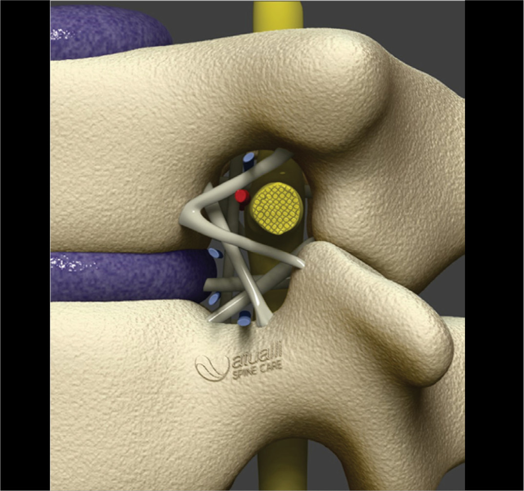
The geometry of the lumbar foramen was described as an oval, round, or inverted teardrop-shaped “window” in the lateral aspect of the lumbar spine. The anatomical limits of the foramen consist of the adjacent vertebral pedicles superiorly and inferiorly, the posteroinferior margin of the upper vertebral body, the intervertebral disc, the posterosuperior notch of the lower vertebral body anteriorly, and the yellow ligament and the superior and inferior articular facets posteriorly. The upper part of the foramen is occupied by neural tissue in more than 50% (Figure 1)16,17.

Although the morphology of the foramen, especially regarding stenotic changes, has been investigated in the literature, there is no agreement on what constitutes a normal lumbar foramen.18 The shape of the lumbar intervertebral foramen differs depending on the direction from which it is viewed. A macroscopic anatomical view of the lumbar spine in healthy individuals shows that the intervertebral foramen cannot be identified from the dorsal side (Figure 2), but becomes gradually visible when viewed laterally at an angle of 30°, and is maximized at a lateral angle of 90° from the left. From the superior intervertebral foramen L1-L2 to the inferior level L4-L5, a progressive widening is observed.19 There is variation in the dimensions of the foramen with the level of the spine, sex, and age. The height of the foramen varies between 19 and 21 mm, and the superior and inferior sagittal diameters of the foramen vary between 7 and 8 mm and between 5 and 6 mm, respectively, being larger in the upper lumbar foramina and smaller in the lower ones.18

There is condensation of the connective tissue within the foramen, which forms the transforaminal ligaments and divides the foramen into compartments, separating the neural structures from the vascular structures. Previously considered anomalous structures, transforaminal ligaments were not widely known, and the criteria for identifying and classifying them are not universal. They are, however, of potential importance during neurological procedures, as their entrapment can traction the nerve root. Transforaminal ligaments are not present in all patients, but when they are, the incidence of all types of ligaments is significantly higher, with the most common type being the superior corporotransverse ligament. By decreasing the total amount of space available for the passage of the spinal nerve, it is concluded that the foraminal ligaments may be the cause of nerve root entrapment, resulting in radicular pain. However, some studies claim that these ligaments do not cause radicular pain, but rather serve to protect nerves and vessels.20-22
It is important to identify the emerging root, as well as the compartments and neurovascular relationships. As the nerve root slides under the medial edge of the pedicle, it takes an oblique direction, leaving the pedicle. The spinal nerve reaches the foraminal exit, curves anterolaterally around the base of the underlying pedicle and the transverse process, and divides into anterior and posterior primary branches. The dorsal root ganglion (DRG) increases in diameter from L1 to L5, with the upper ganglia being more lateral and the lower ones more medial. As the root canal S1 is short, the DRG for S1 runs practically intraforaminal. Postoperative dysesthesia is related to excessive manipulation or damage caused by increased temperature in the ganglion23.
The ligaments in the lumbar intervertebral foramen are classified as follows: bands placed obliquely are considered upper and lower corporotransverse ligaments, while bands that operate transversely are called upper and lower transforaminal ligaments23. The two body-transverse ligaments are mainly distributed in the intervertebral foramen L5-S1: the superior body-transverse ligament attaches from the posterolateral corner of the vertebral body to the accessory process of the transverse process of the same vertebra; the inferior body-transverse ligament connects the same posterolateral corner of one vertebral body to the transverse process below23. The transforaminal ligaments are more superior in the intervertebral foramina L1-L423, the superior transforaminal ligaments attach to the inferior vertebral notches and the inferior ones attach to the superior vertebral notches; the middle transforaminal ligaments cross from the posterolateral corner of a fibrous ring to the yellow ligament behind and the zygapophyseal joint capsule24.
The anatomical location of the transforaminal ligaments led many early studies to conclude that these condensations of the fascia were the cause of nerve root entrapment, resulting in radiating pain, as they decreased the available space for the passage of the spinal nerve21,22,25-28. It has been reported that the cross-sectional area of a foramen decreases by up to 30% due to these fascia bands21,22,28. However, Kuofi, et al.26 mapped the ligaments topographically, establishing their consistent presence in the foramina, and concluded, contrary to previous studies, that they did not cause nerve root entrapment leading to radicular pain. Instead, they protected more nerves and vessels23,25. Clearly, a more in-depth investigation into these structures is necessary.
The possibility of imaging studies for individual anatomical analysis of the intervertebral foramen and its structures enables targeted preoperative planning. Can help choose appropriately sized dilators or endoscopic instruments, assist in locating and determining the amount of articular facet to be resected in the procedure29.
The lumbar arteries from L1-L2 to L4-L5 were divided into three groups of terminal branches in the lateral zone of the lumbar intervertebral foramen: the anterior, spinal, and dorsal branches. The lumbar branch of the iliolumbar artery or the inferior arterial branch L4 entered the foramen L5-S1 in some cases. The previous branch groups were mainly previous branches of the transverse process (anterior branch, anterior artery of the transverse process). The previous branch of the transverse process was the abdominal branch, thick, and formed many muscular branches along the anterior part of the transverse process, with the communicating branches and the adjacent anterior branches of the transverse process anastomosed. The extra-spinal arterial network was made up of the dorsal branch of the interarticular artery and the upper and lower articular arteries. The upper and lower articular arteries crossed in front of the tendinous arch of the intertransverse ligament and ran along the accompanying vein23.
In 1991, Parviz Kambin described a triangular region for safe endoscopic access, referred to as Kambin’s safety triangle.30 The emerging root is the hypotenuse, the upper edge of the lower vertebral body forms one of the legs, and the superior articular process of the caudal vertebra forms the other leg. Using this path, there is a safe route for the passage of cannulas and endoscopic instruments.31 The measurements of Kambin’s triangle in cadavers revealed that the area of the triangle becomes wider as it moves to the lower level of the lumbar spine (L4-L5)32. It is of great importance to have knowledge of the work zone for the TF33,34 approach. The relationship between the course of the emerging root and the intervertebral discs is such that the angle of the nerve root bifurcation becomes more pronounced in the coronal section as it moves towards the L5 nerve root (lower level)35,36.
The Kambin triangle becomes narrow based on the reduction of the height of the intervertebral disc, as occurs in the degeneration of the intervertebral discs and the zygapophyseal joints associated with aging.19 Complications due to erroneous approach during endoscopic access can lead to radicular pain (13-40%) due to root injury or injuries to periradicular arteries that produce epidural hematoma or spinal ischemia if the injury occurs in the Adamkiewicz. Therefore, it is relevant for the surgeon to recognize possible anatomical variations of the spine37.
Ozer et al.38 demonstrated a significant anatomical variation and proposed a classification into three types, according to the appearances of the surgical view and the findings of the study with cadavers. The first type is a closed triangle with no available space between the elements of the triangle, the second type is a narrow triangle, and the third type is the normal triangle, as described by Kambin. In this study, 48 triangles were studied and of these only 10 (20.8%) are classified as type 3; 23 (48%) as type 2, and 15 (31.2%) as type 1. That is, 79.2% of the triangles were considered abnormal.
It is known that the medial border of the triangle is composed of bone tissue (articular process and superior articular facet of the caudal vertebra) and nervous tissue (crossing root and dura) under the bone39,40. However, in most cases, even in young people without facet hypertrophy, the actual working area is small and the bone tissue covers the triangle38. Thus, it becomes necessary to remove a part of the joint process to create enough space for endoscopic action. Foraminoplasty represents an important step for access to the intervertebral foramen or lateral recess41.
In endoscopic spine surgery, it is very common for us to locate ourselves based on the clock hands. For example, when we are approaching the L3-L4 foramen on the right, we have at 3 o’clock the L3 root (emergent), at 6 o’clock the intervertebral disc, at 9 o’clock the pedicle of L4, and at 12 o’clock the superior articular process of L4 (Figure 3). The knowledge of anatomical structures directly impacts the outcome of the endoscopic surgical procedure.

Kambim triangle and its anatomical relations with the emerging root (3h), intervertebral disc (6h), inferior pedicle (9h), and facet joint (12h).
CONCLUSION
The transforaminal access forced the spine surgeon to know in detail the intervertebral foramen and all the structures that pass through it. A detailed preoperative analysis of the foramen allows access through the safety corridor and avoids neurological complications.
-
Study conducted by the Clínica Atualli, São Paulo, SP, Brazil.
REFERENCES
-
1 Hahn BS, Park JY. Incorporating New Technologies to Overcome the Limitations of Endoscopic Spine Surgery: Navigation, Robotics, and Visualization. World Neurosurg. 2021;145:712-21. doi: 10.1016/j.wneu.2020.06.188.
» https://doi.org/10.1016/j.wneu.2020.06.188. -
2 Chen KT, Jabri H, Lokanath YK, Song MS, Kim JS. The evolution of interlaminar endoscopic spine surgery. J Spine Surg. 2020;6(2):502-12. doi: 10.21037/jss.2019.10.06.
» https://doi.org/10.21037/jss.2019.10.06. -
3 Vaishnav AS, Othman YA, Virk SS, Gang CH, Qureshi SA. Current state of minimally invasive spine surgery. J Spine Surg. 2019;5(Suppl 1):S2-S10. doi: 10.21037/jss.2019.05.02.
» https://doi.org/10.21037/jss.2019.05.02. -
4 Huang CC, Fitts J, Huie D, Bhowmick DA, Abd-El-Barr MM. Evolution of Cervical Endoscopic Spine Surgery: Current Progress and Future Directions-A Narrative Review. J Clin Med. 2024;13(7):2122. doi: 10.3390/jcm13072122.
» https://doi.org/10.3390/jcm13072122. -
5 Chen KT, Kim JS, Huang APH, Lin MHC, Chen CM. Current Indications for Spinal Endoscopic Surgery and Potential for Future Expansion. Neurospine. 2023;20(1):33-42. doi: 10.14245/ns.2346190.095.
» https://doi.org/10.14245/ns.2346190.095. -
6 Wirth F, Bergamaschi ECQA, Forti F da S, Bergamaschi JPM. Development of Indications for Endoscopic Spine Surgery: An Overview. Int J Transl Med. 2023;3(3):321-33. doi: 10.3390/ijtm3030023.
» https://doi.org/10.3390/ijtm3030023. -
7 Kim HS, Raorane HD, Wu PH, Yi YJ, Jang IT. Evolution of endoscopic transforaminal lumbar approach for degenerative lumbar disease. J Spine Surg. 2020;6(2):424-37. doi: 10.21037/jss.2019.11.05.
» https://doi.org/10.21037/jss.2019.11.05. -
8 Yeung AT. The Evolution and Advancement of Endoscopic Foraminal Surgery: One Surgeon’s Experience Incorporating Adjunctive Techologies. SAS J. 2007;1(3):108-17. doi: 10.1016/SASJ-2006-0014-RR.
» https://doi.org/10.1016/SASJ-2006-0014-RR. -
9 Yeung A, Lewandrowski KU. Five-year clinical outcomes with endoscopic transforaminal foraminoplasty for symptomatic degenerative conditions of the lumbar spine: a comparative study of inside-out versus outside-in techniques. J Spine Surg. 2020;6(Suppl 1):S66-S83. doi: 10.21037/jss.2019.06.08.
» https://doi.org/10.21037/jss.2019.06.08. -
10 Burkett D, Brooks N. Advances and Challenges of Endoscopic Spine Surgery. J Clin Med. 2024;13(5):1439. doi: 10.3390/jcm13051439.
» https://doi.org/10.3390/jcm13051439. -
11 Choi G, Pophale CS, Patel B, Uniyal P. Endoscopic Spine Surgery. J Korean Neurosurg Soc. 2017;60(5):485-97. doi: 10.3340/jkns.2017.0203.004.
» https://doi.org/10.3340/jkns.2017.0203.004. -
12 Kwon H, Park JY. The Role and Future of Endoscopic Spine Surgery: A Narrative Review. Neurospine. 2023;20(1):43-55. doi: 10.14245/ns.2346236.118.
» https://doi.org/10.14245/ns.2346236.118. -
13 Zhang J, Liu TF, Shan H, Wan ZY, Wang Z, Viswanath O, et al. Decompression Using Minimally Invasive Surgery for Lumbar Spinal Stenosis Associated with Degenerative Spondylolisthesis: A Review. Pain Ther. 2021;10(2):941-59. doi: 10.1007/s40122-021-00293-6.
» https://doi.org/10.1007/s40122-021-00293-6. -
14 Fanous A, Tumialán L, Wang M. Kambin’s triangle: definition and new classification schema. J Neurosurg Spine. 2019;32(3):390-8. doi: 10.3171/2019.8.SPINE181475.
» https://doi.org/10.3171/2019.8.SPINE181475. -
15 Kim DH, Choi G, Lee SH, Fessler RG (eds). Endoscopic Spine Surgery. Georg Thieme Verlag; 2018. doi: 10.1055/b-006-149730.
» https://doi.org/10.1055/b-006-149730. - 16 Gilchrist R V, Slipman CW, Bhagia SM. Anatomy of the Intervertebral Foramen. Pain Physician. 2002;5(4):372-8.
- 17 Stephens MM, Evans JH, O’Brien JP. Lumbar Intervertebral Foramens: An in Vitro Study of Their Shape in Relation to Intervertebral Disc Pathology. Spine (Phila Pa 1976). 1991;16(5):525-9.
- 18 Cinotti G, De Santis P, Nofroni I, Postacchini F. Stenosis of Lumbar Intervertebral Foramen: Anatomic Study on Predisposing Factors. Spine (Phila Pa 1976). 2002;27(3):223-9.
-
19 Uchikado H, Nishimura Y, Hattori G, Ohara Y. Micro-anatomical structures of the lumbar intervertebral foramen for full-endoscopic spine surgery: review of the literatures. J Spine Surg. 2020;6(2):405-14. doi: 10.21037/jss.2019.10.07.
» https://doi.org/10.21037/jss.2019.10.07. -
20 Qian Y, Qin A, Zheng MH. Transforaminal ligament may play a role in lumbar nerve root compression of foraminal stenosis. Med Hypotheses. 2011;77(6):1148-9. doi: 10.1016/j.mehy.2011.09.025.
» https://doi.org/10.1016/j.mehy.2011.09.025. -
21 Min JH, Kang SH, Lee JB, Cho TH, Suh JG. Anatomic Analysis of the Transforaminal Ligament in the Lumbar Intervertebral Foramen. Operative Neurosurgery. 2005;57(Suppl 1):37-41. doi: 10.1227/01.NEU.0000163481.58673.1A.
» https://doi.org/10.1227/01.NEU.0000163481.58673.1A. -
22 Zhao Q, Zhong E, Shi B, Li Y, Sun C, Ding Z. The morphology and clinical significance of the intraforaminal ligaments at the L5-S1 levels. Spine J. 2016;16(8):1001-6. doi: 10.1016/j.spinee.2016.03.048.
» https://doi.org/10.1016/j.spinee.2016.03.048. -
23 Yuan SG, Wen YL, Zhang P, Li YK. Ligament, nerve, and blood vessel anatomy of the lateral zone of the lumbar intervertebral foramina. Int Orthop. 2015;39(11):2135-41. doi: 10.1007/s00264-015-2831-6.
» https://doi.org/10.1007/s00264-015-2831-6. -
24 Umeh R, Fisahn C, Burgess B, Iwanaga J, Moisi M, Oskouian RJ, et al. Transforaminal Ligaments of the Lumbar Spine: A Comprehensive Review. Cureus. 2016;8(10):e811. doi: 10.7759/cureus.811.
» https://doi.org/10.7759/cureus.811. -
25 Kyung-Hoon Kim, Daniel H. Kim, Chapter 1 - Diagnosis and Treatment of Spinal Pain, Editor(s): Daniel H. Kim, Yong-Chul Kim, Kyung-Hoon Kim, Minimally Invasive Percutaneous Spinal Techniques, W.B. Saunders, 2010, Pages 1-28, ISBN 9780702029134, https://doi.org/10.1016/B978-0-7020-2913-4.00001-X
» https://doi.org/10.1016/B978-0-7020-2913-4.00001-X - 26 Amonoo-Kuofi HS, El-Badawi MG, Fatani JA. Ligaments Associated with Lumbar Intervertebral Foramina. 1. Li to L4. J Anat. 1988;156:177-83.
- 27 Amonoo-Kuofi HS, El-Badawi MG, Fatani JA, Butt MM. Ligaments Associated with Lumbar Intervertebral Foramina. 2. The Fifth Lumbar Level. J Anat. 1988;159:1-10.
-
28 Marić DL, Krstonošić B, Erić M, Marić DM, Stanković M, Milošević NT. An anatomical study of the lumbar external foraminal ligaments: appearance at MR imaging. Surg Radiol Anat. 2015;37(1):87-91. doi: 10.1007/s00276-014-1320-8.
» https://doi.org/10.1007/s00276-014-1320-8. -
29 Sclafani JA, Kim CW. Complications Associated with the Initial Learning Curve of Minimally Invasive Spine Surgery: A Systematic Review. Clin Orthop Relat Res. 2014;472(6):1711-7. doi: 10.1007/s11999-014-3495-z.
» https://doi.org/10.1007/s11999-014-3495-z. - 30 Kambin P. Arthroscopic microdiskectomy. Mt Sinai J Med. 1991;58(2):159-64.
- 31 Kambin P, Brager MD. Percutaneous posterolateral discectomy. Anatomy and mechanism. Clin Orthop Relat Res. 1987;(223):145-54.
-
32 Hoshide R, Feldman E, Taylor W. Cadaveric Analysis of the Kambin’s Triangle. Cureus. 2016;8(2):e475. doi: 10.7759/cureus.475.
» https://doi.org/10.7759/cureus.475. -
33 Hardenbrook M, Lombardo S, Wilson MC, Telfeian AE. The anatomic rationale for transforaminal endoscopic interbody fusion: a cadaveric analysis. Neurosurg Focus. 2016;40(2):E12. doi: 10.3171/2015.10.FOCUS15389.
» https://doi.org/10.3171/2015.10.FOCUS15389. -
34 Lertudomphonwanit T, Keorochana G, Kraiwattanapong C, Chanplakorn P, Leelapattana P, Wajanavisit W. Anatomic Considerations of Intervertebral Disc Perspective in Lumbar Posterolateral Approach via Kambin’s Triangle: Cadaveric Study. Asian Spine J. 2016;10(5):821-7. doi: 10.4184/asj.2016.10.5.821.
» https://doi.org/10.4184/asj.2016.10.5.821. -
35 Suh SW, Shingade VU, Lee SH, Bae JH, Park CE, Song JY. Origin of lumbar spinal roots and their relationship to intervertebral discs. J Bone Joint Surg Br. 2005;87-B(4):518-22. doi: 10.1302/0301-620X.87B4.15529.
» https://doi.org/10.1302/0301-620X.87B4.15529. -
36 Wu YS, Lin Y, Zhang XL, Tian NF, Sun LJ, Xu HZ, et al. The Projection of Nerve Roots on the Posterior Aspect of Spine From T11 to L5. Spine (Phila Pa 1976). 2012;37(20):E1232-7. doi: 10.1097/BRS.0b013e318265dd5d.
» https://doi.org/10.1097/BRS.0b013e318265dd5d. -
37 Ahn Y. Transforaminal percutaneous endoscopic lumbar discectomy: technical tips to prevent complications. Expert Rev Med Devices. 2012;9(4):361-6. doi: 10.1586/erd.12.23.
» https://doi.org/10.1586/erd.12.23. -
38 Ozer AF, Suzer T, Can H, Falsafi M, Aydin M, Sasani M, et al. Anatomic Assessment of Variations in Kambin’s Triangle: A Surgical and Cadaver Study. World Neurosurg. 2017;100:498-503. doi: 10.1016/j.wneu.2017.01.057.
» https://doi.org/10.1016/j.wneu.2017.01.057. - 39 Waxenbaum JA, Reddy V, Williams C, Futterman B. Anatomy, Back, Lumbar Vertebrae. StatPearls [Internet]; 2024.
-
40 Kim HS, Wu PH, An JW, Lee YJ, Lee JH, Kim MH, et al. Evaluation of Two Methods (Inside-Out/Outside-In) Inferior Articular Process Resection for Uniportal Full Endoscopic Posterolateral Transforaminal Lumbar Interbody Fusion: Technical Note. Brain Sci. 2021;11(9):1169. doi: 10.3390/brainsci11091169.
» https://doi.org/10.3390/brainsci11091169. -
41 Lokhande PV. Full endoscopic spine surgery. J Orthop. 2023;40:74-82. doi: 10.1016/j.jor.2023.04.010.
» https://doi.org/10.1016/j.jor.2023.04.010.
Edited by
-
Reviewed by:
Erasmo Zardo