 Transposition flap for desmotomy reconstruction in dogs with patellar luxation
ABSTRACT:
This study described a transposition flap that can be used as an adjunctive procedure for medial or lateral patellar luxations and to evaluate its use in dogs retrospectively. Dogs with grade 3 and grade 4 patellar luxation were retrospectively enrolled in this study. Five dogs had medial luxation, and three had lateral luxation. The transposition flap was used in 11 hind limbs with patellar luxation. After deepening the trochlear groove (chondroplasty or wedge recession trochleoplasty), transposition of the tibial tuberosity, and desmotomy, a triangular flap was created with the stretched parapatellar soft tissue in the opposite direction to the patellar luxation. The base of the flap was located approximately 1 cm proximal to the patella and the apex near the tibial tuberosity. After the flap was elevated by cutting through the apex, it was transposed to the desmotomy area, and its apex was sutured to the distal aspect of the desmotomy. The edges of the flap were then sutured to the medial and lateral edges of the desmotomy. All dogs had a good outcome with stabilized patellae and complete functional recovery at least 2 months after surgery. In conclusion, the transposition flap provide soft-tissue coverage to the desmotomy area and can be considered a complementary procedure for the stabilization of patellar luxation. However, the results should be interpreted as preliminary due to the limited sample size, short-term outcomes, and the descriptive nature of the analysis.
Key words:
dog; surgery; patella; soft tissue
RESUMO:
O objetivo do estudo foi descrever um retalho de transposição, que pode ser utilizado como um procedimento complementar para luxações patelares mediais ou laterais, bem como avaliar seu uso em cães de forma retrospectiva. Deste modo, cães com luxação patelar de graus 3 e 4 foram incluídos no estudo e desses cães cinco apresentavam luxação medial e três exibiam luxação lateral. Além disso, o retalho de transposição foi utilizado em 11 membros pélvicos com luxação patelar. Após o aprofundamento do sulco troclear (condroplastia ou trocleoplastia de recessão em cunha), transposição da tuberosidade tibial e desmotomia, um retalho triangular foi criado a partir do tecido parapatelar distendido, na direção oposta à luxação patelar. A base do retalho foi localizada aproximadamente 1 cm proximal à patela, e o ápice, próximo à tuberosidade tibial. Após o retalho ser elevado, com a secção no ápice, ele foi transposto para a área da desmotomia e o ápice suturado à margem caudal da desmotomia. As bordas do retalho foram então suturadas às margens medial e lateral da desmotomia. Todos os cães apresentaram bons resultados, com as patelas estabilizadas e recuperação funcional completa, por pelo menos 2 meses após a cirurgia. Portanto, foi possível concluir que o retalho de transposição fornece cobertura de tecido mole para a área da desmotomia e pode ser considerado um procedimento complementar para a estabilização da luxação patelar. No entanto, os resultados devem ser interpretados como preliminares devido ao tamanho limitado da amostra, a avaliação de curto prazo e a natureza descritiva da análise.
Palavras-chave:
cão; cirurgia; patela; tecido mole
INTRODUCTION
Patellar luxation is an orthopedic disorder observed in various breeds of dogs, with congenital/developmental cases being more common than acquired cases (HARASEN, 2006; DI DONA et al., 2018; PERRY & DÉJARDIN, 2021). The prevalence of patellar luxation can vary from country to country. For example, it was found in 1.30% of 210,824 dogs attending clinics in England (O’NEILL et al., 2016) and 9.2% of 8,694 orthopedic cases in Italian clinics, excluding fractures (BOSIO et al., 2017). The incidence rate of patellar luxation diagnoses in an insured Swedish population was 15.6 cases per 10,000 dog-years at risk (ENGDAHL et al., 2023). The direction of patellar luxation can be medial, lateral, or bidirectional, with medial patellar luxation being diagnosed more frequently in small-breed dogs (ROUSH, 1993; DI DONA et al., 2018; ENGDAHL et al., 2023).
Surgical treatment is most commonly indicated for dogs with lameness or hind limb deformity, with the choice of surgical technique - whether bony, soft tissue, or a combination - depending on factors such as the grade of luxation, severity of skeletal deformity, chronicity, age, size, body weight, and other individual considerations (HARASEN, 2006; PERRY & DÉJARDIN, 2021). Surgical intervention includes bony, soft tissue techniques, or combinations, which can be chosen according to luxation grade, skeletal deformity severity, chronicity, age, size and body weight of the dog, and other factors (HARASEN, 2006; PERRY & DÉJARDIN, 2021). However, a single surgical procedure is not suitable for correcting all alterations (ROUSH, 1993; DI DONA et al., 2018). Several techniques used to correct the luxation of the medial patella can also be used for lateral patellar luxation (ROUSH, 1993).
Deepening of the trochlear groove, tibial tuberosity transposition, and corrective osteotomies of the femur and tibia are bony techniques used to realign the extensor mechanism (PERRY & DÉJARDIN, 2021). Desmotomy, imbrication, anti-rotational sutures, capsulectomy, and quadriceps release are some of the soft tissue procedures that can be used to modify abnormal forces in immature dogs and as an adjunct to bony procedures in mature dogs (HARASEN, 2006; DI DONA et al., 2018).
Despite various surgical procedures to correct patellar luxation, transposition flaps using the stretched parapatellar soft tissue (joint capsule and retinaculum) are still underutilized (PALUMBO, 1971). Therefore, the purpose of this study was to describe a transposition flap that can be used as an adjunctive procedure for medial or lateral patellar luxations and to evaluate its use in dogs retrospectively.
MATERIALS AND METHODS
Dogs with grade 3 and/or grade 4 patellar luxation that were presented to the University Veterinary Hospital, between 2022 and 2023 were enrolled in this study. The inclusion criteria were dogs that underwent transposition flaps and had the same surgeon performing the surgical procedure. Exclusion criteria included incomplete clinical data or absence of follow-up. All owners read and signed a consent form for their dog’s surgery.
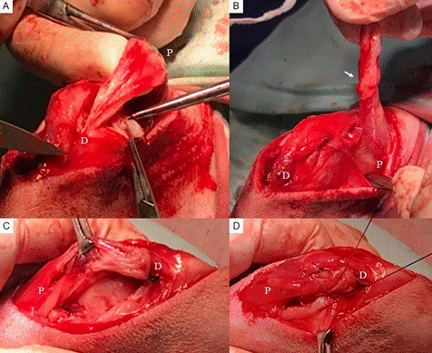
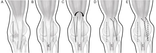
Under general anesthesia, each dog was positioned in dorsal recumbency and aseptically prepared for surgery. Briefly, the approach was medial parapatellar or lateral parapatellar for lateral or medial luxation, respectively. We deepened the trochlear groove (chondroplasty or wedge recession trochleoplasty), transpositioned the tibial tuberosity and fixed with a Kirschner wire according to the luxation direction, and performed a lateral desmotomy for lateral luxation or a medial desmotomy for medial luxation. A triangular flap was then created with the stretched parapatellar soft tissue (retinaculum and joint capsule) opposite the direction of patellar luxation (Figure 1A). The base of the flap was located approximately 1 cm proximal to the patella and adjusted proportionally to the desmotomy area, with the apex near the tibial tuberosity. After the flap was elevated by cutting through the apex, it was transposed to the desmotomy area. Before suturing the flap, the joint capsule was imbricated at the donor site (Figure 1B). The flap apex was then sutured to the distal aspect of the desmotomy, and the edges of the flap were sutured to the medial and lateral edges of the desmotomy using a cruciate mattress suture pattern of monofilament absorbable suture material (Figure 1C and Figure 1D). Figure 2 shows a schematic drawing of the technique. After confirming the stability of the patella during stifle manipulation, subcutaneous tissue, and skin were routinely sutured.
Transposition flap in a dog with medial patellar luxation. (A) Triangular flap created with the stretched parapatellar soft tissue on the lateral side; (B) Released flap after section of the apex region (arrow) and lateral imbrication to close the area; (C) Desmotomy area on the medial side; (D) Flap transposed to the medial desmotomy area being sutured to the edges of the desmotomy. P: Proximal, D: Distal.
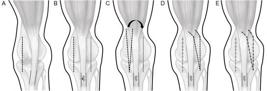
Schematic drawing of the transposition flap for a lateral luxation. (A) Medial parapatellar approach; (B) Transposition of the tibial tuberosity with a Kirschner (arrow) after deepening the trochlear groove and desmotomy on the lateral side; (C) Triangular flap created with the stretched parapatellar soft tissue on the medial side; (D) Flap transposed to the lateral desmotomy and imbrication of the joint capsule on the medial side; (E) Flap transposed sutured to the desmotomy area on the lateral side.
Clinical outcome was evaluated at least 2 months after the surgery. The outcome was based on previously reported criteria (BOSIO et al., 2017), as follows: good - stabilization of the patella and complete functional recovery, fair - recurrence of patellar luxation but with a less severe grade than the original, poor - recurrence of patellar luxation with the same grade or worse. The lameness grading score was as follows: 1 - none, 2 - barely perceptible, 3 - mild, 4 - severe with weight-bearing, 5 - severe with non-weight-bearing (GOH, 2025).
RESULTS AND DISCUSSION
Eight dogs met the inclusion criteria. Five dogs had a medial luxation and three had a lateral luxation. The medial luxation was bilateral in two animals and unilateral in three dogs. The lateral luxation was bilateral in one animal and unilateral in two dogs. The data are presented in table 1. All patellar luxations were classified as developmental, and no animal had a history of trauma. The transposition flap was used in 11 hind limbs with patellar luxation. Dogs with bilateral luxation underwent bilateral surgery in a single session.
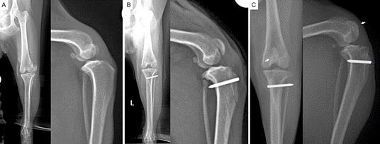
During the surgical procedure, the tibial tuberosity was separated from the tibia in two dogs (nos. 1, 2) due to tissue tension but was reattached with a Kirschner wire. In addition, a tension band was applied to two limbs. In the postoperative period, one dog (no. 7) experienced migration of the Kirschner wire three weeks after surgery. Although, the tibial tuberosity was not completely healed, no displacement was observed and the animal showed no lameness. Radiographic views of lateral patellar luxation before and after the surgery are illustrated in figure 3. The outcome was classified as good in all dogs two months after surgery. At that time, none of the dogs exhibited any signs of lameness.
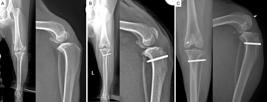
Craniocaudal and mediolateral radiographic views of a lateral patellar luxation (grade 3), respectively, before (A) and immediately after surgery (B). Radiographs taken two months after surgery demonstrated bone healing of the tibial crest, as well as the correct positioning of the patella within the femoral trochlear groove, as indicated by the white arrows (C).
The transposition flap is only useful when a desmotomy is required, as in the present cases. Desmotomy is used to release the contracted tissue on the same side of the patellar luxation, through an incision of the retinaculum and joint capsule (HARASEN, 2006; DI DONA et al., 2018; PERRY & DÉJARDIN, 2021). Due to the luxation grade in the present cases, the patella could not return to the trochlear groove without desmotomy. Other techniques, such as corrective osteotomies of the distal femur and proximal tibia (DI DONA et al., 2018; PERRY & DÉJARDIN, 2021), could avoid desmotomy but were not used due to the owners’ financial constraints.
The stretched parapatellar soft tissues in the opposite direction to the patellar luxation were used for the transposition flap development. Usually, these soft tissues are tightened using imbrication techniques or undergo partial resection to reduce soft tissue laxity (ROUSH, 1993; DI DONA et al., 2018). In the present study, the tissue to be removed during partial resection was used as a transposition flap. Flap transplantation has been described previously in dogs with patellar luxation, but the flap was lifted to preserve both ends of the original tissue (PALUMBO, 1971). Therefore, the desmotomy area is not completely closed.
In the present study, the size and width of the transposition flap were proportional to the extent of tissue laxity and the size of the desmotomy area. Consequently, flap dimensions - including thickness and size - varied depending on the individual animal. Although, it is reported that a releasing incision can be maintained open as synovial fluid leakage is not considered a problem (HARASEN, 2006), the use of a transposition flap can provide soft-tissue coverage to the area and help avoid tissue contraction during healing, which may lead to reluxation in the postoperative period. In addition, a study observed that the frequency of major complications in patellar luxation correction was higher after retinacular or capsular release, which was associated with less proximodistal soft tissue support (ARTHURS & LANGLEY-HOBBS, 2006). Thus, the transposition flap could reduce avulsion forces because if the soft-tissue structures are intact, the tibial tuberosity transposition site can be relieved from partial load-sharing (ARTHURS & LANGLEY-HOBBS, 2006).
In addition to the transposition flap, the imbrication technique was also used. The balance between releasing and tightening of the soft tissue must be done carefully to maintain normal patellar tracking, prevent reluxation or even induce iatrogenic luxation (PERRY & DÉJARDIN, 2021).
No local complications were reported in any of the dogs. The complications observed in the present study, such as detachment of the tibial tuberosity from the tibia during the surgical procedure and postoperative pin migration, were not related to the transposition flap. Reluxation and complications related to the implants are among the most common complications after corrective surgery for patellar luxation, followed by avulsion of the tibial crest (HARASEN, 2006; CASHMORE et al., 2014; DI DONA et al., 2018). Pin migration in case no. 7 may be related to the Kirschner wire direction. A congenital medial patellar luxation study verified that Kirschner wire directed caudodistally, or the absence of a tension band increased the risk of avulsion after surgery (CASHMORE et al., 2014).
Recurrence of luxation was not observed in the present case despite the grades of patellar luxation. Major complications occur more often in grade 3 and grade 4 patellar luxation operations, as seen in the present cases (ARTHURS & LANGLEY-HOBBS, 2006; BOSIO et al., 2017; DI DONA et al., 2018; PERRY & DÉJARDIN, 2021) which occurred in the present cases. The deepening of the trochlear groove and tibial tuberosity transposition were used to minimize the underlying anatomical abnormalities. Tibial tuberosity transposition has been reported to be responsible for the lower frequency of patellar reluxation and postoperative complications (ARTHURS & LANGLEY-HOBBS, 2006).
This study has some limitations. First, the number of dogs was small; although, 11 limbs were operated on. Second, each joint underwent multiple surgical procedures, including the transposition flap, making it difficult to isolate the specific effect of the flap. Additionally, the absence of ultrasound imaging limited the ability to monitor flap changes during the healing process. Therefore, further studies with a larger number of dogs and other imaging techniques are needed.
CONCLUSION
In conclusion, the described method showed that the transposition flap provides soft-tissue coverage to the desmotomy area and can be considered as complementary procedure for the stabilization of patellar luxation. However, the results should be interpreted as preliminary due to the limited sample size, short-term outcomes, and the descriptive nature of the analysis.
ACKNOWLEDGMENTS
We want to thank Financiadora de Estudos e Projetos (FINEP); Grant 01.12.0530.00), Conselho Nacional de Desenvolvimento Científico e Tecnológico (CNPq) - PQ 305813/2023-4), and PROPe, through the Emergency Support for Multiuser Equipment at Universidade Estadual Paulista (Unesp) (Call 06/2024). The study was financed in part by the Coordenação de Aperfeiçoamento de Pessoal de Nível Superior (CAPES), Brasil - Finance code 001.
REFERENCES
-
ARTHURS, G. I.; LANGLEY-HOBBS, S. J. Complications associated with corrective surgery for patellar luxation in 109 dogs. Veterinary Surgery, v.35, n.6, p.559-566, 2006. Available from: <https://onlinelibrary.wiley.com/doi/10.1111/j.1532-950X.2006.00189.x?__cf_chl_tk=UwwRyj245KAu9nRrarWAzLjdf8ZdURwMXZjps.9lLcY-1773751977-1.0.1.1-yujbVPAcCtiGCLEclYW5CWVGZgTuftKCGyenQL2t3JE>. Accessed: Apr. 07, 2025. doi: 10.1111/j.1532-950X.2006.00189.x.
» https://doi.org/10.1111/j.1532-950X.2006.00189.x.» https://onlinelibrary.wiley.com/doi/10.1111/j.1532-950X.2006.00189.x?__cf_chl_tk=UwwRyj245KAu9nRrarWAzLjdf8ZdURwMXZjps.9lLcY-1773751977-1.0.1.1-yujbVPAcCtiGCLEclYW5CWVGZgTuftKCGyenQL2t3JE -
BOSIO, F. et al. Prevalence, treatment and outcome of patellar luxation in dogs in Italy. A retrospective multicentric study (2009-2014). Veterinary and Comparative Orthopaedics and Traumatology, v.30, n.5, p.364-370, 2017. Available from: <Available from: https://www.thieme-connect.com/products/ejournals/abstract/10.3415/VCOT-16-05-0073 >. Accessed: Apr. 07, 2025. doi: 10.3415/VCOT-16-05-0073.
» https://doi.org/10.3415/VCOT-16-05-0073.» https://www.thieme-connect.com/products/ejournals/abstract/10.3415/VCOT-16-05-0073 -
CASHMORE, R. G. et al. Major complications and risk factors associated with surgical correction of congenital medial patellar luxation in 124 dogs. Veterinary and Comparative Orthopaedics and Traumatology, v.27, n.4, p.263-270, 2014. Available from: <Available from: https://www.thieme-connect.com/products/ejournals/abstract/10.3415/VCOT-13-08-0100 >. Accessed: Apr. 07, 2025. doi: 10.3415/VCOT-13-08-0100.
» https://doi.org/10.3415/VCOT-13-08-0100.» https://www.thieme-connect.com/products/ejournals/abstract/10.3415/VCOT-13-08-0100 -
DI DONA, F. et al. Patellar luxation in dogs. Veterinary Medicine (Auckland, N.Z.), v.31, n.9, p.23-32, 2018. Available from: <Available from: https://pmc.ncbi.nlm.nih.gov/articles/PMC6055913/ >. Accessed: Apr. 07, 2025. doi: 10.2147/VMRR.S14254.
» https://doi.org/10.2147/VMRR.S14254.» https://pmc.ncbi.nlm.nih.gov/articles/PMC6055913/ -
ENGDAHL, K. et al. The epidemiology of patellar luxation in an insured Swedish dog population. Preventive Veterinary Medicine, v.220, p.106034, 2023. Available from: <Available from: https://www.sciencedirect.com/science/article/pii/S0167587723001988?via%3Dihub >. Accessed: Apr. 07, 2025. doi: 10.1016/j.prevetmed.2023.106034.
» https://doi.org/10.1016/j.prevetmed.2023.106034.» https://www.sciencedirect.com/science/article/pii/S0167587723001988?via%3Dihub -
GOH, C. S. S. Top questions about orthopedic examinations from general practitioners. Todays Veterinary Practice, v.May/June, p.96-99, 2025. Available from: <Available from: https://todaysveterinarypractice.com/orthopedics/top-questions-about-orthopedic-examinations-from-general-practitioners/ >. Accessed: Apr. 07, 2025.
» https://todaysveterinarypractice.com/orthopedics/top-questions-about-orthopedic-examinations-from-general-practitioners/ -
HARASEN, G. Patellar luxation: pathogenesis and surgical correction. The Canadian Veterinary Journal, v.47, n.10, p.1037-1039, 2006. Available from: <Available from: https://pmc.ncbi.nlm.nih.gov/articles/PMC1571132/ >. Accessed: Apr. 07, 2025. PMID: 17078257.
» https://pmc.ncbi.nlm.nih.gov/articles/PMC1571132/ -
O’NEILL, D. G. et al. The epidemiology of patellar luxation in dogs attending primary-care veterinary practices in England. Canine Genetics and Epidemiology, v.3, p.4, 2016. Available from: <Available from: https://cgejournal.biomedcentral.com/articles/10.1186/s40575-016-0034-0 >. Accessed: Apr. 07, 2025. doi: 10.1186/s40575-016-0034-0.
» https://doi.org/10.1186/s40575-016-0034-0.» https://cgejournal.biomedcentral.com/articles/10.1186/s40575-016-0034-0 - PALUMBO, N. E. A new technic for repair of canine patellar luxations. Modern Veterinary Practice, v.52, p.51-27, 1971.
-
PERRY, K. L.; DÉJARDIN, L. M. Canine medial patellar luxation. Journal of Small Animal Practice, v.62, n.5, p.315-335, 2021. Available from: <Available from: https://onlinelibrary.wiley.com/doi/10.1111/jsap.13311 >. Accessed: Apr. 07, 2025. doi: 10.1111/jsap.13311.
» https://doi.org/10.1111/jsap.13311.» https://onlinelibrary.wiley.com/doi/10.1111/jsap.13311 -
ROUSH, J. K. Canine patellar luxation. Veterinary Clinics of North America: Small Animal Practice, v.23, n.4, p.855-868, 1993. Available from: <Available from: https://www.sciencedirect.com/science/article/abs/pii/S0195561693500876?via%3Dihub >. Accessed: Apr. 07, 2025. doi: 10.1016/s0195-5616(93)50087-6.
» https://doi.org/10.1016/s0195-5616(93)50087-6.» https://www.sciencedirect.com/science/article/abs/pii/S0195561693500876?via%3Dihub
BIOETHICS AND BIOSECURITY COMMITTEE APPROVAL
-
CR-2025-0222.R2
-
We authors of the article entitled “Transposition flap for desmotomy reconstruction in dogs with patellar luxation” declared that, for all relevant purposes, the project that generated the data presented in this paper was not submitted for evaluation to the Ethics Committee of the University/Research Institute. This is because we used cases treated as part of the routine hospital procedures, rather than in an experimental setting. We are also aware of the content of the Brazilian resolutions of the Conselho Nacional de Controle de Experimentação Animal (CONCEA) <http://www.mct.gov.br/index.php/content/view/310553.html> if it involves animals. Thus, the authors assume full responsibility for the presented data and are available for possible questions, should they be required by the competent authorities.
-
DATA AVAILABILITY STATEMENT
The data supporting the findings of this study are available directly from the correspondent author upon request.
-
DECLARATION OF USE OF ARTIFICIAL INTELLIGENCE
In this paper, we did not use AI tools for paper writing and data processing, but only used it to enhance the quality of writing.
Edited by
-
ASSOCIATE EDITOR:
Rudi Weiblen (0000-0002-1737-9817)
-
SCIENTIFIC EDITOR:
Alexandre Pinto Ribeiro (0000-0002-8922-2886)
The data supporting the findings of this study are available directly from the correspondent author upon request.