 Causes and pathological aspects of neuroinflammatory diseases of the central nervous system in cats
ABSTRACT:
Neuroinflammatory diseases in cats can have various causes; however, comprehensive studies on these conditions are limited. This study described the causes and pathological aspects of 60 cases of inflammatory lesions in the central nervous system (CNS) of domestic cats based on a 10-year retrospective analysis of necropsies. Infectious diseases were identified in 47 of 60 cases (78.3%), including viral (28/60, 46.7%), fungal (9/60, 15%), bacterial (5/60, 8.3%) and parasitic (5/60, 8.3%) infections. The main aetiological agent was feline infectious peritonitis virus (27/60, 45%), followed by Cryptococcus sp. (9/60, 15%), Streptococcus sp. (4/60, 6.7%), Toxoplasma gondii (2/60, 3.3%), Gurltia paralysans (2/60, 3.3%), rabies virus (1/60, 1.6%) and Sarcocystis neurona (1/60, 1.6%). The most frequent histopathological lesions were pyogranulomatous and granulomatous meningoencephalitis. Our results highlighted the most common neuroinflammatory diseases affecting the feline CNS and underscore the importance of thorough diagnostic processes. Detailed pathological analysis and supplementary laboratory tests are crucial for the accurate diagnosis of these neurologic conditions.
Key words:
central nervous system; feline; Felis catus; neuropathology; feline infectious peritonitis virus
RESUMO:
As doenças neuroinflamatórias em gatos podem surgir de diversas causas, e estudos abrangentes sobre essas condições são limitados na literatura. Este estudo tem como objetivo descrever as causas e os aspectos patológicos de 60 casos de lesões inflamatórias no sistema nervoso central (SNC) de gatos domésticos, com base em uma análise retrospectiva de 10 anos de necropsias realizadas. Para realizar o objetivo, as doenças infecciosas foram identificadas em 47 dos 60 casos (78,3%), incluindo infecções virais (28/60, 46,7%), fúngicas (9/60, 15%), bacterianas (5/60, 8,3%) e parasitárias (5/60, 8,3%). O principal agente etiológico foi o vírus da peritonite infecciosa felina (27/60, 45%), seguido por Cryptococcus sp. (9/60, 15%), Streptococcus sp. (4/60, 6,7%), Toxoplasma gondii (2/60, 3,3%), Gurltia paralysans (2/60, 3,3%), vírus da raiva (1/60, 1,6%) e Sarcocystis neurona (1/60, 1,6%). Treze casos (21,7%) de encefalite e meningoencefalite tiveram causas desconhecidas. Além disso, as lesões histopatológicas mais frequentes foram as meningoencefalites piogranulomatosas e as granulomatosas. Nossos resultados destacam as doenças neuroinflamatórias mais comuns que afetam o SNC felino e ressaltam a importância de processos diagnósticos minuciosos. A análise patológica detalhada e exames laboratoriais complementares são fundamentais para o diagnóstico preciso dessas condições neurológicas.
Palavras-chave:
sistema nervoso central; felino; Felis catus; neuropatologia; vírus da peritonite infecciosa felina
INTRODUCTION
Neuroinflammatory diseases in animals include a variety of inflammatory lesions in the central nervous system (CNS), that have unique characteristics owing to the organization and anatomical arrangement of this system (MILLER & PORTER, 2022). Inflammation, which may be minor or asymptomatic in other tissues, can lead to death or permanent disability if it involves the CNS (CANTILE & YOUSSEF, 2016). In routine diagnosis, these lesions are traditionally categorized by their composition and the specific tissue affected (encephalitis, myelitis, ependymitis, choroiditis and meningitis) (CANTILE & YOUSSEF, 2016). Recognizing the patterns of CNS inflammation is crucial for identifying its cause and defining differential diagnosis (GUNN-MOORE & REED, 2011). In some instances, serous to suppurative or purulent inflammation can suggest a bacterial infection; whereas, eosinophilic inflammation can be associated with parasitic migrations. Moreover, lymphoplasmacytic and histiocytic inflammation is typically seen with viral or protozoal infections, and granulomatous inflammation is usually caused by fungi, protozoa and certain bacteria (MILLER & PORTER, 2022).
Neuroinflammatory diseases in domestic animals can arise from various causes, either as primary lesions or secondary to a systemic process (MILLER & PORTER, 2022). In cats, infectious agents such as feline infectious peritonitis (FIP) virus, Cryptococcus sp. and Toxoplasma gondii are the predominant causes (BRADSHAW et al., 2004). Occasional bacterial infections may also occur (SCHWAB et al., 2007). However, identifying the exact cause of inflammatory lesions in the CNS can be challenging because pathological findings and complementary tests often yield inconclusive results (BRADSHAW et al., 2004; SCHWAB et al., 2007; NEGRIN et al., 2017; NESSLER et al., 2020). Comprehensive studies on the frequency and pathological aspects of neuroinflammatory diseases in cats are limited. Current literature mainly comprises case reports, general studies on nervous system diseases, and specific disease investigations (BRADSHAW et al., 2004; CHAVES et al., 2018; RISSI, 2018; RODRIGUES et al., 2020). Therefore, this manuscript described the causes and pathological aspects of CNS inflammatory lesions in domestic cats, based on cases diagnosed in southern Brazil.
MATERIALS AND METHODS
Case selection
Electronic reports of cats submitted for necropsy between January 2013 and December 2023 to the Department of Veterinary Pathology at the Universidade Federal do Rio Grande do Sul were searched for cases with inflammatory lesions in the CNS. Inclusion criteria encompassed cases with gross and histological descriptions of encephalitis, myelitis, ependymitis, choroiditis, meningitis, leptomeningitis, meningoencephalitis, or meningomyelitis. Information extracted from records included signalment (sex, breed, and age), concurrent comorbidities, and the status of feline immunodeficiency virus (FIV) and feline leukaemia virus (FeLV) infection. FIV and FeLV status were previously determined through immunohistochemical, serological, or molecular tests (DE MELLO et al., 2023). Formalin-fixed paraffin-embedded tissue blocks of the selected cases were retrieved from our archives. Cases with unavailable CNS tissue or severe autolysis compromising tissue quality were excluded.
Pathologic analysis
Original gross descriptions and photographs of the selected cases were examined. The CNS tissues were anatomically divided into the cerebrum, cerebellum, brainstem and spinal cord. Cerebrum and cerebellum samples were evaluated from all animals; however, samples from all four regions (including the brainstem and spinal cord) were not available for every case. Although, tissue trimming standardization in our lab involves coronal sections of these regions, samples from all regions were not consistently available. Tissues from all cases were sectioned and stained with haematoxylin and eosin (HE). Periodic acid-Schiff reaction (PAS), Alcian blue (AB) and Gram stains were used to highlight intralesional fungi or bacteria when necessary. Histological lesions were classified based on anatomical location, affected tissue (ventriculus, neuropil, and leptomeninges), distribution (focal, multifocal, multifocal to coalescent, focally extensive, and diffuse) and characteristics of the inflammatory infiltrate. Inflammatory infiltrate were classified according to the cell composition and graded as discrete, moderate, or marked. Additional findings, such as fibrin exudation, haemorrhage, fibrinoid vasculitis, and gliosis, were assessed as present or absent.
Diagnostics criteria establishments
The cause of the CNS lesions in all selected cases was determined using histological findings and ancillary tests previously performed by the original veterinary pathologists. These ancillary tests included routine bacterial and fungal culture, direct fluorescent antibody test (DFAT) for rabies, immunohistochemistry (IHC) for specific agents (Table 1) and/or molecular tests (nested PCR and sequencing for Sarcocytis neurona) (HAMMERSCHMITT et al., 2020) on CNS tissues. Cases were categorized based on the etiological agent of the inflammatory process (viral, bacterial, fungal or parasitic) or were grouped into a category with an unknown cause. All cases were tested for FIV and FeLV, in cases with unknown status, testing for FIP was also performed. The diagnostic criteria for each category were as follows: 1) viral diseases: feline coronavirus antigen by IHC or rabies antigen via both IHC and DFAT; 2) fungal diseases: intralesional fungal structures using PAS and AB stains and/or fungal isolation; 3) bacterial diseases: intralesional bacteria through Gram staining or bacterial isolation; 4) parasitic disease: Toxoplasma gondii antigen by IHC; Sarcocystis neurona antigen and DNA through IHC and nested PCR, respectively; or intralesional parasite.
RESULTS
Of the 2,287 cats necropsied during the study period, 72 (3.1%) were diagnosed with inflammatory lesions in the CNS. Sixty cases were included in this study. The selected cats included 34/60 (56.6%) males and 26/60 (43.4%) females, with ages ranging from 30 days to 17 years (mean and median of 4.5 and 1 years, respectively). Previous retroviral status was available for all cats, of which 51.7% (31/60) were FIV- and FeLV-negative, 25% (15/60) were FeLV-positive, 13.3% (8/60) were FIV-positive, and 10% (6/60) were FIV- and FeLV-positive. Seven cats had significant comorbidities, including lymphoma, chronic renal disease, pulmonary carcinoma, hepatic carcinoid, pneumonia and cholangiohepatitis. Viral diseases were most common (28/60, 46.7%), followed by fungal (9/60, 15%), bacterial (5/60, 8.3%), and parasitic diseases (5/60, 8.3%). Thirteen cases had unknown cause (13/60, 21.7%). Table 2 summarizes the pathological aspects of each disease.
Viral
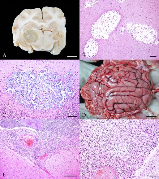
FIP was diagnosed in 27 cases, of which 15 were males and 12 were females. Their ages ranged from 1 month to 16 years (mean and median of 2.5 and 1 year, respectively). Most cats were mixed breed (21/27), followed by two Maine Coons, two Persians and two unidentified breeds. Of the 27 cases with available retroviral status, 10 were FeLV-positive, 2 were FeLV- and FIV positive, 2 were FIV-positive and 13 were FeLV- and FIV-negative. Macroscopically, there was multifocal fibrillar material deposition in the meninges and ventricle surface in 4 of 27 cases and dilatation of the lateral ventricles (Figure 1A) in 4 of 27 cases. Microscopy revealed moderate multifocal perivascular infiltrate of neutrophils, lymphocytes, plasma cells and macrophages (Figure 1B) in the meninges (21/27), cerebrum neuropil (21/27) and ventricles (9/27). Additional findings included fibrin exudation (14/27), fibrinoid vasculitis (8/27) (Figure 1C), fibrin thrombus (5/27) and hemorrhage (2/27) in the lesion areas. IHC for feline coronavirus revealed granular intracytoplasmic immunostaining in the macrophages (Figure 1D) in all cases.

(A) Feline infectious peritonitis. Telencephalon with dilation of the lateral ventricles and accumulation of fibrillar material in the ventricular surface. Bar, 1 cm. (B) Feline infectious peritonitis. Numerous inflammatory cells in the fourth ventricle and perivascular spaces of the leptomeninges and superficial brainstorm. Hematoxylin and eosin (HE). Bar, 50 µm. (C) Feline infectious peritonitis. Blood vessel in the neuroparenchyma with fibrinoid degeneration and fibrin deposition. HE. Bar, 100 µm. (D) Feline infectious peritonitis. Multifocal immunolabelling for FIP virus in cytoplasm of macrophages in the neuroparenchyma. Immunohistochemistry (IHC). Bar, 100 µm. (1E) Rabies. Intracytoplasmic viral inclusion in the cytoplasm of a Purkinje neuron. HE. Bar, 100 µm. (1F) Rabies. Multifocal immunolabelling for rabies virus in the cytoplasm of Purkinje neuron. IHC. Bar, 100 µm.
A 2-year-old male domestic shorthair cat was diagnosed with rabies. This cat had not been vaccinated against rabies and tested negative for FeLV and FIV. No macroscopic lesions were observed. The microscopic lesions were characterized by discrete multifocal perivascular infiltrates of lymphocytes and plasma cells in the neuropil of the telencephalon and brainstem. Eosinophilic intracytoplasmic inclusion bodies (Negri bodies) were observed in the neuronal aggregates and Purkinje neurons (Figure 1E). Granular immunostaining was observed for rabies virus in the cytoplasm and processes of neurons (Figure 1F), primarily in the brainstem and cerebellum. The diagnosis was confirmed using DFAT in an official laboratory.
Fungal
Cryptococcosis was the only fungal neuroinflammatory disease identified in this study. The mean age of the 9 affected cats was 7 years (median, 7,5 years), with 6 males and 3 females. These cats comprised 8 of 9 domestic shorthair and 1 Abyssinian. Of all cases with available retroviral status, three were FIV-positive, one FeLV- and FIV-positive and five FeLV- and FIV-negative. Macroscopically, 6 of 9 cases exhibited focal (2/6) to multifocal (4/6) nodular areas with a gelatinous and white appearance (Figure 2A) in the telencephalon, cerebellum and brainstem. Microscopy of these nodular areas revealed focal to multifocal aggregates of fungal structures (Figure 2B) admixed with discrete infiltrates of foamy vacuolated macrophages, lymphocytes and plasma cells in the meninges and neuropil of the affected tissues. These fungi were characterized by oval to round yeasts (5 to 10 µm in diameter) with a basophilic central structure (6 to 7 µm in diameter), surrounded by a thin basophilic wall (1 to 2 µm) and a clear mucinous thick capsule (10 to 20 µm). The capsule was positive by PAS and AB staining (Figure 2C). Additionally, one case had a fungal culture showing growth of Cryptococcus sp.
(A) Cryptococcosis. Transverse section of the brain with numerous intracerebral gelatinous pale masses. Bar, 2 cm. (B) Cryptococcosis. Neuroparenchyma with spaces filled with fungal yeasts morphologically consistent with Cryptococcus spp. and small number of inflammatory cells. HE. Bar, 50 µm. (C) Cryptococcosis. Neuroparenchyma with fungal yeasts. Alcian Blue stain. Bar, 100 µm. (D) Bacterial meningoencephalitis. Yellow multifocal areas and hyperemia on the surface by telencephalic leptomeninges. Bar, 2 cm. (E) Bacterial meningoencephalitis. Marked inflammatory infiltrate in the leptomeninges with thrombosis, fibrin deposition and aggregates of bacteria. HE. Bar, 100 µm. (F) Bacterial meningoencephalitis. Marked inflammatory infiltrate in the leptomeninges. HE. Bar, 100 µm.
Bacterial
There were 5 cases of bacterial infections in 3 females and 2 males. The ages ranged from 30 days to 2 years (mean and median of 7.4 months and 3 months, respectively). These cats comprised four domestic shorthair and one Maine Coon. Among the five cats with a retroviral status, only one tested positive for both FIV and FeLV. Macroscopically, there was a yellow suppurative exudate (Figure 2D) in the meninges and softening of the nervous tissue, mainly in the cerebellum and brainstem in three of five cases. Additionally, two cats exhibited extensive haemorrhagic areas with friable fibrillar material deposition, which was attributed to trauma. Microscopically, extensive areas of infiltrate composed of intact and degenerate neutrophils and macrophages with fibrin deposition, and bacterial aggregates (Figure 2E and Figure 2F) were observed in the meninges (5/5) and neuropil (3/5) of the affected areas. Areas of thrombosis and haemorrhage (3/5) were also observed. These intralesional bacteria are gram-positive. Bacterial culture was performed in four cases, resulting in the pure growth of Streptococcus canis in two cases and mixed culture of Streptococcus spp. and Staphylococcus spp. or Flavobacterium sp. in the remaining two cases. Unfortunately, it was not possible to determine the entry point of the analysed bacterial cases.
Parasitic
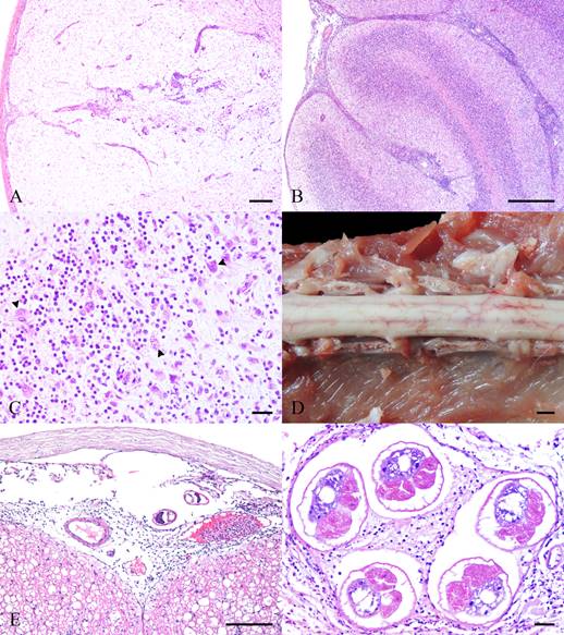
Two male domestic shorthair cats were diagnosed with toxoplasmosis. One cat was 1 year old, and the other was 3 years old. The retroviral status was available for both cats; however, only one tested positive for FeLV. Macroscopically, focal-to-multifocal circular yellow areas were observed in the brain and spinal cord. Histological examination of these areas revealed moderate-to-marked multifocal perivascular infiltrates of lymphocytes and plasma cells (Figure 3A), along with fibrinoid degeneration of the vascular wall. Numerous tachyzoites and occasional oval cystic structures (25 to 40 µm in diameter) filled with bradyzoites were observed in the lesion foci. Additional findings in one case included gliosis and malacia. IHC for T. gondii confirmed the diagnosis in both cases, showing granular immunostaining of cysts and tachyzoites associated with inflammatory foci.

(A) Toxoplasmosis. Neuroparenchyma with marked malacia. HE. Bar, 100 µm. (B) Sarcocystis neurona infection. Cerebellum with marked inflammatory infiltrate in the leptomeninges. HE. Bar, 100 µm. (C) Sarcocystis neurona infection. Moderate inflammatory infiltrate of lymphocytes, plasma cells and macrophages associated with parasitic schizonts in the neuroparenchyma. HE. Bar, 100 µm. (D) Gurltia paralysans infection. Lumbar segment of the spinal cord with small, congested, tortuous blood vessels. Bar, 1 cm. (E) G. paralysans infection. Leptomeninges of the spinal cord with two sections of intravascular parasites and perivascular inflammatory infiltrate. HE. Bar, 100 µm. (F) G. paralysans infection. Leptomeninges of the spinal cord with sections of intravascular parasites and perivascular inflammatory infiltrate. HE. Bar, 100 µm.
A 1-year-old male domestic shorthair cat presented with inflammatory lesions in the CNS caused by S. neurona. The cat tested positive for the FeLV and negative for the FIV. The only significant macroscopical finding was the cerebellar herniation. Microscopic examination revealed marked multifocal-to-coalescent infiltrates of degenerate neutrophils, macrophages and rare lymphocytes (Figure 3B) within the neuropil and meninges of the telencephalon and cerebellum. Associated with these lesions, there were occasional schizonts (15 to 30 µm in diameter) filled with numerous merozoites (Figure 3C). Additionally, moderate multifocal perivascular infiltrates of lymphocytes, plasma cells and macrophages were observed in the perivascular spaces. IHC and molecular tests confirmed the diagnostic of S. neurona. Detailed data of this case have been published elsewhere (HAMMERSCHMITT et al., 2020).
Infection by Gurltia paralysans was observed in two cats (a 9-month-old male domestic shorthair and an 8-year-old female domestic shorthair). The retroviral status was available, and both cats tested positive for FIV and negative for FeLV. Macroscopically, red areas were observed in meninges the spinal cord (Figure 3D). Histological examination revealed multifocal areas with moderate infiltrates by eosinophils and few lymphocytes, plasma cells, neutrophils, and macrophages in the meninges and adjacent neuropils of the brain and spinal cord. Lesion areas displayed blood vessel proliferation, fibrin thrombi and occasional cross-sections of nematode parasites (200 to 300 µm in diameter). These parasites had pseudocoelomic cavity, thin eosinophilic cuticle, coelomyarian musculature, and an intestinal tract composed of multinucleated cells and ovaries (Figure 3E and Figure 3F), compatible with G. paralysans.
Unknown cause
Encephalitis and meningoencephalitis of unknown cause were diagnosed in 13 cases, of which 8 were females and 5 were males. The ages ranged from 3 month to 17 years (mean and median of 9.6 and 10 years, respectively). Most cats were mixed breed (12/13), followed by one unidentified breed. Macroscopically, cerebellar herniation is present in 2 of 13 cases. Microscopically, the lesions were classified as lymphoplasmacytic encephalitis (5/13), granulomatous meningoencephalitis (4/13), lymphoplasmacytic meningoencephalitis (3/13) and suppurative meningoencephalitis (1/13). Retroviral status was available for all cases, with five cats positive for FeLV and all cats negative for FIV and FIP. However, no correlation was found between these agents and the lesions in the nervous system.
DISCUSSION
Neuroinflammatory diseases were uncommon in the studied feline population, comprising only 3.1% of the necropsies performed. This finding is similar to the previously reported frequencies of 3% in cats (EGENVALL et al., 2009) and 4.8% in dogs (ELBERT et al., 2022). Infectious diseases were the predominant cause of nervous system lesions in this study (77.9%), which is particularly noteworthy from a clinical perspective and highlights opportunities for the prevention and treatment of affected cats with neurological clinical signs (ADDIE et al., 2009; PENNISI et al., 2013). Retroviral infections, particularly FeLV, are associated with various infectious diseases, including those affecting the CNS (DE MELLO et al., 2023). Nevertheless, the rates of FIV and FeLV infection varied among the feline diseases identified in our study. Moreover, there was important variability in the ages of the affected cats, with only FIP showing a notably higher frequency in young animals, which is consistent with the existing literature (ADDIE, 2012; SLAVIERO et al., 2024). No specific predispositions were evident for other infectious diseases, and the small sample size precluded correlations.
In our study, a few cats with neuroinflammatory diseases exhibited macroscopic lesions. Cryptococcosis, FIP, bacterial infections and G. paralysans infections were the diagnoses that had some macroscopical lesions. Although changes caused by the FIP virus vary among clinical disease forms, hydrocephalus is the predominant lesion and is associated with ventricular obstruction from inflammation in or around the ventricular system (RISSI, 2018). Primary inflammatory lesions such as fibrin deposition in the meninges or ventricular system are also common in FIP (RISSI, 2018). Cryptococcus sp. infections typically present with gelatinous CNS lesions owing to the characteristics of this fungus and the minimal inflammatory response induced (BERMANN et al., 2023). Although descriptions of bacterial CNS infections in cats are rare, suppurative meningeal exudate is an expected change (BRITTON & DAVIES, 2010). Additionally, the red areas in the spinal cord of G. paralysans infections were the result of vascular proliferation (TOGNI et al., 2013). These findings underscore the importance of recognizing lesion patterns associated with each pathogen, aiding in differential diagnoses during necropsies of cats with neurological clinical signs.
In this present study, we identified various inflammatory lesions in the feline CNS. The main aetiological agent was the FIP, which is consistent with other studies (BRADSHAW, 2004; GUNN-MOORE & REED, 2011) and that highlighted the systemic nature of this highly contagious virus (THAYER et al., 2022). Interestingly, we observed a high number of infections caused by Cryptococcus sp., which is uncommon in other surveys (BRADSHAW, 2004; GUNN-MOORE & REED, 2011; PENNISI et al., 2013). However, the few diagnoses of toxoplasmosis differ from reports that emphasized its higher prevalence in neurological cases in cats (GUNN-MOORE & REED, 2011; KÜNZEL, 2017). Streptococcus sp. have been isolated from a significant number of suppurative meningoencephalitis cases, unlike the usual bacterial species associated with CNS lesions in cats (PRESCOTT et al., 2023). Streptococcus sp. is commensal of the skin, pharynx, upper respiratory tract, and genital tract of cats, and often become opportunistic pathogens due to wounds, trauma, surgical procedures, viral infections, and immunosuppressive conditions (GUNN-MOORE & REED, 2011; PRESCOTT et al., 2023). Additionally, we found one case of rabies virus infection, which was likely due to the adoption of preventive measures (FRYMUS et al., 2009). Finally, G. paralysans, a newly recognized parasite with limited documentation in South America, was also observed in our cases (ROJAS-BARÓN et al., 2022).
The microscopical findings of our results provided interesting insights into the patterns of neuroinflammatory diseases in cats, where some diseases revealed characteristics that could suggest a specific etiology involved. Although most of our FIP cases were characterized by pyogranulomatous inflammation with fibrin exudation and fibrinoid vasculitis, there was variation in the morphological pattern, as reinforced in previous surveys (RISSI, 2018). This variation must be considered in routine diagnosis, which has led to the application of IHC for feline coronavirus in cases of unknown cause (data not shown). A distinctive feature of cryptococcosis is the minimal inflammatory response, admixed with many encapsulated yeast forms that are easily identifiable with routine stains (RODRIGUES et al., 2020). Our bacterial infections manifested as suppurative meningoencephalitis, an inflammatory response to these agents (MILLER & PORTER, 2022). In contrast, the patterns observed in parasitic infections showed variable morphologic patterns, and the intralesional parasitic structures helped in diagnosis. Therefore, we suggested detailed CNS evaluation in cases of suspected parasitic lesions.
Several etiological agents associated with inflammatory lesions in the CNS of cats were not detected in the present study. These agents include the Borna virus, Bartonella henselae, Mycoplasma felis, feline herpesvirus, feline calicivirus, Aujeszky’s disease virus, Aspergillus spp., dematiaceous fungi (such as Cladophialophora bantiana) and parasitic aberrant migration (such as Dirofilaria sp.) (LEIBOVITZ et al., 2008; BEAUCHAMP et al., 2011; GUNN-MOORE & REED, 2011; FAVOLE et al., 2013; MILLER & PORTER, 2022). The potential involvement of some of these agents, particularly the viral agents, could not be ruled out in cases of unknown origin. Employing advanced diagnostical techniques to investigate these cases could help identify the causative pathogens. Furthermore, other differential diagnoses should be considered in animals presenting with neurological signs, including neoplasms lymphomas (MELLO et al., 2019; RISSI, 2023) and lesions resulting from cranial trauma (ADAMANTOS & GAROSI, 2011). The main limitations of this retrospective study included the lack of standardization in sample collection and the absence of complementary tests on fresh material. The retrospective study was conducted at a diagnostic center, thus the sample size was limited.
Different infectious diseases were identified as the primary causes of inflammatory lesions in the CNS of cats. The primary aetiological agent was the FIP virus, followed by Cryptococcus sp. and bacterial infections. Less frequently diagnosed agents included T. gondii, G. paralysans, rabies virus, and S. neurona. This study highlighted the macroscopical and microscopical patterns of lesions associated with each pathogen, providing valuable insights for diagnostic procedures, while reinforcing the importance of systematic CNS sampling during necropsy, appropriate tissue collection and freezing for subsequent testing, and the use of IHC.
ACKNOWLEDGMENTS
This research was supported by the Conselho Nacional de Desenvolvimento Científico e Tecnológico (CNPq) and was financed in part by the Coordenação de Aperfeiçoamento de Pessoal de Ní́vel Superior (CAPES) - Finance code 001.
REFERENCES
-
ADAMANTOS, S.; GAROSI, L. Head trauma in the cat: 1. Assessment and management of craniofacial injury. Journal of Feline Medicine and Surgery, v.13, n.11, p.806-814, 2011. Available from: <Available from: https://doi.org/10.1016/j.jfms.2011.09.002 >. Accessed: May, 21, 2024. doi: 10.1016/j.jfms.2011.09.002.
» https://doi.org/10.1016/j.jfms.2011.09.002.» https://doi.org/10.1016/j.jfms.2011.09.002 - ADDIE, D. D. Feline coronavirus infections. In: GREENE, C. E. (ed.). Infectious Disease of the Dog and Cat. 4.ed. Missouri: Elsevier, p.92-108, 2012.
-
ADDIE, D. et al. Feline infectious peritonitis. ABCD guidelines on prevention and management. Journal of Feline Medicine and Surgery, v.11, n.7, p.594-604, 2009. Available from: <Available from: https://doi.org/10.1016/j.jfms.2009.05.008 >. Accessed: Mar. 13, 2024. doi: 10.1016/j.jfms.2009.05.008.
» https://doi.org/10.1016/j.jfms.2009.05.008.» https://doi.org/10.1016/j.jfms.2009.05.008 -
BEAUCHAMP, D. J. et al. Mycoplasma felis-associated meningoencephalomyelitis in a cat. Journal of Feline Medicine and Surgery, v.13, n.2, p.139-143, 2011. Available from: <Available from: https://doi.org/10.1016/j.jfms.2010.10.004 >. Accessed: Apr. 15, 2024. doi: 10.1016/j.jfms.2010.10.004.
» https://doi.org/10.1016/j.jfms.2010.10.004.» https://doi.org/10.1016/j.jfms.2010.10.004 -
BERMANN, C. D. S. et al. Cryptococcosis in domestic and wild animals: A review. Medical Mycology, v.61, n.4, p.1-14, 2023. Available from: <Available from: https://doi.org/10.1093/mmy/myad016 >. Accessed: Apr. 15, 2024. doi: 10.1093/mmy/myad016.
» https://doi.org/10.1093/mmy/myad016.» https://doi.org/10.1093/mmy/myad016 -
BRADSHAW, J. M. et al. A retrospective study of 286 cases of neurological disorders of the cat. Journal of Comparative Pathology, v.131, n.2-3, p.112-120, 2004. Available from: <Available from: https://doi.org/10.1016/j.jcpa.2004.01.010 >. Accessed: Mar. 10, 2024. doi: 10.1016/j.jcpa.2004.01.010.
» https://doi.org/10.1016/j.jcpa.2004.01.010.» https://doi.org/10.1016/j.jcpa.2004.01.010 -
BRITTON, A. P.; DAVIES, J. L. Rhinitis and meningitis in two shelter cats caused by Streptococcus equi subspecies zooepidemicus. Journal of Comparative Pathology, v.143, n.1, p.70-74, 2010. Available from: <Available from: https://doi.org/10.1016/j.jcpa.2009.12.007 >. Accessed: May, 27, 2024. doi: 10.1016/j.jcpa.2009.12.007.
» https://doi.org/10.1016/j.jcpa.2009.12.007.» https://doi.org/10.1016/j.jcpa.2009.12.007 - CANTILE, C.; YOUSSEF, S. Nervous system. In: MAXIE, M. G. (ed.). Jubb, Kennedy and Palmer’s Pathology of Domestic Animals. 6.ed. St. Louis: Elsevier, v.1, p.251-406, 2016.
-
CHAVES, R. O. et al. Neurological diseases in cats: 155 cases. Pesquisa Veterinária Brasileira, v.38, n.1, p.107-112, 2018. Available from: <Available from: https://doi.org/10.1590/1678-5150-PVB-4320 >. Accessed: Mar. 15, 2024. doi: 10.1590/1678-5150-PVB-4320.
» https://doi.org/10.1590/1678-5150-PVB-4320.» https://doi.org/10.1590/1678-5150-PVB-4320 -
DE MELLO, L. S. et al. Diseases associated with feline leukemia virus and feline immunodeficiency virus infection: A retrospective study of 1470 necropsied cats (2010-2020). Comparative Immunology, Microbiology and Infectious Diseases, v.95, p.101963, 2023. Available from: <Available from: https://doi.org/10.1016/j.cimid.2023.101963 >. Accessed: Jun. 16, 2024. doi: 10.1016/j.cimid.2023.101963.
» https://doi.org/10.1016/j.cimid.2023.101963.» https://doi.org/10.1016/j.cimid.2023.101963 -
EGENVALL, A. et al. Mortality of life-insured Swedish cats during 1999-2006: age, breed, sex, and diagnosis. Journal of Veterinary Internal Medicine, v.23, n.6, p.1175-1183, 2009. Available from: <Available from: https://doi.org/10.1111/j.1939-1676.2009.0396.x >. Accessed: Apr. 18, 2024. doi: 10.1111/j.1939-1676.2009.0396.x.
» https://doi.org/10.1111/j.1939-1676.2009.0396.x.» https://doi.org/10.1111/j.1939-1676.2009.0396.x -
ELBERT, J. A. et al. Neuroinflammatory diseases of the central nervous system of dogs: A retrospective study of 207 cases (2008-2019). Canadian Veterinary Journal, v.63, n.2, p.178-186, 2022. Available from: <Available from: https://pmc.ncbi.nlm.nih.gov/articles/PMC8759338/pdf/cvj_02_178.pdf >. Accessed: May, 26, 2024.
» https://pmc.ncbi.nlm.nih.gov/articles/PMC8759338/pdf/cvj_02_178.pdf -
FAVOLE, P. et al. Epidural dirofilariosis in a paraparetic cat: case report of Dirofilaria immitis infection. Journal of Feline Medicine and Surgery, v.15, n.12, p.1160-1164, 2013. Available from: <Available from: https://doi.org/10.1177/1098612X13492740 >. Accessed: Jun. 06, 2024. doi: 10.1177/1098612X13492740.
» https://doi.org/10.1177/1098612X13492740.» https://doi.org/10.1177/1098612X13492740 -
FRYMUS, T. et al. Feline rabies. ABCD guidelines on prevention and management. Journal of Feline Medicine and Surgery, v.11, n.7, p.585-593, 2009. Available from: <Available from: https://doi.org/10.1016/j.jfms.2009.05.007 >. Accessed: Apr. 14, 2025. doi: 10.1016/j.jfms.2009.05.007.
» https://doi.org/10.1016/j.jfms.2009.05.007.» https://doi.org/10.1016/j.jfms.2009.05.007 -
GUNN-MOORE, D. A.; REED, N. CNS disease in the cat: current knowledge of infectious causes. Journal of Feline Medicine and Surgery, v.13, n.11, p.824-836, 2011. Available from: <Available from: https://doi.org/10.1016/j.jfms.2011.09.004 >. Accessed: Mar. 15, 2024. doi: 10.1016/j.jfms.2011.09.004.
» https://doi.org/10.1016/j.jfms.2011.09.004.» https://doi.org/10.1016/j.jfms.2011.09.004 -
HAMMERSCHMITT, M. E. et al. First molecular characterization of Sarcocystis neurona causing meningoencephalitis in a domestic cat in Brazil. Parasitology Research, v.119, p.675-682, 2020. Available from: <Available from: https://doi.org/10.1007/s00436-019-06570-w >. Accessed: May, 23, 2024. doi: 10.1007/s00436-019-06570-w.
» https://doi.org/10.1007/s00436-019-06570-w.» https://doi.org/10.1007/s00436-019-06570-w -
HENKER, L. C. et al. Pathological, immunohistochemical, and molecular findings of equine protozoal myeloencephalitis due to Sarcocystis neurona infection in Brazilian horses. Tropical Animal Health and Production, v.52, p.3809-3817, 2020. Available from: <Available from: https://doi.org/10.1007/s11250-020-02419-y >. Accessed: May, 23, 2024. doi: 10.1007/s11250-020-02419-y.
» https://doi.org/10.1007/s11250-020-02419-y.» https://doi.org/10.1007/s11250-020-02419-y -
KÜNZEL, F. et al. Meningoencephalitis in cats in Austria: A study of infectious causes, including Encephalitozoon cuniculi. Journal of Feline Medicine and Surgery, v.19, n.2, p.171-176, 2017. Available from: <Available from: https://pmc.ncbi.nlm.nih.gov/articles/PMC10816562/ >. Accessed: May, 29, 2024. doi: 10.1177/1098612X15621352.
» https://doi.org/10.1177/1098612X15621352.» https://pmc.ncbi.nlm.nih.gov/articles/PMC10816562/ -
LEIBOVITZ, K. et al. Bartonella species antibodies and DNA in cerebral spinal fluid of cats with central nervous system disease. Journal of Feline Medicine and Surgery, v.10, n.4, p.332-337, 2008. Available from: <Available from: https://doi.org/10.1016/j.jfms.2007.12.011 >. Accessed: May, 29, 2024. doi: 10.1016/j.jfms.2007.12.011.
» https://doi.org/10.1016/j.jfms.2007.12.011.» https://doi.org/10.1016/j.jfms.2007.12.011 -
MELLO, L. S. et al. Feline lymphoma in the nervous system: pathological, immunohistochemical, and etiological aspects in 16 cats. Pesquisa Veterinária Brasileira, v.39, n.6, p.393-401, 2019. Available from: <Available from: https://doi.org/10.1590/1678-5150-PVB-6295 >. Accessed: May, 31, 2024. doi: 10.1590/1678-5150-PVB-6295.
» https://doi.org/10.1590/1678-5150-PVB-6295.» https://doi.org/10.1590/1678-5150-PVB-6295 - MILLER, A. D.; PORTER, B. F. Nervous system. In: ZACHARY, J. F. (ed.). Pathologic Basis of Veterinary Disease. 7.ed. St. Louis: Elsevier , 2022. p.892-991.
-
NEGRIN, A. et al. Feline meningoencephalomyelitis of unknown origin: A retrospective analysis of 16 cases. Canadian Veterinary Journal, v.58, n.10, p.1073-1080, 2017. Available from: <Available from: https://pmc.ncbi.nlm.nih.gov/articles/PMC5603942/pdf/cvj_10_1073.pdf >. Accessed: May, 23, 2024.
» https://pmc.ncbi.nlm.nih.gov/articles/PMC5603942/pdf/cvj_10_1073.pdf -
NESSLER, J. et al. Meningoencephalomyelitis of unknown origin in cats: A case series describing clinical and pathological findings. Frontiers in Veterinary Science, v.7, p.1-10, 2020. Available from: <Available from: https://doi.org/10.3389/fvets.2020.00291 >. Accessed: Apr. 21, 2024. doi: 10.3389/fvets.2020.00291.
» https://doi.org/10.3389/fvets.2020.00291.» https://doi.org/10.3389/fvets.2020.00291 -
PENNISI, M. G. et al. Cryptococcosis in cats: ABCD guidelines on prevention and management. Journal of Feline Medicine and Surgery, v.15, n.7, p.611-618, 2013. Available from: <Available from: https://doi.org/10.1177/1098612X13489224 >. Accessed: Mar. 18, 2024. doi: 10.1177/1098612X13489224.
» https://doi.org/10.1177/1098612X13489224.» https://doi.org/10.1177/1098612X13489224 - PRESCOTT, J. F. et al. Streptococcal and enterococcal infections. In: SYKES, J. E. (ed.). Greene’s Infectious Diseases of the Dog and Cat. 5.ed. Elsevier, p.597-610, 2023.
-
RISSI, D. R. A retrospective study of the neuropathology and diagnosis of naturally occurring feline infectious peritonitis. Journal of Veterinary Diagnostic Investigation, v.30, n.3, p.392-399, 2018. Available from: <Available from: https://doi.org/10.1177/1040638718755833 >. Accessed: Mar. 29, 2024. doi: 10.1177/1040638718755833.
» https://doi.org/10.1177/1040638718755833.» https://doi.org/10.1177/1040638718755833 -
RISSI, D. R. A review of primary central nervous system neoplasms of cats. Veterinary Pathology, v.60, n.3, p.294-307, 2023. Available from: <Available from: https://journals.sagepub.com/doi/10.1177/03009858231155400?url_ver=Z39.88-2003𝔯_id=ori:rid:crossref.org𝔯_dat=cr_pub%20%200pubmed > Accessed: Apr. 23, 2024. doi: 10.1177/03009858231155400.
» https://doi.org/10.1177/03009858231155400.» https://journals.sagepub.com/doi/10.1177/03009858231155400?url_ver=Z39.88-2003𝔯_id=ori:rid:crossref.org𝔯_dat=cr_pub%20%200pubmed -
RODRIGUES, R. et al. Cats with central nervous system cryptococcosis. Acta Scientiae Veterinariae, v.48, p.1-5, 2020. Available from: <Available from: https://seer.ufrgs.br/index.php/ActaScientiaeVeterinariae/article/view/108026 >. Accessed: May, 19, 2024. doi: 10.22456/1679-9216.108026.
» https://doi.org/10.22456/1679-9216.108026.» https://seer.ufrgs.br/index.php/ActaScientiaeVeterinariae/article/view/108026 -
ROJAS-BARÓN, L. et al. Gurltia paralysans: A neglected angio-neurotropic parasite of Domestic Cats (Felis catus) and Free-Ranging Wild Felids (Leopardus spp.) in South America. Pathogens, v.11, n.7, p.1-17, 2022. Available from: <Available from: https://doi.org/10.3390/pathogens11070792 >. Accessed: May, 12, 2024. doi: 10.3390/pathogens11070792.
» https://doi.org/10.3390/pathogens11070792.» https://doi.org/10.3390/pathogens11070792 -
SCHWAB, S. et al. Non-suppurative meningoencephalitis of unknown origin in cats and dogs: an immunohistochemical study. Journal of Comparative Pathology, v.136, n.2-3, p.96-110, 2007. Available from: <Available from: https://doi.org/10.1016/j.jcpa.2006.11.006 >. Accessed: May, 14, 2024. doi: 10.1016/j.jcpa.2006.11.006.
» https://doi.org/10.1016/j.jcpa.2006.11.006.» https://doi.org/10.1016/j.jcpa.2006.11.006 -
SLAVIERO, M. et al. Pathological findings and patterns of feline infectious peritonitis in the respiratory tract of cats. Journal of Comparative Pathology, v.210, p.15-24, 2024. Available from: <Available from: https://doi.org/10.1016/j.jcpa.2024.02.001 >. Accessed: Jul. 03, 2024. doi: 10.1016/j.jcpa.2024.02.001.
» https://doi.org/10.1016/j.jcpa.2024.02.001.» https://doi.org/10.1016/j.jcpa.2024.02.001 -
THAYER, V. et al. AAFP/EveryCat Feline Infectious Peritonitis Diagnosis Guidelines. Journal of Feline Medicine and Surgery, v.24, n.9, p.905-933, 2022. Available from: <Available from: https://doi.org/10.1177/1098612X221118761 >. Accessed: Jun. 15, 2024. doi: 10.1177/1098612X221118761.
» https://doi.org/10.1177/1098612X221118761.» https://doi.org/10.1177/1098612X221118761 -
TOGNI, M. et al. Aspectos epidemiológicos, clínicos e anatomopatológicos da infecção por Gurltia paralysans em gatos. Pesquisa Veterinária Brasileira, v.33, n.3, p.363-371, 2013. Available from: <Available from: http://dx.doi.org/10.1590/S0100-736X2013000300015 >. Accessed: Apr. 07, 2024. doi: 10.1590/S0100-736X2013000300015.
» https://doi.org/10.1590/S0100-736X2013000300015.» http://dx.doi.org/10.1590/S0100-736X2013000300015
-
CR-2025-0539.R1
-
BIOETHICS AND BIOSECURITY COMMITTEE APPROVAL
The authors confirm that the journal’s ethical policies, as noted on the journal’s author guidelines page, have been adhered to. The samples were submitted as part of routine clinical diagnostic testing in accordance with institutional and national guidelines.
-
DATA AVAILABILITY STATEMENT
All data supporting this study are included within the article.
-
DECLARATION OF USE OF ARTIFICIAL INTELLIGENCE
The authors declare that they have not used any generative artificial intelligence for the writing of this manuscript.
Edited by
-
ASSOCIATE EDITOR:
Rudi Weiblen (0000-0002-1737-9817)
-
SCIENTIFIC EDITOR:
Alexandre Mazzanti (0000-0002-1330-2142)
All data supporting this study are included within the article.