Abstracts
Non-iatrogenic traumatic cervical esophageal perforations are usually hard to manage in the clinical setting, and often require a careful and individualized approach. The low incidence of this particular problem leads to a restricted clinical experience among most centers and justify the lack of a standardized surgical approach. Conservative treatment of esophageal perforation remains a controversial topic, although early and sporadic reports have registered the efficacy of non-operative care, especially following perforation in patients that do not sustain any other kind of injuries, and who are hemodynamically stable and non-septic. We report a case of a patient sustaining a single cervical gunshot wound compromising the cervical esophagus and who was treated exclusively with cervical drainage, enteral support and antibiotics.
Esophageal perforation; Upper gastrointestinal tract; Esophagus; Esophagus; Case reports
Ferimentos traumáticos do esôfago não iatrogênicos são de difícil manejo clínico e requerem condutas individualizadas e cuidadosas. Frente à baixa incidência dessa afecção, a maioria dos centros não possui experiência suficiente para a definição de uma conduta padronizada para o manejo de tais lesões. O tratamento conservador da perfuração do esôfago permanece um tema controverso, embora relatos mais recentes tenham documentado sua eficácia, especialmente após a perfuração, em pacientes que não apresentam outras lesões associadas, instabilidade hemodinâmica ou sinais de sepse. É apresentado aqui o caso de um paciente com ferimento por projétil no esôfago cervical tratado exclusivamente com manejo conservador, tendo sido realizados drenagem da lesão, suporte nutricional por meio de sonda nasoenteral e antibioticoterapia, com evolução satisfatória.
Perfuração esofágica; Trato gastrintestinal superior; Esôfago; Esôfago; Relatos de casos
CASE REPORT
Conservative treatment in isolated penetrating cervical esophageal injury: case report
Marina Gabrielle EpsteinI; Sara Venoso CostaI; Filipe Gusmão CarvalhoI; Aline Fioravanti PasquettiI; Herico Arsie NetoII; Pamella Tung PedrosoII; Cesar Augusto SimõesIII; Jaques PinusIV; Marcelo Augusto Fontenelle Ribeiro JuniorV
IGeneral Surgery Residency Program, Universidade de Santo Amaro - UNISA - São Paulo (SP), Brazil
IIEmergency Service, Hospital Municipal Dr. Moyses Deutsch - São Paulo (SP), Brazil
IIIHead and Neck Surgery Discipline, Universidade de Santo Amaro - UNISA - São Paulo (SP), Brazil; Hospital das Clínicas, Faculdade de Medicina, Universidade de São Paulo - USP, São Paulo (SP), Brazil
IVPediatric Surgery Discipline, Universidade Federal de São Paulo - UNIFESP, São Paulo (SP), Brazil; Pediatric Surgery, Hospital Municipal Dr. Moyses Deutsch - São Paulo (SP), Brazil
VGeneral Surgery Discipline, Universidade de Santo Amaro - UNISA, São Paulo (SP), Brazil; General Surgery Service, Hospital Municipal Dr. Moyses Deutsch - São Paulo (SP), Brazil
Corresponding author Corresponding author: Marcelo Augusto Fontenelle Ribeiro Jr Rua Alceu de Campos Rodrigues, 46 - Vila Nova Conceição Zip code: 04544-000 - São Paulo (SP), Brazil Phone: (55 11) 3845-5820 E-mail: mribeiro@cwaynet.com.br
ABSTRACT
Non-iatrogenic traumatic cervical esophageal perforations are usually hard to manage in the clinical setting, and often require a careful and individualized approach. The low incidence of this particular problem leads to a restricted clinical experience among most centers and justify the lack of a standardized surgical approach. Conservative treatment of esophageal perforation remains a controversial topic, although early and sporadic reports have registered the efficacy of non-operative care, especially following perforation in patients that do not sustain any other kind of injuries, and who are hemodynamically stable and non-septic. We report a case of a patient sustaining a single cervical gunshot wound compromising the cervical esophagus and who was treated exclusively with cervical drainage, enteral support and antibiotics.
Keywords: Esophageal perforation; Upper gastrointestinal tract; Esophagus/injuries; Esophagus/radiography; Case reports
INTRODUCTION
In penetrating wounds of the cervical region, esophageal damage in the cervical portion occurs in 4 to 10% of cases, and corresponds to about 70% of injuries to the organ. Penetrating wounds in the thorax compromise the thoracic esophagus in about 0.5 to 2%(1,2). When treatment is established in the first 24 hours, time considered early by most authors, death occurs in about 25% of patients. After this period of time, diagnosis is considered late and mortality rises 50%. When the treatment is done in the first 24 hours, the gold standard period of time, the mortality rate is around 25%. After this interval of time, it is considered a late diagnosis and the mortality rate may achieve 50%(3).
CASE REPORT
MAM, a 27-year-old white man was admitted to the Emergency Service of Hospital Municipal Dr. Moysés Deutsch. The patient was victim of a gunshot wound. Upon admission he had blood in airways, was hemodynamically stable with no changes in consciousness level. In addition, the patient had a projectile entry hole in the left cervical region alongside the sternocleidomastoid muscle in zone two and saliva was dripping from this hole. An orotracheal intubation was performed, and shortly afterwards the patient became hemodynamically unstable. A chest x-ray showed no evidence of pneumothorax. Besides, the presence of a bullet at the danger space was observed. For that reason, considering the patient' clinical condition, an exploratory cervicotomy was advised, and a lesion was found in the posterior cervical esophagus measuring approximately 2cm. Due to the unfavorable location as well as difficulties in approach, it was decided to insert in parallel to the wound a nasoenteric tube, a nasogastric tube, and a laminar drain. A protective tracheostomy was done at the third tracheal ring, and when the procedure was finished, the patient was transferred to the intensive care unit, and there he was treated with an anticholinergic and antibiotics. After hemodynamic stabilization, a CT scan (Figure 1) showed a bullet located anteriorly to the C3/C4 vertebral bodies, which was causing artifacts that degraded the local image, moderate pneumomediastinum and bilateral pneumothorax that probably resulted from barotrauma, and were promptly drained. On the second postoperative day, an upper digestive endoscopy was performed to reinsert the nasoenteric tube, which had been accidentally removed. The endoscopy also revealed an oval-shaped esophageal perforation measuring about 20mm in the posterior region, located approximately 24cm from the superior dental arch (Figure 2). The patient progressed favorably with the tracheostomy that was removed on the 5th postoperative day, as well as the chest tube, on the 15th day. After that, a methylene blue test was done.
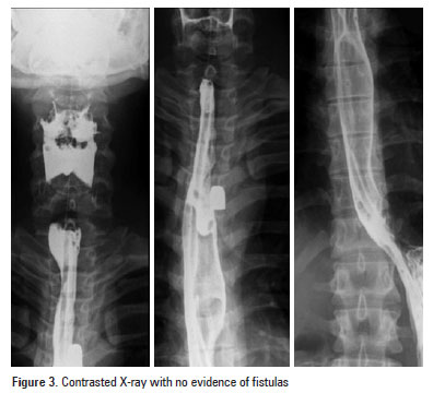
A contrast examination (Figure 3), performed on the 19th day after surgery, showed no extravasation, and the patient was authorized to receive oral nutrition, and on the 20th postoperative day he was discharged.
DISCUSSION
Nonoperative treatment criteria for esophageal perforations were first defined by Cameron et al., in 1979, and later on modified by Altorjay et al., in 1997(1). Nonoperative treatment is the treatment of choice in cases of suspicious or limited perforation, instrumental injury in the cervical region, drained cavity in the esophagus, and non obstructive neoplasms(4,5).
Posteriorly to the retropharyngeal space is located the danger space, which is bound by the alar fascia anteriorly and the prevertebral fascia posteriorly. It extends from the base of the skull and goes freely through the entire posterior mediastinum to the level of the diaphragm, in which the two fascial layers are combined(6). Thus, the danger space provides the most important anatomic route for contiguous spread between the neck and the chest.
Retropharyngeal abscesses are among the most serious deep space infections, since the infection can extend directly into the anterior or posterior regions of the superior mediastinum, or into the entire length of the posterior mediastinum via the danger space(7). Patients who are managed nonoperatively are monitored from 7 to 10 days without oral nutrition, receive total parenteral nutrition, and broad spectrum antibiotics. If a patient remains stable after this time, an esophagogram with gastrograffin contrast is done to check whether the perforation is closed. If no evidence of contrast extravasation is found, the patient starts oral nutrition(5,6). In this report the option to introduce a nasoenteric tube during the intraoperative time was done because it was feasible and safe under the direct supervision of the surgeon. The patient had no other injuries that compromised his digestive tract, and the placement of a nasoenteric tube is currently the best option for patients to have an adequate nutritional support. Enteral nutritional decreases the bacterial translocation and attenuates the inflammatory response mediated by cytokines during the acute phase so reducing the risk of multiple organ dysfunction syndrome.
Patients with delayed diagnosis represent a serious problem. They are usually in a bad clinical condition, need aggressive and definitive treatment, and require multiple surgical procedures with a worse outcome(8). Complications of cervical and esophagopleural fistulas, and pleural empyema are described in the literature(6,9). The treatment consists of extended drainage to eliminate the fistulas and to treat the pleural infection(9). The adequate moment to apply the conservative treatment is not easy to define and a judicious evaluation of the clinical situation is always imperative. In the early phase it is difficult to predict if the perforation effects are limited, or will progress to mediastinitis, pleural empyema or sepsis. The surgeon's experience is fundamental to apply the conservative treatment based on the clinical situation, laboratorial tests and imaging tools or if necessary, according to all current standards, to proceed to an aggressive surgical treatment that may include a damage control approach before the definitive treatment(9). The surgical options at the acute phase are: the exteriorization of the cervical esophagus, the drainage of the thoracic and mediastinal cavities, and the abdominal approach to proceed with the placement of an enteral feeding tube. The tube placement is preferably done by a jejunostomy because sometimes a gastric tube might be inserted lately as part of the reconstruction procedure.
Received on:Oct 30, 2011
Accepted on: Mar 3, 2012
- 1. Vogel SB, Rout WR, Martin TD, Abbitt PL. Esophageal perforation in adults: aggressive, conservative treatment lowers morbidity and mortality. Ann Surg. 2005;241(6):1016-21; discussion 1021-3.
- 2. Weber AL, Siciliano A. CT and MR imaging evaluation of neck infections with clinical correlations. Radiol Clin North Am. 2000;38(5):941-68.
- 3. Marsico GA, Azevedo DE, Guimarães CA, Mathias I, Azevedo LG, Machado T. Perfurações de esôfago. Rev Col Bras Cir. 2003;30(3):216-23.
- 4. Altorjay A, Kiss J, Vörös A, Bohák A. Nonoperative management of esophageal perforations. Is it justified? Ann Surg. 1997;225(4):415-21.
- 5. Ribeiro Junior MA, Alves LD. Ferimentos cervicais: revisão de literatura. Emerg Clín. 2008;14:12-8.
- 6. Shaffer HA Jr, Valenzuela G, Mittal RK. Esophageal perforation. A reassessment of the criteria for choosing medical or surgical therapy. Arch Intern Med. 1992;152(4):757-61.
- 7. Brinster CJ, Singhal S, Lee L, Marshall MB, Kaiser LR, Kucharczuk JC. Evolving options in the management of esophageal perforation. Ann Thorac Surg. 2004;77(4):1475-83.
- 8. Fonseca AZ, Ribeiro MA Jr, Frazão M, Costas MC, Spinelli L, Contrucci O. Esophagectomy for a traumatic esophageal perforation with delayed diagnosis. World J Gastrointest Surg. 2009;1(1):65-7.
- 9. Reynolds SC, Chow AW. Severe soft tissue infections of the head and neck: a primer for critical care physicians. Lung. 2009;187(5):271-9.
Corresponding author:
Publication Dates
-
Publication in this collection
22 Jan 2013 -
Date of issue
Dec 2012
History
-
Received
30 Oct 2011 -
Accepted
03 Mar 2012


