 Moraxella peritoneal dialysis-related infections: a systematic review
Abstract
Introduction: Moraxella species, although infrequent in peritoneal dialysis (PD)-associated infections, are established pathogens. Moraxella catarrhalis is recognized as a cause of upper and lower respiratory tract infections and is increasingly associated with β-lactamase production. The role of Moraxella species in PD-related infections warrants further investigation.
Methods: A comprehensive search of PubMed and Google Scholar was conducted for cases of PD-associated infections caused by Moraxella. Inclusion criteria required a diagnosis of peritonitis or exit-site infection confirmed by culture. Demographic data, clinical presentation, microbiological identification, antimicrobial resistance, therapeutic regimens, and outcomes were analyzed.
Results: Fourteen cases of Moraxella-related peritonitis in PD patients were identified between 1987 and 2024 from 13 studies, with no reports of exitsite infections. The mean age was 56 years; diabetic nephropathy was the most common underlying condition (four cases). M. catarrhalis (n = 6) and Moraxella osloensis (n = 5) were the most frequent pathogens. The most commonly reported methods were bacterial growth in culture media, followed by species identification using biochemical profiling or automated systems (n = 10), and advanced confirmatory techniques. Clinical presentation included abdominal pain, fever, and clouding of the dialysis fluid. Most isolates were susceptible to cephalosporins, aminoglycosides, and fluoroquinolones. The majority of patients (86%) retained their catheters after appropriate treatment, usually with intraperitoneal cephalosporins, achieving complete symptom resolution within a few days. Overall prognosis was favorable, with no mortality.
Conclusion: Moraxella species, although rare, should be considered in the differential diagnosis of PD-associated infections. Identification using advanced techniques allows effective treatment and favorable outcomes, often without the need for catheter removal.
Keywords:
Moraxella; Peritonitis; Peritoneal Dialysis; Anti-Infective Agents; Susceptibility; Review
Resumo
Introdução: Espécies de Moraxella, embora raras em infecções associadas à diálise peritoneal (DP), são patógenos estabelecidos. Moraxella catarrhalis causa infecções respiratórias, frequentemente associadas à produção de β-lactamase. Seu papel na DP merece investigação.
Métodos: Buscou-se nas bases PubMed e Google Scholar casos de infecções por Moraxella em DP. Critérios de inclusão: diagnóstico de peritonite ou infecção no local de saída confirmados por cultura. Analisaramse dados demográficos, clínicos, microbiologia, resistência, terapêutica e desfechos.
Resultados: Catorze casos de peritonite relacionada à Moraxella em pacientes em DP foram identificados entre 1987 e 2024, a partir de 13 estudos, sem relatos de infecções no local de saída. A idade média foi 56 anos; a nefropatia diabética foi a condição de base mais comum (quatro casos). M. catarrhalis (n = 6) e Moraxella osloensis (n = 5) foram os patógenos mais frequentes, com crescimento bacteriano apoiado em meios de cultura, seguido de identificação por perfis bioquímicos ou sistemas automatizados (n = 10), e técnicas confirmatórias avançadas empregadas quando relatadas. A apresentação clínica incluiu dor abdominal, febre e turbidez do líquido de diálise. A maioria dos isolados mostrou suscetibilidade a cefalosporinas, aminoglicosídeos e fluoroquinolonas. A maior parte dos pacientes (86%) manteve os cateteres após tratamento adequado, geralmente com administração intraperitoneal de cefalosporinas, com resolução completa dos sintomas em poucos dias. O prognóstico geral foi favorável, sem mortalidade.
Conclusões: Espécies de Moraxella, embora raras, devem constar no diagnóstico diferencial das infecções relacionadas à DP. Sua identificação por técnicas avançadas permite tratamento eficaz e desfechos favoráveis, frequentemente sem necessidade de remoção do cateter.
Descritores:
Moraxella; Peritonite; Diálise Peritoneal; Suscetibilidade; Anti-Infecciosos; Revisão
Introduction
Peritoneal dialysis (PD) provides important benefits for patients with end-stage renal disease (ESRD), but its long-term efficacy is frequently compromised by infections, with peritonitis and exit-site infections being the most critical determinants of technique failure. While gram-positive organisms are the predominant causative pathogens, there is a growing recognition of infections caused by less common bacteria, including Moraxella species1,2,3.
Moraxella species are aerobic, gram-negative, oxidase-positive diplococci. The organism originally known as Branhamella catarrhalis was formally reclassified under the genus Moraxella in 1984, thereby receiving its current name, Moraxella catarrhalis4. This taxonomic change was later supported by DNA-hybridization studies and 16S rRNA gene sequencing, which demonstrated a closer genetic relationship to the genus Moraxella2,5. Although typically commensals of the human respiratory tract and mucosal surfaces, these bacteria have increasingly been recognized as rare yet significant pathogens in cases of peritonitis among patients undergoing peritoneal dialysis. Furthermore, M. catarrhalis has been identified as a causative agent of PD-associated peritonitis6. Additionally, other species such as Moraxella osloensis have also been implicated, albeit less frequently1. The emergence of these species as causative pathogens of PD-associated peritonitis is noteworthy, given their rarity and the diagnostic challenges they present.
The pathogenesis of Moraxella-induced peritonitis in PD patients is not fully elucidated. Proposed mechanisms include direct contamination during catheter insertion or manipulation, hematogenous spread from distant sites, or translocation from the gastrointestinal tract3,6,7. Accurate identification of Moraxella species requires initial growth on conventional culture media, followed by advanced methods for species-level confirmation. Techniques such as matrix-assisted laser desorption/ionization time-of-flight mass spectrometry (MALDI-TOF MS) and 16S ribosomal RNA (16S rRNA) gene sequencing are ideal for correct species identification, especially when phenotypic similarities with other gramnegative organisms may lead to misclassification2.
Moraxella species are generally resistant to penicillins due to the high prevalence of β-lactamase production; therefore, penicillins cannot be considered reliable treatment options. Resistance patterns may vary, and empirical therapy should be guided by antimicrobial susceptibility testing, local antibiograms, and individual patient factors5. The management approach often includes intraperitoneal antibiotic administration, with the duration tailored to clinical response8.
This review aims to synthesize and critically evaluate the existing literature on Moraxella-associated infections in PD patients, with a focus on clinical manifestations, diagnostic challenges, microbiological assessment, treatment strategies and outcomes. By analyzing reported cases, we seek to deepen the understanding of these uncommon infections and inform clinical practice to improve patient care.
Methods
Study Framework and Protocol Registration
This systematic review was structured in accordance with the PRISMA 2020 guidelines for transparent reporting of systematic evidence syntheses9. The review protocol was prospectively registered in the PROSPERO international prospective registry of systematic reviews (ID: CRD420251042629; accessible at: https://www.crd.york.ac.uk/PROSPERO/view/CRD420251042629, accessed April 29, 2025).
Selection Parameters
We included published case reports documenting infectious complications in patients undergoing peritoneal dialysis (PD), specifically peritonitis and/or exit-site infections (ESIs), attributed to Moraxella species. The diagnosis of peritonitis was established when at least two of the following criteria were met: (i) clinical features consistent with peritonitis, such as abdominal pain and/or cloudy dialysis effluent; (ii) peritoneal fluid white cell count >100/µL with >50% polymorphonuclear leukocytes after a dwell time of at least 2 hours; (iii) positive dialysis effluent culture8. Exit-site infections were defined as the presence of purulent exudate at the catheter exit-site, irrespective of the presence or absence of pericatheter erythema. Tunnel infections, often clinically silent, were considered when inflammation was observed along the catheter’s subcutaneous course, particularly in conjunction with ESI8. Only culture-confirmed infections due to Moraxella species from dialysate, exit-site exudate, or tunnel tissue were included. Pathogen identification was required to be based on conventional culture-based biochemical methods, while additional confirmatory techniques such as MALDI-TOF MS and/or 16S rRNA gene sequencing were accepted when reported.
Data Acquisition Strategy
An extensive search of the medical literature was conducted using two electronic databases: PubMed and Google Scholar. The search covered all literature published up to May 1, 2025. Keywords included: “Moraxella” or “Branhamella” associated with “peritonitis”, “exit-site infection”, and “peritoneal dialysis”. No language restrictions were applied. Reports in languages other than English were translated using Google Translate (https://translate. google.gr, last accessed May 31, 2025).
Screening and Eligibility Assessment
Two stages of screening were applied. Initially, titles and abstracts were reviewed independently by two investigators (JD and AP) for potential relevance. The full texts of eligible articles were then assessed independently by three additional reviewers (CA, VK, and NP). Any discrepancies were resolved via discussion among all reviewers (JD, CA, VK, AP, NP) to ensure consensus. Reference lists of included studies were manually reviewed to identify additional cases.
Data Extraction and Management
All relevant case data were extracted into a structured database. No reports were excluded based on completeness, although some lacked specific clinical or microbiological details. Extracted variables included publication year, geographic origin, patient demographics (age, sex), primary renal diagnosis leading to ESRD, prior episodes of PD-related infections, clinical presentation, laboratory and microbiological findings, diagnostic modalities, antimicrobial susceptibility profiles, treatment regimens (drug type, route, duration), catheter management strategies, and clinical outcomes.
Ethical Compliance
This article is based on previously published data and does not contain any new studies with human participants or animals performed by any of the authors. Therefore, ethical approval and informed consent were not applicable.
Data Synthesis and Statistical Analysis
A centralized dataset was constructed using Microsoft Excel (version 5.2.3790.1830, Microsoft Corporation, Redmond, WA, USA). Quantitative variables were summarized descriptively. Statistical analysis was performed using GraphPad Instat (version 3.10; GraphPad Software, San Diego, CA, USA).
Results
Our systematic search yielded 1,026 records, of which 114 remained following removal of duplicates. After title and abstract review, 13 case reports/series were included for full-text assessment, as depicted in the PRISMA flow diagram (Figure 1). Ultimately, a total of 14 distinct cases of PD-associated infections caused by Moraxella species were identified, all of which were cases of peritonitis. All infections originated in the outpatient setting and none were associated with hospital care. These cases spanned the period from 1987 to 2024 and were reported across seven different countries in North America, Europe, and Asia, with the majority originating from the United States.
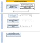
The PRISMA flow diagram of our literature search and selection process applied during the overview.
The included cases involved 9 male and 5 female patients with a wide age distribution (range: 22–83 years; median: 56 years). All patients were undergoing PD for a dialysis vintage period ranging from 12 to 84 months (Table 1). Diabetic nephropathy was the most frequently reported primary renal pathology (n = 4), followed by single cases of hypertensive renal disease, membranous nephropathy, malignant nephrosclerosis, and chronic glomerulonephritis in patients with available data. Notably, none of the patients were reported to have significant comorbidities, immunosuppression, or other systemic health issues apart from the underlying nephrological condition necessitating PD. This may suggest that Moraxella-associated peritonitis can also occur in immunocompetent individuals. A history of prior peritonitis episodes was noted in the majority (56%; 5/9) of cases, typically involving common pathogens such as Staphylococcus aureus or Streptococcus species.
Reported cases of peritoneal dialysis-associated infections caused by Moraxella species, listed chronologically from oldest to most recent
Among the 14 patients with Moraxella-associated peritonitis, information on the presence of fever was available for 12. Of these, 7 (58%) were afebrile, while 5 (42%) exhibited febrile responses, with temperatures ranging from 37.8 to 38.6°C or simply noted as “fever”. Peritoneal white blood cell (WBC) counts showed considerable variability, ranging from 260 to 10,528 cells/mm3. A predominance of polymorphonuclear cells (PMNs) was observed in 8 cases (>90%), with the highest recorded proportion reaching 99%. One case had a notably low PMN percentage (15%), and differential counts were unavailable in two cases. Peripheral blood WBC counts were available only in 6 cases and ranged from 1,675 to 10,500 × 106/L. In most of these, a neutrophilic predominance was noted, with PMN percentages ranging from 78 to 92%, except for one case with a notably low neutrophil proportion (37%).
M. catarrhalis was the predominant isolate (n = 6), followed by M. osloensis (n = 5), while Moraxella phenylpyruvica and Moraxella nonliquefaciens were each identified in a single case (Table 2). An additional case involved an unidentified Moraxella species. Conventional culture methods supported bacterial growth (e.g., BacT/Alert systems, chocolate agar, blood agar), followed by species identification through biochemical profiling or automated identification systems (n = 10). In more recent reports, advanced confirmatory techniques such as MALDI-TOF MS (n = 4) and/or 16S rRNA gene sequencing (n = 3) were additionally employed, reflecting the evolution of microbiological diagnostic capabilities over time. Five cases reported polymicrobial infections, including co-isolation of Enhydrobacter aerosaccus, Kluyvera ascorbata, Rhizobium radiobacter, mixed Streptococci/ Enterococci and diphtheroids. These cases emphasized the need for comprehensive microbiological workup, particularly in patients with atypical clinical courses.
Analysis of microbiological data from 14 peritoneal dialysis-related peritonitis cases attributed to Moraxella species
Antibiotic sensitivity data were available for 11 isolates. Most isolates were susceptible to third-generation cephalosporins (e.g., cefotaxime, ceftriaxone, ceftazidime), aminoglycosides (e.g., gentamicin, tobramycin, amikacin), macrolides (e.g., erythromycin, clarithromycin), and fluoroquinolones (e.g., ciprofloxacin, levofloxacin). However, resistance to penicillin and ampicillin was noted in a few cases, particularly among M. catarrhalis and M. osloensis strains, consistent with their known production of β-lactamase enzymes.
In polymicrobial infections, the choice of antibiotic therapy was guided by the most resistant co-isolated organism. Notably, in one case involving co-isolation with Rhizobium radiobacter, resistance to multiple β-lactams necessitated the use of intraperitoneal ciprofloxacin and intravenous meropenem.
All patients with complete reported data received intraperitoneal antibiotics as part of their initial treatment protocol. The most commonly used agents (e.g., cefazolin, ceftazidime) were often in combination with aminoglycosides or fluoroquinolones (Table 3). Treatment durations ranged from 10 to 28 days, with a median of 21 days. Catheter removal was required in only two cases, one involving Moraxella phenylpyruvica and the other M. osloensis, both of which resulted in a transition to hemodialysis. In all 14 cases, the infection was resolved without mortality or long-term complications. Notably, catheter salvage was achieved in 86% (12/14) of patients, reaffirming the generally favorable prognosis of Moraxella-related PD infections when promptly recognized and adequately treated.
Therapeutic strategies and clinical outcomes in reviewed cases of Moraxella-associated peritonitis in patients undergoing peritoneal dialysis
The overall prognosis was favorable across all reported cases. No infection-related mortality was documented. Clinical resolution of peritonitis symptoms was typically achieved within few days of antibiotic initiation. Laboratory parameters, including peritoneal fluid white blood cell counts and serum inflammatory markers, showed rapid improvement in most cases.
Discussion
This systematic review comprehensively examines the role of Moraxella species as etiological agents in peritoneal dialysis-associated infections, with a specific focus on peritonitis. The exclusive documentation of Moraxella-related cases as peritonitis emphasizes their clinical infrequency, the inherent diagnostic complexities, and the generally favorable therapeutic outcomes reported. The findings underscore the importance of maintaining a high index of suspicion for atypical pathogens such as Moraxella species, which, despite their rarity, deserve clinical recognition in the differential diagnosis of PD-related peritonitis. Their identification has historically been overlooked, likely due to limitations of traditional diagnostic methodologies and their commensal nature in mucosal sites.
A consistent observation across cases was the predominance of M. catarrhalis and M. osloensis, both traditionally associated with respiratory tract colonization. Their isolation from peritoneal fluid raises intriguing questions regarding pathogenic mechanisms. Several routes of peritoneal cavity colonization have been proposed, including direct contamination during PD exchanges, translocation from the gastrointestinal tract, and hematogenous spread from distant mucosal foci3,6,7. The presence of co-infecting organisms in some cases may suggest a polymicrobial origin or reflect compromised mucosal barriers.
Importantly, the review highlights the evolving microbiological armamentarium in detecting Moraxella species. While earlier cases relied solely on biochemical testing and growth on selective media, more recent cases incorporated MALDI-TOF MS and 16S rRNA gene sequencing for accurate specieslevel identification19. These advancements have markedly enhanced the sensitivity and specificity of pathogen detection and may partly explain the increased detection rate of Moraxella species in more recent years2. Nevertheless, despite technological progress, diagnostic delays remain possible in low-resource settings. From a clinical standpoint, heightened awareness is necessary for early recognition of Moraxella-associated PD peritonitis. Infections due to fastidious organisms may present with subacute symptoms or atypical laboratory profiles, leading to misclassification or underdiagnosis.
The antimicrobial susceptibility profile of Moraxella isolates in PD-associated infections was generally favorable. Most strains exhibited susceptibility to cephalosporins, aminoglycosides and fluoroquinolones, consistent with the known antibiotic profiles of Moraxella species. In particular, M. catarrhalis, despite its ability to produce β-lactamase enzymes, demonstrated preserved susceptibility to cephalosporins. Nonetheless, resistance to penicillin and ampicillin was reported in few isolates of both M. catarrhalis and M. osloensis, emphasizing the importance of individualized antimicrobial therapy guided by susceptibility testing2,5.
Despite the presence of isolates with variable susceptibility to antibiotics, clinical outcomes were uniformly favorable, with all 14 patients achieving resolution of infection. Most infections were successfully managed without catheter removal, a noteworthy outcome considering that catheter salvage is a critical determinant of long-term PD continuation. Only two patients (14%) required catheter removal, both of whom eventually resumed peritoneal dialysis following switch to hemodialysis. This reaffirms prior observations that even non-traditional pathogens can be effectively managed with early intervention and appropriate therapy20.
Therapeutic strategies varied in drug choice and duration, reflecting empirical decisions tailored to patient condition and local practices. Notably, intraperitoneal cephalosporins, often in combination with aminoglycosides or fluoroquinolones, were the most commonly employed regimens. Treatment duration was typically aligned with standard ISPD guidelines for gram-negative peritonitis, ranging from 14 to 21 days8. The consistent recovery in our cohort suggests that Moraxella infections in PD patients follow a benign course when managed according to protocol.
An additional point of consideration is the potential impact of polymicrobial infections. In 5 of the 14 reviewed cases (~36%), Moraxella species were co-isolated with other microorganisms in peritoneal fluid cultures, complicating the interpretation of microbiological findings and the determination of the primary causative agent. The co-isolated organisms including Enhydrobacter aerosaccus, Kluyvera ascorbate, and Rhizobium radiobacter are rarely reported or poorly characterized as etiological agents in PD-related peritonitis. Among them, R. radiobacter has occasionally been implicated in PD peritonitis, primarily in immunocompromised hosts or in the context of catheter-related infections21. In contrast, K. ascorbata and E. aerosaccus are considered organisms of low virulence, with only anecdotal or exceptional associations with human disease and virtually no established link to PD-associated infections. Mixed Streptococci/Enterococci and diphtheroids, although more frequently encountered in clinical specimens, are often regarded as contaminants, albeit with potential pathogenicity in selected cases8. As a result, the pathogenic role of Moraxella in these polymicrobial episodes remains unclear. Whether it acts as a true pathogen or merely as a commensal cannot be determined without more detailed clinical, inflammatory, and microbiological data. Nevertheless, these mixed infections may obscure diagnosis and delay targeted therapy. Importantly, in all polymicrobial cases, culture-guided antimicrobial treatment led to clinical resolution, underscoring the critical importance of thorough microbiological evaluation, particularly in cases with atypical presentations or suboptimal initial response to empirical therapy22.
Our study has certain unavoidable limitations, primarily due to its reliance on previously published case reports rather than original patient data. As a result, it is susceptible to publication bias and may not represent the complete range of clinical scenarios. In addition, heterogeneity in reporting, particularly with regard to susceptibility testing and treatment regimens, may limit generalizability. Nonetheless, this review provides the most comprehensive analysis to date on Moraxella peritonitis in PD and offers clinically relevant guidance.
In conclusion, although rare, Moraxella species are recognized pathogens in the setting of PD-associated peritonitis. Diagnosis should be based on clinical features, peritoneal fluid analysis, and culture results. Prompt identification using conventional culture methods, with advanced confirmatory techniques when available, along with appropriate antimicrobial therapy and judicious catheter management, can result in uniformly favorable clinical outcomes. As diagnostic tools become more sophisticated, clinicians should consider them and integrate them into laboratory practice. Further research is warranted to define the true incidence of Moraxella peritoneal dialysis-related infections, optimal management strategies, and the long-term impact on peritoneal dialysis outcomes.
Acknowledgments
The authors would like to thank the library services of Aristotle University of Thessaloniki and Hippokration Hospital for their support in literature retrieval.
Data Availability
No new data were generated or analyzed in this study.
References
-
1. Badrising S, Bakker L, Lobatto S, van Es A. Peritonitis in a peritoneal dialysis patient due to Rhizobium radiobacter and Moraxella osleonsis: case report and literature review. Perit Dial Int. 2014;34(7):813–5. doi: https://doi.org/10.3747/pdi.2013.00238. PubMed PMID: 25520491.
» https://doi.org/10.3747/pdi.2013.00238 -
2. Yamada A, Kasahara K, Ogawa Y, Samejima K, Eriguchi M, Yano H, et al. Peritonitis due to Moraxella osloensis: A case report and literature review. J Infect Chemother. 2019;25(12):1050–2. doi: https://doi.org/10.1016/j.jiac.2019.05.018. PubMed PMID: 31196771.
» https://doi.org/10.1016/j.jiac.2019.05.018 -
3. Torigoe K, Yoshidome A, Otsuka E, Tsuji K, Yamashita A, Kitamura M, et al. A case of peritoneal dialysis-related peritonitis due to Moraxella osloensis. Cureus. 2024;16(11):e74294. doi: https://doi.org/10.7759/cureus.74294. PubMed PMID: 39717310.
» https://doi.org/10.7759/cureus.74294 - 4. Bovre K. The genus Moraxella In: Krieg NR, Holt JG, editors. Bergey’s manual of systematic bacteriology. Baltimore, MD: Williams & Wilkins Co; 1984. p. 296–303. (Vol. 1).
-
5. Verduin CM, Hol C, Fleer A, van Dijk H, van Belkum A. Moraxella catarrhalis: from emerging to established pathogen. Clin Microbiol Rev. 2002;15(1):125–44. doi: https://doi.org/10.1128/CMR.15.1.125-144.2002. PubMed PMID: 11781271.
» https://doi.org/10.1128/CMR.15.1.125-144.2002 -
6. Sadjadi SA, Obedoza P, Annamarju P. Moraxella catarrhalis peritonitis. Am J Case Rep. 2012;13:19–21. doi: https://doi.org/10.12659/AJCR.882358. PubMed PMID: 23569477.
» https://doi.org/10.12659/AJCR.882358 -
7. Dasgupta MK. Biofilms and infection in dialysis patients. Semin Dial. 2002;15(5):338–46. doi: https://doi.org/10.1046/j.1525-139X.2002.00084.x. PubMed PMID: 12358638.
» https://doi.org/10.1046/j.1525-139X.2002.00084.x -
8. Li PK, Chow KM, Cho Y, Fan S, Figueiredo AE, Harris T, et al. ISPD peritonitis guideline recommendations: 2022 update on prevention and treatment. Perit Dial Int. 2022;42(2):110–53. doi: https://doi.org/10.1177/08968608221080586. PubMed PMID: 35264029.
» https://doi.org/10.1177/08968608221080586 -
9. Haddaway NR, Page MJ, Pritchard CC, McGuinness LA. PRISMA2020: an R package and Shiny app for producing PRISMA 2020-compliant flow diagrams, with interactivity for optimised digital transparency and Open Synthesis. Campbell Syst Rev. 2022;18(2):e1230. doi: https://doi.org/10.1002/cl2.1230. PubMed PMID: 36911350.
» https://doi.org/10.1002/cl2.1230 -
10. Damani NN, Chin AT. Branhamella catarrhalis peritonitis in a continuous ambulatory peritoneal dialysis patient. Nephron J. 1987;45(2):160–1. doi: https://doi.org/10.1159/000184102. PubMed PMID: 3561626.
» https://doi.org/10.1159/000184102 -
11. MacArthur RD. Branhamella catarrhalis peritonitis in two continuous ambulatory peritoneal dialysis patients. Perit Dial Int. 1990;10(2):169–71. doi: https://doi.org/10.1177/089686089001000210. PubMed PMID: 2131805.
» https://doi.org/10.1177/089686089001000210 -
12. Contreras MR, Ash SR, Swick SD, Grutzner J. Peritonitis due to Moraxella (Branhamella) catarrhalis in a diabetic patient receiving peritoneal dialysis. South Med J. 1993;86(5):589–90. doi: https://doi.org/10.1097/00007611-199305000-00023. PubMed PMID: 8488414.
» https://doi.org/10.1097/00007611-199305000-00023 - 13. Velusamy L, Mohanty MJ. Moraxella and Kluyvera peritonitis in a CAPD patient with human immunodeficiency virus. Perit Dial Int. 2003;23(6):611–2. PubMed PMID: 14703211.
-
14. Cirillo P, Melfitano A, Pepe V, Delli Carri P, Gesualdo L. Peritonitis due to Moraxella phenylpyruvica. Perit Dial Int. 2009;29(3):358–9. doi: https://doi.org/10.1177/089686080902900323. PubMed PMID: 19458311.
» https://doi.org/10.1177/089686080902900323 -
15. Fandos JM, Mañez MB. Peritonitis due to Moraxella nonliquefaciens. Perit Dial Int. 2014;34(6):674–5. doi: https://doi.org/10.3747/pdi.2013-00148. PubMed PMID: 25228220.
» https://doi.org/10.3747/pdi.2013-00148 - 16. Hernández-Egido S, Puerta-Mateo A, Cores-Calvo O, Ruiz-Ferraras E. Peritonitis por Moraxella osloensis: a propósito de un caso y revisión de la literatura [Moraxella osloensis peritonitis: case report and review]. Rev Esp Quimioter. 2016;29(3):161–3. PubMed PMID: 27015822.
-
17. Adapa S, Gumaste P, Konala VM, Agrawal N, Garcha AS, Dhingra H. Peritonitis due to Moraxella osloensis: an emerging pathogen. Case Rep Nephrol. 2018;2018:4968371. doi: https://doi.org/10.1155/2018/4968371. PubMed PMID: 30671269.
» https://doi.org/10.1155/2018/4968371 -
18. Pilcevic D. Successful treatment of CAPD peritonitis caused by Moraxella catarrhalis. Serb J Exp Clin Res. 2018;19(1): 58–9. doi: https://doi.org/10.1515/sjecr-2016-0098.
» https://doi.org/10.1515/sjecr-2016-0098 -
19. Dichtl K, Klugherz I, Greimel H, Luxner J, Köberl J, Friedl S, et al. A head-to-head comparison of three MALDI-TOF mass spectrometry systems with 16S rRNA gene sequencing. J Clin Microbiol. 2023;61(10):e0191322. doi: https://doi.org/10.1128/jcm.01913-22. PubMed PMID: 37732759.
» https://doi.org/10.1128/jcm.01913-22 -
20. Salzer WL. Peritoneal dialysis-related peritonitis: challenges and solutions. Int J Nephrol Renovasc Dis. 2018;11:173–86. doi: https://doi.org/10.2147/IJNRD.S123618. PubMed PMID: 29928142.
» https://doi.org/10.2147/IJNRD.S123618 -
21. Roy S, Basuli D, Rahman EU, Adapa S, Reddy SN. Rhizobium radiobacter-Induced Peritonitis: a case report and literature analysis. J Med Cases. 2022;13(9):471–4. doi: https://doi.org/10.14740/jmc3999. PubMed PMID: 36258704.
» https://doi.org/10.14740/jmc3999 -
22. Sahlawi MA, Wilson G, Stallard B, Manera KE, Tong A, Pisoni RL, et al. Peritoneal dialysis-associated peritonitis outcomes reported in trials and observational studies: a systematic review. Perit Dial Int. 2020;40(2):132–40. doi: https://doi.org/10.1177/0896860819893810. PubMed PMID: 32063197.
» https://doi.org/10.1177/0896860819893810
Edited by
-
Editorial Responsibility
Editor-in-chief: Miguel C. Riella https://orcid.org/0000-0003-4181-613X.Associate Editor: Thyago Proença de Moraes https://orcid.org/0000-0002-2983-3968.