Abstract
Objective
To review the main acute complications of inflammatory bowel disease in order to present the state of the art of their respective diagnosis and treatment.
Methods
A bibliographic search was conducted in Medline database using the following keywords: “inflammatory bowel disease”, “Colitis Ulcerative”, “Crohn Disease”, “emergency” among others that had their variation evaluated by the MESH. Articles from the last 10 years conducted with humans, written in Portuguese or English, and published in journals with impact factor greater than 1 were selected.
Results
After carrying out the search phrase and selecting the filters, 20 articles were selected to be included in the research. The most common acute complications were evaluated, focusing on their current propaedeutic and management aspects.
Conclusion
Most emergencies related to inflammatory bowel disease should be treated non-operatively firstly, prioritizing patient hemodynamic state. In selected cases of life-threatening complications emergent operative treatment are mandatory. The timing of procedure is the most important aspect. As general rule, in Crohn’s Disease, operative treatment should be postponed as much as possible and the resection as small as possible. In case of ulcerative rectocolitis, if the hemodynamic state of the patient allows, proctocolectomy should be expedited with curative intention.
Keywords:
Inflammatory bowel diseases; Emergencies; Complications
Resumo
Objetivo
Revisar as principais complicações agudas das doenças inflamatórias intestinais, a fim de apresentar o estado da arte de seus respectivos diagnósticos e tratamentos.
Métodos
Foi realizada uma pesquisa bibliográfica no banco de dados Medline, utilizando as seguintes palavras-chave: “doença inflamatória intestinal”, “Colite Ulcerativa”, “Doença de Crohn”, “emergência” entre outras que tiveram sua variação avaliada pelo MESH. Artigos dos últimos 10 anos realizados com seres humanos, escritos em português ou inglês, e publicados em periódicos com fator de impacto maior que um foram selecionados.
Resultados
Após a construção da frase de pesquisa e seleção dos filtros, 20 artigos foram selecionados para inclusão no estudo. As complicações agudas mais comuns foram avaliadas, enfocando seus atuais aspectos propedêuticos.
Conclusão
A maioria das emergências relacionadas à doença inflamatória intestinal deve ser tratada primariamente de forma não cirúrgica, priorizando a hemodinâmica do paciente. Em casos selecionados de complicações potencialmente fatais, tratamento cirúrgico de emergência é mandatório. O momento do procedimento é o aspecto mais importante. Como regra geral, na Doença de Crohn, o tratamento cirúrgico deve ser adiado ao máximo com ressecção menor possível. No caso de retocolite ulcerativa, se o estado hemodinâmico do paciente permitir, a proctocolectomia deve ser realizada com intenção curativa.
Palavras-chave:
Doença inflamatória intestinal; Emergências; Complicações
Background
Inflammatory Bowel Diseases (IBDs) comprise chronic inflammatory bowel disorders, having Crohn’s Disease (CD) and Ulcerative Rectocolitis (URC) as main representatives, both considered to be related to abnormal immune response. In URC, inflammation is diffuse and restricted to the mucosa, causing its continuous involvement and affecting the rectum, although it can extend to the entire colon. In CD, the lesions are discontinuous, affecting all layers of the intestinal wall (transmural) and located in any segment of the gastrointestinal tract.11 Ballou S, Hirsch W, Singh P, Rangan V, Nee J, Iturrino J, et al. Emergency department utilisation for inflammatory bowel disease in the United States from 2006 to 2014. Aliment Pharmacol Ther. 2018;47:913-21.,22 Yarur AJ, Mandalia AB, Dauer RM, Czul F, Deshpande AR, Kerman DH, et al. Predictive factors for clinically actionable computed tomography findings in inflammatory bowel disease patients seen in the emergency department with acute gastrointestinal symptoms. J Crohns Colitis. 2014;8:504-12.
The clinical picture of both diseases may resemble. The diagnosis is made through clinical, endoscopic, radiological, and histological data. Among the acute complications that require urgent or emergency care includes: infectious complications such as intra-abdominal abscesses, colitis caused by Clostridium and Cytomegalovirus (CMV), anomalies of motor function of intestinal transit progression such as Toxic Megacolon (TM), intestinal hemorrhage and intestinal obstructions and perforations. About 20 % of patients with UC and 80 % of CD will undergo surgical intervention during their lifetime. The surgery offers a good quality of life for UC patients, the same outcome is less noted in patients with CD, whose will go undergone some surgical interventions in the majority of cases. Laparoscopic approach has replaced laparotomy in tertiary centers. Laparoscopic proctocolectomy and restorative IPAA is becoming the standard of care in the treatment of UC and laparoscopic ileo-cecal resection is already the new gold standard in the treatment of stenotic CD of terminal ileum. The timing of surgery is a key issue for proper management of IBD patients. Laparoscopic approach is less studied in the emergency scenario, but the option increases specially in tertiary centers. The study aimed a narrative and comprehensive reviewed of the main emergencies observed in patients with IBD, in the last 10 years highlighting its temporal changes.33 Bemelman WA, Warusavitarne J, Sampietro GM, Serclova Z, Zmora O, Luglio G, et al. ECCO-ESCP Consensus on Surgery for Crohn’s Disease. J Crohns Colitis. 2018;12:1-16.–77 Gralnek IM, Neeman Z, Strate LL. Acute lower gastrointestinal bleeding. N Engl J Med. 2017;376:1054-63.
Method
A bibliographic search was conducted in the MedLine database using the following keywords: “inflammatory bowel disease”[ti] OR “Colitis, Ulcerative”[ti] OR “Crohn Disease”[ti] AND (emergencies OR emergency OR urgency OR peritonitis OR “Secondary Peritonitis” OR “Toxic Megacolon” OR “Intestinal Perforations” OR “Perforation, Intestinal” OR “Perforations, Intestinal” OR “Hemorrhage, Gastrointestinal” OR “Gastrointestinal Hemorrhages” OR hematochezia OR “gastrointestinal bleeding” OR “Intestinal Obstructions” OR “Obstruction, Intestinal” OR “intestinal obstruction”). From a total of 595 citations, studies in non-humans and languages other than English or Portuguese were excluded. Articles from the last 10 years were selected. Of 197 citations obtained, a new filter evaluated the impact factor of the scientific journal, excluding articles in journals with an impact factor lower than one, and adherence to the subject. Twenty articles remained and were included in the research.
Results/Discussion
Toxic megacolon
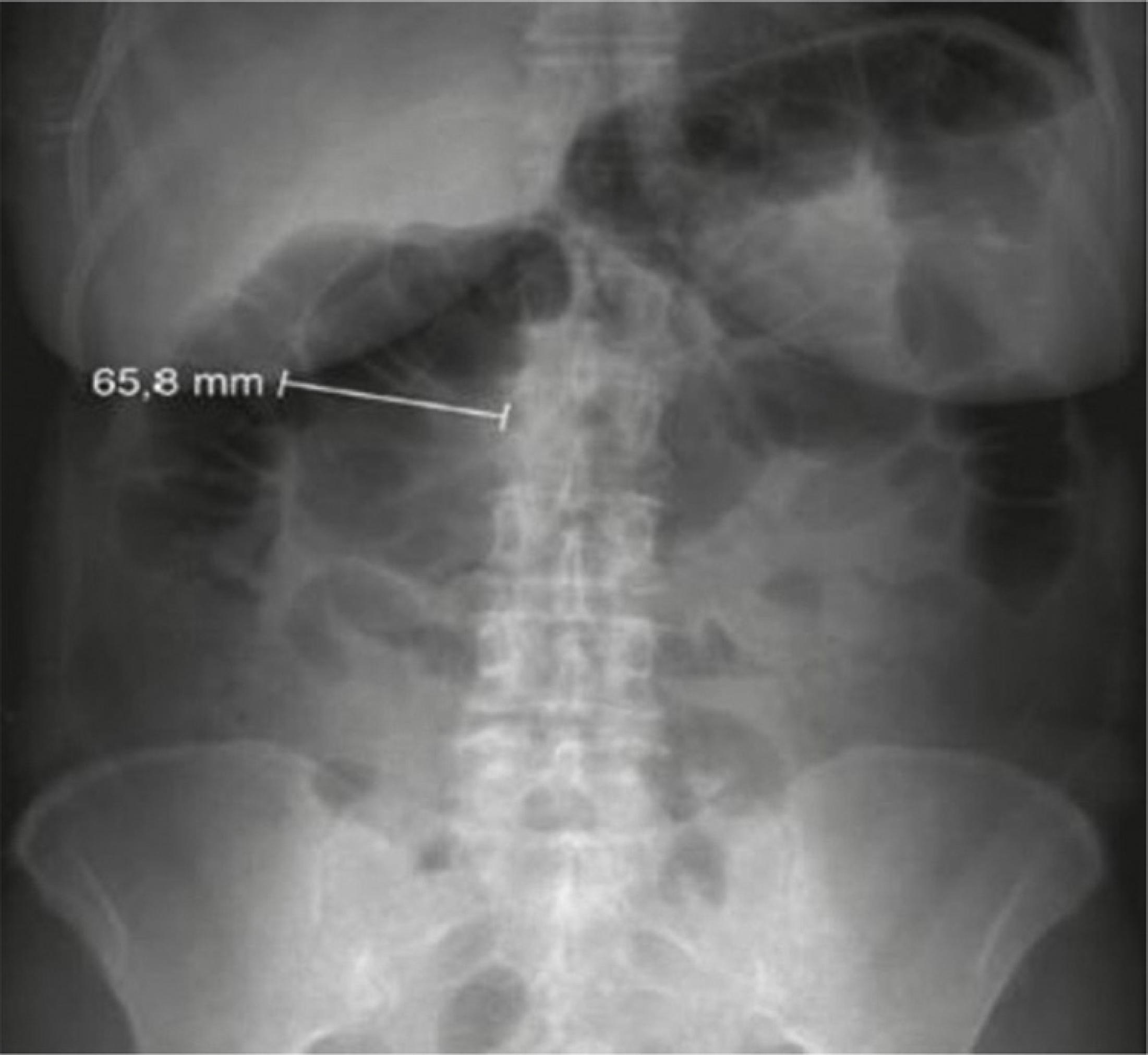
TM is defined as colonic dilatation greater than 6 cm, in the presence of acute colitis and signs of severe systemic inflammatory response.88 Woodhouse E. Toxic megacolon: a review for emergency department clinicians. J Emerg Nurs. 2016;42:481-6. About 10 % of patients admitted with ulcerative colitis and 1%–5% of those with CD develop TM.66 Frazer C, Hussey L, Bemker M. Gastrointestinal motility problems in critically ill patients. Crit Care Nurs Clin North Am. 2018;30:109-21.
The exact pathophysiology of TM remains unclear, but many studies postulate that it involves the presence of infection in a colon with previous ulcers, causing relaxation of the colonic smooth muscle and decreased gastrocolic reflex leading to significant dilatation of the affected colonic segment. Infection by CMV, Shigella, Salmonella, Entamoeba, enterohemorrhagic, Escherichia coli, and Clostridium difficile (C-diff ‒ become more common due to the indiscriminate use of antibiotics) should be investigated (Fig. 1).66 Frazer C, Hussey L, Bemker M. Gastrointestinal motility problems in critically ill patients. Crit Care Nurs Clin North Am. 2018;30:109-21.,88 Woodhouse E. Toxic megacolon: a review for emergency department clinicians. J Emerg Nurs. 2016;42:481-6.
The diagnosis of TM is established when we had 3 of the 4 criteria below: 1) Temperature > 38.6 °C, 2) Tachycardia (> 120 bpm), 3) Leukocyte count > 10,500 mm3 with left shift, and 4) Anemia (hemoglobin or hematocrit < 60 % of normal) (Table 1).88 Woodhouse E. Toxic megacolon: a review for emergency department clinicians. J Emerg Nurs. 2016;42:481-6.,99 Jalan KN, Sircus W, Card WI, Falconer CW, Bruce CB, Crean GP, et al. An experience with ulcerative colitis: toxic dilation in 55 cases. Gastroenterology. 1969;57:68-82.
Diagnostic imaging examinations include plain abdominal radiography, which reveals colonic distension > 6 cm. Abdominal ultrasound and contrast-enhanced computed tomography are also useful in determining the cause or detecting perforation. Blood cultures are necessary to safely identify the pathogens involved, and empirical therapy should not be postponed (Fig. 2).66 Frazer C, Hussey L, Bemker M. Gastrointestinal motility problems in critically ill patients. Crit Care Nurs Clin North Am. 2018;30:109-21.
TM is a life-threatening complication. The diagnosis and prompt treatment addressing the correction of hypovolemia, electrolyte disorders, abdominal compartment syndrome; administration of antimicrobials and operative treatment, is the priority and should be cautiously evaluated.88 Woodhouse E. Toxic megacolon: a review for emergency department clinicians. J Emerg Nurs. 2016;42:481-6.
Operative treatment should be considered in those cases that do not respond to intensive and antimicrobial care after 48–72 h or when the perforation is diagnosed. The technique of choice yet is total proctocolectomy with closure of the remaining rectum and terminal ileostomy. In elective condition, a second approach could be performed with an ileorectal anastomosis, if the rectum remains preserved or an ileoanal anastomosis with ileal reservoir, if the rectum is compromised (Table 2). 66 Frazer C, Hussey L, Bemker M. Gastrointestinal motility problems in critically ill patients. Crit Care Nurs Clin North Am. 2018;30:109-21.,88 Woodhouse E. Toxic megacolon: a review for emergency department clinicians. J Emerg Nurs. 2016;42:481-6.
Gastrointestinal bleeding
Massive gastrointestinal bleeding is one of the most difficult complications of IBD that requires emergency treatment; however, it is not so frequent. In cases of URC, the bleeding is usually diffuse and associated with extensive colitis. On the other hand, in cases of CD, it is more intensive, but its source is localized.1010 Kim KJ, Han BJ, Yang SK, Na SY, Park SK, Boo SJ, et al. Risk factors and outcome of acute severe lower gastrointestinal bleeding in Crohn’s disease. Dig Liver Dis. 2012;44:723-8.
Hemodynamic stabilization of the patient is the priority attitude of acute care surgeon. In a stable patient, the site and etiology of hemorrhage should be investigated. However, in unstable hemodynamic patients, despite resuscitative measures, emergency operative treatment is indicated. Once the cause of bleeding is known, specific management and prevention against new bleeding episodes should be immediately implemented (Table 3).77 Gralnek IM, Neeman Z, Strate LL. Acute lower gastrointestinal bleeding. N Engl J Med. 2017;376:1054-63.,1111 Burg MD, Riccoboni ST, Nusbaum J, Gupta N. Management of inflammatory bowel disease flares in the emergency department [digest]. Emerg Med Pract. 2017;19:S1-2.
Main aspects in the treatment of massive gastrointestinal hemorrhage in intestinal bowell disease.
The bleeding should be classified according to the location: a clinical diagnosis of hematemesis and melena supports the hypothesis of High Gastrointestinal Bleeding (HGB), while enterorrhagia is indicative of Low Gastrointestinal Bleeding (LGB). However, massive HGB can manifest with hematochezia, and LGB with slow transit can cause melena. The passage of a nasogastric catheter, followed by gastric lavage, may be useful: the return blood or “coffee-ground” material strongly supports the hypothesis of HGB.77 Gralnek IM, Neeman Z, Strate LL. Acute lower gastrointestinal bleeding. N Engl J Med. 2017;376:1054-63.,1212 Burg MD, Riccoboni ST. Management of inflammatory bowel disease flares in the emergency department. Emerg Med Pract. 2017;19:1-20.
It is worth of note, that LGB is defined as hemorrhage into the gastrointestinal tract distal to the ligament of Treitz. In most cases of CD, the bleeding source is located in the small intestine, and its precise identification is essential to limit bowel resection. This can be achieved with careful exploration of small bowel, looking for a “bleeding tattoo” in the intestinal wall, or by an intra-operative enteroscopy. In addition, in difficult cases, because patient’s clinical status, transcatheter visceral angiography with embolization, may control the bleeding and postponed the operative treatment. This is especially indicated, when a significative bleeding is the first manifestation of the disease, the non-operative treatment failed and patient clinical heath status is not good (angiography).1313 Speir EJ, Ermentrout RM, Martin JG. Management of acute lower gastrointestinal bleeding. Tech Vasc Interv Radiol. 2017;20:258-62.
In colonic bleeding, colonoscopy is the method of choice for identifying the source of bleeding, being used for diagnosis and therapy purposes. It is possible to examine the colon, without its previous cleaning, as the blood has a cathartic effect. Thus, when colonoscopy is unable to diagnosis the source or the bleeding is so massive, other diagnostic methods may be indicated. The scintigraphy with marked red blood cells may guide the performance of angiography. It is important take in mind, that contrasting tests (as opaque enema or intestinal transit) are contraindicated.44 He B, Yang J, Xiao J, Gu J, Chen F, Wang L, et al. Diagnosis of lower gastrointestinal bleeding by multi-slice CT angiography: a meta-analysis. Eur J Radiol. 2017;93:40-5.,77 Gralnek IM, Neeman Z, Strate LL. Acute lower gastrointestinal bleeding. N Engl J Med. 2017;376:1054-63.,1010 Kim KJ, Han BJ, Yang SK, Na SY, Park SK, Boo SJ, et al. Risk factors and outcome of acute severe lower gastrointestinal bleeding in Crohn’s disease. Dig Liver Dis. 2012;44:723-8.,1313 Speir EJ, Ermentrout RM, Martin JG. Management of acute lower gastrointestinal bleeding. Tech Vasc Interv Radiol. 2017;20:258-62.
Scintigraphy is a sensitive modality, capable of detecting small continuous or intermittent bleeding with hemorrhagic flow from 0.1 mL/min. However, it has the disadvantage of inaccurate localization of source of bleeding. The great value of scintigraphy is to guide the need for arteriography for additional information: if positive, arteriography is indicated; if negative, arteriography is not indicated. 44 He B, Yang J, Xiao J, Gu J, Chen F, Wang L, et al. Diagnosis of lower gastrointestinal bleeding by multi-slice CT angiography: a meta-analysis. Eur J Radiol. 2017;93:40-5.,77 Gralnek IM, Neeman Z, Strate LL. Acute lower gastrointestinal bleeding. N Engl J Med. 2017;376:1054-63.,1313 Speir EJ, Ermentrout RM, Martin JG. Management of acute lower gastrointestinal bleeding. Tech Vasc Interv Radiol. 2017;20:258-62.
Arteriography is a slightly less sensitive examination, as it requires a hemorrhagic flow of 0.5–1.0 mL/min but has the advantage of accurate localization of the bleeding site and enables therapy through vasopressin injection or embolization of the bleeding artery. Therefore, some authors argue that it can be used directly, without the need for prior scintigraphy in cases of massive continuous bleeding.44 He B, Yang J, Xiao J, Gu J, Chen F, Wang L, et al. Diagnosis of lower gastrointestinal bleeding by multi-slice CT angiography: a meta-analysis. Eur J Radiol. 2017;93:40-5.,77 Gralnek IM, Neeman Z, Strate LL. Acute lower gastrointestinal bleeding. N Engl J Med. 2017;376:1054-63.,1313 Speir EJ, Ermentrout RM, Martin JG. Management of acute lower gastrointestinal bleeding. Tech Vasc Interv Radiol. 2017;20:258-62.
The operative treatment of choice cases of URC, with heavy diffuse bleeding, is the proctocolectomy with primary ileorectal anastomosis, or terminal ileostomy and closure of the rectum or its exteriorization through the mucous fistula. However, if the source of bleeding is the rectum, its resection is necessary. In cases of CD, minimal resections should be performed due to the high risk of recurrence and other complications, attempting primary segment anastomosis whenever the patient's clinical condition allows.1010 Kim KJ, Han BJ, Yang SK, Na SY, Park SK, Boo SJ, et al. Risk factors and outcome of acute severe lower gastrointestinal bleeding in Crohn’s disease. Dig Liver Dis. 2012;44:723-8.,1111 Burg MD, Riccoboni ST, Nusbaum J, Gupta N. Management of inflammatory bowel disease flares in the emergency department [digest]. Emerg Med Pract. 2017;19:S1-2.
Intestinal obstruction
Intestinal obstruction is more frequent in CD than in URC. In both cases, it may originate in a benign or malignant process. During the natural course of CD, the patient may have a predominance of the fibrostenosing phenotype, responsible for high intestinal obstructions, usually due to benign annular lesions. Colonic obstruction due to CD is rare and may be associated with the presence of a tumor. Meanwhile, in cases of URC, the presence of obstruction due to stenosis should always raise the possibility of underlying colonic neoplasm as a primary cause of the obstruction.1414 Guo Z, Cao L, Gong J, Li Y, Gu L, Zhu W, et al. Effect of a clinical pathway in patients with Crohn’s disease complicated with intestinal obstruction. Zhonghua Wei Chang Wai Ke Za Zhi. 2017;20:53-7.
The clinical picture of intestinal obstruction usually begins with progressively worsening abdominal pain occurring in the epigastrium, periumbilical region, or, in distal obstructions, hypogastrium. Upon abdominal auscultation, at the beginning, hydro-aerial noises may be exacerbated, showing the presence of fighting peristalsis that evolves with a decrease or absence of peristaltic sounds. Moreover, the patient may experience nausea and vomiting. Conversely, abdominal distension is more frequent in cases of low intestinal obstruction. Impaired elimination of flatus and stool is also expected, above all complete intestinal obstruction. Rectal touch is an important step and can detect the presence of rectal tumors and assist in differential diagnoses such as fecaloma, foreign body, and uterine or ovarian tumors. During palpatory examination, a palpable mass represents inflammatory plastron or tumors. When the patient develops with loop distress, bacteremia, or peritonitis, it is possible to notice changes in vital signs such as fever, tachycardia, and tachypnea ‒ in most severe cases, septic shock may occur.1414 Guo Z, Cao L, Gong J, Li Y, Gu L, Zhu W, et al. Effect of a clinical pathway in patients with Crohn’s disease complicated with intestinal obstruction. Zhonghua Wei Chang Wai Ke Za Zhi. 2017;20:53-7.
In cases of obstructive acute abdomen, the first imaging is non-contrast radiology. Changes suggestive of obstruction are loop dilatation and appearance of circular folds in “coin stack” (small intestine obstruction); and three or more hydro-aerial levels at different heights shown in radiography in the anteroposterior view with the patient in orthothastic position; and absence of gas in the colon and rectum (indicate a complete obstruction). The study of intestinal transit with contrast is usually indicated for semi-occlusion or higher recurrent sets. Computed tomography of the abdomen should be used to clarify doubtful cases and diagnose complications.22 Yarur AJ, Mandalia AB, Dauer RM, Czul F, Deshpande AR, Kerman DH, et al. Predictive factors for clinically actionable computed tomography findings in inflammatory bowel disease patients seen in the emergency department with acute gastrointestinal symptoms. J Crohns Colitis. 2014;8:504-12.,1111 Burg MD, Riccoboni ST, Nusbaum J, Gupta N. Management of inflammatory bowel disease flares in the emergency department [digest]. Emerg Med Pract. 2017;19:S1-2.,1414 Guo Z, Cao L, Gong J, Li Y, Gu L, Zhu W, et al. Effect of a clinical pathway in patients with Crohn’s disease complicated with intestinal obstruction. Zhonghua Wei Chang Wai Ke Za Zhi. 2017;20:53-7.
Severe cases with radiological features of complete intestinal obstruction, signs of peritonitis, loop distress, or strangulation should be urgently undergone a operative treatment first, after clinical and hemodynamic stabilization. The benefit of operative treatment should be balanced with the risks associated with surgery, patient's co-morbidities, and presence or absence of strangulation. In cases of partial intestinal obstruction, it is possible opting for non-operative treatment first, while wait the clinical evolution, which should be carefully monitored. However, if the patient does not improve after 48 h, laparotomy should be expedite (Table 4).1212 Burg MD, Riccoboni ST. Management of inflammatory bowel disease flares in the emergency department. Emerg Med Pract. 2017;19:1-20.,1515 Hajibandeh S, Hajibandeh S, Panda N, Khan RMA, Bandyopadhyay SK, Dalmia S, et al. Operative versus non-operative management of adhesive small bowel obstruction: a systematic review and meta-analysis. Int J Surg. 2017;45:58-66.
The operative treatment of patients with stenosis due to CD, resection of the compromised segment is usually necessary, although recurrence of the disease at the site of resection is common. Wide lumen stapled ileocolic side-to-side [functional end-to-end] anastomosis is the preferred technique. However, patients with multiple stenosis or undergoing extensive resections are at risk of developing short bowel syndrome. In such cases, a therapeutic option is performing stenosis plastic, if bowel disease is up to 68 cm of extension. Nevertheless, the option is inappropriate for patients with acute inflammation of the intestinal bowel. Other alternative treatments are endoscopic procedures such as stenting and balloon dilation, especially if the disease involves the colon. In cases of stenosis due to URC with associated risk of neoplasm, bowel resection should be performed. In addition, remind that appendectomy of a normal looking appendix, in patients with terminal ileitis has a high risk of intra-abdominal septic complications and fistulas.33 Bemelman WA, Warusavitarne J, Sampietro GM, Serclova Z, Zmora O, Luglio G, et al. ECCO-ESCP Consensus on Surgery for Crohn’s Disease. J Crohns Colitis. 2018;12:1-16.,1212 Burg MD, Riccoboni ST. Management of inflammatory bowel disease flares in the emergency department. Emerg Med Pract. 2017;19:1-20.
Intestinal perforation and peritonitis
This complication is observed in both CD and URC. In CD frequently develops in the terminal ileum; and in URC, it is usually associated with TM.11 Ballou S, Hirsch W, Singh P, Rangan V, Nee J, Iturrino J, et al. Emergency department utilisation for inflammatory bowel disease in the United States from 2006 to 2014. Aliment Pharmacol Ther. 2018;47:913-21.,1616 Pant C, Deshpande A, Fraga-Lovejoy C. Emergency department visits related to inflammatory bowel disease: results from nationwide emergency department sample. J Pediatr Gastroenterol Nutr. 2015;61:282-4. In cases of intestinal perforation blocked by omentum and/or neighboring structures (flegmon and abscess), the first option is image-guided drainage (ultrasound or computed tomography) followed by operative or non-operative treatment. Thus, it is possible to operate the patient electively, decreasing the morbidity and mortality rates.1111 Burg MD, Riccoboni ST, Nusbaum J, Gupta N. Management of inflammatory bowel disease flares in the emergency department [digest]. Emerg Med Pract. 2017;19:S1-2.,1717 Van Ruler O, Boermeester MA. Surgical treatment of secondary peritonitis: a continuing problem. German version. Chirurg. 2016;87:13-9.
The presence of diffuse peritonitis implies in immediate operative treatment with cautiously evaluation of the diseased segment. So, the operative technique varies according to the site and cause of the perforation: small intestine ‒ should opt for resection of the smallest possible intestinal loop segment, since the probability of CD recurrence is high – at the same economic approach should be done for colonic perforation in CD cases. If the colon is the etiology of perforation ‒ when caused by a complication of URC, total colectomy associated with ileostomy should be implemented, and the intestinal transit should be reconstructed later.1111 Burg MD, Riccoboni ST, Nusbaum J, Gupta N. Management of inflammatory bowel disease flares in the emergency department [digest]. Emerg Med Pract. 2017;19:S1-2.,1717 Van Ruler O, Boermeester MA. Surgical treatment of secondary peritonitis: a continuing problem. German version. Chirurg. 2016;87:13-9.,1818 Maseda E, Gimenez MJ, Gilsanz F, Aguilar L. Basis for selecting optimum antibiotic regimens for secondary peritonitis. Expert Rev Anti Infect Ther. 2016;14:109-24.
Intra-abdominal abscesses
Intra-abdominal abscesses are most often observed in CD. They may be an initial presentation of the disease or develop at any time of its evolution. Patients may present with oligosymptomatic clinical complaints to severe sepsis. Typically, the clinical set is characterized by fever, sometimes with chills, spontaneous localized abdominal pain rigidity, rebound tenderness sometimes similar to acute appendicitis. The laboratory shows leukocytosis with left shift and increased PCR.55 De Groof EJ, Carbonnel F, Buskens CJ, Bemelman WA. Abdominal abscess in Crohn’s disease: multidisciplinary management. Dig Dis. 2014;32:103-9.,1919 Clancy C, Boland T, Deasy J, McNamara D, Burke JP. A meta-analysis of percutaneous drainage versus surgery as the initial treatment of Crohn’s disease-related intra-abdominal abscess. J Crohns Colitis. 2016;10:202-8.
Suspected cases require hospitalization and early empirical antibiotic therapy (ciprofloxacin and metronidazole are a good first-line option, and cephalosporin’s may be used in combination with metronidazole and amoxicillin. The imaging investigation by computed tomography or magnetic resonance imaging is option of first choice. Once the abscess is identified, the next step is to choose the best drainage alternative. Available procedures include tomography-guided percutaneous drainage, with catheter maintenance for washing or operative treatment of the viscera. Puncture and analysis of the fluid are important step for identifying the microorganism and guiding therapy.55 De Groof EJ, Carbonnel F, Buskens CJ, Bemelman WA. Abdominal abscess in Crohn’s disease: multidisciplinary management. Dig Dis. 2014;32:103-9.,1111 Burg MD, Riccoboni ST, Nusbaum J, Gupta N. Management of inflammatory bowel disease flares in the emergency department [digest]. Emerg Med Pract. 2017;19:S1-2.,1818 Maseda E, Gimenez MJ, Gilsanz F, Aguilar L. Basis for selecting optimum antibiotic regimens for secondary peritonitis. Expert Rev Anti Infect Ther. 2016;14:109-24.,1919 Clancy C, Boland T, Deasy J, McNamara D, Burke JP. A meta-analysis of percutaneous drainage versus surgery as the initial treatment of Crohn’s disease-related intra-abdominal abscess. J Crohns Colitis. 2016;10:202-8.
Percutaneous drainage should be a temporary approach, because most cases will need operative treatment for removing the compromised intestinal segment responsible for the abscess. 55 De Groof EJ, Carbonnel F, Buskens CJ, Bemelman WA. Abdominal abscess in Crohn’s disease: multidisciplinary management. Dig Dis. 2014;32:103-9.,1919 Clancy C, Boland T, Deasy J, McNamara D, Burke JP. A meta-analysis of percutaneous drainage versus surgery as the initial treatment of Crohn’s disease-related intra-abdominal abscess. J Crohns Colitis. 2016;10:202-8.,2020 Roberts BW. CT-guided Intra-abdominal abscess drainage. Radiol Technol. 2015;87, 187CT-203CT; quiz 204CT-7.
Conclusion
Most emergencies related to IBD should be treated non-operatively firstly, prioritizing patient hemodynamic state. In selected cases of life-threatening complications, refractory to clinical resuscitative measures, persistent hemodynamic instability, more aggressive approaches are mandatory, including emergent operative treatment. As general rule, in case of CD, operative treatment should be postponed as much as possible and the resection as small as possible, i.e., restrict to the area of complication. In the case of URC, in turn, if the hemodynamic status of the patient allows, proctocolectomy should be expedited with curative intention; otherwise, resection and diversion should be implanted
-
⋆
Institution: Faculdade de Ciências Médicas e da Saúde de Juiz de Fora ‒ SUPREMA.
References
-
1Ballou S, Hirsch W, Singh P, Rangan V, Nee J, Iturrino J, et al. Emergency department utilisation for inflammatory bowel disease in the United States from 2006 to 2014. Aliment Pharmacol Ther. 2018;47:913-21.
-
2Yarur AJ, Mandalia AB, Dauer RM, Czul F, Deshpande AR, Kerman DH, et al. Predictive factors for clinically actionable computed tomography findings in inflammatory bowel disease patients seen in the emergency department with acute gastrointestinal symptoms. J Crohns Colitis. 2014;8:504-12.
-
3Bemelman WA, Warusavitarne J, Sampietro GM, Serclova Z, Zmora O, Luglio G, et al. ECCO-ESCP Consensus on Surgery for Crohn’s Disease. J Crohns Colitis. 2018;12:1-16.
-
4He B, Yang J, Xiao J, Gu J, Chen F, Wang L, et al. Diagnosis of lower gastrointestinal bleeding by multi-slice CT angiography: a meta-analysis. Eur J Radiol. 2017;93:40-5.
-
5De Groof EJ, Carbonnel F, Buskens CJ, Bemelman WA. Abdominal abscess in Crohn’s disease: multidisciplinary management. Dig Dis. 2014;32:103-9.
-
6Frazer C, Hussey L, Bemker M. Gastrointestinal motility problems in critically ill patients. Crit Care Nurs Clin North Am. 2018;30:109-21.
-
7Gralnek IM, Neeman Z, Strate LL. Acute lower gastrointestinal bleeding. N Engl J Med. 2017;376:1054-63.
-
8Woodhouse E. Toxic megacolon: a review for emergency department clinicians. J Emerg Nurs. 2016;42:481-6.
-
9Jalan KN, Sircus W, Card WI, Falconer CW, Bruce CB, Crean GP, et al. An experience with ulcerative colitis: toxic dilation in 55 cases. Gastroenterology. 1969;57:68-82.
-
10Kim KJ, Han BJ, Yang SK, Na SY, Park SK, Boo SJ, et al. Risk factors and outcome of acute severe lower gastrointestinal bleeding in Crohn’s disease. Dig Liver Dis. 2012;44:723-8.
-
11Burg MD, Riccoboni ST, Nusbaum J, Gupta N. Management of inflammatory bowel disease flares in the emergency department [digest]. Emerg Med Pract. 2017;19:S1-2.
-
12Burg MD, Riccoboni ST. Management of inflammatory bowel disease flares in the emergency department. Emerg Med Pract. 2017;19:1-20.
-
13Speir EJ, Ermentrout RM, Martin JG. Management of acute lower gastrointestinal bleeding. Tech Vasc Interv Radiol. 2017;20:258-62.
-
14Guo Z, Cao L, Gong J, Li Y, Gu L, Zhu W, et al. Effect of a clinical pathway in patients with Crohn’s disease complicated with intestinal obstruction. Zhonghua Wei Chang Wai Ke Za Zhi. 2017;20:53-7.
-
15Hajibandeh S, Hajibandeh S, Panda N, Khan RMA, Bandyopadhyay SK, Dalmia S, et al. Operative versus non-operative management of adhesive small bowel obstruction: a systematic review and meta-analysis. Int J Surg. 2017;45:58-66.
-
16Pant C, Deshpande A, Fraga-Lovejoy C. Emergency department visits related to inflammatory bowel disease: results from nationwide emergency department sample. J Pediatr Gastroenterol Nutr. 2015;61:282-4.
-
17Van Ruler O, Boermeester MA. Surgical treatment of secondary peritonitis: a continuing problem. German version. Chirurg. 2016;87:13-9.
-
18Maseda E, Gimenez MJ, Gilsanz F, Aguilar L. Basis for selecting optimum antibiotic regimens for secondary peritonitis. Expert Rev Anti Infect Ther. 2016;14:109-24.
-
19Clancy C, Boland T, Deasy J, McNamara D, Burke JP. A meta-analysis of percutaneous drainage versus surgery as the initial treatment of Crohn’s disease-related intra-abdominal abscess. J Crohns Colitis. 2016;10:202-8.
-
20Roberts BW. CT-guided Intra-abdominal abscess drainage. Radiol Technol. 2015;87, 187CT-203CT; quiz 204CT-7.
Publication Dates
-
Publication in this collection
23 Mar 2020 -
Date of issue
Jan-Mar 2020
History
-
Received
20 June 2019 -
Accepted
28 Oct 2019 -
Published
24 Dec 2019