 Membrane-free bone regeneration using biphasic calcium phosphate, heterologous fibrin biopolymer, and photobiomodulation: an in vivo study
Abstract
Background: Although bone tissue possesses inherent regenerative capacity, critical-sized defects require grafts for complete functional repair. This in vivo study evaluated the bone repair process using laser photobiomodulation therapy (PBM) in defects filled with a combination of hydroxyapatite, β-tricalcium phosphate and heterologous fibrin biopolymer (HFB).
Methods: Thirty male rats were divided into three groups: biomaterial alone (BG), biomaterial + HFB (BBG), and biomaterial + HFB + PBM (BBPG). A 5-mm circular calvarial osteotomy was performed and filled according to each protocol. In BBPG, an 830-nm laser was applied immediately post-surgery and three times weekly until euthanasia at 14 or 42 days. Analyses included micro-CT, histomorphology, histomorphometry, and polarized light microscopy of collagen fibers.
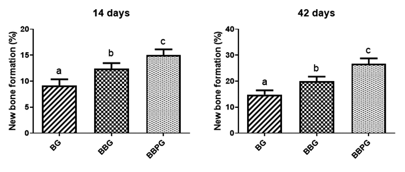
Results: Micro-CT showed centripetal bone regeneration restricted to defect margins, with biomaterial particles persisting centrally. Histologically, new bone progressed from immature trabecular architecture at day 14 to a mature lamellar conformation by day 42, notably in BBPG. All groups showed a significant temporal increase in new bone percentage. BBPG demonstrated superior bone growth at 42 days (26.64 ± 2.15%) compared to BG (14.85 ± 1.63%) and BBG (20.05 ± 1.70%). The birefringence of the collagen fibers showed a color transition from red to yellowish-green during the analyzed periods.
Conclusion: The combination of the biomaterial, fibrin biopolymer and photobiomodulation significantly enhanced bone defect repair and matrix maturation without barrier membranes, presenting high translational potential for cost-effective clinical applications in regenerative medicine.
Keywords:
Biocompatible materials; Bone regeneration; Photobiomodulation; Fibrin sealant; Biopolymers
Background
When bone tissue sustains an injury, it possesses an inherent capacity to regenerate, ultimately restoring both its architecture and functionality. The healing process begins immediately with the inflammatory phase, marked by clot formation [1, 2]. During this stage, various signaling molecules, including inflammatory mediators, angiogenic agents, and growth factors, are released, activating immune cells [3]. As collagen networks develop, they provide a scaffold for the formation of new bone, initially producing what is known as primary or immature bone [4]. This is followed by the remodeling phase, in which bone resorption and regeneration gradually reshape the tissue until its original structure and function are fully reestablished [5].
Bone defects differ in terms of shape, size, mechanical integrity, and vascularization, factors that can either facilitate or impede the healing process [6]. Minor defects typically heal without complication, whereas critical-size defects (CSDs) often lead to the development of fibrous tissue rather than new bone, compromising full regeneration [7]. Bone restoration may be necessary in a range of clinical contexts, including traumatic injuries, congenital abnormalities, and the surgical removal of tumors [8, 9].
In cases involving extensive bone defects, the application of implants and bone substitutes becomes essential to support the healing process [10, 11]. Grafting techniques are employed to enhance or accelerate local regeneration, using bone sourced from various origins: autografts (from the patient), allografts (from a donor of the same species), or xenografts (from a different species) [12]. Although autogenous grafts are considered the gold standard due to their biocompatibility and osteogenic potential, their use is constrained by the need for dual surgical procedures and associated risks [13, 14]. As a result, research has focused on developing synthetic alternatives, including alloplastic biomaterials such as hydroxyapatite, beta-tricalcium phosphate (β-TCP), bioactive polymers, and specialized bioglasses designed to mimic the properties of natural bone [15-19].
To promote satisfactory repair, certain bioproducts or biopharmaceuticals are required to provide a three-dimensional scaffolding system for the biomaterials, such as fibrin biopolymer (also called heterologous fibrin sealant). This material is composed of thrombin and fibrin, which, in addition to providing this biological scaffold, also acts as a sealant, hemostatic, and healing agent in the biological processes of tissue repair [20-23].
To accelerate morphofunctional recovery, physical therapies such as low-level laser therapy (LLLT), now more commonly referred to as photobiomodulation therapy (PBM), play a significant role in bone repair [24]. PBM utilizes low-intensity light, typically within specific wavelength ranges, to target injured tissues, exerting biomodulatory effects at the cellular level [25, 26]. In the context of bone defect healing, PBM has been shown to reduce inflammation, promote vascular proliferation, and stimulate osteoblast activity and osteogenesis [27, 28]. These effects collectively contribute to faster and higher-quality regeneration of the treated area [29-31].
Previous studies integrating bone repair therapies with heterologous fibrin and photobiomodulation have been carried out with various objectives, such as the stabilization of autogenous bone grafts [32], the evaluation of new biomaterials in guided bone regeneration (GBR) using membranes as epithelial growth barriers, in rat calvaria [33, 34] or long bone repair under different photobiomodulation protocols [35, 36]. In the present study, a photobiomodulation protocol was combined with a particulate synthetic biomaterial and a 100% heterologous fibrin biopolymer purified from snake venom, without the use of protective barriers such as the membranes of GBR membranes.
This experimental protocol was designed to evaluate the bone regeneration process using photobiomodulation (PBM) therapy by integrating and analyzing the key components involved in effective tissue repair. The study focused on defects treated with a composite material combining hydroxyapatite and β-tricalcium phosphate, which was further enhanced by the addition of a heterologous fibrin biopolymer to form a novel biocomplex.
Methods
Experimental design
Thirty adult male Wistar rats (Rattus norvegicus), aged 90 days and with a mean body weight of approximately 285 g, were obtained from the Central Animal Facility of the University of Marília (Unimar). The experimental protocol was reviewed and approved by the Institutional Animal Care and Use Committee (CEUA-Unimar), under protocol number 007/2018.
Upon arrival, the animals were housed in standard polypropylene cages, with four rats per cage, under controlled environmental conditions. The bioterium maintained a 12-hour light/dark cycle regulated by an automated timer, with artificial illumination. Ambient temperature was stabilized at 22 ± 1 °C using air conditioning and an exhaust ventilation system, and monitored daily using a calibrated room thermometer. This study was conducted in accordance with the ARRIVE guidelines (Animal Research: Reporting of In Vivo Experiments), ensuring transparency and reproducibility in animal research. All procedures adhered to the ethical principles established by the National Centre for the Replacement, Refinement, and Reduction of Animals in Research (NC3Rs), aiming to minimize animal use and suffering while maximizing scientific integrity.
All animals were randomly assigned to three experimental groups to ensure unbiased distribution:
-
biomaterial (BG);
-
biomaterial + fibrin biopolymer (BBG);
-
biomaterial + fibrin biopolymer + PBM (BBPG)
Surgical Procedures
For the experimental surgical intervention, animals were anesthetized via intramuscular administration of a combination of tiletamine hydrochloride and zolazepam hydrochloride (10 mg/kg; Telazol®, Fort Dodge Laboratories, IA, USA). All procedures were conducted under the supervision of a licensed veterinary professional to ensure compliance with ethical and welfare standards.
Prior to surgery, the cranial region, specifically the frontal-parietal area between the external auricular pinnae, was shaved using an electric clipper (Philips® Multigroom QG3250, São Paulo, Brazil). Body weight was recorded using a precision scale (MicroNal®, São Paulo, Brazil). Individual identification was performed by perforating the external ear pinnae with Ainsworth® punch pliers (Golgran®, São Paulo, Brazil). The shaved region, including adjacent fur, was disinfected using a 10% topical solution of polyvinylpyrrolidone-iodine (PVPI; Povidine®, Vic Pharma Indústria e Comércio Ltda, São Paulo, Brazil).
Each surgical procedure was carried out independently on a sterile, cork-covered wooden surface, with all instruments replaced between specimens to prevent cross-contamination.
Animals were gently restrained in the prone position on the surgical table, ensuring minimal stress or trauma. A 4-cm semilunar skin incision was made using a No. 15 carbon steel scalpel blade (Embramax®, São Paulo, Brazil). The periosteum was carefully elevated with a syndesmotome and retracted along with surrounding soft tissues to expose the parietal bone surface.
A standardized circular osteotomy (5.0 mm diameter) was performed at the center of the parietal bone using a trephine drill (Neodent®, Curitiba, Brazil) mounted on a 500 Kavo electric contra-angle handpiece (KaVo® Dental Excellence, Joinville, Brazil), connected to an electric micromotor (KaVo® Dental Excellence, Joinville, Brazil). The procedure was executed at low rotational speed (1500 rpm) under continuous irrigation with sterile 0.9% saline solution to mitigate thermal damage and prevent bone necrosis. The resulting bone fragment was smooth-edged and intact, with preservation of the dura mater and underlying cerebral structures.
In the BG group, cranial defects were filled with a blood clot combined with the GenPhos XP® (Baumer, Mogi Mirim, Brazil) biomaterial. In contrast, animals in the BBG and BBPG groups received a composite of heterologous fibrin biopolymer and GenPhos XP® biomaterial. The biomaterial was weighed using an analytical precision balance (MicroNal® Equipamentos de Precisão, São Paulo, Brazil) to ensure a standardized mass of approximately 0.04 mg. Following complete polymerization of the fibrin biopolymer with the biomaterial, the resulting biocomplex was carefully transferred to the defect site without exerting pressure on the underlying brain tissue.
Surgical site closure involved meticulous repositioning of the soft tissues, ensuring that the periosteum adequately covered the defect area. The skin was sutured using simple interrupted stitches with 4-0 silk suture material (Ethicon®, Johnson & Johnson, São Paulo, Brazil). The region was then gently cleansed with gauze moistened in a 2% topical chlorhexidine solution (Riohex®, Farmacêutica Rioquímica, São José do Rio Preto, Brazil).
Postoperatively, animals were placed in lateral recumbency within individual cages and exposed to incandescent light to facilitate anesthetic recovery. Immediately following surgery, a single dose of antibiotic (Flotril® 2.5%, Schering-Plough, Rio de Janeiro, Brazil) was administered intramuscularly at 0.2 mL/kg. Analgesia was initiated with intramuscular dipyrone (Analgex V®, Agener União, São Paulo, Brazil) at a dose of 0.06 mL/kg and maintained for three consecutive days. Subsequently, analgesic support was continued with oral paracetamol (Medley®, São Paulo, Brazil) at a dose of 200 mg/kg, administered as six drops per animal diluted in drinking water until the time of euthanasia.
Throughout the experimental period, animals were closely monitored for signs of pain and distress. Behavioral assessments included evaluation of apathy, depression, aggression, or hyperexcitability, particularly when these traits deviated from baseline behavior. Activity levels were observed for changes ranging from hypoactivity to hyperactivity, with attention to gait, posture, and facial expression. Additional parameters included general appearance, food and water intake, clinical symptoms, spontaneous behavior, and responses to external stimuli.
GenPhos XP® biomaterial
GenPhos XP® (Baumer S.A., Mogi Mirim, Brazil) represents a fully synthetic biphasic calcium phosphate ceramic, formulated as 70% hydroxyapatite [HA; Ca₁₀(PO₄)₆(OH)₂] and 30% β-tricalcium phosphate [β-TCP; Ca₃(PO₄)₂] granules (0.50-0.75 mm), characterized by interconnected macroporosity (70-240 µm) and a tailored resorption profile of 7-9 months for guided bone regeneration in dentoalveolar and maxillofacial procedures.
The biphasic matrix couples the inherent structural resilience and osteoconductive mimicry of the bone's mineral component, ensuring scaffold longevity, with the accelerated hydrolytic breakdown of β-TCP's under osteoclast- and macrophage-derived acidity, thereby promoting neovascularization and sequential substitution by autologous bone matrix [31].
Heterologous fibrin biopolymer (HFB)
The heterologous fibrin biopolymer was manufactured and provided by the Center for the Study of Venoms and Venomous Animals at Unesp (Cevap, Botucatu, Brazil). Its components and application formulas are listed in patent BR 102014011432-7, issued on July 6, 2022, by the Brazilian National Institute of Industrial Property. The biopolymer consists of three separate solutions, previously thawed, mixed, and homogenized before application. Fraction 1 is thrombin-like (gyroxin), obtained from the venom of Crotalus durissus terrificus; the diluent is calcium chloride; and fraction 2 is fibrinogen (cryoprecipitate) from the blood of Bubalus bubalis (buffalo). The proportion used was 1:1:2 (fraction 1, diluent and fraction 2, respectively).
For each animal, the preparation of the heterologous fibrin biopolymer followed a standardized protocol. Initially, the GenPhos XP® biomaterial was weighed using an analytical balance and stored in an Eppendorf Tubex® 3810x microtube (Eppendorf AG, Hamburg, Germany). Subsequently, 10 µL of fraction 1 (thrombin-like enzyme) was pipetted into a separate microtube. In the second microtube, 10 µL of the calcium chloride diluent was combined with 20 µL of fraction 2 (fibrinogen derived from Bubalus bubalis), and the mixture was gently homogenized.
The contents of both microtubes were then transferred to a sterile plastic container to initiate polymerization. To prevent cross-contamination and ensure precision, the micropipette tip was replaced for each individual solution during the preparation process.
Photobiomodulation (PBM)
Animals assigned to the BBPG group received photobiomodulation therapy using a gallium-aluminum-arsenide (GaAlAs) laser device. To facilitate precise irradiation of the calvarial region, animals were gently restrained manually, allowing full exposure of the surgical site without the need for anesthetic intervention.
The laser protocol employed continuous wave emission at a wavelength of 830 nm, with an output power of 30 mW and an energy density of 6 J/cm². Irradiation was performed at four equidistant points arranged in a cross-shaped configuration over the defect area, with each site receiving 24 s of exposure. The laser beam had a spot size of 0.116 cm², resulting in a power density of 258.6 mW/cm². Each irradiation point received 0.72 J of energy (24 s exposure), totaling 2.88 J per session across four points. The emitter was positioned in direct contact with the skin at a perpendicular angle (90°), yielding a total application time of 96 s per session.
Treatment commenced immediately after surgery and was administered three times per week until the endpoint of the experiment. Laser output calibration was performed directly on the device (Laserpulse®, IBRAMED, Amparo, Brazil) prior to each session. The therapeutic parameters were based on the protocol established by de Oliveira Gonçalves et al. [32], and utilized in previous research [33, 34], ensuring methodological consistency with prior studies.
Sample collection and euthanasia
At 14 and 42 days postoperatively, five animals from each experimental group were weighed and subsequently euthanized. The procedure was conducted in a calm and isolated environment to minimize stress and avoid visual or auditory exposure to other animals. Euthanasia was performed via intraperitoneal administration of sodium thiopental (2.5% solution) at a dose of 150 mg/kg, applied to the lower left abdominal quadrant. To enhance analgesic coverage, lidocaine hydrochloride was co-administered at a dose of 10 mg/kg.
Following euthanasia, the calvarial defect region was carefully excised using a conical carbide burr mounted on a low-speed surgical handpiece (Dabi Atlante®, Ribeirão Preto, Brazil), ensuring preservation of the supraperiosteal soft tissues. The harvested specimens were immediately immersed in 10% neutral buffered formalin (pH 7.2) for fixation over a period of seven days. After fixation, samples were forwarded for histological processing.
Confirmation of death was followed by proper disposal procedures: the carcasses were placed in white biological waste bags, frozen, and sent to an authorized disposal facility in accordance with institutional biosafety protocols.
Microtomography
Microtomographic images of each specimen were acquired using a SkyScan 1174 computed microtomography system (Bruker-microCT®, Kontich, Belgium). The reconstruction process was performed with the 64Bits270013 platform and the NRecon® software (version 1.6.8.0, SkyScan, 2011, Bruker-micro-CT®), generating approximately 1000-1100 axial slices per sample, based on standardized anatomical reference parameters [35].
Histomorphology and histomorphometry
Following specimen collection, samples were rinsed under running water for 24 h and subsequently subjected to a demineralization protocol using an EDTA-based solution. The solution was composed of 4.13% Tritiplex® III (Merck KGaA, Hessen, Germany) and 0.44% sodium hydroxide (Labsynth, São Paulo, Brazil), with weekly replacement over a six-week period. To monitor the progression of demineralization, radiographic assessments were performed during each solution change using Insight IP-21 F-Speed periapical film (Carestream®, Carestream Health, New York, USA).
Upon completion of demineralization, specimens were dehydrated through a graded ethanol series, cleared in xylene, and embedded in Histosec® paraffin (Merck, Hessen, Germany). Semi-serial coronal sections were obtained from the central region of the defect using a Leica® RM2245 semi-automatic microtome (Leica Biosystems®, Wetzlar, Germany). Sections were cut at a thickness of 5 µm, with six slides prepared per specimen, each containing four sections. These were stained with hematoxylin and eosin (HE), Masson’s trichrome (MT) and Picrosirius red (PR) for histological evaluation.
Histomorphological analysis was performed across all specimens, encompassing the entire defect area to assess bone regeneration patterns. Parameters evaluated included the presence of granulation tissue, inflammatory infiltrate, and the quality and maturity of newly formed bone, as well as the extent of defect filling by regenerated tissue.
For quantitative assessment, four semi-serial sections from each surgical site were examined using an Olympus® light microscope (Olympus Corporation, Tokyo, Japan). Images were captured using a 10× objective lens coupled to a digital camera, with acquisition software configured to a resolution of 4080 × 3072 pixels and a spot resolution of 30%. Image analysis was conducted using ImageJ® software version 1.50d (Wayne Rasband®, National Institutes of Health, USA; Java 1.7_67, 64-bit).
From the semi-serial sections, two central slices, representing the largest diameter of the defect and spaced 300 µm apart, were selected for morphometric analysis. The area of newly formed bone was measured, and the percentage of bone regeneration was calculated [36].
Statistical analysis
Statistical analyses were conducted using GraphPad Prism version 8 (GraphPad Software Inc., 2018; San Diego, CA, USA). To evaluate the temporal effect on the percentage of newly formed bone within each experimental group, an unpaired Student’s t-test was applied. Intergroup comparisons at identical time points were performed using one-way analysis of variance (ANOVA) for independent samples, followed by Tukey’s post hoc test to identify specific differences. Statistical significance was established at a threshold of p < 0.05 for all tests. Each group and time point comprised five samples (n = 5). Prior to hypothesis testing, Bartlett’s test was employed to assess the homogeneity of variances and confirm the normal distribution of the data.
Results
Microtomography
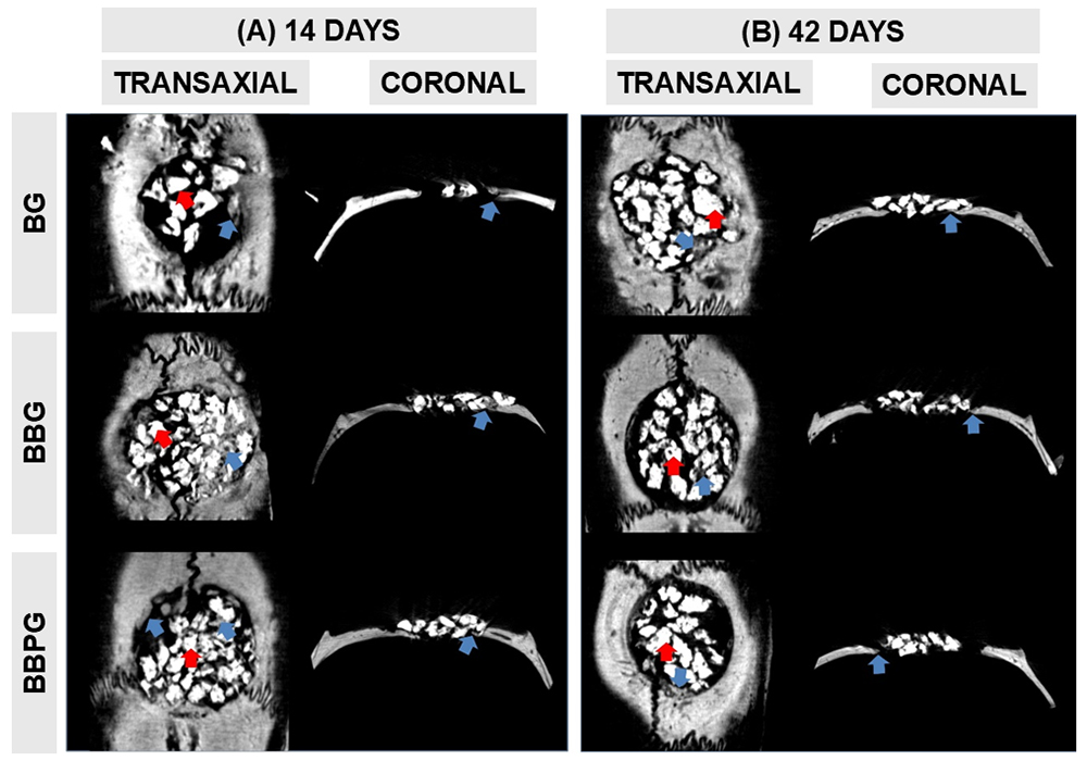
At the 14-day mark, two-dimensional micro-computed tomography (micro-CT) scans in both transaxial and coronal planes revealed findings consistent with histological analysis. All samples exhibited a centripetal pattern of bone regeneration, characterized by increased radiodensity at the lateral margins of the defect. Hyperdense biomaterial particles were visible in grayscale, occupying the surgical site (Figure 1 A ).
By 42 days, all experimental groups showed deposition of partially mineralized bone tissue adjacent to the dura mater. However, this bone formation remained localized to the defect borders throughout the observation period. Biomaterial particles persisted in all specimens, and complete osseous closure of the defect was not achieved (Figure 1 B ).
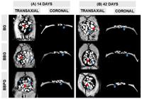
Representative micro-computed tomographic (micro-CT) images displaying transaxial and coronal views of calvarial bone defects across experimental groups at (A) 14 and (B) 42 days post-surgery. BG: defects treated with biomaterial alone; BBG: defects treated with biomaterial combined with heterologous fibrin biopolymer; BBPG: defects treated with biomaterial and heterologous fibrin biopolymer, followed by photobiomodulation therapy. Blue arrows indicate newly formed bone tissue, while red arrows denote biomaterial particles within the defect site. 5-mm osteotomy.
Histomorphology
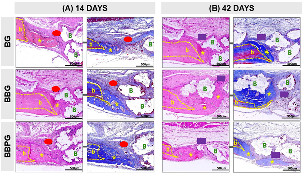
At 14 days post-intervention, histological analysis across all experimental groups revealed a consistent pattern of bone regeneration, initiating from the defect margins and progressing centripetally toward the wound center. The newly formed bone exhibited a trabecular architecture, densely populated by osteocytes and lined with active osteoblasts. The central region of the defect was predominantly occupied by granulation tissue, characterized by a high cellular content including inflammatory cells, neovascular structures, fibroblasts, and dispersed biomaterial particles (Figure 2 A ).
By day 42, all specimens demonstrated complete coverage of the defect by fibrous connective tissue. Biomaterial particles were either encapsulated by dense collagen fibers or integrated into the mineralized bone matrix. The inflammatory infiltrate appeared sparsely distributed, indicative of a resolving inflammatory response. In the BBPG animals, the newly formed bone displayed a lamellar structure, suggestive of advanced maturation and ongoing remodeling. The surrounding connective tissue exhibited a pronounced collagenous composition (Figure 2 B ).
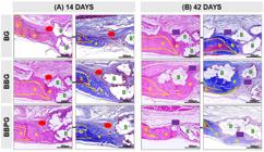
Histological sections of calvarial bone defects stained with hematoxylin and eosin (HE) and Masson’s trichrome (MT) at (A) 14 and (B) 42 days post-surgery. Across all experimental groups, newly formed bone tissue (indicated by asterisks) was consistently observed at the defect margins (yellow b). Reactive tissue (red ellipse) was present surrounding biomaterial particles (green B) at both time points. By day 42, the defect areas were predominantly occupied by fibrous connective tissue (purple rectangle), residual biomaterial, and a sparse distribution of inflammatory cells. Images captured at 10× magnification.
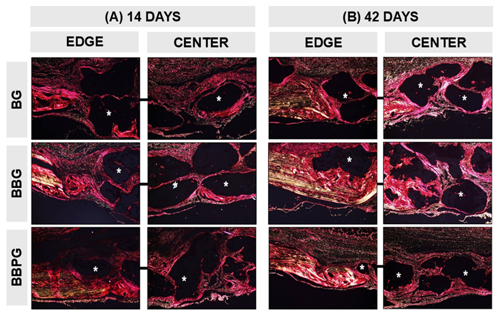
Picrosirius red (PR) staining, examined under polarized light microscopy, enabled assessment of the maturation dynamics of the newly formed bone matrix. At 14 days, all specimens exhibited thin type I collagen fibers within the nascent bone matrix, displaying intense red birefringence. Notably, in the photobiomodulation group (BBPG), the collagen fibers appeared more organized and thicker, with a yellowish-red hue, indicative of advanced matrix maturation (Figure 3 A ).
By 42 days, the initial type I collagen fibers laid down during the proliferative phase were progressively replaced by broader type III fibrils, as evidenced by a shift toward yellow-green birefringence under polarized light. Residual GenPhos XP® biomaterial particles observed at the conclusion of the experimental period appeared as darkfield structures (Figure 3 B ).
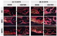
Polarized light microscopy images illustrate the birefringence of collagen fibers in coronal sections of rat calvarial bone defects, captured at the defect margins and central regions. Samples were stained with Picrosirius red and analyzed at (A) 14 and (B) 42 days post-surgery. BG: defects treated with biomaterial alone; BBG: defects treated with biomaterial combined with heterologous fibrin biopolymer; BBPG: defects treated with biomaterial and heterologous fibrin biopolymer, followed by photobiomodulation therapy. Biomaterial particles are indicated by asterisks. Images acquired using a 10× objective on an inverted light microscope.
Histomorphometric
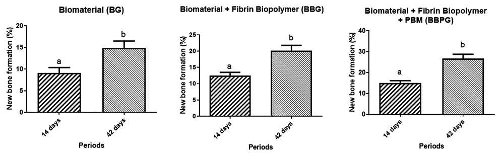
A statistically significant increase in the percentage of new bone formation was observed across all experimental groups (BG, BBG, and BBPG) when comparing the two time points - 14 and 42 days post-intervention (Figure 4).
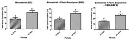
Quantitative analysis of new bone formation (%) in each experimental group (BG, BBG, and BBPG) at 14 and 42 days post-surgery. Data are presented as mean ± standard deviation. Distinct lowercase letters denote statistically significant differences between groups and time points (p < 0.05), as determined by an unpaired Student's t-test.
Comparative analysis of the percentage of newly formed bone revealed statistically significant differences among all experimental groups (BG, BBG, and BBPG) at both 14 and 42 days (Figure 5 and Table 1; p < 0.05).
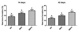
Percentage of new bone formation in each experimental group (BG, BBG, and BBPG) at 14 and 42 days post-surgery. Data are expressed as mean ± standard deviation. Distinct lowercase letters indicate statistically significant differences (p < 0.05), as determined by one-way ANOVA for independent samples followed by Tukey’s post hoc test.
Discussion
This study evaluated in vivo the repair of a critical-sized calvarial bone defect using a heterologous fibrin biopolymer as a three-dimensional scaffold combined with a high-purity, synthetic biphasic calcium phosphate ceramic graft (70% hydroxyapatite and 30% β-tricalcium phosphate) manufactured in Brazil, without the use of traditional guided bone regeneration (GBR) barrier membranes. Additionally, this study investigated the local therapeutic effects of adjunctive photobiomodulation (PBM) therapy to accelerate the healing cascade.
Micro-computed tomography (micro-CT) was utilized to qualitatively monitor the progression of bone regeneration within the critical-sized defects. At 14 days post-surgery, an increased radiodensity was evident along the lateral margins of the defects, accompanied by hyperdense biomaterial particles occupying the surgical site. These particulate scaffolds facilitated centripetal bone formation, which occurred in close structural association with their surfaces. By day 42, the deposition of partially mineralized bone matrix was noted at the defect edges, aligning with findings from previous investigations utilizing structurally similar biphasic ceramics [37-39]. Overall, complete osseous closure was not achieved in any specimen by the 42-day mark. This incomplete closure is an expected biological outcome given the critical nature of the 5.0-mm defect model, which inherently lacks the capacity for spontaneous physiological healing within the animal's lifespan [40, 41].
Histological evaluation remains a foundational method for assessing the qualitative characteristics of tissue healing and new bone architecture. In the present study, histological analysis at 14 days revealed abundant granulation tissue characterized by inflammatory cell infiltration, active neovascularization, fibroblast proliferation, and dispersed biomaterial particles occupying the central region of the defect. By day 42, the inflammatory response had subsided across all groups, indicating a physiological progression toward a resolving remodeling phase. Notably, specimens from the BBPG group exhibited a more densely collagenized connective tissue matrix and newly formed bone with lamellar structural features, reflecting an advanced stage of bone maturation. These observations corroborate established literature indicating that the seamless structural integration of newly formed bone with residual biomaterial particles serves as an index of active osteoregenerative activity [42-44]. Furthermore, these qualitative histological findings strongly support the microtomographic and quantitative histomorphometric analyses.
The progression of bone neoformation was quantitatively assessed through histomorphometric analysis. A statistically significant temporal increase in the percentage of newly formed bone was observed across all experimental groups between the 14- and 42-day time points. Notably, the tri-therapeutic approach in the BBPG group demonstrated superior bone regeneration, yielding mean values of 15.02 ± 1.10% at day 14 and 26.64 ± 2.15% at day 42. These findings underscore the enhanced therapeutic efficacy achieved by combining advanced bioproducts (biphasic ceramic and fibrin scaffold) with PBM therapy, reinforcing the potential of this combinatorial protocol to accelerate bone matrix deposition within challenging clinical defect models [22, 45, 46].
The extracellular matrix plays a pivotal role in regulating cellular signaling, recruitment, and behavior during bone healing, with its structural integrity largely dependent on the organization of its collagenous components. In this study, the maturation dynamics of the newly formed bone matrix were assessed using polarized light birefringence imaging of Picrosirius red-stained coronal sections. At the 14-day time point, the collagen matrix exhibited predominantly thin type I collagen fibers, characteristic of the proliferative phase of inflammation [47, 48]. In contrast, specimens from the photobiomodulation group (BBPG) displayed highly organized, thicker collagen bundles, indicating accelerated matrix remodeling [26, 49]. By day 42, a gradual transition was observed across the groups, with type I fibers being increasingly replaced by broader type III fibrils [50]. These patterns demonstrate that adjunctive physical therapies, such as PBM, facilitate rapid collagen cross-linking and enhance the structural organization of the regenerating osteoid matrix [51, 52].
The BBPG group received photobiomodulation therapy utilizing a gallium-aluminum-arsenide (GaAlAs) laser. This modality is widely recognized in the literature for its ability to enhance angiogenesis and stimulate osteoblast proliferation, thereby promoting osteogenesis [53]. Additionally, it exerts anti-inflammatory effects at the local site, collectively contributing to accelerated and more effective bone regeneration within the biostimulated region [54].
One potential limitation of this study is the absence of a negative control group left to heal with a blood clot alone. However, it is well established that critical-size defects are fundamentally incapable of spontaneous regeneration, even when surgically stabilized, making a therapeutic intervention mandatory to evaluate active regeneration [55]. Furthermore, animal allocation into the experimental groups was guided by the ethical principles of the "3Rs" (replacement, reduction, and refinement) to minimize animal numbers while ensuring statistical validity [56]. These principles continue to serve as a cornerstone in contemporary biomedical research, ensuring responsible and humane use of animal models.
While a previous study by our research group confirmed the separate utilities of these modalities [33], the structural and biological interaction of this particulate biomaterial with both the heterologous fibrin biopolymer and PBM, specifically in the absolute absence of traditional guided bone regeneration barriers, remained to be elucidated. Eliminating costly collagen or synthetic membranes [57] could substantially reduce the financial burden of maxillofacial and dental bone grafting procedures, making advanced bone regeneration therapies more accessible.
Conclusions
This study demonstrated the novel combinatorial application of a heterologous fibrin biopolymer (HFB), hydroxyapatite/β-tricalcium phosphate, and laser photobiomodulation (PBM) therapy for bone repair in the absolute absence of traditional guided bone regeneration (GBR) barrier membranes.
Based on the qualitative micro-computed tomography, structural histomorphology, quantitative histomorphometry, and polarized light birefringence imaging of collagen fibers, it can be concluded that the combination of biphasic calcium phosphate with heterologous fibrin biopolymer and laser photobiomodulation therapy exerts a beneficial effect on bone regeneration. The combined protocol significantly accelerated bone matrix deposition and structural maturation within critical-sized calvarial defects. These findings highlight its high translational potential as a promising, cost-effective, and efficient therapeutic strategy in regenerative medicine and bone grafting procedures.
Acknowledgments
The authors express their sincere gratitude to Cirilo Francisco Santos Neto (University of Marília, Marília, Brazil) for his expert technical support in preparing the histological slides. Additionally, the authors thank dental students Gabrielle Caroline Rodrigues, Victória Lopes de Oliveira, Isabela de Oliveira Santos, Larissa Souza Barbosa, Thiago Borges Jacob, and Luana Aparecida de Carvalho Moreira for their valuable assistance during the experimental phases of this study.
References
-
1. Duda GN, Geissler S, Checa S, Tsitsilonis S, Petersen A, Schmidt-Bleek K. The decisive early phase of bone regeneration. Nat Rev Rheumatol. 2023 Feb;19(2):78-95. doi: 10.1038/s41584-022-00887-0.
» https://doi.org/10.1038/s41584-022-00887-0 -
2. Schmidt-Bleek K, Kwee BJ, Mooney DJ, Duda GN. Boon and bane of inflammation in bone tissue regeneration and its link with angiogenesis. Tissue Eng Part B Rev. 2015 Aug;21(4):354-64. doi: 10.1089/ten.TEB.2014.0677.
» https://doi.org/10.1089/ten.TEB.2014.0677 -
3. Yang N, Liu Y. The role of the immune microenvironment in bone regeneration. Int J Med Sci. 2021;18(16):3697-707. doi: 10.7150/ijms.61080.
» https://doi.org/10.7150/ijms.61080 -
4. Carvalho MS, Cabral JMS, da Silva CL, Vashishth D. Bone matrix non-collagenous proteins in tissue engineering: creating new bone by mimicking the extracellular matrix. Polymers (Basel). 2021 Mar 30;13(7):1095. doi: 10.3390/polym13071095.
» https://doi.org/10.3390/polym13071095 -
5. Šromová V, Sobola D, Kaspar P. A brief review of bone cell function and importance. Cells. 2023 Nov 5;12(21):2576. doi: 10.3390/cells12212576.
» https://doi.org/10.3390/cells12212576 -
6. Ma Q, Miri Z, Haugen HJ, Moghanian A, Loca D. Significance of mechanical loading in bone fracture healing, bone regeneration, and vascularization. J Tissue Eng. 2023 May 22;14:20417314231172573. doi: 10.1177/20417314231172573.
» https://doi.org/10.1177/20417314231172573 -
7. Wei J, Chen X, Xu Y, Shi L, Zhang M. Significance and considerations of establishing standardized critical values for critical size defects in animal models of bone tissue regeneration. Heliyon. 2024;10(15):e33768. doi: 10.1016/j.heliyon.2024.e33768.
» https://doi.org/10.1016/j.heliyon.2024.e33768 -
8. Migliorini F, La Padula G, Torsiello E, Spiezia F, Oliva F, Maffulli N. Strategies for large bone defect reconstruction after trauma, infections or tumour excision: a comprehensive review of the literature. Eur J Med Res. 2021;26(1):130. doi: 10.1186/s40001-021-00593-9.
» https://doi.org/10.1186/s40001-021-00593-9 -
9. Wang B, Feng C, Liu Y, Mi F, Dong J. Recent advances in biofunctional guided bone regeneration materials for repairing defective alveolar and maxillofacial bone: a review. Jpn Dent Sci Rev. 2022;58(1):233-48. doi: 10.1016/j.jdsr.2022.07.001.
» https://doi.org/10.1016/j.jdsr.2022.07.001 -
10. Xue N, Ding X, Huang R, Jiang R, Huang H, Pan X, Hu J, Ren L. Bone tissue engineering in the treatment of bone defects. Orthop Surg. 2022 Oct;14(10):2321-34. doi: 10.1111/os.13378.
» https://doi.org/10.1111/os.13378 -
11. Guo N, Leu MC. Additive manufacturing: technology, applications and research needs. Front Mech Eng. 2013;8(3):215-43. doi: 10.1007/s11465-013-0248-8.
» https://doi.org/10.1007/s11465-013-0248-8 -
12. Battafarano G, Rossi M, De Martino V, Marampon F, Borro L, Secinaro A, Rizzo A, Della Rocca G. Strategies for bone regeneration: from graft to tissue engineering. Int J Mol Sci. 2021 Dec 9;22(24):13283. doi: 10.3390/ijms222413283.
» https://doi.org/10.3390/ijms222413283 -
13. Zhang J, Zhang W, Yue W, Qin W, Zhao Y, Xu G. Research progress of bone grafting: a comprehensive review. Int J Mol Sci . 2025 Apr 14;26(8):4214. doi: 10.3390/ijms26084214.
» https://doi.org/10.3390/ijms26084214 -
14. McAllister BS, Haghighat K. Bone augmentation techniques. J Periodontol. 2007 Mar;78(3):377-96. doi: 10.1902/jop.2007.060048.
» https://doi.org/10.1902/jop.2007.060048 -
15. Fukuba S, Okada M, Nohara K, Iwata T. Alloplastic bone substitutes for periodontal and bone regeneration in dentistry: current status and prospects. Materials (Basel). 2021 Feb 25;14(5):1074. doi: 10.3390/ma14051074.
» https://doi.org/10.3390/ma14051074 -
16. Pomini KT, Cestari TM, Santos German J, de Oliveira Rosso MP, de Oliveira Gonçalves JB, Buchaim DV, Barraviera B, Ferreira RS Jr. Influence of experimental alcoholism on the repair process of bone defects filled with beta-tricalcium phosphate. Drug Alcohol Depend. 2019 Mar 1;197:197-206. doi: 10.1016/j.drugalcdep.2019.01.022.
» https://doi.org/10.1016/j.drugalcdep.2019.01.022 -
17. de Moraes R, Plepis AM de G, Martins V da CA, Garcia CF, Galdeano EA, Maia FLM, da Cunha MR, Munhoz MA de S, Fernandes VA dos R, Beraldo RA. Viability of collagen matrix grafts associated with nanohydroxyapatite and elastin in bone repair in the experimental condition of ovariectomy. Int J Mol Sci . 2023 Oct 28;24(21):15684. doi: 10.3390/ijms242115684.
» https://doi.org/10.3390/ijms242115684 -
18. Nogueira DMB, de Oliveira Rosso MP, Buchaim DV, Zangrando MSR, Buchaim RL. Update on the use of 45S5 bioactive glass in the treatment of bone defects in regenerative medicine. World J Orthop. 2024;15(2):204-14. doi: 10.5312/wjo.v15.i2.204.
» https://doi.org/10.5312/wjo.v15.i2.204 -
19. Nogueira DMB, Rosso MP de O, Santos PS da S, Sousa- Neto MD, Silva-Sousa AC, Soares CT, Buchaim DV, Bueno CR de S, Brito FEO de, Barraviera B. Biological behavior of bioactive glasses SinGlass 45S5 and SinGlass High F18 in the repair of critical bone defects. Biomolecules. 2025 Jan 15;14(1):118. doi: 10.3390/biom14010118.
» https://doi.org/10.3390/biom14010118 -
20. Buchaim DV, Cassaro CV, Shindo JVTC, Coletta BBD, Pomini KT, De Oliveira Rosso MP, Sousa CR, Barraviera B, Ferreira RS Jr. Unique heterologous fibrin biopolymer with hemostatic, adhesive, sealant, scaffold and drug delivery properties: a systematic review. J Venom Anim Toxins incl Trop Dis. 2019 Nov 11;25:e20190038. doi: 10.1590/1678-9199-JVATITD-2019-0038.
» https://doi.org/10.1590/1678-9199-JVATITD-2019-0038 -
21. Kempe PRG, Chiarotto GB, Barraviera B, Ferreira RS, de Oliveira ALR. Neuroprotection and immunomodulation by dimethyl fumarate and a heterologous fibrin biopolymer after ventral root avulsion and reimplantation. J Venom Anim Toxins incl Trop Dis . 2020 Oct 5;26:e20201. doi: 10.1590/1678-9199-JVATITD-2020-0001.
» https://doi.org/10.1590/1678-9199-JVATITD-2020-0001 -
22. Buchaim DV, Andreo JC, Pomini KT, Barraviera B, Ferreira RS, Duarte MAH, Basso Alves Resende T de C, Coletta BB Della, Costa NP da, Buchaim RL. A biocomplex to repair experimental critical size defects associated with photobiomodulation therapy. J Venom Anim Toxins incl Trop Dis . 2022 Jul 28;28:e20220. doi: 10.1590/1678-9199-JVATITD-2022-0020.
» https://doi.org/10.1590/1678-9199-JVATITD-2022-0020 -
23. Rossi J de O, Araujo EMC, Camargo MEC, Ferreira Junior RS, Barraviera B, Miglino MA, Buchaim RL, Buchaim DV. Effectiveness of the association of fibrin scaffolds, nanohydroxyapatite, and photobiomodulation with simultaneous low-level red and infrared lasers in bone repair. Materials (Basel) . 2024 Sep 11;17(17):4351. doi: 10.3390/ma17184351.
» https://doi.org/10.3390/ma17184351 -
24. Obradović RR, Kesić LG, Peevska S. Influence of low-level laser therapy on biomaterial osseointegration: a mini-review. Lasers Med Sci. 2009;24(3):447-51. doi: 10.1007/s10103-009-0641-z.
» https://doi.org/10.1007/s10103-009-0641-z -
25. Sergio LPS, Campos VMA, Vicentini SC, Mencalha AL, de Paoli F, Fonseca AS. Low-intensity red and infrared lasers affect mRNA expression of DNA nucleotide excision repair in skin and muscle tissue. Lasers Med Sci . 2016;31(3):429-35. doi: 10.1007/s10103-015-1895-2.
» https://doi.org/10.1007/s10103-015-1895-2 -
26. Ribeiro LNS, de Figueiredo FAT, da Silva Mira PC, Arnez MFM, Matsumoto MAN, de Menezes LM, Bueno CRA, Buchaim DV. Low-level laser therapy (LLLT) improves alveolar bone healing in rats. Lasers Med Sci . 2022 Oct;37(7):2961-9. doi: 10.1007/s10103-022-03567-5.
» https://doi.org/10.1007/s10103-022-03567-5 -
27. Reis CHB, Buchaim DV, Ortiz A de C, Fideles SOM, Dias JA, Miglino MA, Issa JPM, Buchaim RL. Application of fibrin associated with photobiomodulation as a promising strategy to improve regeneration in tissue engineering: a systematic review. Polymers (Basel) . 2022 Dec 27;14(1):99. doi: 10.3390/polym14010099.
» https://doi.org/10.3390/polym14010099 -
28. Amaroli A, Colombo E, Zekiy A, Aicardi S, Benedicenti S, De Angelis N. Interaction between laser light and osteoblasts: photobiomodulation as a trend in the management of socket bone preservation - a review. Biology (Basel). 2020 Feb 29;9(2):39. doi: 10.3390/biology9020039.
» https://doi.org/10.3390/biology9020039 -
29. Statkievicz C, Toro LF, de Mello- Neto JM, de S DP, Casatti CA, Issa JPM, Ribeiro MS, Lopes-Martins RAB, Parizotto NA. Photomodulation multiple sessions as a promising preventive therapy for medication-related osteonecrosis of the jaws after tooth extraction in rats. J Photochem Photobiol B Biol. 2018 May;182(5):47-57. doi: 10.1016/j.jphotobiol.2018.03.015.
» https://doi.org/10.1016/j.jphotobiol.2018.03.015 -
30. Escudero JSB, Perez MGB, de Oliveira Rosso MP, Buchaim DV, Pomini KT, Campos LMG, de Oliveira JB, Sales JGDM, Rodrigues Camargo MEC. Photobiomodulation therapy (PBMT) in bone repair: a systematic review. Injury. 2019 Nov;50(11):1853-61. doi: 10.1016/j.injury.2019.10.006.
» https://doi.org/10.1016/j.injury.2019.10.006 -
31. Paini S, Bighetti ACC, Cestari TM, Arantes RVN, Santos PS, Mena-Laura EE, Garlet GP, Taga R, Assis GF. Concentration-dependent effects of latex F1-protein fraction incorporated into deproteinized bovine bone and biphasic calcium phosphate on the repair of critical-size bone defects. J Biomed Mater Res B Appl Biomater. 2020 Nov;108(8):3270-85. doi: 10.1002/jbm.b.34664.
» https://doi.org/10.1002/jbm.b.34664 -
32. de Oliveira Gonçalves JB, Buchaim DV, de Souza Bueno CR, Pomini KT, Barraviera B, Ferreira RSJ, Issa JPM, Buchaim RL. Effects of low-level laser therapy on autogenous bone graft stabilized with a new heterologous fibrin sealant. J Photochem Photobiol B Biol . 2016 Nov;162(9):663-8. doi: 10.1016/j.jphotobiol.2016.07.023.
» https://doi.org/10.1016/j.jphotobiol.2016.07.023 -
33. Bento M, Moscatel M, Pagani BT, Flavia B, Trazzi DM, Pascon T, Aranda-Garcia A, Marques MM, Reis CHB, Buchaim DV. Heterologous fibrin biopolymer as a key scaffold for bone regeneration: synergistic effects with photobiomodulation and membrane therapy. Gels. 2026 Jan 6;12(1):56. doi: 10.3390/gels12010056.
» https://doi.org/10.3390/gels12010056 -
34. Della Coletta BB, Jacob TB, Moreira LA de C, Pomini KT, Buchaim DV, Eleutrio RG, Shimano MM, Shimano AC, Reis CHB, Buchaim RL. Photobiomodulation therapy on the guided bone regeneration process in defects filled by biphasic calcium phosphate associated with fibrin biopolymer. Molecules. 2021 Jan 13;26(2):405. doi: 10.3390/molecules26020405.
» https://doi.org/10.3390/molecules26020405 -
35. de Oliveira Rosso MP, Oyadomari AT, Pomini KT, Coletta BB Della, Shindo JVTC, Ferreira RSJ, Barraviera B, Buchaim DV. Photobiomodulation therapy associated with heterologous fibrin biopolymer and bovine bone matrix helps to reconstruct long bones. Biomolecules. 2020 Oct 15;8(10):383. doi: 10.3390/biomolecules8100383.
» https://doi.org/10.3390/biomolecules8100383 -
36. Rodrigues SJ Jr, dos Santos LC, Buchaim DV, Duarte MAH, Alcalde MP, Barraviera B, Buchaim RL. Efficacy of three-dimensional bioactive composites in long bone repair with photobiomodulation. Materials (Basel) . 2025 Jan 15;14(2):218. doi: 10.3390/ma14020218.
» https://doi.org/10.3390/ma14020218 -
37. Umoh JU, Sampaio A V., Welch I, Pitelka V, Goldberg HA, Underhill TM, Geuens T, Frank CA. In vivo micro-CT analysis of bone remodeling in a rat calvarial defect model. Phys Med Biol. 2009 May 7;54(9):2847-61. doi: 10.1088/0031-9155/54/9/028.
» https://doi.org/10.1088/0031-9155/54/9/028 -
38. Zhao N, Qin L, Liu Y, Zhai M, Li D. Improved new bone formation capacity of hyaluronic acid-bone substitute compound in rat calvarial critical size defect. BMC Oral Health. 2024 Aug 24;24(1):994. doi: 10.1186/s12903-024-04679-8.
» https://doi.org/10.1186/s12903-024-04679-8 -
39. Otto L, Wolint P, Bopp A, Woloszyk A, Becker AS, Boss A, Beerenwinkel N, Pellegata NS, Meyrat BM, Kappeler C. 3D-microtissue derived secretome as a cell-free approach for enhanced mineralization of scaffolds in the chorioallantoic membrane model. Sci Rep. 2021 Jan 21;11(1):1861. doi: 10.1038/s41598-021-81132-8.
» https://doi.org/10.1038/s41598-021-81132-8 -
40. Perez JR, Kouroupis D, Li DJ, Best TM, Kaplan L, Correa D. Tissue engineering and cell-based therapies for fractures and bone defects. Front Bioeng Biotechnol. 2018 Mar 23;6:105. doi: 10.3389/fbioe.2018.00105.
» https://doi.org/10.3389/fbioe.2018.00105 -
41. Sparks DS, Saifzadeh S, Savi FM, Dlaska CE, Berner A, Henkel J, Stoddart MJ, Smit T, Grijpma DW, Richards RG, Eglin D, Alini M, Grad S. A preclinical large-animal model for the assessment of critical-size load-bearing bone defect reconstruction. Nat Protoc. 2020 May;15(8):2877-924. doi: 10.1038/s41596-019-0271-2.
» https://doi.org/10.1038/s41596-019-0271-2 -
42. Allo BA, Costa DO, Dixon SJ, Mequanint K, Rizkalla AS. Bioactive and biodegradable nanocomposites and hybrid biomaterials for bone regeneration. J Funct Biomater. 2012 Sep 21;3(3):432-63. doi: 10.3390/jfb3030432.
» https://doi.org/10.3390/jfb3030432 -
43. Szwed-Georgiou A, Pociski P, Kupikowska-Stobba B, Urbaniak MM, Rusek-Wala P, Szustakiewicz K, Gostomski L, Suchecki M, Walesiak-Kędzierska A, Juzwa W. Bioactive materials for bone regeneration: biomolecules and delivery systems. ACS Biomater Sci Eng. 2023 Sep 11;9(9):5222-54. doi: 10.1021/acsbiomaterials.3c00373.
» https://doi.org/10.1021/acsbiomaterials.3c00373 -
44. Shah SA, Sohail M, Nakielski P, Rinoldi C, Zargarian SS, Kosik-Kozio A, Pierini F, Eltohamy M, Ullah S, Hasany M, Rainer A, Calabrese G, Giuffrida R, Barbani N, Brucale M, De Sanctis F, Battista E, Ristori C, Biscarini F, Paulin T, Chen X, Cho KY, Bianco A. Integrating micro- and nanostructured platforms and biological drugs to enhance biomaterial-based bone regeneration strategies. Biomacromolecules. 2025 Feb 10;26(2):140-62. doi: 10.1021/acs.biomac.4c01062.
» https://doi.org/10.1021/acs.biomac.4c01062 -
45. de Oliveira Rosso MP, Oyadomari AT, Pomini KT, Coletta BB Della, Shindo JVTC, Ferreira RSJ, Barraviera B, Buchaim DV. Photobiomodulation therapy associated with heterologous fibrin biopolymer and bovine bone matrix helps to reconstruct long bones. Biomolecules. 2020 Oct 15;8(10):383. doi: 10.3390/biomolecules8100383.
» https://doi.org/10.3390/biomolecules8100383 -
46. Moscatel MBM, Pagani BT, Trazzi BF de M, Reis CHB, Ribeiro CA, Buchaim DV, Gomes DMDC, Leite NP, Bueno CRA, Carvalho JC. Effects of photobiomodulation in association with biomaterials on the process of guided bone regeneration: an integrative review. Ceramics. 2025 Mar 7;8(1):94. doi: 10.3390/ceramics8010094.
» https://doi.org/10.3390/ceramics8010094 -
47. Casalechi HL, De Farias Marques AC, Da Silva EAP, Aimbire F, Marcos RL, Lopes-Martins RAB, Silva DF. Analysis of the effect of phototherapy in model with traumatic Achilles tendon injury in rats. Lasers Med Sci . 2014;29(3):1075-81. doi: 10.1007/s10103-013-1475-3.
» https://doi.org/10.1007/s10103-013-1475-3 -
48. Gerbi MEMDM, Miranda JM, De Arruda JAA, Moreno LMM, Carneiro VSM, Brasilino NC, Barbosa FF, Lima Ribeiro MS, Anbinder AL. Photobiomodulation therapy in bone repair associated with bone morphogenetic proteins and guided bone regeneration: a histomorphometric study. Photomed Laser Surg. 2018 Sep;36(8):581-8. doi: 10.1089/pho.2018.4485.
» https://doi.org/10.1089/pho.2018.4485 -
49. Pretel H, Lizarelli RFZ, Ramalho LTO. Effect of low-level laser therapy on bone repair: histological study in rats. Lasers Surg Med. 2007;39(10):788-96. doi: 10.1002/lsm.20575.
» https://doi.org/10.1002/lsm.20575 -
50. Alhaskawi A, Dong Y, Zou X, Zhou W, Ezzi SHA, Goutham Kota V, Xia Y, Ye C, Sun Y, Yue B. Advancements in biomaterials and scaffold design for tendon repair and regeneration. J Appl Biomater Funct Mater. 2025 Sep;23(3):2280800024687654. doi: 10.1177/2280800024687654.
» https://doi.org/10.1177/2280800024687654 -
51. Sataray-Rodriguez A, Ojeda ZC, Montes AM, Jones K, Umerani A, Arriaga LS, Camargo LS, Zamudio-Cuevas Y, López-Martínez S, Manzo-Merino J. Optimizing low-level light therapy for skin rejuvenation: efficacy of wavelengths and treatment parameters in collagen synthesis and aging signs. Mod Res Inflamm. 2025 Mar;14(1):64-78. doi: 10.4236/mri.2025.141007.
» https://doi.org/10.4236/mri.2025.141007 -
52. Lawrence J, Sorra K. Photobiomodulation as medicine: low-level laser therapy (LLLT) for acute tissue injury or sport performance recovery. J Funct Morphol Kinesiol. 2024 May 20;9(2):69. doi: 10.3390/jfmk9020069.
» https://doi.org/10.3390/jfmk9020069 -
53. Bai J, Li L, Kou N, Bai Y, Zhang Y, Lu Y, Zou X, Zhao N, Jia X, Zhou W, Ye C, Sun Y, Yue B. Low level laser therapy promotes bone regeneration by coupling angiogenesis and osteogenesis. Stem Cell Res Ther. 2021 Dec 27;12(1):693. doi: 10.1186/s13287-021-02758-z.
» https://doi.org/10.1186/s13287-021-02758-z -
54. Babtan A, Ilea A, Feurdean C, Ceci S, Pula B, Candrea S, Roman DL, Bogdan C, Citu CM, Iabotarow DE. Biostimulation with low-level laser therapy and its effects on soft and hard tissue regeneration. Literature review. J Mind Med Sci. 2022;9(2):228-37. doi: 10.25122/jmms-2022-0217.
» https://doi.org/10.25122/jmms-2022-0217 -
55. Nauth A, Schemitsch E, Norris B, Nollin Z, Watson JT. Critical-size bone defects: is there a consensus for diagnosis and treatment? J Orthop Trauma. 2018;32(Suppl 1):S7-11. doi: 10.1097/BOT.0000000000001115.
» https://doi.org/10.1097/BOT.0000000000001115 -
56. MacArthur Clark J. The 3Rs in research: a contemporary approach to replacement, reduction and refinement. Br J Nutr. 2018 Aug;120(s1):S1-7. doi: 10.1017/S0007114517002227.
» https://doi.org/10.1017/S0007114517002227 -
57. Duarte ND, Frigerio PB, Estefania G, Chica A, Okamoto R, Buchaim RL, Reis CHB, Cruzat VF, Krause GC, Tirapegui J. Biomaterials for guided tissue regeneration and guided bone regeneration: a review. Dent J (Basel). 2025 Jan 16;13(1):79. doi: 10.3390/dj13010079
» https://doi.org/10.3390/dj13010079
-
Availability of data and materials
All data generated or analyzed during this study are included in this article.
-
Funding
RSFJ is a National Council for Scientific and Technological Development (CNPq) PQ1D research fellow (grant no. 301608/2022-9). RLB is a CNPq PQ1C research fellow (grant no. 302545/2025-5). MAHD is a CNPq PQ1B research fellow (grant no. 304153/2024-9). This work was also supported by the Center for Translational Science and Biopharmaceutical Development (FAPESP grant no. 2021/11936-3) and utilized the lyophilization multi-user equipment (EMU - FAPESP grant no. 2023/16514-5).
-
Ethics approval
The experimental protocol was reviewed and approved by the Institutional Animal Care and Use Committee of the University of Marília (CEUA/UNIMAR, protocol no. 007/2018). This study was conducted in strict compliance with the ARRIVE (Animal Research: Reporting of In Vivo Experiments) guidelines and adhered to the ethical principles established by the National Centre for the Replacement, Refinement, and Reduction of Animals in Research (NC3Rs). All animal care and surgical procedures additionally conformed to the guidelines of the Brazilian National Council for the Control of Animal Experimentation (CONCEA) and Federal Law no. 11.794/2008.
-
Consent for publication
Not applicable.
All data generated or analyzed during this study are included in this article.