 Dengue virus serotype 4 infection in human fatal cases: histopathological investigation
Abstract
BACKGROUND Dengue, the disease caused by dengue virus (DENV), is responsible for over 300 million cases worldwide. Although DENV-4 usually presents with milder symptoms, some cases can result in death.
OBJECTIVES The present study aims to characterise the immunohistopathological profile of hepatic, pulmonary, cardiac and renal involvement in fatal cases of DENV-4 that occurred between 2011 and 2013 in Brazil.
METHODS To this end, tissue samples from the patients were subjected to histopathological analysis using bright-field microscopy and cytokine detection by immunohistochemistry.
FINDINGS Cells stained for tumour necrosis factor-alpha (TNF-α) and interferon-gamma (IFN-γ) were observed in the liver, lungs and kidneys. In the liver, the most common finding was periportal necrosis, along with steatosis and mononuclear cell infiltration. In the lungs, pneumocyte necrosis was only observed in areas of alveolar oedema. Septal and alveolar haemorrhage were present, along with thickening of the alveolar walls. In the kidneys, glomerular capillary congestion was observed in most of the analysed cases. Inflammatory cell infiltration was observed in the cortical region, along with proximal and distal tubule necrosis. Finally, in the heart, despite limited local cytokine expression, the cardiac tissue remained morphologically preserved.
MAIN CONCLUSIONS Although the findings reported in this study are consistent with those described in fatal dengue cases, the absence of cardiac alterations indicates that DENV-4 may cause a milder infection in this organ.
Key words:
DENV-4; fatal cases; liver; lung; heart; kidney
The dengue virus (DENV) belongs to the genus Orthoflavivirus, family Flaviviridae, which has four antigenically distinct serotypes: DENV-1, -2, -3 and -4.1 DENV is an arthropod-borne virus (arbovirus) transmitted by haematophagous mosquitoes of the Aedes genus, more specifically Aedes aegypti and Aedes albopictus. Infection with any of the four serotypes results in the disease known as dengue, an acute febrile illness characterised by its broad spectrum of clinical manifestations.2
DENV infection ranges from asymptomatic and subclinical cases to mild, self-limiting fever and very severe cases of haemorrhage and plasma leakage.3 When present, clinical manifestations occur after an incubation period of three to fifteen days.4 Although the vast majority of symptomatic cases present with mild symptoms such as fever, headache, retro-orbital pain, myalgia, arthralgia, leucopoenia and thrombocytopenia, around 5% of dengue infected patients develop the severe form of the disease, termed severe dengue (SD), a condition associated with increased vascular permeability, severe thrombocytopenia and haemorrhage in the skin and other organs.5 The evolution of SD can also lead to the development of hypotension, hypovolaemic shock and circulatory failure, which, if not treated promptly, can result in the patient's death within 8 to 24 h.6 Previous studies suggest an increased incidence of DENV infections resulting in the involvement of different systems, such as gastrointestinal, hepatic, respiratory, cardiac, neurological and renal, in adults and children.7,8,9,10
Dengue is prevalent in tropical and subtropical regions and is endemic in more than 120 countries. Epidemiological studies suggest that around 50 to 200 million people are infected annually, with 500,000 severe cases and more than 40,000 deaths associated with the disease.2,11 In Latin America alone, dengue is responsible for around 1.5 million cases every year,12 although it is estimated that almost half of the world's population lives in places where there is a real risk of infection.13 In Brazil, between the years of 1951-1960 and 2013-2022, the average transmission potential for dengue by Ae. aegypti has increased by 94.5%.14 This increase is the result of the interaction between various factors, such as climate change, population growth, uncontrolled urbanisation, migration, the evolution of the virus, inefficient health systems and socioeconomic inequality, among others.15,16
In 2023, 1,530,940 probable cases of dengue were detected in Brazil, which represents an increase of 16.5% in the number of cases compared to 2022. By epidemiological week 35 of 2023, 1,458 cases of SD had been confirmed, with 946 deaths from dengue.17 The number of probable cases further increased in 2024, with 6,215,201 detected between epidemiological weeks 1 and 26. During this same time period, 82,908 cases of SD were confirmed, an increase of 307.8% when compared to the previous year. The number of fatal cases of dengue also saw an increase in 2024, with 4,269 confirmed deaths.18 Regarding serotype distribution, DENV-1 represents the largest number of cases, with 39,816, followed by DENV-2, with 4,144, and lastly by DENV-3, with 46.17,18 Although DENV-4 has not been identified among the cases tested by the Brazilian Ministry of Health since 2018, circulation of this serotype can still be observed in the emergence of sporadic cases, such as in the states of Rio de Janeiro, Mato Grosso do Sul and Minas Gerais.19,20,21
In Brazil, although DENV-4 was first detected in 1981-82,22 in the northern region of the country, this serotype remained absent from the Brazilian epidemiological scenario for almost three decades, until it re-emerged in the State of Roraima, in 2010.23 After its reintroduction, DENV-4 quickly spread to other states in the country, favouring not only the increase of dengue cases in subsequent years, due to the high number of individuals susceptible to this new serotype, but also the risk of developing more severe manifestations in individuals previously infected with DENV-1, -2 or -3.24 While serotype 4 was not very prevalent among the cases analysed by the Ministry of Health of Brazil until 2015, its presence could be detected in more than two thirds of Brazilian states, and it has been responsible for a large number of epidemics since its reintroduction, mainly due to the population's susceptibility to the new serotype.25
DENV-4 cases usually present milder symptoms and are less associated with hospitalisations when compared to other serotypes.26,27 However, factors such as secondary infections by heterologous DENV serotypes28 or the interaction between dengue and other comorbidities often result in fatal cases,29 and can prove to be important aggravating factors during the course of the disease. In Brazil, the circulation of DENV-4 has been associated with mild cases,30 however, severe and fatal cases resulting from infection with this serotype have also been reported.8,9,31 In this context, this study aims to investigate the histopathological and inflammatory profile of four fatal cases of DENV-4, the latest serotype to be reintroduced in Brazil.
MATERIALS AND METHODS
Ethics statement - All the procedures carried out in this study comply with the principles and regulations established by the resolution 196/96 of the National Health Council (Brazil) and were previously approved by the Human Research Ethics Committee of the Oswaldo Cruz Institute (Fiocruz) with Certificate of Submission for Ethical Appraisal Nº 57221416.0.1001.5248. The study was conducted in accordance with the Declaration of Helsinki, and approved by the Ethics Committee of Federal University of Ceará (Certificate of Submission for Ethical Appraisal Nº 58571016.0.0000.5054, approved 15/07/2017).
Liver, lung, heart and kidney samples from human fatal cases of dengue were kindly provided by Dr Fernanda Capelo Barroso Dr Emília Tomé de Souza from the Federal University of Ceará. These samples come from a project approved by the Human Research Ethics Committee of the Federal University of Ceará with Certificate of Submission for Ethical Appraisal Nº 58571016.0.0000.5054. Informed consent was obtained from all subjects and/or their legal guardian(s).
Human fatal cases - The liver, lung, heart and kidney samples analysed in this study were obtained from four fatal cases (Table) of dengue from the 2012 epidemic in the State of Ceará, and were received by spontaneous demand. Laboratory diagnosis with confirmation of the DENV-4 as the infecting serotype was previously carried out by the Central Laboratory of the State of Ceará through viral isolation (fresh tissue) followed by indirect immunofluorescence with specific monoclonal antibody for each serotype.32 Out of the four cases, only one, case #4, had been previously infected with DENV.
Bright field microscopy - The tissue samples were dehydrated in increasing concentrations of ethanol, clarified in xylene and embedded in paraffin. The resulting paraffin blocks were then sectioned into 4 µm slices using a microtome, mounted in glass slides, stained with haematoxylin and eosin (H&E) and finally analysed in a bright field microscope.
Immunohistochemistry - Tissue samples 4 µm thick were obtained in a microtome and mounted in glass slides. The peroxidase immunohistochemistry protocol was carried out as described by Rabelo et al.33 Briefly, these paraffin-embedded samples were deparaffinised and rehydrated. Antigen retrieval was performed by heating the tissue in citrate buffer (pH 6.0) at 90ºC. Subsequently, endogenous peroxidase activity was blocked and tissue sections were incubated with a Protein Blocker solution (Dako, USA). The samples were then incubated overnight with the primary antibodies tumour necrosis factor-alpha (TNF-α) (Sc-52746; Santa Cruz Biotechnology, TX, USA) or interferon gamma (IFN-γ) (250707; Abbiotec, CA, USA) at a concentration of 1:200 in a dark chamber at 4ºC. The following day, the tissue samples were incubated with a secondary horseradish peroxidase-conjugated antibody (Spring Bioscience, USA) for 15 min. The reaction was revealed using diaminobensidine as a chromogen and the sections were counterstained with Harris' haematoxylin. For negative control of the immunohistochemistry reaction, samples were incubated only with the secondary horseradish peroxidase-conjugated antibody.
RESULTS
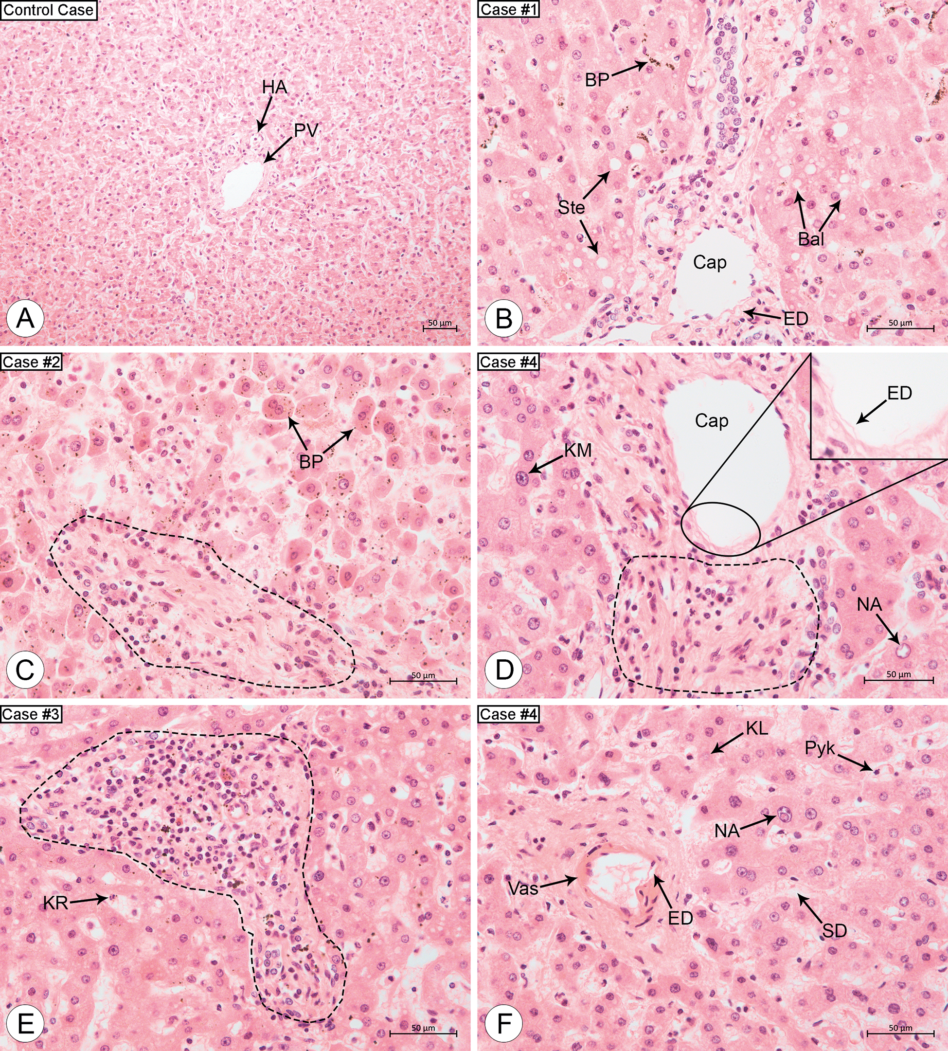
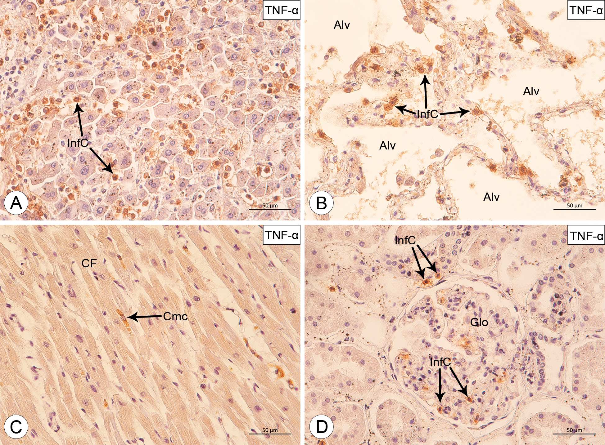
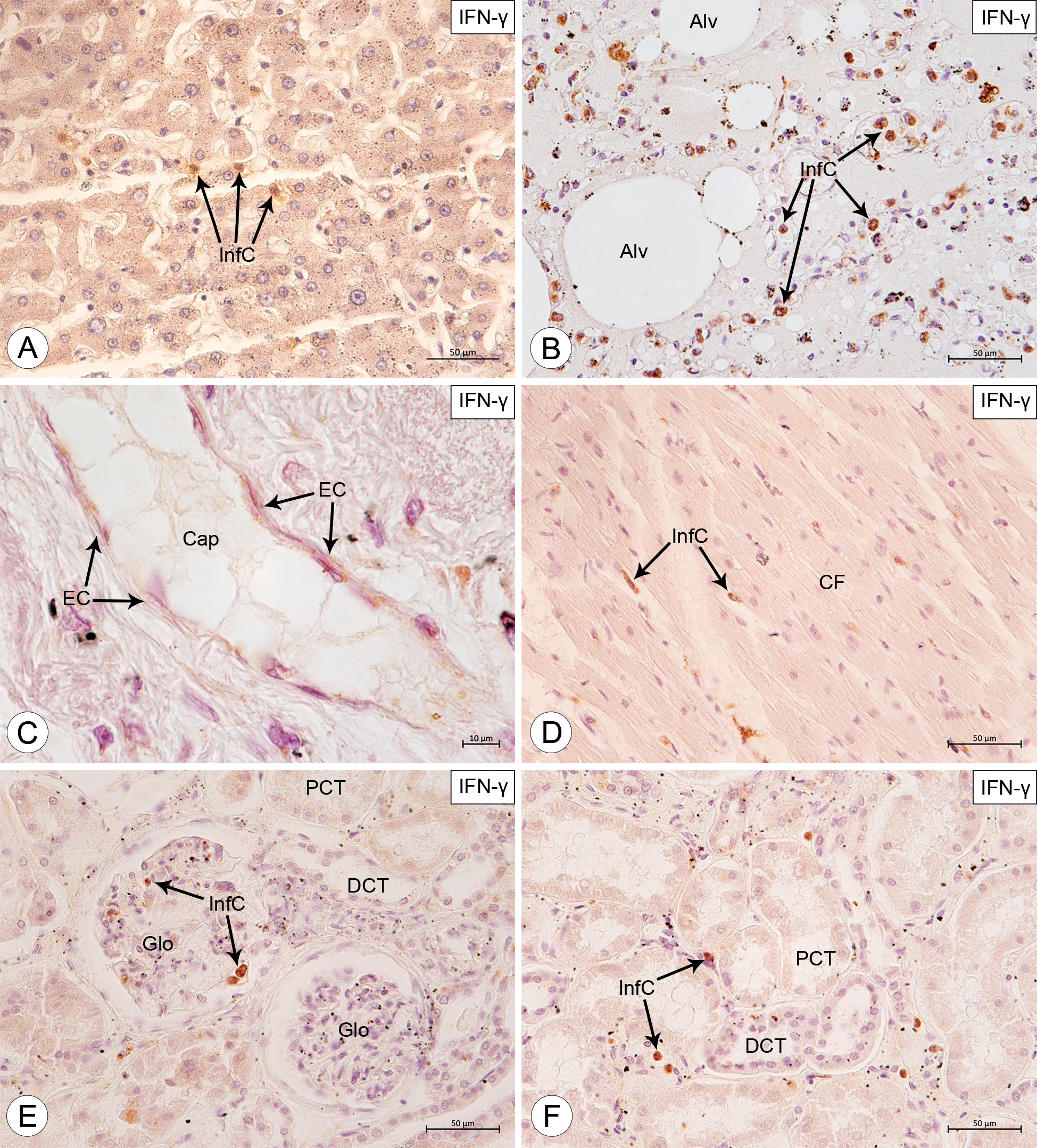
Histopathology of the liver of DENV-4 infected fatal cases - The main finding in the liver was necrosis (Fig. 1C-E). These extensive areas of hepatocytes undergoing the process of cell death were predominantly located in the centrilobular zone of the hepatic lobule. Also in this zone, signs of ballooning degeneration could be observed in hepatocytes (Fig. 1B), as well as dilation of the sinusoidal capillaries (Fig. 1F). Round, well-defined vacuoles were present in the cytoplasm of hepatocytes (Fig. 1B). This finding, called steatosis, signals the excessive accumulation of lipids inside the cell, usually causing lateralisation of the nucleus, and was often present in regions of hepatocyte necrosis. Hepatocytes near areas of necrosis had biliary pigments accumulated in the cytoplasm (Fig. 1B-C), which, in higher concentrations, can lead to cell death. Different types of nuclear degeneration, such as karyorrhexis (Fig. 1E), karyolysis (Fig. 1F) and pyknosis (Fig. 1F) were observed in necrotic hepatocytes. Many nuclei presented irregular morphology (Fig. 1F), and karyomegaly was observed in many hepatocytes (Fig. 1D). Vasculitis was observed in some capillaries (Fig. 1F), often with the detachment of the endothelial wall (Fig. 1B, D). Mononuclear cell infiltration was also observed in areas of necrosis, with the presence of Kupffer cells (Fig. 1C-E). Furthermore, the cytokine TNF-α was detected in a considerable proportion of inflammatory cells in the sinusoidal capillaries (Fig. 2A), while IFN-γ was also detected, albeit in fewer inflammatory cells (Fig. 3A). No areas of haemorrhage were observed in any of the analysed livers. Overall, hepatic morphology of the studied areas was significantly altered, particularly due to the disruption of the parenchymal integrity, caused by the necrosis of the tissue.
In contrast, the liver of a patient that was not infected with DENV-4 presented well-defined hepatic lobules, with uniform hepatic cords and healthy hepatocytes. The structures of the triad region had a normal aspect, with no alterations in the morphology of the biliary duct, the hepatic artery or the portal vein (Fig. 1A). The central vein and the surrounding parenchyma also presented the regular aspect of a well-functioning liver.
micrographs of liver sections stained with haematoxylin and eosin. (A) Liver of a patient not infected with dengue virus 4 (DENV-4). HA: hepatic artery; PV: portal vein (B, C, D, E, F). Livers of patients infected with DENV-4. Bal: ballooning degeneration; BP: biliary pigment; Cap: capillary; dashed lines: areas of hepatocyte necrosis; ED: endothelial detachment; HA: hepatic artery; KL: karyolysis; KM: karyomegalia; KR: karyorrhexis; NA: nuclear atypia; Pyk: pyknosis; SD: sinusoidal capillary dilation; Ste: steatosis; Vas: vasculitis. Magnification: A: 200X; B, C, D, E, F: 400X.
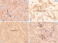
micrographs of immunostained organs incubated with anti-tumour necrosis factor-alpha (TNF-α) antibody. (A) Liver of a patient infected with dengue virus 4 (DENV-4). InfC: inflammatory cell stained for TNF-α. (B) Lung of a patient infected with DENV-4. Alv: alveolus. (C) Heart of a patient infected with DENV-4. CF: cardiac fibres; Cmc: cardiomyocyte stained for TNF-α. (D) Kidney of a patient infected with DENV-4. Glo: glomerulus. Magnification: A, B, C, D: 400X.
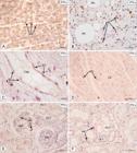
micrographs of immunostained organs incubated with anti-interferon-gamma (IFN-γ) antibody. (A) Liver of a patient infected with dengue virus 4 (DENV-4). InfC: inflammatory cell stained for IFN-γ. (B, C) Lungs of patients infected with DENV-4. Alv: alveolus; Cap: capillary; EC: endothelial cell. (D) Heart of a patient infected with DENV-4. CF: cardiac fibres. (E, F) Kidneys of patients infected with DENV-4. Glo: glomerulus; DCT: distal convoluted tubule; PCT: proximal convoluted tubule. Magnification: C: 400X; A, B, D, E, F: 200X.
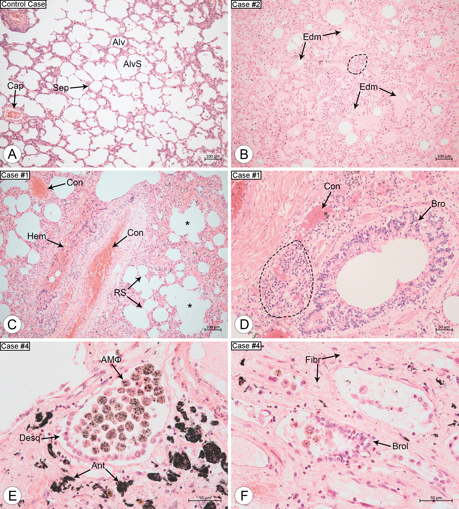
Histopathology of the lung of DENV-4 infected fatal cases - In the lung, several areas of oedema were observed, sometimes completely occluding the alveolar space (Fig. 4B). This exudate presented small lymphocytic content, with alveolar macrophages in its midst. In some analysed fields, few alveoli were unobstructed, leaving a diminished functional area for gas exchange. In addition, blood vessels of large and small calibre showed signs of vascular congestion, with alveolar macrophages among the stagnant erythrocytes (Fig. 4C-D). The cytokine IFN-γ was detected in endothelial cells close to areas of inflammatory cell infiltration (Fig. 3C). Thickening of the alveolar septa was widely observed, mainly due to the infiltration of mononuclear cells (Fig. 4B, D) and to haemorrhage (Fig. 4C), as well as ruptured alveolar septa (Fig. 4C), bronchitis (Fig. 4D) and bronchiolitis (Fig. 4F), which would further impair the gas exchange. Due to this, many alveoli showed signs of compensatory hyperinflation, being overly expanded (Fig. 4C). The presence of TNF-α and IFN-γ in inflammatory cells in the pulmonary parenchyma was confirmed through immunostaining (Figs 2B, 3B). The presence of alveolar macrophages was observed in the alveolar septa, the alveolar space and in the respiratory bronchioles (Fig. 4E). The desquamation of the bronchiolar epithelium was a common finding (Fig. 4E). In one particular case (case #4), carbon particles were observed, both deposited in the bronchiolar interstitium and phagocytosed by alveolar macrophages, with the appearance of anthracosis (Fig. 4E), possibly associated with the patient's lifestyle. Extensive areas of fibrosis were also present in this case (Fig. 4F).
The lung of a patient that was not infected with DENV-4 presented healthy bronchioles and alveolar sacs, with unobstructed alveoli and thin septa (Fig. 4A). No noteworthy morphological alterations were observed in the pulmonary parenchyma. Capillaries and blood vessels also had a normal aspect.
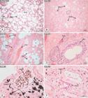
micrographs of lung sections stained with haematoxylin and eosin. (A) Lung of a patient not infected with dengue virus 4 (DENV-4). Cap: capillary; Sep: septum; Alv: alveolus; AlvS: alveolar sac. (B, C, D, E, F) Lungs of patients infected with DENV-4. Asterisk (*): compensatory alveolar hyperinflation; AMΦ: alveolar macrophage; Ant: anthracosis; Bro: bronchitis; Brol: bronchiolitis; Con: vascular congestion; Dashed line: inflammatory cell infiltration; Desq: cell desquamation; Edm: oedema; Fib: fibrosis; Hem: haemorrhage; RAS: ruptured septum. Magnification: A, B, C: 100X; D: 200X; E, F: 400X.
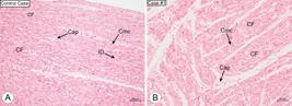
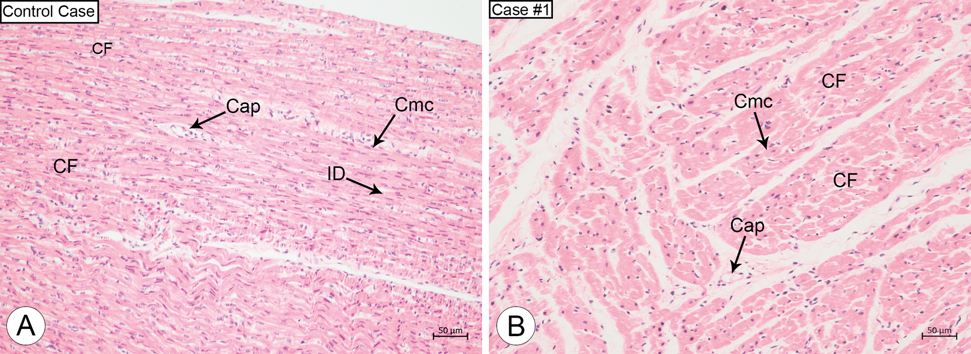
micrographs of heart sections stained with haematoxylin and eosin. (A) Heart of a patient not infected with dengue virus 4 (DENV-4). Cap: capillary; CF: cardiac fibres; Cmc: cardiomyocyte; ID: intercalated disk. (B) Heart of a patient infected with DENV-4. Magnification: A, B: 200X.
Histopathology of the heart of DENV-4 infected fatal cases - Overall, the analysed hearts presented normal morphology, with no alterations indicating a pathological process. The cardiac fibres, the cardiomyocytes and the capillaries, showed no abnormal characteristics (Fig. 5B). Very few cardiomyocytes in the cardiac parenchyma were stained for TNF-α and, in a similar scenario, few inflammatory cells stained for IFN-γ were observed (Figs 2C, 3D).
Likewise, the heart of a patient that was not infected with DENV-4 presented a healthy appearance, with a similar morphology to the infected patient (Fig. 5A).
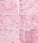
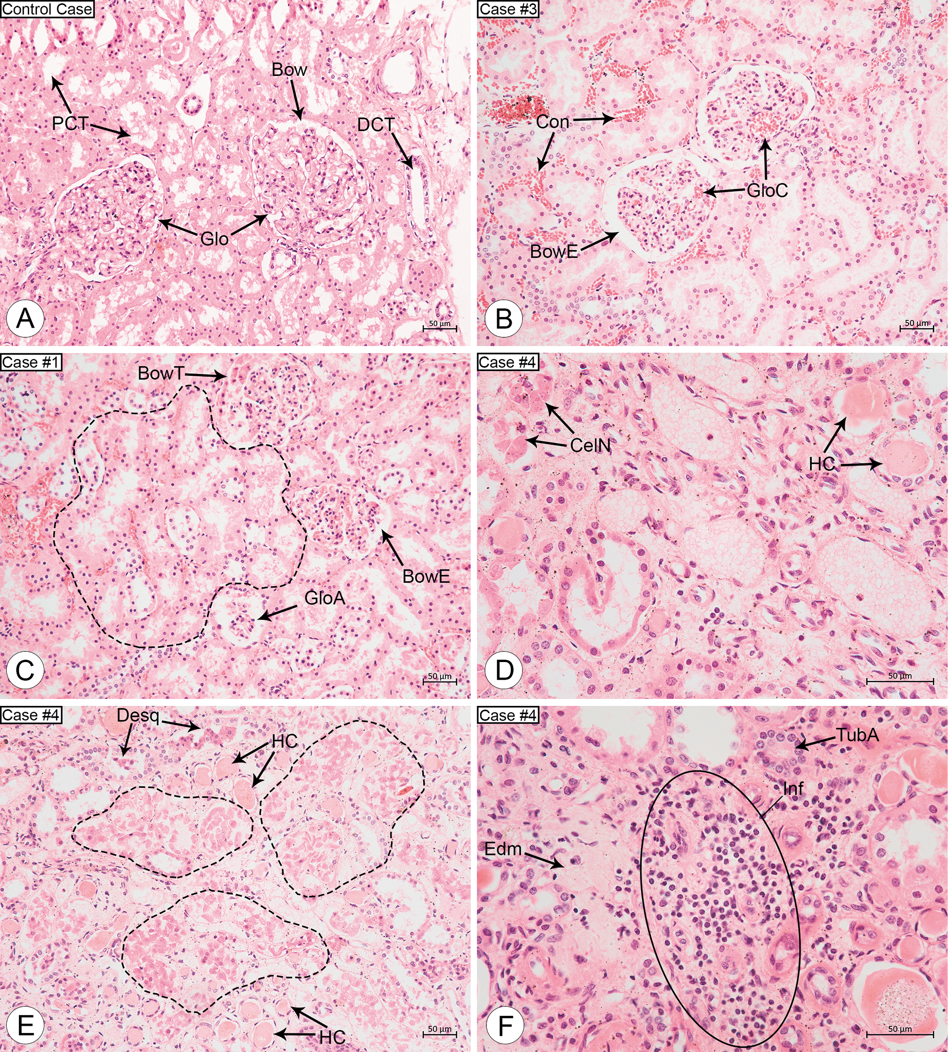
micrographs of kidney sections stained with haematoxylin and eosin. (A) Kidney of a patient not infected with dengue virus 4 (DENV-4). Bow: bowman's space; DCT: distal convoluted tubule; Glo: glomerulus; PCT: proximal convoluted tubule. (B, C, D, E, F) Kidneys of patients infected with DENV-4. BowE: bowman's space enlargement; BowT: bowman's capsule thickening; CelN: tubular cell necrosis; Dashed line: area of tubular necrosis; Desq: cell desquamation; Edm: oedema; GloA: glomerular atrophy; GloC: glomerular congestion; HC: hyaline cast; Inf: inflammatory cell infiltration; TubA: tubular atrophy. Magnification: A, B, C, E: 200X; D, F: 400X.
Histopathology of the kidney of DENV-4 infected fatal cases - In the cortical region of the kidney, the structure of the Malpighian corpuscle was noticeably altered, with signs of glomerular capillary congestion (Fig. 6B), glomerular atrophy (Fig. 6C), enlargement of the Bowman's space (Fig. 6B-C) and thickening of the parietal epithelium of the Bowman's capsule (Fig. 6C). The most common finding was tubular necrosis, with the death of tubular cells that form the epithelium of both the proximal and distal convoluted tubules (Fig. 6C-E). These areas of necrosis would often be close to morphologically altered Malpighian corpuscles. The degree of tubular damage varied, with some tubules only presenting a partial desquamation of the epithelium (Fig. 6E), while others were devoid of functional cells and entirely filled with cellular debris (Fig. 6C). Tubular atrophy was also observed, with extreme reduction of the luminal space (Fig. 6F). The formation of hyaline casts, structures composed of condensed plasma proteins, was regularly observed in the lumen of renal tubules (Fig. 6D-6E), suggesting an altered capillary permeability in the glomerulus. Infiltration of inflammatory cells was observed in some cases, close to areas of tubular necrosis and oedema (Fig. 6F), but was sparse and not present throughout the entirety of the renal parenchyma. Still, some inflammatory cells stained for TNF-α and IFN-γ were detected both in the glomerular capillaries and in renal capillaries (Figs 2D, 3E-F). The medullary region of the kidney presented a regular morphology, with no noteworthy alterations.
In comparison, the kidney of a patient that was not infected with DENV-4 showed a regular morphology, made up of healthy Malpighian corpuscles and a normal tubular structure, with no pathological alterations present (Fig. 6A).
DISCUSSION
During DENV infection, alterations in the liver are among the most commonly observed and are usually associated with cases of SD.34,35 Human patients present jaundice, acute hepatitis, hepatomegaly, liver failure and elevated serum albumin levels. Macroscopically, several areas of vascular congestion as well as several haemorrhagic foci can be observed. In bright field microscopy, several areas of inflammatory infiltrate can be seen near the portal space, as well as focal vacuolisation of the cytoplasm of hepatocytes, areas of vascular congestion, the presence of inflammatory infiltrate in sinusoidal capillaries, hyperplasia of Kupffer cells and lipid inclusions in hepatocytes.7,36,37 Other alterations normally observed on the cellular level include micro and macrogoticular steatosis, focal areas of necrosis with presence of a mononuclear inflammatory infiltrate, mitochondrial engorgement, characteristic of the apoptotic process, haemorrhage and oedema.7
In this study, histopathological analysis of the livers of fatal human cases revealed classic findings of dengue, such as steatosis, inflammatory cell infiltration, hepatocyte necrosis and dilation of sinusoidal capillaries. Kupffer cell hyperplasia, which has been previously described in human fatal cases of dengue,7 is certainly a result of the increased activity of these cells during DENV infection, since macrophages play a large role in activating the inflammatory response and vasodilation through the release of cytokines and chemokines.38 Dilation of the sinusoidal capillaries, generally located in the periportal or perivascular region, suggests a predisposition related to vascularisation disorders.39 It is known that hypertension in the central vein can result in an increase in sinusoidal fenestrae, a reduction in portal venous inflow and, as a consequence of the change in pressure change, promote periportal necroinflammatory activity.40 Furthermore, the obstruction of the portal vein, leading to a decreased blood flow velocity, has also been associated with the dilation of the sinusoidal capillaries.41 The accumulation of biliary pigments in the cytoplasm of hepatocytes, while not a widespread alteration, could indicate an initial presentation of cholestasis, which has been described as a rare hepatic manifestation caused by dengue infection.42 Balloon-like degeneration of hepatocytes is one of the main findings in non-alcoholic steatohepatitis,43 although it is not a commonly described finding in dengue. This form of cell death may be associated with a late and irreversible stage of lipid accumulation in the cytoplasm.44
Pulmonary manifestations in human cases of DENV infection are uncommon, however, alterations such as peribronchial thickening, bilateral pulmonary congestion, pleural effusion, pulmonary oedema, haemorrhage and pneumonitis have already been reported in cases of dengue and SD.45,46,47 Atypical manifestations include acute respiratory distress syndrome and pulmonary dysfunction.37 Histopathological and ultrastructural analysis of lung tissue from fatal cases of dengue revealed interstitial pneumonia, thickening of the alveolar septum, diffuse infiltration of mononucleated and polymorphonucleated cells, areas of alveolar congestion, appearance of a hyaline membrane, type II pneumocyte hyperplasia, platelet recruitment, areas of haemorrhage and oedema, and the presence of viral particles in the endothelium.7,9,36,45
The most consistent findings in the fatal cases analysed in this study were observed in the lungs. Haemorrhage, septal thickening, characterising pneumonitis, inflammatory cell infiltration and vascular congestion were observed in all the cases of death. Curiously, the presence of oedema was not detected in the case diagnosed with SD. Even so, in this specific case, vascular congestion was observed in almost all the vessels analysed, suggesting the presence of alterations in the pulmonary circulatory system. Pulmonary fibrosis, observed in one of the fatal cases, is a chronic regenerative process associated with a range of factors, such as smoking, gastro-oesophageal reflux, the use of some medications, diabetes mellitus, exposure to occupational risks and various infectious agents, such as the Epstein-Barr virus, cytomegalovirus and human herpes viruses 7 and 8,48 although it has never been directly associated with DENV infection. Pulmonary haemorrhage was also observed in some of the cases, most notably in the interalveolar septum, though it was also present in the alveoli, which could have led to the ruptured septa observed. Pulmonary haemorrhage has already been reported in fatal human cases,7,9,36 usually associated with haemoptysis.45,49 It is important to note that the presence of oedema is not necessarily a histopathological finding associated with DENV infection, and may be a common complication of fluid replacement in dengue patients.50 Even so, oedema, accompanied by haemorrhage, is a finding suggestive of alterations in vascular permeability and of plasma.51
To date, the symptomatology and histopathology of the manifestations caused in the heart by DENV are still poorly studied.52 Abnormal aspects of cardiac function observed in DENV infection include sinus tachycardia, increased jugular pressure, altered heart rhythm and interstitial oedema,53 as well as bilateral ventricular systolic dysfunction, the latter observed more commonly in cases of SD.54 Cases of myocarditis are rare in DENV infections and are often asymptomatic, although they are among the most common findings observed in cardiac tissue.55,56 Myocarditis can have serious clinical consequences, such as arrhythmia, heart failure and cardiogenic shock.57,58 The X-ray of a human patient with a confirmed case of dengue revealed progressive cardiomegaly59,60 and histopathological and ultrastructural analysis of cardiac tissue from fatal cases of dengue revealed the presence of nuclear and mitochondrial alterations, degeneration of cardiac fibres, haemorrhage, interstitial oedema, diffuse inflammatory infiltrate and the presence of macrophages in the myocardium.7,52
In the fatal cases analysed in this study, no noteworthy alterations were observed in the heart. This may be explained by the rarity of cardiac involvement during dengue,37,61 since most patients do not show myocardial damage or signs of cardiomyocyte infection by the virus.54
Even though the role of the kidneys during dengue infection is not thoroughly understood, serious manifestations such as acute kidney injury and glomerulonephritis have been reported.62,63 It is believed that renal damage can be caused by multiple factors, such as haemolysis, haemodynamic alterations induced by the virus, deposition of immune complexes in the glomerulus and deposition of muscle proteins resulting from rhabdomyolysis.63,64,65 Histopathological alterations include congestion of glomerular capillaries, focal haemorrhage, oedema, inflammatory cell infiltration, hydropic degeneration, micro abscess formation, thrombus formation in the glomerulus, and acute tubular necrosis.7,8,9,66 Acute kidney injury, a condition characterised by a sudden drop in kidney function, is one of the most frequently reported findings, although the factors that cause its onset during dengue are mostly unexplored.67,68,69 The histopathology of the kidney during dengue is characterised by necrosis of the proximal and distal tubules, with dilation of the lumen, loss of the brush border, simplification of the tubular epithelium and loss of the nucleus.70
In this study, tubular necrosis was observed in all cases analysed. Even though the viral antigen has been previously detected in the epithelium of tubular cells,71 the capacity of DENV to infect these cells has not been demonstrated yet. It has been suggested that the damage to the renal tubules is caused by ischaemia, due to a decreased blood flow to the kidney, on account of the significant blood volume loss during SD.7 In a likely manner, tubular atrophy, characterised by a notable reduction of the tubular lumen, is also associated with the ischaemic process of small blood vessels of the kidney.72 Also observed was the formation of hyaline casts in the tubular space, which is a common occurrence in the kidney. The ones observed in this study appear to be formed mostly by uromodulin — a strong immunomodulatory protein — due to their well-defined round morphology and pale-pink colour when stained with H&E. While normally described as a benign process, hyaline cast formation has been previously associated with tubular and glomerular injury.72,73,74 In turn, the obstruction of the renal tubules, be it by the reduction of the lumen due to atrophy or the overaccumulation of proteins, may alter the hydrostatic pressure gradient across the glomerular capillaries, leading to an enlargement of the Bowman's space.75 The congestion of the glomerular capillaries is possibly the result of the endothelial alterations caused by SD and are probably aggravated by the atrophy of the glomerulus.63,76 It is also possible that the reduction of the glomerular lumen facilitates the deposition of immune complexes in the glomerulus. Although the importance of immune complexes during dengue-related kidney disease has been widely discussed,76,77,78,79 a clear consensus has yet to be established on how exactly the pathological process occurs.
Finally, also in this study, a large number of mononuclear inflammatory cells marked for TNF-α were detected in liver, lung and kidney of DENV-4 infected patients. Inflammatory cells marked for IFN-γ were also detected in these same organs, albeit to a lesser degree. In the lung, specifically, endothelial cells were marked for IFN-γ.
Various cytokines and chemokines are related to tissue damage, being secreted by macrophages and T lymphocytes during the attempt to contain DENV infection.80,81 Furthermore, TNF-α is capable of altering the vascular permeability of capillaries, triggering cases of plasma leakage,82 and is detected in higher levels during secondary DENV infections. Likewise, IFN-γ levels are also higher during secondary DENV infection and during SD.83,84,85 While it has been suggested that IFN-γ secretion plays an important part in the hyperpermeability so characteristic of SD,86 this cytokine appears to be downregulated by DENV replication and viral RNA production.87 Additionally, a previous study conducted in mice lacking interferon receptors demonstrated that IFN-γ plays a role in eliciting a protective response from the initial systemic dissemination of DENV following infection.88 Even still, these cytokines seem to correlate with disease severity and could be used as disease severity markers.89
In the analysed fatal human cases infected with DENV-4, the findings were consistent with what is described in the literature, particularly regarding hepatic, pulmonary and renal alterations. Still, no cardiac alterations were observed, reinforcing either their rarity or a diminished tropism of this serotype for heart cells.
Although hepatic histopathological manifestations are among the most commonly reported during DENV-4 infection, in this study isolated areas of hepatocyte necrosis and inflammation were observed, suggesting a milder infection in the liver. Furthermore, due to severe alterations in the lung, such as haemorrhage and oedema, which can prove fatal even when properly treated, this study suggests that pulmonary manifestations may also play an important role in more severe cases of dengue.
The kidney, on the other hand, is, supposedly, not a viral replication site, but is still largely affected by the systemic manifestations of dengue, presenting tubular and glomerular alterations that can lead to renal failure. Furthermore, even though DENV-4 infection is said to cause a milder form of the disease, this study shows that it is still able to evolve into more severe cases of dengue, that can even lead to death.
Despite being absent from the epidemiological reports of the past four years in Brazil, isolated sporadic cases of DENV-4 have been identified throughout the country, indicating a silent circulation, that goes mostly undetected due to the high prevalence of other serotypes. Ultimately, despite being less associated with hospitalisations and deaths when compared to other serotypes, DENV-4 should still be treated as one of the four cornerstones of dengue, equally as important as DENV-1, DENV-2 and DENV-3, particularly due to factors such as secondary infections with heterologous serotypes or the other comorbidities that can often result in fatal cases.
The main limitation of this study is the limited clinical information and lack of in-depth laboratory diagnosis of the DENV-4 fatal cases. Unfortunately, since the samples were received by spontaneous demand, long after the end of the epidemic, it is not possible for this data to be retrieved.
ACKNOWLEDGEMENTS
To Oswaldo Cruz Institute for providing us with its technology and facilities during the course of the research. We would also like to thank Dr Fernanda Capelo Barroso from the Federal University of Ceará, for providing the human cases used in this study.
-
Financial support: FAPERJ [to FBS (grant number E-26/202.003/2016)], CNPq [to FBS and OMB (grants number 302462/2018-0 and 301992/2017-7, respectively)], CNPq for the student fellowships.
DATA AVAILABILITY
The data that supports the findings of this study were generated at Oswaldo Cruz Foundation/Oswaldo Cruz Institute and is available from the corresponding author on request.
REFERENCES
-
1 ICTV - International Committee on Taxonomy of Viruses. Genus: Flavivirus - Flaviviridae - positive-sense RNA Viruses. 2024 [cited 2025 Jun 18]. Available from: https://ictv.global/report/chapter/flaviviridae/flaviviridae
» https://ictv.global/report/chapter/flaviviridae/flaviviridae -
2 WHO - World Health Organization. About dengue. 2025 [cited 2025 Jun 18]. Available from: https://www.cdc.gov/dengue/about/index.html
» https://www.cdc.gov/dengue/about/index.html - 3 Wang WH, Urbina AN, Chang MR, Assavalapsakul W, Lu PL, Chen YH, et al. Dengue hemorrhagic fever - A systemic literature review of current perspectives on pathogenesis, prevention and control. J Microbiol Immunol Infect. 2020; 53(6): 963-78.
- 4 Bäck AT, Lundkvist A. Dengue viruses - an overview. Infect Ecol Epidemiol. 2013; 3: 10.3402/iee.v3i0.19839.
-
5 WHO - World Health Organization. Handbook for clinical management of dengue. World Health Organization. 2012. Available from: https://www.who.int/publications/i/item/9789241504713
» https://www.who.int/publications/i/item/9789241504713 - 6 Zompi S, Harris E. Animal models of dengue virus infection. Viruses. 2012; 4(1): 62-82.
- 7 Póvoa TF, Alves AM, Oliveira CA, Nuovo GJ, Chagas VL, Paes MV. The pathology of severe dengue in multiple organs of human fatal cases: histopathology, ultrastructure and virus replication. PLoS One. 2014; 9(4): e83386.
- 8 Nunes PCG, Rioja LDS, Coelho JMCO, Salomão NG, Rabelo K, José CC, et al. Renal injury in DENV-4 fatal cases: viremia, immune response and cytokine profile. Pathogens. 2019; 8(4): 223.
- 9 Cunha MS, Coletti TM, Guerra JM, Ponce CC, Fernandes NCCA, Résio RA, et al. A fatal case of dengue hemorrhagic fever associated with dengue virus 4 (DENV-4) in Brazil: genomic and histopathological findings. Braz J Microbiol. 2022; 53(3): 1305-12.
- 10 Oliveira LLS, Alves FAV, Rabelo K, Moragas LJ, Mohana-Borges R, de Carvalho JJ, et al. Immunopathology of renal tissue in fatal cases of dengue in children. Pathogens. 2022; 11(12): 1543.
-
11 CDC - Centers for Disease Control and Prevention. Areas with risk of dengue. 2025 [cited 2025 Jun 19]. Available from: https://www.cdc.gov/dengue/areas-with-risk/?CDC_AAref_Val=https://www.cdc.gov/dengue/areaswithrisk/around-the-world.html.
» https://www.cdc.gov/dengue/areas-with-risk/ - 12 Fischer C, de Oliveira-Filho EF, Drexler JF. Viral emergence and immune interplay in flavivirus vaccines. Lancet Infect Dis. 2020; 20(1): 15-7.
-
13 PAHO - Pan-American Health Organization. Dengue. 2025 [cited 2025 Jun 19]. Available from: https://www3.paho.org/data/index.php/en/mnu-topics/indicadores-dengue-en.html
» https://www3.paho.org/data/index.php/en/mnu-topics/indicadores-dengue-en.html - 14 Hartinger SM, Palmeiro-Silva YK, Llerena-Cayo C, Blanco-Villafuerte L, Escobar LE, Diaz A, et al. The 2023 Latin America report of the lancet countdown on health and climate change: the imperative for health-centred climate-resilient development. Lancet Reg Health Am. 2024; 33: 100746.
- 15 Katzelnick LC, Coloma J, Harris E. Dengue: knowledge gaps, unmet needs, and research priorities. Lancet Infect Dis. 2017; 17(3): e88-e100.
- 16 de Sousa SC, Carneiro M, Eiras ÁE, Bezerra JMT, Barbosa DS. Factors associated with the occurrence of dengue epidemics in Brazil: a systematic review. Rev Panam Salud Publica. 2021; 45: e84.
-
17 SVS/MS - Secretaria de Vigilância em Saúde/Ministério da Saúde. Boletim Epidemiológico. Monitoramento das arboviroses urbanas: semanas epidemiológicas 1 a 35 de 2023. 2023 [cited 2025 Jun 22]. Available from: https://www.gov.br/saude/pt-br/centrais-de-conteudo/publicacoes/boletins/epidemiologicos/edicoes/2023/boletim-epidemiologico-volume-54-no-13/view
» https://www.gov.br/saude/pt-br/centrais-de-conteudo/publicacoes/boletins/epidemiologicos/edicoes/2023/boletim-epidemiologico-volume-54-no-13/view -
18 SVS/MS - Secretaria de Vigilância em Saúde/Ministério da Saúde. Boletim Epidemiológico. Monitoramento das arboviroses e balanço de encerramento do Comitê de Operações de Emergência (COE) Dengue e outras Arboviroses 2024. 2024 [cited 2025 Jun 22]. Available from: https://www.gov.br/saude/pt-br/centrais-de-conteudo/publicacoes/boletins/epidemiologicos/edicoes/2024/boletim-epidemiologico-volume-55-no-11.pdf
» https://www.gov.br/saude/pt-br/centrais-de-conteudo/publicacoes/boletins/epidemiologicos/edicoes/2024/boletim-epidemiologico-volume-55-no-11.pdf -
19 SES/RJ - Secretaria de Estado de Saúde/Rio de Janeiro. Boletim Epidemiológico da Dengue - Rio de Janeiro: painel de indicadores. 2024 [cited 2025 Jun 22]. Available from: http://sistemas.saude.rj.gov.br/tabnetbd/dash/dash_dengue.htm
» http://sistemas.saude.rj.gov.br/tabnetbd/dash/dash_dengue.htm -
20 SES/MS - Secretaria de Estado de Saúde/Mato Grosso do Sul. Boletim Epidemiológico. Dengue. Semana epidemiológica 47/2024. 2024 [cited 2025 Jun 22]. Available from: https://www.vs.saude.ms.gov.br/wp-content/uploads/2024/05/Boletim-Epidemiologico-Dengue-SE-18-2024.pdf
» https://www.vs.saude.ms.gov.br/wp-content/uploads/2024/05/Boletim-Epidemiologico-Dengue-SE-18-2024.pdf -
21 SES/MG - Secretaria de Estado de Saúde/Minas Gerais. Boletim Epidemiológico. Arboviroses urbanas (Dengue, Chikungunya e Zika). Nº 323 - Semana epidemiológica 20/2024. 2024 [cited 2025 Jun 22]. Available from: https://www.saude.mg.gov.br/images/1_noticias/05_2024/03-mai-jun-jul/20-05-BO_Arbo%20323.pdf
» https://www.saude.mg.gov.br/images/1_noticias/05_2024/03-mai-jun-jul/20-05-BO_Arbo%20323.pdf - 22 Osanai CH, Travassos da Rosa AP, Tang AT, do Amaral RS, Passos AD, Tauil PL. Surto de dengue em Boa Vista, Roraima nota prévia. Rev Inst Med Trop São Paulo. 1983; 25(1): 53-4.
- 23 Temporao JG, Penna GO, Carmo EH, Coelho GE, Azevedo RSS, Nunes MRT, et al. Dengue virus serotype 4, Roraima State, Brazil. Emerg Infect Dis. 2011; 17(5): 938-40.
- 24 Salles TS, Sá-Guimarães TE, de Alvarenga ESL, Guimarães-Ribeiro V, de Meneses MDF, de Castro-Salles PF, et al. History, epidemiology and diagnostics of dengue in the American and Brazilian contexts: a review. Parasit Vectors. 2018; 11(1): 264.
- 25 Fares RC, Souza KP, Añez G, Rios M. Epidemiological Scenario of Dengue in Brazil. Biomed Res Int. 2015; 2015: 321873.
- 26 Balmaseda A, Hammond SN, Pérez L, Tellez Y, Saborío SI, Mercado JC, et al. Serotype-specific differences in clinical manifestations of dengue. Am J Trop Med Hyg. 2006; 74(3): 449-56.
- 27 Kosasih H, Alisjahbana B, Nurhayati, de Mast Q, Rudiman IF, Widjaja S, et al. The epidemiology, virology and clinical findings of dengue virus infections in a cohort of Indonesian adults in western Java. PLoS Negl Trop Dis. 2016; 10(2): e0004390.
- 28 Halstead SB. Pathogenesis of dengue: dawn of a New Era. F1000Res. 2015; 4: F1000 Faculty Rev-1353.
- 29 Kumar KJ, Chandrashekar A, Basavaraja CK, Kumar HC. Acute pancreatitis complicating dengue hemorrhagic fever. Rev Soc Bras Med Trop. 2016; 49(5): 656-9.
- 30 Faria NRC, Solorzano VEF, de Souza LJ, Nogueira RMR, de Bruycker-Nogueira F, Chouin-Carneiro T, et al. Analysis of clinical and laboratory alterations related to dengue case severity: comparison between serotypes 2 and 4 in Brazil. Am J Trop Med Hyg. 2017; 97(1): 137-45.
- 31 Heringer M, Souza TMA, Lima MDRQ, Nunes PCG, Faria NRDC, de Bruycker-Nogueira F, et al. Dengue type 4 in Rio de Janeiro, Brazil: case characterization following its introduction in an endemic region. BMC Infect Dis. 2017; 17(1): 410.
- 32 Gubler DJ, Kuno G, Sather GE, Velez M, Oliver A. Mosquito cell cultures and specific monoclonal antibodies in surveillance for dengue viruses. Am J Trop Med Hyg. 1984; 33(1): 158-65.
- 33 Rabelo K, de Souza LJ, Salomão NG, Machado LN, Pereira PG, Portari EA, et al. Zika induces human placental damage and inflammation. Front Immunol. 2020; 11: 2146.
- 34 Leowattana W, Leowattana T. Dengue hemorrhagic fever and the liver. World J Hepatol. 2021; 13(12): 1968-76.
- 35 Wongtrakul W, Charatcharoenwitthaya K, Karaketklang K, Charatcharoenwitthaya P. Incidence of acute liver failure and its associated mortality in patients with dengue infection: a systematic review and meta-analysis. J Infect Public Health. 2024; 17(8): 102497.
- 36 Basílio-de-Oliveira CA, Aguiar GR, Baldanza MS, Barth OM, Eyer-Silva WA, Paes MV. Pathologic study of a fatal case of dengue-3 virus infection in Rio de Janeiro, Brazil. Braz J Infect Dis. 2005; 9(4): 341-7.
- 37 Gulati S, Maheshwari A. Atypical manifestations of dengue. Trop Med Int Health. 2007; 12(9): 1087-95.
-
38 Dissanayake HA, Seneviratne SL. Liver involvement in dengue viral infections. Rev Med Virol. 2018; 28(2). doi: 10.1002/rmv.1971.
» https://doi.org/10.1002/rmv.1971 - 39 Cullen JM, Stalker MJ. Liver and biliary system. Jubb, Kennedy & Palmer's pathology of domestic animals. Vol. 2. 2016: 258-352.e1.
- 40 Hilscher M, Sanchez W. Congestive hepatopathy. Clin Liver Dis (Hoboken). 2016; 8(3): 68-71.
- 41 Brancatelli G, Furlan A, Calandra A, Burgio MD. Hepatic sinusoidal dilatation. Abdom Radiol (NY). 2018; 43(8): 2011-22.
- 42 Gunasekera AM, Eranthaka U, Priyankara D, Kalupahana R. A rare case of acute liver failure with intrahepatic cholestasis due to dengue hemorrhagic fever: CytoSorb® and plasma exchange aided in the recovery: case report. BMC Infect Dis. 2022; 22(1): 938.
- 43 Caldwell S, Ikura Y, Dias D, Isomoto K, Yabu A, Moskaluk C, et al. Hepatocellular ballooning in NASH. J Hepatol. 2010; 53(4): 719-23.
- 44 Saxena R. Practical hepatic pathology: a diagnostic approach. 2nd ed. Philadelphia: Elsevier; 2018.
- 45 Marchiori E, Ferreira JL, Bittencourt CN, de Araújo Neto CA, Zanetti G, Mano CM, et al. Pulmonary hemorrhage syndrome associated with dengue fever, high-resolution computed tomography findings: a case report. Orphanet J Rare Dis. 2009; 4: 8.
- 46 de Almeida RR, Paim B, de Oliveira SA, Souza Jr AS, Gomes ACP, Escuissato DL, et al. Dengue hemorrhagic fever: a state-of-the-art review focused in pulmonary involvement. Lung. 2017; 195(4): 389-95.
- 47 Marchiori E, Hochhegger B, Zanetti G. Pulmonary manifestations of dengue. J Bras Pneumol. 2020; 46(1): e20190246.
- 48 Zisman DA, Keane MP, Belperio JA, Strieter RM, Lynch JP 3rd. Pulmonary fibrosis. Methods Mol Med. 2005; 117: 3-44.
- 49 Sharma SK, Gupta BS, Devpura G, Agarwal A, Anand S. Pulmonary haemorrhage syndrome associated with dengue haemorrhagic fever. J Assoc Physicians India. 2007; 55: 729-30.
- 50 Yuvarajan S, Durga K, Rakesh J. Importance of pulmonary complications in dengue. Int J Bas App Med Sc. 2015; 5(1): 322-7.
- 51 Koyama H, Chierakul W, Charunwatthana P, Sanguanwongse N, Phonrat B, Silachamroon U, et al. Lung ultrasound findings of patients with dengue infection: a prospective observational study. Am J Trop Med Hyg. 2021; 105(3): 766-70.
- 52 Shivanthan MC, Navinan MR, Constantine GR, Rajapakse S. Cardiac involvement in dengue infection. J Infect Dev Ctries. 2015; 9(4): 338-46.
- 53 Gupta VK, Gadpayle AK. Subclinicical cardiac involvement in dengue haemorrhagic fever. J Indian Acad Clin Med. 2010; 11(2): 107-11.
- 54 Yacoub S, Griffiths A, Chau TT, Simmons CP, Wills B, Hien TT, et al. Cardiac function in Vietnamese patients with different dengue severity grades. Crit Care Med. 2012; 40(2): 477-83.
- 55 Aslam M, Aleem NA, Zahid MF, Rahman AJ. Unusual presentation of dengue fever: a child with acute myocarditis. Sultan Qaboos Univ Med J. 2016; 16(1): e101-4.
- 56 Araiza-Garaygordobil D, García-Martínez CE, Burgos LM, Saldarriaga C, Liblik K, Mendoza I, et al. Neglected tropical diseases and other infectious diseases affecting the heart (the NET‐Heart) project. Dengue and the heart. Cardiovasc J Afr. 2021; 32(5): 276-83.
- 57 Sheetal S, Jacob E. A study on the cardiac manifestations of dengue. J Assoc Physicians India. 2016; 64(5): 30-4.
- 58 Khan AA, Khan FU, Akhtar SA, Ghaffar R. Dengue beyond fever-fatal dengue myocarditis and complete heart block: a case report and brief overview of cardiac manifestations of dengue fever. SAGE Open Med Case Rep. 2023; 11: 2050313X231193983.
- 59 Lee IK, Lee WH, Liu JW, Yang KD. Acute myocarditis in dengue hemorrhagic fever: a case report and review of cardiac complications in dengue-affected patients. Int J Infect Dis. 2010; 14(10): e919-22.
- 60 Pereda MG, López M, Mariluz M. Dengue complicado y miocarditis: comunicación de un caso [Myocarditis and complicated dengue: a case report]. Rev Chilena Infectol. 2015; 32(2): 238-9. Spanish.
- 61 Yacoub S, Wertheim H, Simmons CP, Screaton G, Wills B. Cardiovascular manifestations of the emerging dengue pandemic. Nat Rev Cardiol. 2014; 11(6): 335-45.
- 62 Arif A, Razzaque MRA, Kogut LM, Tebha SS, Shahid F, Essar MY. Expanded dengue syndrome presented with rhabdomyolysis, compartment syndrome, and acute kidney injury: a case report. Medicine (Baltimore). 2022; 101(7): e28865.
- 63 Bignardi PR, Pinto GR, Boscarioli MLN, Lima RAA, Delfino VDA. Acute kidney injury associated with dengue virus infection: a review. J Bras Nefrol. 2022; 44(2): 232-7.
- 64 Oliveira JF, Burdmann EA. Dengue-associated acute kidney injury. Clin Kidney J. 2015; 8(6): 681-5.
- 65 Mallhi TH, Khan AH, Adnan AS, Sarriff A, Khan YH, Jummaat F. Incidence, characteristics and risk factors of acute kidney injury among dengue patients: a retrospective analysis. PLoS One. 2015; 10(9): e0138465.
- 66 Limonta D, Falcón V, Torres G, Capó V, Menéndez I, Rosario D, et al. Dengue virus identification by transmission electron microscopy and molecular methods in fatal dengue hemorrhagic fever. Infection. 2012; 40(6): 689-94.
- 67 Eswarappa M, Reddy SB, John MM, Suryadevara S, Madhyashatha RP. Renal manifestations of dengue viral infection. Saudi J Kidney Dis Transpl. 2019; 30(2): 394-400.
- 68 Ismail J, Sankar J. Acute kidney injury in dengue - not unprecedented. Indian J Pediatr. 2020; 87(12): 993-4.
- 69 Diptyanusa A, Phumratanaprapin W. Predictors and outcomes of dengue-associated acute kidney injury. Am J Trop Med Hyg. 2021; 105(1): 24-30.
- 70 Gaut JP, Liapis H. Acute kidney injury pathology and pathophysiology: a retrospective review. Clin Kidney J. 2020; 14(2): 526-36.
- 71 Jessie K, Fong MY, Devi S, Lam SK, Wong KT. Localization of dengue virus in naturally infected human tissues, by immunohistochemistry and in situ hybridization. J Infect Dis. 2004;189(8): 1411-8.
- 72 Lusco MA, Fogo AB, Najafian B, Alpers CE. AJKD atlas of renal pathology: tubular atrophy. Am J Kidney Dis. 2016; 67(6): e33-4.
- 73 Yu CL, Tsai CY. The physiologic and pathologic roles of Tamm-Horsfall glycoprotein in urinary system. Gaoxiong Yi Xue Ke Xue Za Zhi. 1995; 11(9): 481-95.
- 74 Dvanajscak Z, Cossey LN, Larsen CP. A practical approach to the pathology of renal intratubular casts. Semin Diagn Pathol. 2020; 37(3): 127-34.
- 75 Tobar A, Ori Y, Benchetrit S, Milo G, Herman-Edelstein M, Zingerman B, et al. Proximal tubular hypertrophy and enlarged glomerular and proximal tubular urinary space in obese subjects with proteinuria. PLoS One. 2013; 8(9): e75547.
- 76 Lizarraga KJ, Nayer A. Dengue-associated kidney disease. J Nephropathol. 2014; 3(2): 57-62.
- 77 Wiwanitkit V. Immune complex: does it have a role in pathogenesis of renal failure in dengue infection? Ren Fail. 2005; 27(6): 803-4.
- 78 Upadhaya BK, Sharma A, Khaira A, Dinda AK, Agarwal SK, Tiwari SC. Transient IgA nephropathy with acute kidney injury in a patient with dengue fever. Saudi J Kidney Dis Transpl. 2010; 21(3): 521-5.
- 79 Moi ML, Takasaki T, Kurane I. Detection of virus-antibody immune complexes in secondary dengue virus infection. Methods Mol Biol. 2018; 1604: 331-7.
- 80 Fink J, Gu F, Vasudevan SG. Role of T cells, cytokines and antibody in dengue fever and dengue haemorrhagic fever. Rev Med Virol. 2006; 16(4): 263-75.
- 81 Srikiatkhachorn A, Mathew A, Rothman AL. Immune-mediated cytokine storm and its role in severe dengue. Semin Immunopathol. 2017; 39(5): 563-74.
- 82 Imad HA, Phumratanaprapin W, Phonrat B, Chotivanich K, Charunwatthana P, Muangnoicharoen S, et al. Cytokine expression in dengue fever and dengue hemorrhagic fever patients with bleeding and severe hepatitis. Am J Trop Med Hyg. 2020; 102(5): 943-50.
- 83 Ngwe Tun MM, Nguyen TTT, Ando T, Dumre SP, Soe AM, Buerano CC, et al. Clinical, virological, and cytokine profiles of children infected with dengue virus during the outbreak in southern Vietnam in 2017. Am J Trop Med Hyg. 2020; 102(6): 1217-25.
- 84 Dash MK, Samal S, Rout S, Behera CK, Sahu MC, Das B. Immunomodulation in dengue: towards deciphering dengue severity markers. Cell Commun Signal. 2024; 22(1): 451.
- 85 Masyeni S, Wardhana IMW, Nainu F. Cytokine profiles in dengue fever and dengue hemorrhagic fever: a study from Indonesia. Narra J. 2024; 4(1): e309.
- 86 Hernández SIC, Puerta-Guardo HN, Aguilar HF, Mateos SG, Martinez IL, Ortiz-Navarrete V, et al. Primary dengue virus infections induce differential cytokine production in Mexican patients. Mem Inst Oswaldo Cruz. 2016; 111(3): 161-7.
- 87 Calderón-Peláez MA, Coronel-Ruiz C, Castellanos JE, Velandia-Romero ML. Endothelial dysfunction, HMGB1, and dengue: an enigma to solve. Viruses. 2022; 14(8): 1765.
- 88 Prestwood TR, Morar MM, Zellweger RM, Miller R, May MM, Yauch LE, et al. Gamma interferon (IFN-γ) receptor restricts systemic dengue virus replication and prevents paralysis in IFN-α/β receptor-deficient mice. J Virol. 2012; 86(23): 12561-70.
- 89 Patro ARK, Mohanty S, Prusty BK, Singh DK, Gaikwad S, Saswat T, et al. Cytokine signature associated with disease severity in dengue. Viruses. 2019; 11(1): 34.
Edited by
-
Handling editor:
Adeilton Alves Brandão | https://orcid.org/0000-0001-5877-607X
FIRST REVIEW ROUND - REVIEWERS COMMENTS
About the reviewerREVIEWER #1
This is an interesting manuscript aiming to investigate the histopathological and inflammatory profile of four fatal cases of DENV-4.
I have some questions related to methods and cases.
- At first, I understood that all four patients had DENV-4.
However, in lines 113,114, one of them had other sorotypes. So there are 3 with DENV-4 and 1 without DENV-4?
- Why do the authors chose only TNF-alpha and IFN-gamma to demonstrate inflammation?
-Lines 318 and 319, correct: IFN (not INF)
Line 68 - check the number (6.215.20?)
Line 93 - put in order: 8,9,31
Line 122 - What was th temperature of antigen retrieval?
Line 152 - Please add the code and brand of antibodies
REVIEWER #2
Rasinhas et al. describe histopathological findings in multiple organs from four DENV-4–positive patients.
While the study addresses an important topic—given that little is known about dengue pathogenesis and no animal model fully reproduces human disease—the manuscript lacks some key information that would strengthen its impact and clarity.
Additional details on methodology, as well as minor improvements in organization and terminology, are needed to enhance the overall quality and scientific rigor of the work.
The abstract is generally well structured and clearly summarizes the study objectives, methods, and main findings.
The topic is relevant and contributes to understanding the organ-specific pathology associated with DENV-4 infection.
However, I don’t think the last sentence regarding findings in the heart is necessary.
I would change to “while cardiac tissue remained morphologically preserved despite limited local cytokine expression.”
The introduction is well structured and provides sufficient background on dengue pathogenesis.
However, in line 37, the authors should update the Orthoflavivirus nomenclature according to the latest ICTV classification.
The methodology section requires more detailed description. For example, the authors do not specify how DENV-4 was confirmed after cell isolation—was it by immunohistochemistry, PCR, immunofluorescence assay, sequencing, or another method?
Additionally, they mention that one patient had a secondary dengue infection; it is unclear how this was determined.
Please provide details on the diagnostic criteria or assays used to identify secondary infection.
Clarifying these points will improve the reproducibility and rigor of the study.
The results section provides valuable insights into the histopathological findings across multiple organs from four patients.
However, it is unclear whether the reported findings were observed in all patients or varied between individuals.
Were there differences related to patient age or the presence of a secondary dengue infection?
Clarifying this information would help to better understand the organ-specific lesions associated with secondary infections.
I also recommend including a summary table that compiles the main histopathological findings for each organ and each patient, which would enhance clarity and facilitate comparison.
The discussion section needs to be revised.
The statement “Although the liver is a primary target for DENV-4 infection” (lines 336-338) should be revised for accuracy.
Current evidence indicates that while the liver is an important target organ affected during DENV infection, it is not necessarily the primary site of viral replication.
Early viral replication typically occurs in mononuclear phagocytic and endothelial cells before systemic dissemination.
Also, the sentence “it appears that the lungs are more susceptible to the virus, presenting histopathologic alterations, namely hemorrhage and edema, that can prove fatal even when properly treated” should be reconsidered.
While pulmonary involvement (e.g., edema, hemorrhage, and alveolar damage) has been reported in severe dengue cases, the lungs are not generally considered more susceptible to DENV infection than other organs.
Additionally, some in-text references within parentheses appear out of order.
Please review and ensure that all citations are correctly numbered and sequential.
AUTHORS’ RESPONSE TO THE REVIEWERS
Response to Reviewers
Reviewer: 1
- At first, I understood that all four patients had DENV-4.
However, in lines 113,114, one of them had other sorotypes. So there are 3 with DENV-4 and 1 without DENV-4?
Author’s Reply: To clarify, all four patients were infected with DENV-4, but only one patient had been previously infected by DENV (confirmed via ELISA, with anti-DENV IgG detection).
The three remaining patients had never been infected with DENV before. This section has been revised and corrected (lines 111-114).
- Why do the authors choose only TNF-alpha and IFN-gamma to demonstrate inflammation?
Author’s Reply: Unfortunately, other significant pro inflammatory cytokines known to have a role during DENV infection, such as IL-6 and IL-8 were not readily available, and for that reason we opted to use only TNF-α, which is a central mediator of inflammation and IFN-γ, which modulates antiviral response, to evaluate the inflammatory response.
-Lines 318 and 319, correct: IFN (not INF)
Author’s Reply: The section has been revised and corrected.
Line 68 - check the number (6.215.20?)
Author’s Reply: The section has been revised and corrected.
Line 93 - put in order: 8,9,31
Author’s Reply: The section has been revised and corrected.
Line 122 - What was th temperature of antigen retrieval?
Author’s Reply: The antigen retrieval was performed at 90ºC. This information has been added to the manuscript (line 122).
Line 152 - Please add the code and brand of antibodies
Author’s Reply: The code and brand of each antibody is as follows - TNF-α: Sc-52746 (Santa Cruz Biotechnology, TX, USA);
IFN-γ: 250707 (Abbiotec, CA, USA). This information has been added to the manuscript (lines 124-126).
Reviewer: 2
- The abstract is generally well structured and clearly summarizes the study objectives, methods, and main findings.
The topic is relevant and contributes to understanding the organ-specific pathology associated with DENV-4 infection.
However, I don’t think the last sentence regarding findings in the heart is necessary.
I would change to “while cardiac tissue remained morphologically preserved despite limited local cytokine expression.”
Author’s Reply: The section has been revised and rewritten (lines 31-32).
- The introduction is well structured and provides sufficient background on dengue pathogenesis.
However, in line 37, the authors should update the Orthoflavivirus nomenclature according to the latest ICTV classification.
Author’s Reply: According to ICTV, dengue virus (Orthoflavivirus denguei) is classified as belonging to the Flaviviridae family and the Orthoflavivirus genus.
This information is already included in the text (Line 37).
- The methodology section requires more detailed description. For example, the authors do not specify how DENV-4 was confirmed after cell isolation — was it by immunohistochemistry, PCR, immunofluorescence assay, sequencing, or another method?
Author’s Reply: Serotype confirmation following viral isolation was performed through indirect immunofluorescence, performed by the Central Laboratory of the State of Ceará.
This information has been added to the manuscript (Lines 111-114).
- Additionally, they mention that one patient had a secondary dengue infection; it is unclear how this was determined.
Please provide details on the diagnostic criteria or assays used to identify secondary infection.
Clarifying these points will improve the reproducibility and rigor of the study.
Author’s Reply: Detection of DENV antibodies was performed by the Central Laboratory of the State of Ceará, through ELISA.
Anti DENV-IgG detection confirmed the patient had previously been infected by a different serotype of DENV.
This section has been revised and corrected (lines 113-114).
- The results section provides valuable insights into the histopathological findings across multiple organs from four patients.
However, it is unclear whether the reported findings were observed in all patients or varied between individuals.
Were there differences related to patient age or the presence of a secondary dengue infection?
Clarifying this information would help to better understand the organ-specific lesions associated with secondary infections.
I also recommend including a summary table that compiles the main histopathological findings for each organ and each patient, which would enhance clarity and facilitate comparison.
Author’s Reply: Although the intensity of some findings, such as necrosis, steatosis, edema, hemorrhage and tubular necrosis varied across all cases, there were no significant differences in findings regarding either patient age or secondary infection.
Even though case #4 presented a secondary DENV infection, the apparent severity of the infection could not be perceived in the tissue, as the profile was similar among all cases.
Most alterations described were observed in all cases, and when not, this was specified in the text, such as the finding of an excessive amount of carbon particles in the lung of case #4.
This information has been added to the text (line 174).
Additional information regarding the histopathology micrographs, i.e. to which case each image belongs to (figures 1, 2, 3 and 4).
Still, due to the qualitative nature of the study we have opted to maintain a more descriptive approach.
- The discussion section needs to be revised. The statement “Although the liver is a primary target for DENV-4 infection” (lines 336-338) should be revised for accuracy.
Current evidence indicates that while the liver is an important target organ affected during DENV infection, it is not necessarily the primary site of viral replication.
Early viral replication typically occurs in mononuclear phagocytic and endothelial cells before systemic dissemination.
Also, the sentence “it appears that the lungs are more susceptible to the virus, presenting histopathologic alterations, namely hemorrhage and edema, that can prove fatal even when properly treated” should be reconsidered.
While pulmonary involvement (e.g., edema, hemorrhage, and alveolar damage) has been reported in severe dengue cases, the lungs are not generally considered more susceptible to DENV infection than other organs.
Author’s Reply: The paragraph has been rewritten (lines 337-342).
- Additionally, some in-text references within parentheses appear out of order.
Please review and ensure that all citations are correctly numbered and sequential.
Author’s Reply: This information has been revised and corrected.
- peer review recommendation: accept
REVIEWERS COMMENTS
About the reviewerREVIEWER #1
Authors have addressed all reviewers questions and is now suitable for publication.
However, reference number 32 for the IFA test is actually Gubler, D. J., Kuno, G., Sather, G. E., Velez, M. & Oliver, A. Mosquito cell cultures and specific monoclonal antibodies in surveillance for dengue viruses.
Am. J. Trop. Med. Hyg. 33, 158–165 (1984). Please correct.
REVIEWER #2
I believe all the considerations that were previously mentioned were adequately answered and corrected in the new version of the manuscript.
- peer review recommendation: accept