Biotechnological Potential of Isolated Bacteria from Post-Consumer LED Lamps for Solubilization of Critical Metals
Abstract
This study evaluates the biotechnological potential of isolated microorganisms for the solubilization of critical and valuable metals in LED lamp waste through a biometallurgical process, called bioleaching or biomineration, which occurs with the production of metabolites by microorganisms capable of transforming metals from the solid phase to the liquid phase. Seven different bacteria from LED waste were isolated. Among them, six samples were identified as belonging to the Bacillus genus. The pH increase indicates that the solubilization of the metals occurred through cyanogenesis. Cellular viability was monitored throughout the process, with viable cells existing for up to 30 days of experiment. The Scanning Electron Microscopy images showed changes in the LED particles size after the bioleaching process, confirming the degradation of the material from the metabolites produced by the microorganism during the process.
Keywords:
Bioleaching; Microbial metabolism; Cell viability
1. Introduction
Energy-efficient adaptation strategies developed by governments around the world make consumers switch from conventional lighting technologies to light-emitting diodes (LEDs)1. This is because LED lighting technology offers a significantly higher energy efficiency than incandescent lamps, as well as zero mercury (Hg) induced toxicity, which is different from fluorescent lighting2.
According to Grand View Research, the LED lighting global market was worth US$ 81.48 billion in 2023 and should grow at a compound annual growth rate (CAGR) of 11.0% from 2023 to 2030. The increase in the civil construction area in many developing and developed countries, alongside governmental laws that limit the use of inefficient lighting systems and governmental initiatives that support the decrease of the price of LED lighting, are the main factors predicted to boost the market3. This valorization happens due to the fact that lamp LEDs are reservoirs of critical and valuable metals. Rare earth metals, for example, are a group of 17 elements used in various products in modern society, such as LEDs, magnets, or catalytic converters in cars4,5, and the primary production of rare earth oxides worldwide totaled 214,000 tons in 2020. China plays an important role in its production (57%) and particularly in the refining of rare earths (85%)6.
However, as the consumption rate increases, so does the amount of LED lighting waste. Thus, LED waste, when directly deposited in landfills or incinerated, generates environmental pollution, in addition to wasting critical and valuable metals that add value to the waste7,8. A recent study indicates that LED lighting waste will be responsible for almost 45% of the total lighting waste in the electronical waste flow until 20309.
In this sense, the extraction of rare elements and critical and valuable metals can be carried out using bacterial processes, such as bioleaching. This is an innovative technique to extract valuable metals from waste electrical and electronic equipment (WEEE) that uses microorganisms and their metabolic byproducts10. The bioleaching procedure is advantageous in that it is not only environmentally friendly but can also improve metal recovery. The amount of critical and valuable metals recovered is many times greater than the amount of metal found in a high-grade ore. For example, natural gold ores have gold content of 0.5 to 13.5 g/ton, while e-waste has gold content of 10 to 10,000 g/ton11,12. Thus, the recovery of metals of high added value in WEEEs through the bioleaching process favors technological development and, at the same time, is in line with the Sustainable Development Goals, especially Goal 12, which is related to Responsible Consumption and Production.
Various microorganisms are used in the bioleaching process. Generally, iron and sulfur oxidizing bacteria, fungi, and cyanogenic bacteria are involved in the bioleaching of metals present in WEEE13. Considering their dietary needs, the three main groups of microbes involved in metal bioleaching are: autotrophic bacteria (such as Thiobacilli sp.), heterotrophic bacteria (such as Pseudomonas aeruginosa sp., Bacillus sp.), and heterotrophic fungi (such as Aspergillus sp., Penicillium sp.)12. The most frequently used microorganisms that convert WEEE metal fractions into water-soluble phases are aerobic mesophilic and chemolithotrophic bacteria, also known as Fe/S oxidizing bacteria14. For the recovery of metal fractions from WEEE, a variety of groups of alkalophilic and acidophilic organisms has been used, along with the products of their metabolic processes12. Usually, the bioleaching process, which occurs in an alkaline medium, is promoted by cyanogenesis — a process in which cyanide-producing bacteria produce this substance during the oxidative decarboxylation of glycine, a source of carbon present in the medium15,16.
In this sense, this study investigates the biotechnological potential of isolated microorganisms from lamp LEDs for application in the bioleaching process of critical and valuable metals from WEEEs, seeking a sustainable approach to the recovery of high added value materials for the maintenance of technological development.
2. Methodology
2.1. Obtaining and processing LEDs from lamps
After the post-consumer LED lamps were disassembled, the LEDs were separated from the modules. For the microorganism isolation procedure, the LEDs were macerated, with mortar and pestle, and weighed.
To perform the characterization and other experiments, the LED lamp modules were obtained through a partnership with a Screening Unit (SU) in Porto Alegre. The LEDs removed from the modules were grinded in an analytical cutting mill from Janke & Kunkel, for about 5 minutes.
2.2. LED characterization
The determination of the chemical composition of the LED was performed by XRF analysis in a Shimadzu Energy Dispersive X-Ray Fluorescence Spectrometer, model EDX 720, located in the Laboratory of Characterization and Recovery of Materials, at UNISINOS.
The structure of the crushed LED, as well as the composition of the critical and valuable metals, was also evaluated by Scanning Electron Microscopy (SEM) with EDS (Energy Dispersive Spectroscopy) attached and visualized at various magnifications using SEM, model Mira3 Tescan, from the Graduate Program in Materials Science of the Federal University of Pernambuco.
2.3. Isolation and maintenance of microorganisms
The culture medium used for the isolation and growth of microorganisms was Nutrient Broth (NB), with the following composition: 3 g/L yeast extract; 1.5 g/l meat extract; 5 g/l peptone; 8 g/L sodium chloride. The medium was prepared and autoclaved. 0.6 g of LED (used as described in session 2.1) were added to test tubes containing the culture medium (CM) and incubated in a microbiological greenhouse, at 28ºC, for 15 days. The experiments were performed in triplicate. After incubation, a serialized dillution was carried out to isolate colonies with different morphological aspects.
After isolated, the cultures were maintained in a nutrient agar (NA) medium, with the following composition: 3 g/L yeast extract; 1.5 g/L meat extract; 5 g/L peptone; 8 g/l sodium chloride; 15 g/l agar. The tubes were stored in a refrigerator at 4 °C and transplants were made every 3 months to maintain the culture.
2.4. Partial characterization of isolated microorganisms
Gram staining was performed to analyze the Gram nature (positive or negative) and the shape of the isolated microorganisms. Gram's technique17 became known as Gram staining, which is widely employed in microbiology to identify bacterial groups through the thickness of the cell wall, composed of peptidoglycan. The macroscopic characteristics of the colonies were also identified.
Microbial identification using ionization by matrix-assisted laser desorption-time of flight MS (MALDI-TOF MS) is based on the analysis by MALDI-TOF-MS of microbial lysates18,19. This approach is a new method, which is faster than conventional methods. For example, conventional methods require the isolation and cultivation of microorganisms from specimens using various isolation mediums. The resulting isolates are then identified by techniques such as biochemical properties analysis, direct detection of antigens, or amplification/detection of genes carried by microorganisms. The MALDI microbial identification system consists primarily of a computer equipped with MALDI-TOF MS, a microbial mass spectrum library, and analysis software. The obtained MALDI mass spectra are combined with the microbial mass spectrum library to determine the species19,20.
For identification by MaldTof of the isolated bacteria, the samples were transferred to Petri dishes by depletion, incubated for 20 hours and forwarded to LabMaldi — the Microbiological Analysis Laboratory of the Institute of Basic Health Sciences of the Federal University of Rio Grande do Sul. The materials used were: dissolved BTS, dissolved HCCA, wooden application sticks, and a 0.5-10 µL micropipette 1. For each sample, an isolated colony was spread as a thin film directly on a spot on a MALDI target plate, using a sterile sample applicator (wooden application stick). 1 µL of BTS was added at each of the assigned BTS QC positions and the spots were dried at room temperature. Each sample position and BTS QC position was overlaid with 1 µL of HCCA matrix solution. When the matrix was crystallized and completely dry, the prepared MALDI target plate was ready to be analyzed in the MALDI Biotyper21. Table 1 shows how this score is interpreted.
2.5. Bioleaching process
The medium used for the process was NB (nutrient broth). All microorganisms applied in the process were previously acclimatized. The preinoculum was prepared using NB medium, without addition of the LEDs. Three elevations of the microorganism were added to 100 mL Erlenmeyer flasks, containing 50 mL of medium, which were previously sterilized and incubated in an orbital shaker, for 24 hours, 150 rpm of agitation, and 37 °C. After the incubation period, the Optical Density of the medium was determined in a spectrophotometer (model) at 600 nm wavelength in order to determine the concentration of cells. The O. D. used for inoculum in the bioleaching process was 0.8, equivalent to a cell concentration of 107 CFU/mL.
To prepare the process, 500ml Erlenmeyer flasks, containing 250ml of NB medium, were sterilized in an autoclave containing 5g/L and 10g of ground LED. Then, 10% of the preinoculum was added to the medium containing LEDs and the samples were incubated in an orbital shaker for 30 days, at 150 rpm of shaking and 37°C. At the end of the process, the samples were centrifuged for 15 minutes, at 5000 rpm, and filtered to obtain the cell-free liquid. All experiments were conducted in triplicate.
2.6. Monitoring analyses
Monitoring analyses were performed to evaluate possible changes in chemical and biological characteristics in the bioleaching process. Every 10 days, aliquots of 20 mL were removed from the medium, in which the bioleaching process was taking place, and pH — measured in an Orion automatic pH meter, model 310 — and cell viability — evaluated from serial dilution and plating to count viable cells present in the medium — analyses were performed.
2.7. Scanning electron microscopy
For the characterization of the LEDs after the bioleaching process, the samples were washed with distilled water to improve the visualization of the sample’s surface, due to the accumulation of cells that adhere to the metal surface during the process. Thus, after the end of the bioleaching process, the samples were centrifuged (500 rpm, 15 minutes). The supernatant was reserved for analysis and 50 mL of metal were added to the distilled water. This material was centrifuged again under the same conditions, the supernatant was discarded and the washing process was done two more times. Finally, the samples went to the stove, to dry at 60 °C for 30 minutes, and prepared for the readings in the Scanning Electron Microscope (SEM), Mira3 Tescan model, from the Graduate Program in Materials Science of the Federal University of Pernambuco.
3. Results and Discussion
3.1. LED characterization
Table 2 shows the qualitative chemical composition, found by XRF, of the LED after grinding, in which can be seen that the LEDs are composed of high added value metals. The main elements in their composition are titanium (Ti), silicon (Si), tin (Sn), copper (Cu), and yttrium (Y). In lower concentration, there are also other importante elements, such as gallium (Ga), silver (Ag), calcium (Ca), aluminum (Al), bromine (Br), lead (Pb), among others. This characterization is crutial to determine which elements of interest—valuable and critical metals—are presente in the LED waste, confirming the importance in using this waste. Many authors also observed the presence of these elements in characterizations performed by XRF22-24.
Result of the qualitative chemical composition via XRF of elements present in the crushed LED waste used in the bioleaching process.
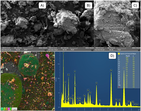
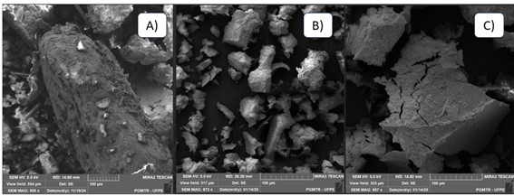
Figure 1 shows images of the ground LED after processing in a scanning electron microscopy, under different approximations. The structure of the metal before the bioleaching process is granular and the surface is even (without cracks), indicating that the grinding process reduced the particle size, but did not modify the structure of the material. Furthermore, the images indicate particles with variable sizes, smaller than 300 µm. These images will be relevant when comparing with the same material after the bioleaching process. These images are relevant for comparison with the same material after the bioleaching process, as parameters such as particle size, surface presentation, and structure of the material are verified to identify the production of metabolites that may have solubilized the material throughout the process. A recent study by Peixoto et al.25 presents SEM images to characterize printed circuit board waste before a bioleaching process to remove copper. Waghmode et al.26 evaluated, through SEM, characteristics such as particle size and shape of WEEE before the bioleaching process. The images also showed the polymorphic nature of the waste under different magnifications.
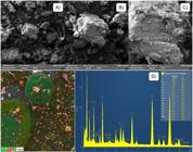
SEM images of the LED waste after processing in different magnitudes: A) 2000x; B) 1000x; C) 5000x; and D) grinded LED magnified 73x in EDS layer (left); graph of the sum spectrum of maps with all elements present in the LED (right).
Furthermore, Figure 1 shows the chemical elements in the grinded LED through Energy Dispersive X-Ray Spectroscopy (EDS). The elements seen in this fraction of the sample are Cu, Fe, Ti, Si, Ag, and Sn. These elements were also detected in the XRF analysis, confirming the presence of metals of interest in the LED sample used. Abraham et al.27 also evaluated the elements in the surface of the metal before the bioleaching process to confirm the presence of the metals of interest in the sample.
3.2. Isolation and partial identification of microorganisms
Identifying a microorganism as a species depends on the fulfillment of assigned specific characteristics, in which recognizing the source or origin of the organism (environment and location, for example) is essential. An improper performance of a preliminary test can confuse and harm this entire process. In the laboratory routine, a systematic reasoning is used, aiming to reduce the cost and time required to release the report and impacting the diagnosis28,29.
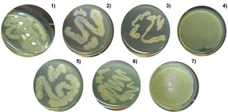
Seven colonies with different morphological characteristics were isolated (Figure 2), then named in a sequence from 1 to 7. Then, the following characterization processes were carried out: Gram Staining, which helps identify bacterial groups through the thickness of the cell wall, composed of peptideoglycan, in addition to showing the shape of the cell through the optical microscope; the presence of endospores — which are structures of the bacteria whose function is to provide resistance and ensure the survival of the organism in an inappropriate environment — in the microbial cell was also evaluated, as the presence of this structure in the cells indicates that the microorganism has a greater ability to develop in an environment with concentrations of LED; and, finally, the MaldTof analysis was carried out to identify, at the proteomic level, the genus and species of the isolated microorganisms.
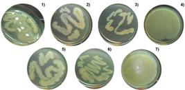
Macroscopic images of colonies of isolated microorganisms on lamp LEDs. The numbers correspond to the respective samples.
Table 3 shows the results of Gram Staining, the shape of the cells, the presence of endospores, and the results of the bacterial identification analyses by MaldTof, as well as the score that represents the interpretation of the results. All isolated bacteria belong to the Gram-positive group. Gram-positive bacteria employ several defense mechanisms, including changes in their cellular wall structure and the production of enzymes, which confer greater resistance in inhospitable environments30. These characteristics are very favorable for the bioleaching process, as one of the main limitations for the commercialization of this method of metal recovery is the toxicity that the elements present to microorganisms.
The morphology of the cells of all isolated microorganisms is bacillus, which is a rod shape. Samples 2, 3, 4, 5, and 7 have endospores, which are structures that also contribute to a greater resistance of microbial cells.
The MaldTof analysis, used to identify isolated microorganisms, is a technique that uses software that determines a score according to the compatibility of the sample data with the microbial mass spectrum library. To interpret the results, we must compare the scores found for each sample with the reliability determined by the software. Table 3 shows that, at the genus level, sample 6 was securely identified, belonging to the Bacillus genus. However, the score for sample 1 is not reliable for genus and species identification. Regarding samples 2, 3, 4, 5, and 7, the scores suggest that they belong to the Bacillus genus, widely described in the literature for its high potential for solubilization of metals in electronic waste31-33. The low accuracy of the identification of microorganisms by this technique can be attributed to the fact that the Bacillus genus may present spores in its morphology, which would difficult the identification at a proteomic level. In this case, further analysis with younger cultures is suggested. Another factor, which may have limited the reliability of the identification by MaldTof, is the possibility of having isolated microorganisms that are not yet in the database. Molecular identification analysis will be performed later to ensure the identification.
A study by Surányi et al.34 used the MALD-TOF technique for a quick identification of 235 bacterial isolates. In this study, the authors were unable to identify a third of the isolates through this technique, even at the genus level, reinforcing the need to apply a second method for the bacteria that were not identified.
3.3. Bioleaching process and parameter monitoring
Several metabolic processes occur during the exposure of metal waste with microbial cells. The monitoring analyses point to chemical and biological transformations, which are occurring during the experiment, indicating the metabolic routes that microorganisms are using for the solubilization of metals. Furthermore, cell viability ensures that microbial cells are active and remain viable for the continuity of the test.
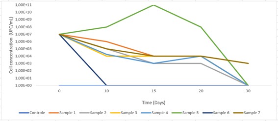
All experiments started with a concentration of 107 CFU/mL, except for the control-condition, which was not subjected to the addition of microorganisms Figure 3 shows the variation in the concentration of viable cells throughout the process. In all conditions, cell viability decreased over time, apart from sample 6, which showed an increase in cell concentration for the first 15 days of the experiment. In contrast, sample 6 showed a complete decrease in the concentration of viable cells in the first 10 days of the experiment, indicating that the metals present in the LED were toxic to this microorganism, causing the death of its cells. The remaining samples remained viable for 20 days of the experiment, except for sample 7, which maintained a concentration of 103 CFU/mL until the end of the experiment (30 days), indicating a greater resistance of this microorganism to longer periods of exposure to LED.
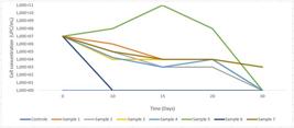
Khezerloo et al.35 conducted a study on the bioleaching process of critical and valuable metals from LED television screens. In their work, the highest yields of indium, aluminum, and strontium were obtained in periods longer than 10 days. This result indicates that microorganisms with greater resistance to longer periods of time have the potential for higher yields of metal solubilization. They also evaluated the viability of cells throughout the bioleaching process of metals in the LEDs. In the study, the concentration of viable cells also reduced considerably during the process. After 30 days of experiment, only 125 cells of A. ferrooxidans remained viable.
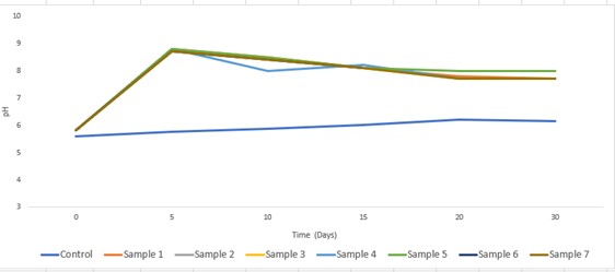
Figure 4 shows the pH changes in the medium of the bioleaching process over the 30 days of the experiment for all evaluated microorganisms. The main parameter influencing the leachability of metals is the pH of the surrounding environment36-39. Relatively small changes in the pH value can cause an increase and a decrease in the concentration of leached substances by several orders of magnitude39. The graph shows a similar pH variation between the different samples (conditions with addition of microorganism) over time. The control-condition (without the addition of microorganism) maintained the pH practically stable during the 30 days of testing (5.6 – 6.2). For the other conditions, the pH of the medium at the beginning of the experiment was slightly acidic (5.8), but, after the first 5 days of the experiment, it increased to values between 8.7 and 8.8. These values were slightly reduced over time and, at the end of the 30 days of incubation, the pH averaged around 7.7, except for sample 5, which had a final pH of 8.0. These results show that the microorganisms may have developed alkaline metabolites through the process of cyanogenesis performed by cyanogenic bacteria16,40. The Bacillus genus is described in the literature by its ability to consume a carbon source and transform it into cianide, performing the bioleaching process in alkaline conditions41-43.
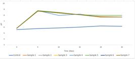
Evaluation of the pH during the bioleaching process for the different isolated microorganisms.
Li et al.16 published a study on the process of gold bioleaching in printed circuit board waste in an alkaline medium by bacteria. The results show better gold solubilization yields in pH ranges between 7 and 9.
3.4. Scanning electron microscopy
Scanning electron microscopy was also performed to examine size, shape, and structure of the LED surface after the bioleaching process. In Figure 5A, we see a LED sample in control condition; that is, the entire procedure was carried out without adding microorganisms, for comparative effect. In this image, the LED surface does now show significant cracks or changes when compared to the initial sample before the bioleaching process (Figure 1). This result indicates that, even with the 30 days of the experiment, the absence of the microorganism did not cause any effects in the morphology of the metal. However, in Figure 5B, in which sample 5 can be seen, there are significant changes in particle size when compared to the control sample (Figure 5A). Furthermore, many particles have different shapes, confirming this process of degradation of the metal. This result indicates that the microorganisms during the bioleaching process released metabolites that solubilized the metal. In Figure 5B, sample 7 is shown, in which particles with many cracks, compared to the control sample, are seen: the metal has a decreased size and a more fragile appearance, indicating, once more, that leaching metabolites were produced during the incubation time of the samples.
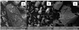
Scanning electron microscopy images: A) LED after incubation without the addition of microorganism; B) LED after bioleaching process with sample 5; and C) LED after bioleaching process with sample 7.
Pourhossein and Mousavi44 carried out a study regarding the bioleaching process of copper, nickel, and gallium in LED lamps waste. Scanning electron microscopy was used to confirm the bioleaching process. The authors confirmed that the reduction in size and change in the shape of the particles are parameters to evaluate metal degradation. Arshadi et al.45 investigated copper and nickel bioleaching from each bacteria. In this study, the samples of scanning electron microscopy also revealed metal degradation after the process. Abraham et al.27 evaluated the bioleaching of metals in WEEEs by Bacillus liqueniforms. In this study, the scanning electron microscopy evaluated the appearance of crack in the metal surface during the incubation period, also confirming the process of solubilization of the metals by this parameter. Pourhossein et al.46 evaluated, through SEM, changes in the morphology of the WEEE used after the bioleaching by bacteria process. According to the authors, the changes in the metal from the leachates produced by the microorganism in the process are a parameter that indicates solubilization of the metals.
4. Conclusions
The characterizations prove the presence of critical and valuable elements in the LED, which makes this WEEE a product of high added value due to the presence of metals that are important for the productive chain of many sectors of the industry. The isolated microorganisms were partially identified as from the Bacillus genus, according to the applied techniques. The monitoring analyses carried out during the bioleaching process demonstrate that microorganisms 5 and 7 stood out in their resistance to metals in the LED, as well as in the production of metabolites that were responsible for the solubilization of metals, in alkaline medium, through the process of cyanogenesis. The SEM images of the residual metals from samples 5 and 7 showed reductions and changes in the particles’ shapes, as well as significant cracks in the waste, which indicates the production of microbial metabolites that promoted the solubilization of the metal. In this sense, the isolated microorganisms of the post-consumer lamp LEDs submitted to the bioleaching process showed biotechnological potential for application in the solubilization of critical and valuable metals in LED WEEE. This study is innovative in the isolation of microorganisms from this WEEE for the application in the bioleaching process, seeking a sustainable approach for the solubilization of high added value metals. Thus, it allows the later recovery of these metals, in order to insert them in the productive chain, using a tool that is more sustainable when compared to hydroleaching methods, which use synthetic acid and alkaline solutions that generate and significant impacts to the environment.
5. Acknowledgments
The authors thank Capes and CNPq for the doctoral and postdoctoral scholarships, CNPq (DT2) for the research grants, and CNPq and FINEP for the resources for the project.
-
Data Availability
Available data - The entire dataset supporting the results of this study was published in the article itself.
6. References
- 1 Prescient & Strategic Intelligence. High power LED market research report by wattage, application, geographical outlook. Dublin; 2019. p. 28-34.
- 2 Zissis G, Bertoldi P, Serrenho T. Atualização sobre o status do mercado mundial de iluminação LED desde 2018. Luxemburgo: Serviço das Publicações da União Europeia; 2021.
- 3 Gran View Research. LED Lighting Market Size & Share, Industry Report, 2030. San Francisco; 2023.
- 4 Krishnamurthy N, Gupta CK. Extractive metallurgy of rare earths. 2nd ed. Boca Raton: CRC Press; 2015.
- 5 Lima IB. Leal W Fo. Rare earths industry: technological, economic and environmental implications. 1st ed. Amsterdam: Elsevier; 2016.
-
6 Bundesanstalt für Geowissenschaften und Rohstoffe [homepage on the Internet]. Hannover: BGR; 2023 [cited 2023 Mar 6]. Available from: www.bgr.bund.de
» www.bgr.bund.de - 7 Haiying Z, Zhao Y, Qi J. Characterization of heavy metals in fly ash from municipal solid waste incinerators in Shanghai. Process Saf Environ Prot. 2010;88(2):114-24.
- 8 Peer M, Fehn T, Hofmann A, Berninger B, Kunz W. Recycling of critical metals from light‐emitting diodes. Chemieingenieurtechnik. 2024;97(1-2):110-15.
- 9 Gunawardhana L, Karunanayake N, Indika Thushari P. Metal characterization of LED lamp waste: hazard classification and potential toxicity impacts evaluation. J Hazard Toxic Radioact Waste. 2024;28(2):04023048.
- 10 Habibi A, Kourdestani SS, Hadadi M. Biohydrometallurgy as an environmentally friendly approach in metals recovery from electrical waste: a review. Waste Manag Res. 2020;38(3):232-44.
- 11 Ivanus RC. Bioleaching of metals from electronic scrap by pure and mixed culture of Acidithiobacillus ferrooxidans and Acidithiobacillus thiooxidans Metal Int. 2010;15(4):62-70.
- 12 Jaiswal M, Srivastava S. A review on sustainable approach of bioleaching of precious metals from electronic wastes. J Hazard Mater Adv. 2024;14:100435.
- 13 Adetunji AI, Oberholster PJ, Erasmus M. Bioleaching of metals from e-waste using microorganisms: a review. Minerals. 2023;13(6):828.
- 14 Needhidasan S, Samuel M, Chidambaram R. Electronic waste: an emerging threat to the environment of urban India. J Environ Health Sci Eng. 2014;12:1-9.
- 15 Blumer C, Haas D. Mechanism, regulation, and ecological role of bacterial cyanide biosynthesis. Arch Microbiol. 2000;173:170-7.
- 16 Li J, Wen J, Guo Y, An N, Liang C, Ge Z. Bioleaching of gold from waste printed circuit boards by alkali-tolerant Pseudomonas fluorescens Hydrometallurgy. 2020;194:105260.
- 17 Gram C. Ueber die isolirte farbung der schizomyceten in schnitt-und trockenpraparaten. Fortschr Med. 1884;2:185-9.
- 18 Bizzini A, Greub G. Matrix-assisted laser desorption ionization time-of-flight mass spectrometry, a revolution in clinical microbial identification. Clin Microbiol Infect. 2010;16(11):1614-9.
- 19 Tsuchida S, Nakayama T. MALDI-based mass spectrometry in clinical testing: Focus on bacterial identification. Appl Sci. 2022;12(6):2814.
- 20 Han SS, Oliveira YS, Choi SK. Cenário atual e desafios na identificação direta de microrganismos usando MALDI TOF MS. Microorganismos. 2021;9:1917.
- 21 UFRGS: Universidade Federal do Rio Grande do Sul. Laboratório de Análises Microbiológicas Através de Maldi-Tof. Técnicas de análise: preparação de amostras. Porto Alegre; 2024.
- 22 De Oliveira RP, Benvenuti J, Espinosa DCR. A review of the current progress in recycling technologies for gallium and rare earth elements from light-emitting diodes. Renew Sustain Energy Rev. 2021;145:111090.
- 23 Rebello RZ, Lima MTWDC, Yamane LH, Siman RR. Characterization of end-of-life LED lamps for the recovery of precious metals and rare earth elements. Resour Conserv Recycling. 2020;153:104557.
- 24 Illés IB, Kékesi T. A comprehensive aqueous processing of waste LED light bulbs to recover valuable metals and compounds. Sustain Mater Techno. 2023;35:e00572.
- 25 Peixoto RAO, Vieira RB, Resende MM, Cardoso VL. Remoção de cobre em placas de circuito impresso de computadores utilizando o processo de biolixiviação. OLEL. 2024;22(7):e5728.
- 26 Waghmode MS, Gunjal AB, Patil NN. Bioleaching of electronic waste. Pollution. 2021;7(1):141-52.
- 27 Abraham J, Chatterjee A, Sharma J. Isolation and characterization of Bacillus licheniformis strain for bioleaching of heavy metals. J Appl Biotechnol Rep. 2020;7(5):139-144.
- 28 Sumita NM, Andriolo A, Ferreira CES, Campana GA, Oliveira GF, Brazão FV, et al., editors. Recomendações da Sociedade Brasileira de Patologia Clínica/Medicina Laboratorial (SBPC/ML): boas práticas em microbiologia clínica. Barueri: Manole; 2015. 274 p.
- 29 Gemelli T. Manual prático de microbiologia clínica. Porto Alegre: Unisinos; 2020.
- 30 Rajput P, Nahar KS, Rahman KM. Evaluation of antibiotic resistance mechanisms in gram-positive bacteria. Antibiotics. 2024;13(12):1197.
- 31 Zhang Q, Ma L, Peng Y, Yan X. Sustainable bioleaching of heavy metals from coal tailings using Bacillus inaquosorum B. 4: mechanistic insights and environmental implications. J Environ Chem Eng. 2024;12(5):113400.
- 32 Dong Y, Zan J, Lin H. Bioleaching of vanadium from stone coal vanadium ore by Bacillus mucilaginosus: influencing factors and mechanism. Int J Miner Metall Mater. 2024;31(8):1828-38.
- 33 Dong Y, Chong S, Lin H. Bioleaching and biosorption behavior of vanadium-bearing stone coal by Bacillus mucilaginosus Int J Miner Metall Mater. 2023;30(2):283-92.
- 34 Surányi BB, Taczman-Brückner A, Mohácsi-Farkas C, Engelhardt T. Rapid identification of bacteria from agricultural environment using MALDI-TOF MS. Acta Aliment. 2023;52(1):113-20.
- 35 Khezerloo S, Nasirpour N, Pourhossein F, Mousavi SM. Bioleaching of indium from spent light-emitting diode monitors and selective recovery followed by solvent extraction. J Environ Manage. 2023;335:117520.
- 36 Sloot HA, Zomeren A. Characterisation leaching tests and associated geochemical speciation modelling to assess long term release behaviour from extractive wastes. Mine Water Environ. 2012;2(31):92-103.
- 37 Król A. Problems of assessment of heavy metals leaching from construction materials to the environment. ACEE. 2011;4(3):71-6.
-
38 Saveyn H, Eder P, Garbarino E, Muchova L, Hjelmar O, Van Der Sloot H, et al. Study on methodological aspects regarding limit values for pollutants in aggregates in the context of the possible development of end-of-waste criteria under the EU Waste Framework Directive [Internet]. Luxembourg: Publications Office of the European Union; 2014 [cited 2023 Mar 6]. Available from: https://publications.jrc.ec.europa.eu/repository/handle/JRC91036
» https://publications.jrc.ec.europa.eu/repository/handle/JRC91036 - 39 Król A, Mizerna K, Bożym M. An assessment of pH-dependent release and mobility of heavy metals from metallurgical slag. J Hazard Mater. 2020;384:121502.
- 40 Li J, Liang C, Ma C. Bioleaching of gold from waste printed circuit boards by Chromobacterium violaceum J Mater Cycles Waste Manag. 2015;17:529-39.
- 41 Faraji F, Mahandra H, Ghahreman A. Evaluation of different amino acids on growth and cyanide production by Bacillus megaterium for gold recovery. Sustainability. 2022; 14(15):9639.
- 42 Rosario CGA, Vallenas-Arévalo AT, Arévalo SJ, Espinosa DCR, Tenório JAS. Biodegradation of cyanide using a Bacillus subtilis strain isolated from artisanal gold mining tailings. Braz J Chem Eng. 2023;40(1):129-36.
- 43 Thakur P, Kumar S. Exploring bioleaching potential of indigenous Bacillus sporothermodurans ISO1 for metals recovery from PCBs through sequential leaching process. Waste Manag Res. 2023;41(7):1255-66.
- 44 Pourhossein F, Mousavi SM. Enhancement of copper, nickel, and gallium recovery from LED waste by adaptation of Acidithiobacillus ferrooxidans Waste Manag Res. 2018;79:98-108.
- 45 Arshadi M, Yaghmaei S, Esmaeili A. Evaluating the optimal digestion method and value distribution of precious metals from different waste printed circuit boards. J Mater Cycles Waste Manag. 2020;22:1690-98.
- 46 Pourhossein F, Mousavi SM, Beolchini F. Innovative bio-acid leaching method for high recovery of critical metals from end-of-life light emitting diodes. Resour Conserv Recycling. 2022;182:106306.
Edited by
-
Associate Editor:
Hamilton Abreu.
-
Editor-in-Chief:
Luiz Antonio Pessan.
Available data - The entire dataset supporting the results of this study was published in the article itself.