Artificial Intelligence Applied to the Production of Biopolymers with a Sustainability Approach and the Use of the Extended Finite Element Method
Abstract
The development of sustainable materials, such as high-performance biopolymers, faces the challenge of high quality control costs, given that their mechanical properties depend on the fiber arrangement within the matrix. To overcome this barrier, this work proposes the use of Artificial Intelligence, specifically Computer Vision, to create a virtualization of the material's microstructure from simple images. This virtual representation serves as a basis for the efficient generation of numerical models using the Finite Element Method (FEM) and the Extended Finite Element Method (XFEM). In this context, the paper introduces the aiMat framework, which integrates Computer Vision and FEM/XFEM modules to optimize the process. The main contribution of this work, therefore, derives from the harmonious integration of these techniques, establishing a more efficient and accessible workflow for modeling composite materials.
Keywords:
Biopolymers; Artificial Intelligence; Computer Vision; XFEM and aiMat
1. Introduction
Interest in biopolymers has been on the rise in recent decades, given the range of applications from healthcare to the food industry1,2. In particular, polymers composed of chitosan have emerged as a promising alternative for the production of green hydrogen3. Consequently, it is extremely important to facilitate studies in this area in order to extend the applications of the material.
Neural networks have evolved rapidly and have shown positive results in several areas. In medicine, advances in tomography images for studying the brain4, bone tissue5, and cells6,7 have brought significant improvements to analysis processes. In materials, applications related to the Finite Element Method with applications in uncertainty analysis of the models created are gaining prominence8.
Accordingly, developing tools to extend new methodologies is a necessary step to increasingly leverage the development of materials, making them safer and more economical. For this reason, a new tool (aiMat) is discussed that links Computer Vision techniques and the Extended Finite Element Method applied to the analysis of thin films formed from chitosan reinforced with sisal fibers.
2. Materials and Methods
2.1. Biopolymers
A composite is a structural material consisting of two or more constituents combined at a macroscopic level and which are not soluble in each other. They consist of two phases: the matrix phase, the base material in which the reinforcement phase is incorporated. The materials in the matrix phase are generally continuous and the material in the reinforcement phase is dispersed and may be in the form of fibers, particles or flakes. The matrix and reinforcement phases are chemically different and separated by a distinct interface.
Chitosan is a polymer produced by the deacetylation of chitin, obtained mainly from crustacean shells. Due to its biocompatible, biodegradable, antibacterial properties and the ease of forming thin films, it has been studied for various uses such as food packaging and as a biomaterial for medical applications. Natural fibers can be used as mechanical reinforcement, in addition to maintaining the biodegradable properties of the material. To incorporate cellulose fibers, the mercerization process is necessary, which, through an alkaline treatment, removes lignin, hemicellulose and other impurities from the surface of the fiber that hinder interaction with the polymer matrix9.
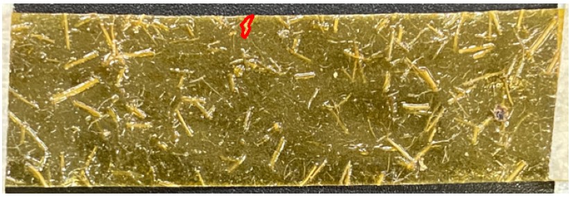
Length and diameter are characteristics of the fiber that directly influence the mechanical properties of the biocomposite. The images used were manually annotated10, after production. It is important to highlight that throughout production, the fibers are randomly added to the matrix phase. One of the control parameters used is the opening of the sieve before inserting the sisal fibers into the chitosan films.
2.2. Neural networks
The principles governing modern deep learning techniques are solidified from multilayer perceptron (MLP) neural networks. MLP networks, as universal approximators, follow Equation A11. In principle, they can be used to approximate any type of function. Problems involving nonlinearity require the use of previously defined nonlinear activation functions .
Algorithms for updating the weights and bias are based on gradient methods. Commonly called stochastic gradient descent (SGD), it has an implementation for updating the parameters as evidenced by Equation B. This considers weight decay , momentum (), dampening () and Nesterov is the portion of the gradient that considers both dampening and weight decay in the previous iteration of the update.
Convolutional neural networks10 play a fundamental role in reducing the parameters required in applications with a large number of inputs. This is especially true in images that often have many pixels even when less dense (HD). Equation C shows the spatial convolution process (two-dimensional) while Equation D is an extension to include the number of channels (three-dimensional).
is the input matrix and is the kernel matrix. The kernel is the learnable parameter, which will be used as a layer of the network. m and n refer to the height and width indices of the image while and are the indices of the feature map formed after the convolution process. The feature map is the set of new features generated after the convolutional process.
Among the main convolutional neural network (CNN) architectures, AlexNet12, revolutionized computer vision by winning the ImageNet competition, demonstrating the effectiveness of deep networks and the use of GPUs for training. VGGNet13 popularized the concept of networks with small stacked convolutional layers, showing that the depth of networks can be efficiently increased. ResNet14, introduced the technique of residual blocks, solving the degradation problem in very deep networks and enabling the training of networks with more than 100 layers. For segmentation tasks, U-Net15, stands out due to its skip connections between encoding and decoding layers, which preserve crucial spatial information, and is widely used for semantic segmentation in biomedical images, especially in contexts with limited labeled data.
2.3. Extended finite element method (XFEM)
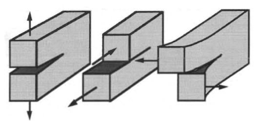
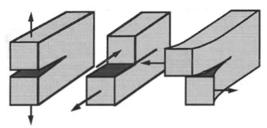
Modeling using XFEM presents 3 fracture modes that define the form of crack propagation that can be used to investigate the toughness of chitosan films. Mode I describes propagation due to axial loading. Mode II simulates the application of pure shear force in relation to the longitudinal part of the body. Mode III considers shear force, but applied to the thickness. Figure 1 represents the discussion.
The equation describing crack width, first defined by Griffith and improved by Irwin, is described by Equation E.
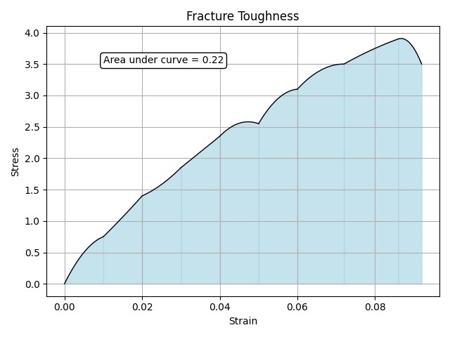
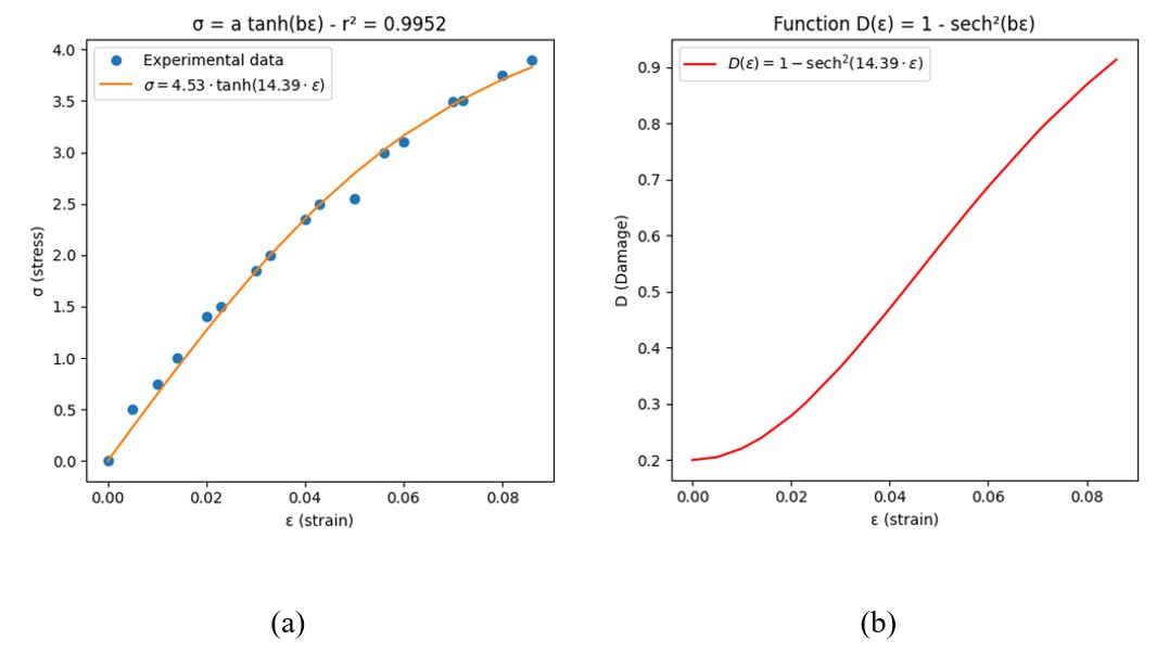
For the initial analysis, a homogeneous model was used. Its fracture energy was calculated using Figure 2, while the plastic parameters were obtained from the constitutive approximation presented in Figure 3.
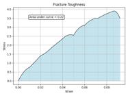
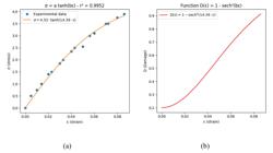
In the Figure 3 the mechanical behavior of fiber-reinforced polymer composites is described by a hyperbolic tangent function parameterized by the coefficients a and b, subject to the condition (Equation F):
where E is the elastic modulus16. By evaluating the rate of change of elasticity and applying the fundamental principles of continuum damage mechanics, the damage variable can be expressed as17,18:
where is the degraded (or effective) elastic modulus of the damaged material.
Accordingly, the degradation of elasticity follows the form:
where ε is the strain anddenotes the hyperbolic secant function. When applied to the composite investigated in this study, this relationship yields the numerical behavior depicted in Figure 2b. Regarding the elastic parameters, Table 1 provides the values used in the model.
2.4. Applied computational vision
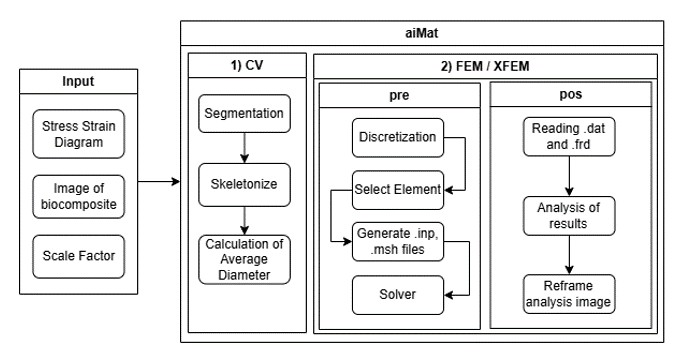
The computer vision methodology using the U-Net network is quite promising and advances in the area tend to improve results. In this sense, adding such contributions in an automated way to slow processes such as geometry construction and parameter assignment in programs involving finite elements is a fundamental task. Thus, aiMat was created containing 2 modules, one for processing related to computer vision while the second deals with FEM and XFEM (Figure 4). The process used to obtain the geometry occurs through an fbd file with its own syntax21. With this, it is possible to call mesh generators such as TETGEN or NETGEN to automate the obtaining of nodes and connectivities.
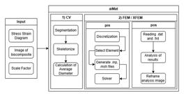
The solver is called using a subprocess on the host machine. This request is made using the inp file which implements the behavior of the solver, commonly used in commercial programs such as Calculix and Abaqus. Due to the modularity of the application, it is possible to use other tools such as NASTRAN and Ansys.
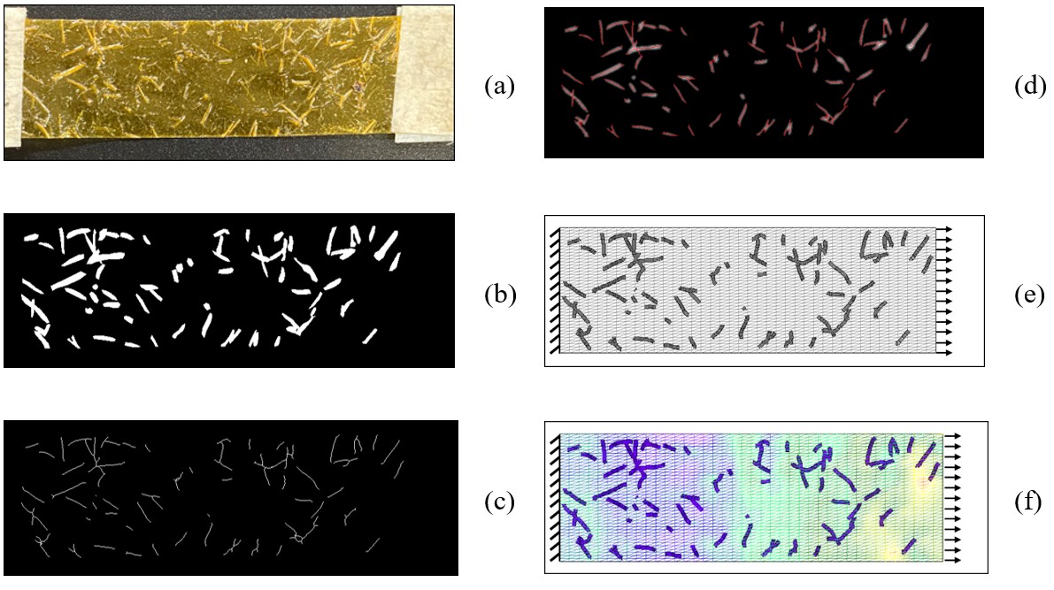
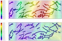
Figure 5 details the list of processes developed in aiMat. First, an input image is segmented by U-Net to determine the arrangement of fibers. Then, a skeleton is generated and is used to create fibers considering each pixel as a node. Both processes are shown in Figure 5a-c. After that, a Euclidean matrix (d) is generated to compute the distances from each contour of the image to its borders, providing a pixel level strategy to compute fiber diameters along with the positions of the skeleton pixels. For simplification, an average diameter was used for each fiber contour. It is important to emphasize, however, that the algorithm provides the diameter for each element on a pixel-by-pixel basis, yielding a precise approximation of varying fiber diameters. Figure 5e shows the boundary conditions: fixed in the left edge and a load22 (1,75 kN/m) applied to the nodes on the right edge; while (f) equivalent plastic strain field without XFEM.
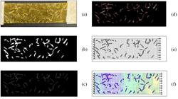
Following the Euclidean matrix calculation – Figure 5d – a Finite Element Mesh is constructed. Table 2 shows the element types used for both matrix and fiber. The connection between fiber and matrix was made using master slave contact.
To ensure the reliability of the numerical results and their independence from the mesh resolution, a comprehensive mesh convergence study was performed for both the matrix and fiber phases.
For the matrix phase, a sensitivity analysis was conducted by varying the element discretization, with the results summarized in Table 3. The data reveals that while stress and displacement values fluctuate with a coarser mesh, they converge to stable results as the mesh is refined. A discretization of 40x40 (1600 elements) was identified as providing an optimal balance between computational accuracy and efficiency.
A subsequent analysis was performed for the fiber reinforcement, as presented in Table 4. Since the fibers were modeled as beam elements following a path determined by the image skeleton, increasing the number of elements along this predefined path had a minimal impact on the overall stress and displacement fields. As a result, the initial discretization of 2106 elements were deemed sufficient for accurately representing fiber behavior.
3. Results and Discussion
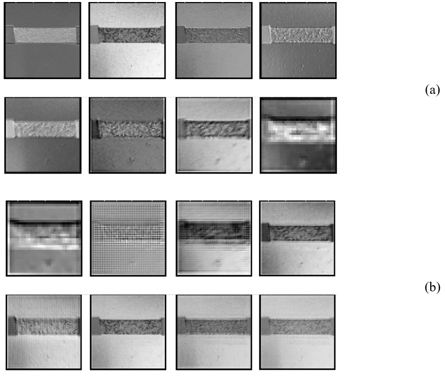
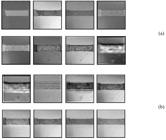
Regarding the U-Net segmentation, Figure 6a and 6b display the respective encoder and decoder feature maps generated by the convolution process (Equation D). While this process reduces the spatial dimensions of the input, it allows the network to extract more abstract features, thus enriching the information used for prediction. Each map reveals important characteristics learned from the fiber images and helps illustrate the outcome of the learning process, which was guided by an optimizer (Equation B).
Subsequently, the aiMat algorithm is employed to automatically generate the model geometry. This involves a discretization process that uses lines derived from the image skeleton to define the fibers, alongside a calculation of their respective diameters. The resulting discretized model provides results comparable to the original sample, as illustrated in Figure 7. Furthermore, since precisely identifying the fiber locations is more challenging than defining the overall matrix boundaries, the matrix geometry was simplified in this step to a rectangular domain containing the fiber network.
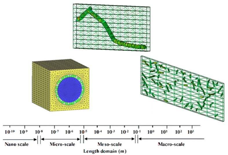
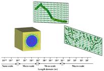
The simulations available in the framework have 3 scale levels: Macro-scale, Meso-scale and Micro-scale (Figure 8).
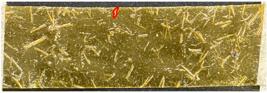
To identify the most loaded fiber, the coordinates of the most stressed node were checked against the corresponding fiber contour in the segmentation mask. With this, it is possible to identify in the input figure (Figure 9) the procedure performed by the simulation, for example, of a tensile test22.
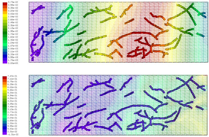
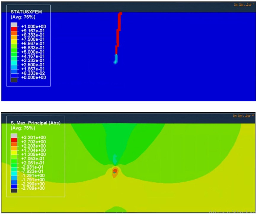
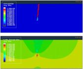
As an example, a homogeneous analysis using Fracture Mode I is presented in Figure 10, showing the crack propagation via the STATUSXFEM parameter and the maximum principal stresses.
The presented features illustrate the different analyses possible by merging Computer Vision with FEM and XFEM simulations. As demonstrated (Figures 5-7), the overall results validate the proposed workflow, confirming that this integration is a viable and efficient approach to automate the modeling of biocomposites. Furthermore, analyses concerning the model production pipeline showed that the time required for creating these models was significantly reduced compared to previous methodologies23.
4. Conclusions
This work addressed the challenge of modeling biocomposites, where the random arrangement of reinforcement fibers determines mechanical performance and elevates quality control costs. To overcome this barrier, the integrated computational framework aiMat was presented, which successfully unites Computer Vision (CV) with numerical simulation. The developed methodology allows for the automated creation of complex model geometries directly from simple images, optimizing the analysis process.
Through the application of a U-Net neural network for image segmentation, aiMat demonstrated its capability to transform 2D images into detailed simulations of mechanical behavior via FEM and XFEM, including stress distribution and fracture propagation. This automation drastically reduces the time required for model creation, making the virtual testing of materials with complex microstructures, such as chitosan films reinforced with sisal, more accessible. The ability to analyze fibers with a non-unidirectional arrangement supports the development of more cost-effective production techniques.
The principal contribution of this study is the development and validation of aiMat as a robust workflow for the virtual characterization of biopolymers. By harmoniously integrating AI and simulation techniques, the work offers a tool to accelerate research in materials science. As for next steps, future work will focus on adding the matrix phase to the CV module to allow for a more direct comparison of the results with data from destructive tests found in the literature.
5. Acknowledgments
This study was financed in part by the Coordenação de Aperfeiçoamento de Pessoal de Nível Superior - Brasil (CAPES) - Finance Code 001
-
Data Availability
The comprehensive dataset that underpins the findings of this study can be obtained by submitting a request to the corresponding author, [Audelis Marcelo].
6. References
- 1 Silva JLM. Estudo das propriedades mecânicas de membranas modificadas de quitosana [monograph]. Mossoró: Universidade Federal Rural do Semiárido; 2018.
- 2 Rotta J, Barreto PLM, Minatti E. Determinação das propriedades mecânicas, difratométricas e térmicas de filmes de quitosana e hidroxipropilmetilcelulose (HPMC). In: 10º Congresso Brasileiro de Polímeros; 2009 Oct 13-17; Foz do Iguaçu. Proceedings. São Paulo: ABPol; 2009.
- 3 Melo SLS. Compósitos de quitosana aditivados com nanocelulose e óxido de grafeno (Go) para eletrolisadores pem [thesis]. Fortaleza: Universidade Federal do Ceará; 2024.
-
4 Trutti AC, Fontanesi L, Mulder MJ, Bazin P-L, Hommel B, Forstmann BU. A probabilistic atlas of the human ventral tegmental area (VTA) based on 7 Tesla MRI data. Brain Struct Funct. 2021;226(4):1155-67. http://doi.org/10.1007/s00429-021-02231-w
» http://doi.org/10.1007/s00429-021-02231-w -
5 Warin K, Limprasert W, Paipongna T, Chaowchuen S, Vicharueang S. Determination of the oral carcinoma and sarcoma in contrast enhanced CT images using deep convolutional neural networks. Sci Rep. 2025;15(1):21672. http://doi.org/10.1038/s41598-025-06318-w
» http://doi.org/10.1038/s41598-025-06318-w - 6 Gupta A, Dhanakshirur RR, Jain K, Garg S, Yadav N, Seth A, et al. Deep learning for detecting and subtyping renal cell carcinoma on contrast-enhanced CT scans using 2D neural network with feature consistency techniques. Indian J Radiol Imaging. 2024;35(3):395-401. http://doi.org/10.1055/s-0044-1800804.
-
7 Weng W, Zhu X. INet: convolutional networks for biomedical image segmentation. IEEE Access. 2021;9:16591-603. http://doi.org/10.1109/ACCESS.2021.3053408
» http://doi.org/10.1109/ACCESS.2021.3053408 -
8 Sharma A, Mukhopadhyay T, Rangappa SM, Siengchin S, Kushvaha V. Advances in computational intelligence of polymer composite materials: machine learning assisted modeling, analysis and design. Arch Comput Methods Eng. 2022;29(5):3341-85. http://doi.org/10.1007/s11831-021-09700-9
» http://doi.org/10.1007/s11831-021-09700-9 -
9 Lim GJH, Koh JJ, Chan KK, Koh XQ, Jintara P, Sutrisnoh NAA, et al. Reinforced chitosan polymer electrolyte for long‐life and robust laminated Li‐Zn structural batteries. Adv Funct Mater. 2025;35(31):2423149. http://doi.org/10.1002/adfm.202423149
» http://doi.org/10.1002/adfm.202423149 - 10 Marcelo AO Jr, Gomes AB, Guerra EPM, Deus EP. Development of artificial intelligence applied to the production of biopolymers with a focus on sustainability. In: Semião JFLC, Sousa NMS, Cruz RMS, Prates GND, editors. Advances in Sustainability Science and Technology - INCREaSE 2023. Proceedings. Cham: Springer; 2023. p. 204-18. http://doi.org/10.1007/978-3-031-44006-9_15.
- 11 Le Cun Y, Boser B, Denker JS, Henderson D, Howard RE, Hubbard W, et al. Handwritten digit recognition with a back-propagation network. Holmdel: AT&T Bell Laboratories; 1989. p. 396-404.
- 12 Krizhevsky A, Sutskever I, Hinton GE. ImageNet classification with deep convolutional neural networks. In: Pereira F, Burges CJ, Bottou L, Weinberger KQ, editors. Advances in Neural Information Processing Systems 25 (NIPS 2012). Proceedings. New York: Curran Associates; 2012.
- 13 Simonyan K, Zisserman A. Very deep convolutional networks for large-scale image recognition. arXiv. 2014. In press.
- 14 He K, Zhang X, Ren S, Sun J. Deep residual learning for image recognition. arXiv. 2015. Preprint.
- 15 Ronneberger O, Fischer P, Brox T. U-Net: convolutional networks for biomedical image segmentation. In: Navab N, Hornegger J, Wells W, Frangi A, editors. Medical Image Computing and Computer-Assisted Intervention – MICCAI 2015. Proceedings. Cham: Springer; 2015. p. 234-41. (vol. 9351). http://doi.org/10.1007/978-3-319-24574-4_28.
- 16 Lu G. HDPE wood-plastic composite material model subject to damage [thesis]. Washington: Washington State University; 2002.
-
17 Lemaitre J, Dufailly J. Damage measurements. Eng Fract Mech. 1987;28(5-6):643-61. http://doi.org/10.1016/0013-7944(87)90059-2
» http://doi.org/10.1016/0013-7944(87)90059-2 -
18 Chaboche JL. Continuous damage mechanics: a tool to describe phenomena before crack initiation. Nucl Eng Des. 1981;64(2):233-47. http://doi.org/10.1016/0029-5493(81)90007-8
» http://doi.org/10.1016/0029-5493(81)90007-8 -
19 Costa MME, Melo SLS, Deus EP. Synthesis of polymeric composites reinforced with unidirectional and bidirectional bamboo fibers. Universal Journal of Materials Science. 2018;6(4):133-8. http://doi.org/10.13189/ujms.2018.060403
» http://doi.org/10.13189/ujms.2018.060403 -
20 Aryaei A, Jayatissa AH, Jayasuriya AC. Nano and micro mechanical properties of uncross-linked and cross-linked chitosan films. J Mech Behav Biomed Mater. 2012;5(1):82-9. http://doi.org/10.1016/j.jmbbm.2011.08.006
» http://doi.org/10.1016/j.jmbbm.2011.08.006 - 21 Wittig K. CalculiX user’s manual: CalculiX GraphiX, version 2.22. Röhrmoos: Guido Dhondt and Klaus Wittig; 2024.
- 22 ASTM: American Society for Testing and Materials. Test method for tensile properties of thin plastic sheeting. West Conshohocken: ASTM; 2018. http://doi.org/10.1520/D0882-18.
-
23 Malashin IP, Martysyuk D, Nelyub V, Borodulin A, Gantimurov A, Tynchenko V. Deep learning for property prediction of natural fiber polymer composites. Sci Rep. 2025;15(1):27837. http://doi.org/10.1038/s41598-025-10841-1
» http://doi.org/10.1038/s41598-025-10841-1
Edited by
-
Associate Editor:
Leonardo Gondim de Andrade e Silva.
-
Editor-in-Chief:
Luiz Antonio Pessan.
The comprehensive dataset that underpins the findings of this study can be obtained by submitting a request to the corresponding author, [Audelis Marcelo].