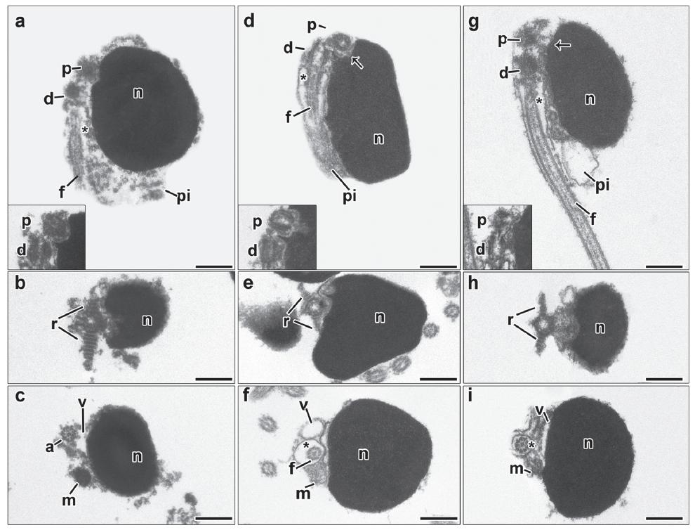
Fig. 1
Spermiogenesis process representative for Lebiasina and Piabucina. Figure subunits are longitudinal sections of spermatids corresponding to (a) Lebiasina aff. uruyensis1, (b) L. aff. uruyensis1, (c) Piabucina boruca, and (d) P. elongata. The flagellum is initially lateral to the nucleus. A slightly movement of the nucleus towards the flagellar axis is present. Consequently the centriolar complex and the nuclear fossa is superolateral. During spermiogenesis, the formation of two striated rootlets occurs on opposite sides of the distal centriole. c: cytoplasm, d: distal centriole, f: flagellum, n: nucleus, p: proximal centriole, pi: midpiece, r: striated rootlet, asterisk: cytoplasmic canal, arrow: nuclear fossa. Bar = 0.5 μm.
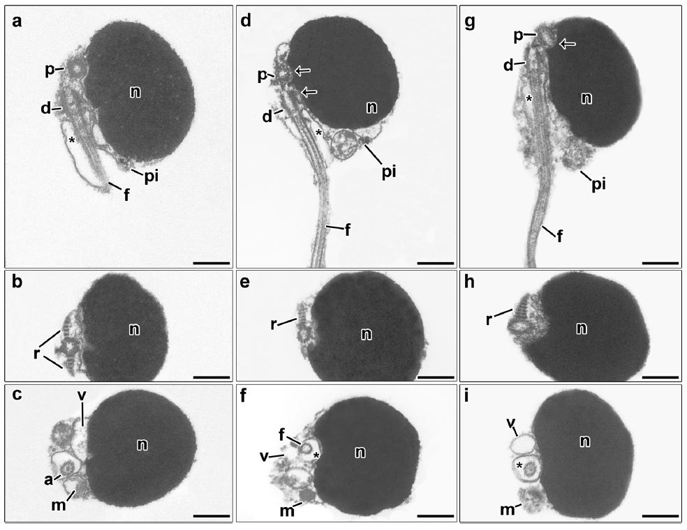
Fig. 2
Spermatozoon of Lebiasina aff. festae (a, b, c), L. bimaculata (d, e, f) and L. erythrinoides (g, h, i). a, d, g: longitudinal sections. b, c, e, f, h, i: transverse sections from top to posterior region. The nucleus (n) of all species is drop-shaped and slightly elongated towards the flagellar axis. The flagellum (f) lies lateral to the nucleus. The centriolar complex (p, d) and nuclear fossa (arrow) is superolateral. Note the presence of the striated rootlets (r) on opposite sides of the distal centriole (d). The proximal centriole (p) is slightly oblique relative to the distal centriole (d) (a-inset, d-inset, g-inset). The cytoplasmic canal is present (asterisk). The midpiece (pi) is short, asymmetrical, and contains the oblong mitochondria (m) and vesicles (v). a: axoneme, d: distal centriole, f: flagellum, m: mitochondria, n: nucleus, p: proximal centriole, r: striated rootlet, v: vesicle, asterisk: cytoplasmic canal, arrow: nuclear fossa. Bar = 0.5 μm.
Fig. 3
Spermatozoon of Lebiasina melanoguttata (a, b, c), L. aff. uruyensis1 (d, e, f) and L. aff. uruyensis2 (g, h, i). a, d, g: longitudinal sections. b, c, e, f, h, i: transverse sections from top to posterior region. The nucleus (n) of all species is dropshaped and slightly elongated towards the flagellar axis. The flagellum (f) lies lateral to the nucleus. The centriolar complex (p, d) and nuclear fossa (arrow) is superolateral. Note the presence of the striated rootlets (r) on opposite sides of the distal centriole (d). The proximal centriole (p) is slightly oblique relative to the distal centriole (d) (a-inset, d-inset, g-inset). The cytoplasmic canal is present (asterisk). The midpiece (pi) is short, asymmetrical, and contains the oblong mitochondria (m) and vesicles (v). a: axoneme, d: distal centriole, f: flagellum, m: mitochondria, n: nucleus, p: proximal centriole, r: striated rootlet, v: vesicle, asterisk: cytoplasmic canal, arrow: nuclear fossa. Bar = 0.5 μm.
Fig. 4
Spermatozoon of Piabucina boruca (a, b, c), P. elongata (d, e, f) and P. panamensis (g, h, i). a, d, g: longitudinal sections. b, c, e, f, h, i: transverse sections from top to posterior region. The nucleus (n) of all species is drop-shaped and slightly elongated towards the flagellar axis. The flagellum (f) lies lateral to the nucleus. The centriolar complex (p, d) and nuclear fossa (arrow) is superolateral. Note the presence of the striated rootlets (r) on opposite sides of the distal centriole (d). The proximal centriole (p) is slightly oblique relative to the distal centriole (d) (a-inset, d-inset, g-inset). The cytoplasmic canal is present (asterisk). The midpiece (pi) is short, asymmetrical, and contains the oblong mitochondria (m) and vesicles (v). a: axoneme, d: distal centriole, f: flagellum, m: mitochondria, n: nucleus, p: proximal centriole, r: striated rootlet, v: vesicle, asterisk: cytoplasmic canal, arrow: nuclear fossa. Bar = 0.5 μm.
Fig. 5
Schematic representation of spermatozoa of Lebiasina and Piabucina. Both spermatozoa are very similar and share primarily the lateral nucleus, superolateral centriolar complex, striated rootlets, oblong mitochondria and some vesicles in the midpiece.