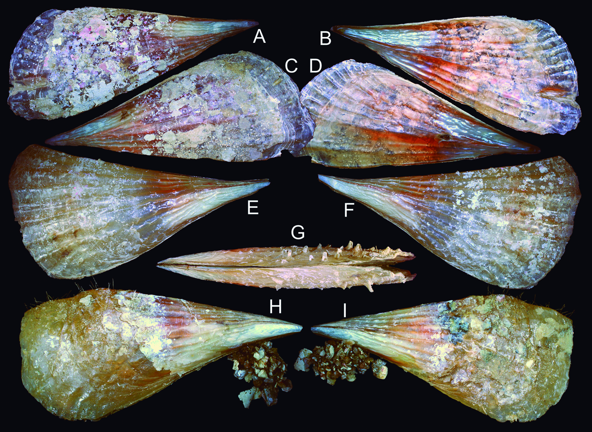
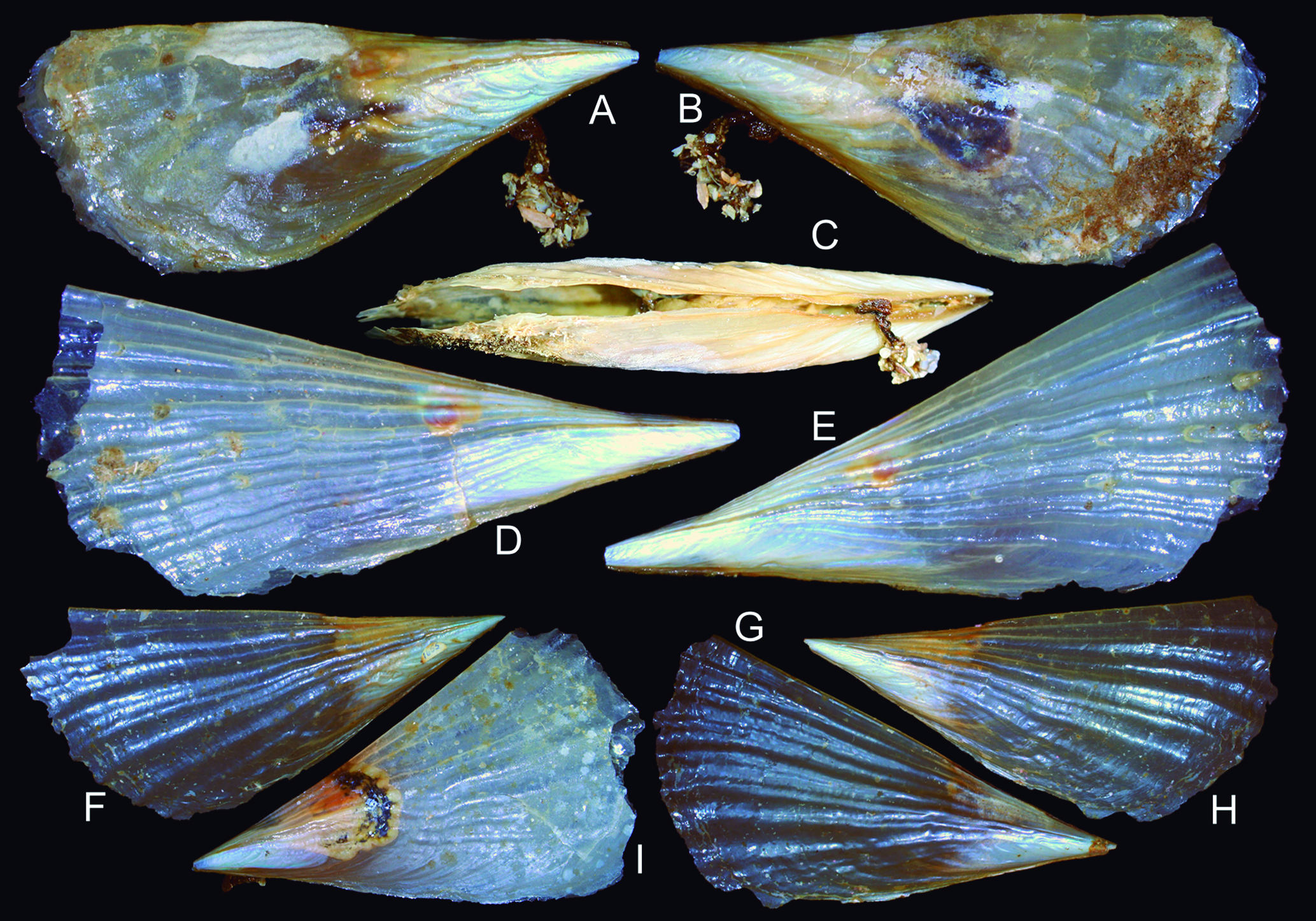
New species, misidentifications and problematic taxonomy of some Atlantic South American marine mollusks: a review
Abstract
In preparation for compiling a catalogue of the South Atlantic American malacofauna, this study addresses the taxonomy of selected species within less intricate groups to ensure accurate classification within the catalogue. The following nomenclatural acts are undertaken for this purpose: New species descriptions: 1, Tectura iguypis; 2, Nacella mirim; 3, Calliostoma soror; 4, Abyssochrysos quasilissus; 5, Caecum jonesae; 6, Sinum striotis; 7, Agaronia sterica; 8, Voluta melodica; 9, Phrontis rocas; 10, Goniofusus phoenix; 11, Dolicholatirus etherius; 12, Lightbournus rendatus; 13, Terebra joculosa; 14, Neoterebra potiguar; 15, Trimusculus pifius; 16, Brevinucula overa; 17, Ennucula ipepa; 18, Tindaria ruru; 19, Barbatia pehenguis; 20, Bathyarca arcadia; 21, Bentharca celeris; 22, Electroma electra; 23, Brachidontes ynous; 24, Pinna trindadis; 25, Pinna nembia; 26, Pinna pereria; 27, Servatrina amazonica; 28, Warrana culmen; 29, Lucinoma apocalyptica; 30, Cyrenoida implexa; 31, Hiatella marisqueira; 32, Cyrtopleura angelicalis; 33, Lyonsiella angulosa; 34, Graptacme obtura. Species revalidated: 1, Fissurella itapema; 2, Cranopsis watsoni; 3, Pedicularia tibia; 4, Semicassis iheringi; 5, Anachis veleda; 6, Terebra chilensis; 7, Iselica anomala; 8, Barbatia cancellaria; 9, Acar domingensis; 10, Limopsis paucidentata; 11, Paracratis borealis; 12, Crassostrea mangle; 13, Abra americana; 14, Choristodon typicum; 15, Pseudochama radians; 16, Polyschides quadridentatum. New synonyms: 1, Fissurella clenchi; 2, Natica castanea; 3, Cyphoma macumba; 4, Voluta thevenini; 5, Pleuroploca granulilabris; 6, Polyschides xavante; 7, Polyschides noronhensis. New combinations: 1, Falsilunatia limbata; 2, Goniofusus damasoi. Redescriptions/taxonomy commented: 1, Nacella mytilina; 2, Fissurella rosea; 3, Calliostoma depictum; 4, Depressiscala nautlae; 5, Depressiscala niditella; 6, Scala scipio; 7, Abyssochrysos brasilianus; 8, Caecum antillarum; 9, Cheilea equestris; 10, Sinum perspectivum; 11, Pseudocyphoma intermedium; 12, Cyphoma signatum; 13, Coronium; 14, Voluta ebraea; 15, Nassarius albus; 16, Goniofusus brasiliensis; 17, Goniofusus strigatus; 18, Leucozonia nassa; 19, Anachis lyrata; 20, Terebra gemmulata; 21, Turbonilla obsoleta; 22, Brevinucula verrillii; 23, Tindaria amabilis; 24, Bathyarca glomerula; 25, Bentharca asperula; 26, Crassostrea mangle; 27, Pinna carnea; 28, Kalolophus antillarum; 29, Warrana besnardi; 30, Chama cristella; 31, Hiatella solida; 32, Cyrtopleura costata; 33, Polyschides tetraschistus. Extension of distribution: 1, Amaea mitchelli; 2, Tindaria cytherea; 3, Lyratellina juttingae. Change of genus: 1, Naticarius cayennensis. New replacement name: 1, Turbonilla absalaoi. Validity of species discussed: 1, Cheilea atlantica; 2, Cheilea americana. Families revalidated: 1, Trichotropidae. Fossil species proposed to remain fossil-only: 1, Cranopsis granulata; 2, Iselica globosa; 3, Limopsis aurita; 4, Limopsis minuta; 5, Abra longicallus. All 104 nomenclatural acts are thoroughly justified, discussed, and mostly illustrated. Several of these acts have co-authors. https://zoobank.org/6DB1BC9F-A0E9-4C36-8BEF-EF8A375F0693.
Keywords
Gastropoda; Bivalvia; Scaphopoda; Taxonomy; Systematics; Mollusc fauna
INTRODUCTION
A project focused on inventories of South American Mollusca species has produced several catalogues as outcomes. Among them, two have already been published: one addressing fossils from Brazil (Simone & Mezzalira, 1994), and another concerning land and freshwater species within Brazil (Simone, 2006). Presently, the project aims to compile a catalogue encompassing all South American marine species from both the Pacific and Atlantic coasts. Due to the considerable number of species, the resulting catalogue will be divided into two volumes. The first volume will cover species along the Atlantic coast, ranging from Colombia to the southern tip of the continent (including the Magellan region of Chile and Argentina). The second volume will encompass Pacific species, spanning from Colombia to Chile.
During the process of surveying data for all species, including the search for type specimens, an important realization emerged: certain species, particularly from Brazil, are misidentified or have taxonomic issues. Consequently, this paper is dedicated to enhancing or rectifying the taxonomy of less complex cases, i.e., cases where resolution does not necessitate a survey equivalent to a PhD dissertation to be solved.
This paper also serves to substantiate the taxonomy of certain higher taxa based on the author’s phylogenetic studies, which have been overlooked in MolluscaBase (2023-2024) but will evidently be applied in the catalogue.
Another point of concern pertains to species described as fossils but, for inexplicable reasons, are considered living. Several cases are even more complex, as the fossils originate from significantly ancient strata, suggesting an isolated species existence over millions of years and described from remote locations, such as Italy (far from the Western Atlantic). Additionally, in several instances, living specimens do not entirely correspond to those depicted in the original descriptions. In such cases, the optimal taxonomic approach seems to involve maintaining the fossil species as fossils and considering the living species as a distinct entity. In all these cases, depicted here, the introduction of a new name was unnecessary, as the oldest synonym from the list could be reinstated.
The taxonomy adopted in this paper usually aligns with MolluscaBase (2024), influenced by the classification system proposed by Simone (2011), which is further supported and supplemented by others (e.g., Simone, 2021, 2024b; Pastorino & Simone, 2021 on Neogastropoda; Simone & Amaral, 2021 on Bivalvia). The classification is mostly based on phenotypic features, and is more minutely explained elsewhere (Simone, 2011, 2024a, b).
Material and methods
Most of the studied material was sourced from the MZSP collection, supplemented by several other institutions from Europe, the USA, and South America. A comprehensive or partial (when dealing with a large amount of material) list of examined specimens accompanies each description or species approach. In some cases, only references are provided. The taxonomy adheres primarily to that of MolluscaBase (2023), with influence from the phylogenetic classification proposed by Simone (2011, 2024b) and subsequent publications (e.g., Simone, 2021). Short lists of synonyms have also been provided, so that can be included in broader taxonomic revisions in the future.
Although the author’s primary method of taxonomic research has been morpho-anatomy, the present paper predominantly adopts a conchological approach. This shift is prompted by the necessity to publish these nomenclatural acts prior to catalogue publication, with the understanding that these species will be further studied in future papers, incorporating complementary data. Nevertheless, anatomical information is included for several species, in some cases to a sufficient extent to support the taxonomic decisions presented.
Some species have tomography images included. In these cases, the Micro CT scanning was done for shell images, using a Phoenix v|tome|x S240 scanner (General Electric), with 1,000 projections, 0.1 mm copper filter, exposure time = 0.2 s, voltage = 70 kV, current = 200 µA, skipping = 1, averaging = 3, and voxel size = 8.18-10.69 µm.
Abbreviations: BMNH, The Natural History Museum, London, UK; FFCLRP, Faculdade de Filosofia, Ciências e Letras de Ribeirão Preto da Universidade de São Paulo, Brazil; col, collector; fms, fathoms; GMNH, Muséum d’Histoide Naturelle de Nenève, Suisse; H, height; IBUFRJ, Departamento de Zoologia, Instituto de Biologia, Universidade Federal do Rio de Janeiro, Brazil; L, length; leg, donor; MCZ, Museum of Comparative Zoology, Harvard University, USA; MD55, Expedición R.V. Marión-Dufresne MD55, MNHN, Bouchet, Leal & Métivier col. [CB: chalut Blake (trawl); CP: chalut à perche (otter trawl); DC: drague Charcot-Picard (dredger); SY, carottier boîte ¼ m2 Sypan (coring box)]; MZSP, Museu de Zoologia da Universidade de São Paulo, Brazil; N, north; MNHN, National Museum d’Histoire Naturelle, Paris, France; o.t., otter trawl; RV, research vessel; S, south; SE southeast; spm, specimen(s); sta, station; USNM & NMNH, National Museum of Natural History, Smithsonian Institution, Washington DC, USA; W, west or width.
RESULTS
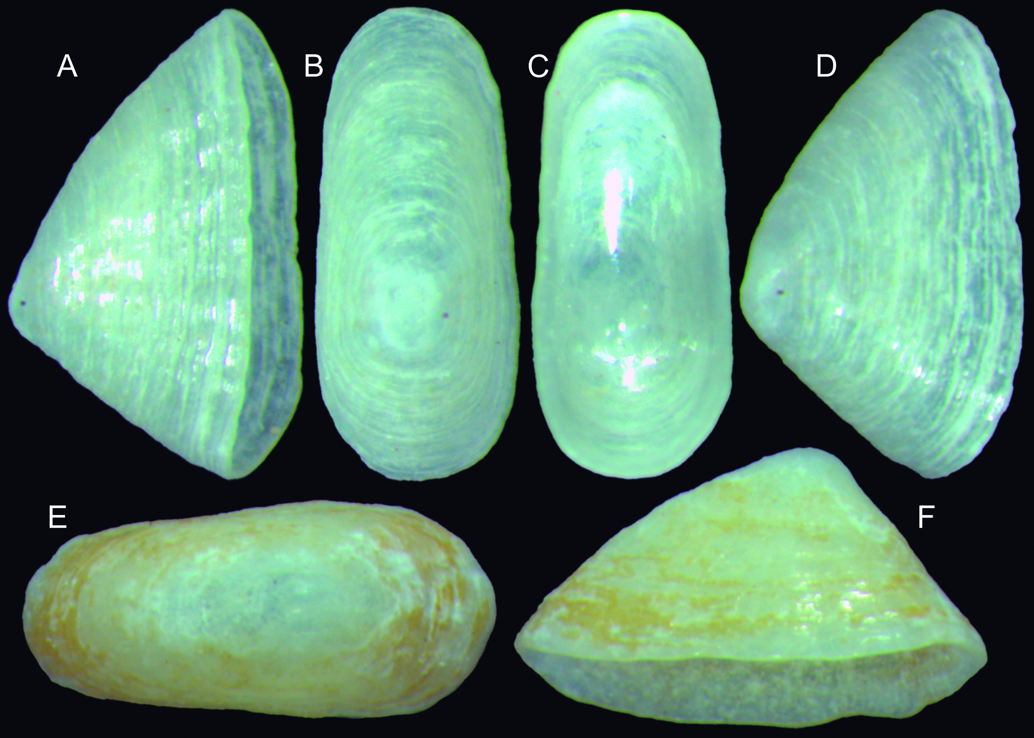
Systematics Class Gastropoda Subclass Patellogastropoda Superfamily Lottioidea Family Lottiidae Genus Tectura Gray, 1847 Tectura iguypis new species (Fig. 1A F, 3C) https://zoobank.org/24FD74FE-FDDF-4D4B-A99B-E9B0AAE5FEDC
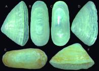
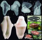
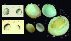
Tectura iguypis shells of type specimens: (A D) Holotype 165600 (L 2.6 mm); (A) right-slightly ventral view; (B) dorsal view; (C) ventral view; (D) right view; (E F) Paratype MZSP 165664 (L 3.1 mm), dorsal and left views.
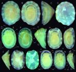
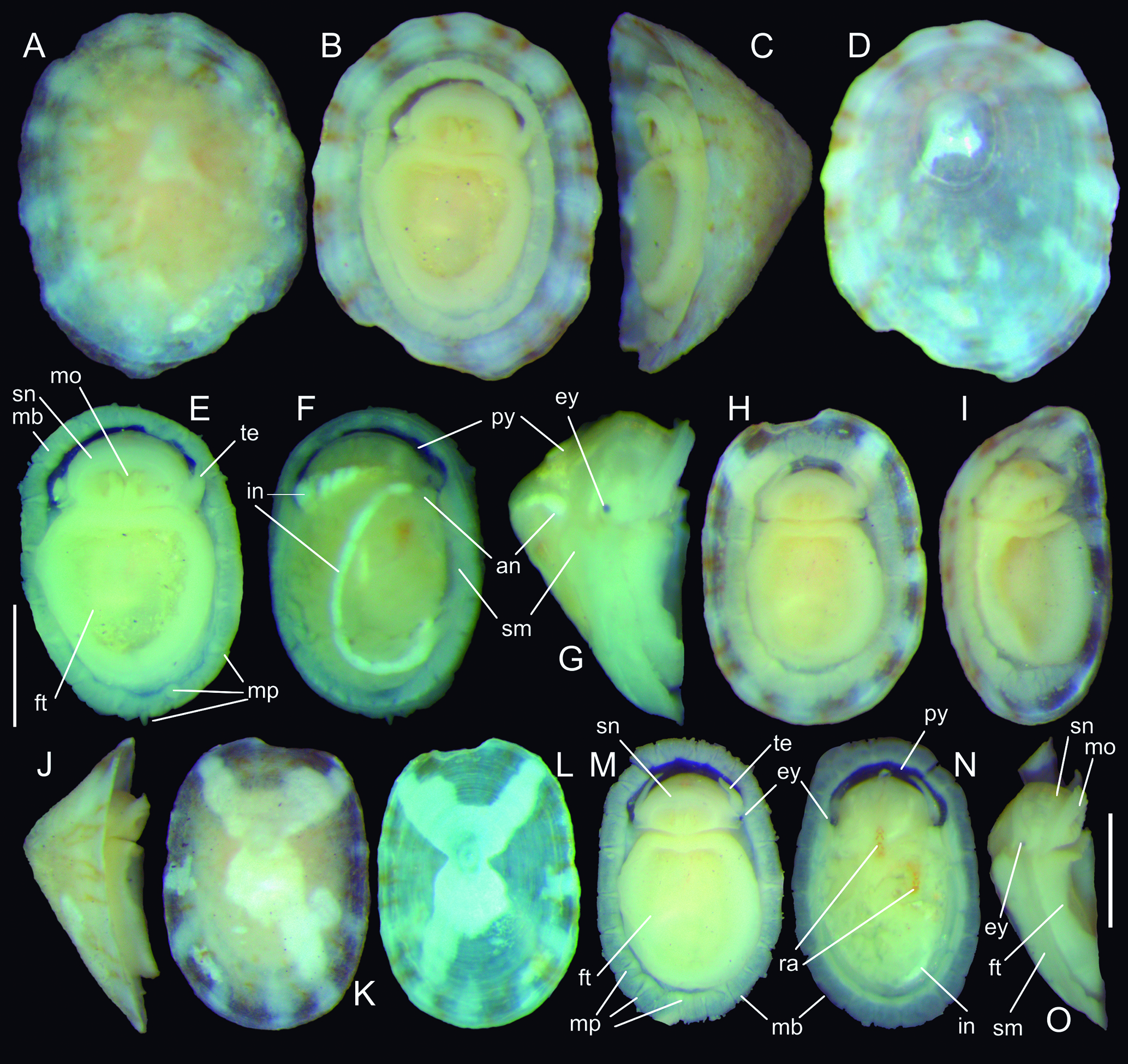
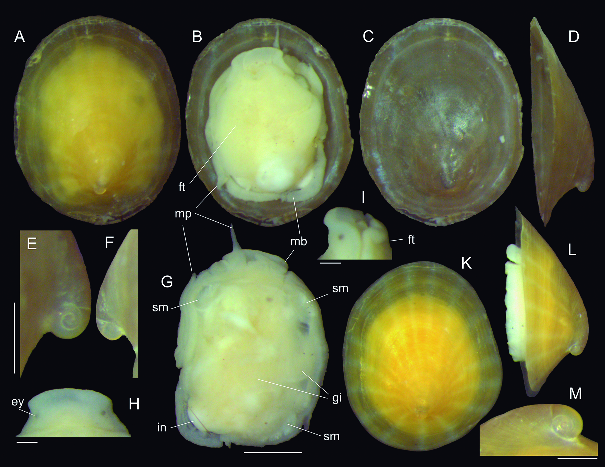
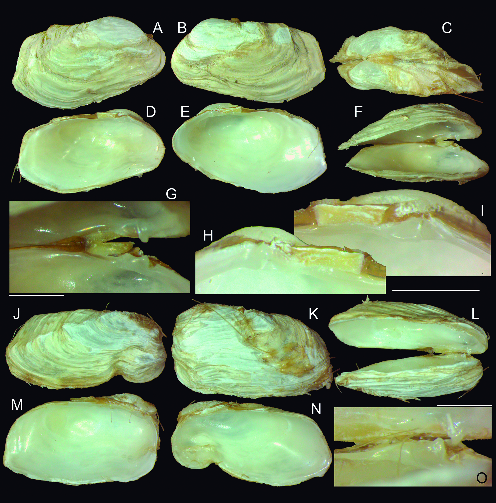
Nacella mirim shells and anatomy of type specimens: (A G) Holotype 165666 (L 2.2 mm); (A) dorsal view; (B) whole ventral view; (C) left-slightly ventral view; (D) shell, ventral view; (E) extracted specimen, ventral view; (F) same, dorsal view; (G) same, right view; (H O) paratype MZSP 100718 (L 2.1 mm); (H) whole ventral view; (I) right-ventral view; (J) right view; (K) dorsal view; (L) shell, ventral-inner view; (M) extracted specimen, ventral specimen; (N) same, dorsal view, first layer of glands and intestinal loops removed to expose radular sac; (O) same, right view, pallial cavity removed. Scales: = 0.5 mm. Lettering: an, anus; ey, eye; ft, foot sole; in, intestinal loop; mb: mantle border; mo, mouth; mp, mantle papillae; py, pallial cavity; ra, radurar sac; sm, shell muscle; sn snout; te, cephalic tentacle.
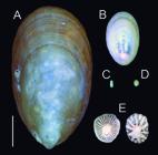
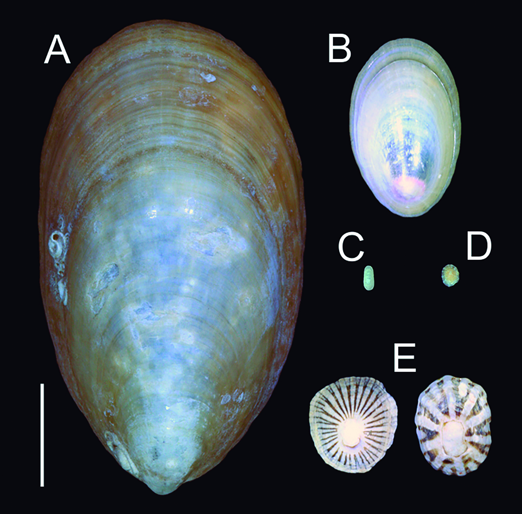
Confrontation among shells of Tectura iguypis, Nacella mirim, N. mytilina, and Lottia leucopleura in a same scale: (A) N. mytilina, MZSP 8587 from Santa Cruz, Argentina; (B) same, MZSP 155805 Tierra del Fuego, Argentina; (C) T. iguypis, holotype; (D) N. mirim, holotype; (E) L. leucopleura, MZSP 162468 from W Jeremie, Haiti. Scale: = 10 mm.
Types: Holotype MZSP 165600, shell. Paratypes: MZSP 165664, 1 shell, MZSP 165665, 5 shells from type locality. BRAZIL. Bahia; Cairu, Praia de Garapuá (Morro de São Paulo), 13°29′32″S 38°54′21″W, MZSP 100771, 1 spm [Petronio Alves Coelho-Filho col., Petrobras, 2011].
Type locality: BRAZIL. Espírito Santo; Guarapari, Praia do Morro, 20°39′42″S 40°29′41″W [Simone col., 15.i.1982].
Diagnosis: East Brazilian species with lateral compression, monochromatic, subcentral apex, lacking additional sculpture.
Description: Shell up to 3 mm, elongated (~2.4 times longer than wide), tall (~1.8 times longer than tall); laterally compressed; almost same width along its length, with posterior region slightly broader than anterior region (Fig. 1B, C, E). Walls thin, slightly translucent. Color whitish (Fig. 1A-D) to yellowish (Fig. 1E-F), uniform. Apex rounded, central in dorsal view (Fig. 1B, E), slightly dislocated posteriorly in lateral view (Fig. 1A, D, F). Protoconch not seen (eroded). Profile with angle ~100°, anterior surface slightly convex; lateral and posterior surface practically planar-straight. Outer surface only possessing growth lines and concentric undulations, no other additional sculpture. Shell edges slightly convex laterally, concave anterior and posteriorly (Fig. 1A, F). Internal surface glossy (Fig. 1C), muscle scars not visible.
Anatomy: Unknown.
Etymology: The specific epithet is a Latinization from native Guarani language word iguýpe, meaning down under, an allusion to the southern occurrence of the species.
Distribution: Coast of Bahia to Espírito Santo.
Measurements (L, W, H in mm): Holotype MZSP 165600 (Fig. 1A-D): 2.6 by 1.1 by 1.4. Paratype: MZSP 165664 (Fig. 1E-F): 3.1 by 1.4 by 1.6.
Remarks: This species have been misidentified as Nacella mytilina (Helbling, 1779) in collections, a species much larger (N. mytilina easily reaches 30-40 mm), with terminal apex (Fig. 3A-B), that occur much more southwards, in Argentina. The minuteness (Fig. 3C) and the lateral compression of the limpet shell easily suggests the genetic attribution to Tectura iguypis, and the species is the first report of the genus in South Atlantic. Tectura virginea (Müller, 1776) occurs in NE Atlantic and Mediterranean, T. iguypis differs by the lateral compression (T. virginea is dorso-ventrally compressed), in lacking color mosaic in shell, and apex close to posterior edge. The remaining Tectura species occur in Pacific Ocean. Two of them also have lateral compression and elongation, supposedly to fit in elongated leaves of intertidal and subtidal algae. One of them is T. depicta (Hinds, 1842), from California, from which T. iguypis differs in lacking color mosaic of brown bands, by taller shell, and by apex more centrally located. The other is T. paleacea (Gould, 1853), which also occurs in California region, from which T. iguypis differs in having a more angulate shell, an anterior region shorter and not so convex, a lighter color (T. paleacea is brown), and in lacking delicate radial sculpture.
Superfamily Patelloidea Family Nacellidae Genus Nacella Schumacher, 1817 Nacella mirim new species (Figs. 2A-O, 3D) https://zoobank.org/40815F89-F6F6-467D-AE98-0EB62AE4712E
Types: Holotype MZSP 165666. Paratypes: MZSP 100736, 5 spm, MZSP 100903, 13 spm, MZSP 100916, 5 spm, MZSP 100718, 10 spm, all from type locality. BRAZIL. Espírito Santo; Guarapari, Praia do Morro, 20°39′42″S 40°29′41″W, MZSP 165667, 3 shells [Simone col., 15.i.1982].
Type locality: BRAZIL. Bahia; Cairu, Praia de Garapuá (Morro de São Paulo), 13°29′32″S 38°54′21″W [Petronio Alves Coelho-Filho col., Petrobras, 2011].
Diagnosis: E Brazilian species with up to ~5 mm, generally pigmented with a slight large white X spot; edge with intercalated white and brown bands. Aped dislocated anteriorly. Presence of eyes.
Description: Shell up to 5 mm, elliptic (~1.3 times longer than wide), conic (4-2.5 times longer than tall); anterior and posterior edges similar-sized (Fig. 2A, D, K, L). Walls thin, slightly translucent. Color beige to light brown, in base, with large white X image more (Fig. 2K, L) or less (Fig. 2A, D) nitid in different specimens, plus intercalated brown and white bands near edges (Fig. 2B, H, L). Apex rounded, central slightly dislocated anteriorly (Fig. 2C, J). Protoconch not seen (eroded). Profile with angle 100-110°, anterior and posterior surfaces varying from planar to weakly convex. Outer surface only possessing growth lines and very weak concentric undulations, no other additional sculpture. Shell edges slightly concave laterally (Fig. 2C, J). Internal surface glossy (Fig. 2D, L), muscle scars not visible.
Snout large (Fig. 1E, M: sn), with ~⅓ of head-foot size. Foot (ft) plane, sole concave, edges simple, 2-3 times wider than snout. Pair of cephalic tentacles located in lateral side of snout (Fig. 2B, E, G, H, I, M-O: te), possessing small dark eye in outer base (ey), lacking ommatophore. Mantle edge (mb) thick, colorless; possessing ~20 small papillae (mp), equidistantly distributed along its ventral surface, their base located in inner region, tapering externally. Shell muscle (sm) horseshoe-shaped, narrow, sightly broader anteriorly. Pallial cavity (py) shallow, restricted only to head region; lacking gill or any other structure. Visceral mass fulfilled by yellowish gonad and digestive gland, and several intestinal, narrow loops, with final region fulfilled by white matter (Fig. 2F, N: in). Radular sac (Fig. 2N) relatively rigid, with docoglossan radula.
Etymology: The specific epithet is a Latinization from native Tupi-Guarani language word mirim, meaning little, an allusion to the small size of the species.
Distribution: Coasts of Alagoas to Espírito Santo.
Habitat: From intertidal up to ~10 m, usually found sorting sediment.
Measurements (L, W, H in mm): Holotype MZSP 165666 (Fig. 1A-D): 2.2 by 1.7 by 0.9. Paratype: MZSP 100718, 2.1 by 1.5 by 0.5.
Additional material examined: BRAZIL. Alagoas; Japaratinga (F. Hartmann col.), off Bitingui, 09°07′05.53″S 35°15′45.82″W, 5 m, MZSP 166753, 2 shells, off Praia do Salgado, 09°05′53.63″S 35°13′48.08″W, 10 m, MZSP 166754, 30 shells. Bahia; Cairu, Praia de Garapuá (Morro de São Paulo), 13°29′32″S 38°54′21″W, MZSP 100717, 2 spm, MZSP 100719, 1 spm, MZSP 100732, 1 spm, MZSP 100734, 1 spm, MZSP 100775, 1 spm, MZSP 100815, 2 spm, MZSP 100833, 2 spm, MZSP 100849, 2 spm, MZSP 100901, 2 spm, MZSP 100904, 1 spm, MZSP 100908, 1 spm, MZSP 100933, 8 spm, MZSP 101290, 3 spm, MZSP 101292, 1 spm; MZSP 101303, 3 spm [Petronio Alves Coelho-Filho col., Petrobras, 2011]; Abrolhos, Recife de Sebastião Gomes, 17°58′03″S 38°42′13″W, 3-8 m (Ex coleção Vanin, IOUSP col., vii.2007.
Remarks:N. mirim is another species usually found identified as N. mytilina in collections, as well as Lottia leucopleura (Gmelin, 1791), a species from the Caribbean. The minuteness of N. mirim, i.e., up to 5 mm, easily distinguishes it from both species, which possibly do not occur in Brazilian coast. A set of anatomical features, such as absence of gill in pallial cavity (Fig. 2B, E, F, H, M, N), the absence of secondary gill surrounding the foot (Fig. 2G, O), the papillae in the mantle edge (Fig. 2E, M: mp), and the docoglossate radula (Fig. 2N: ra), indicate the generic attribution of the species. The presence of eyes is an extraordinary discover. The presence of cephalic eyes is something practically absent in patellogastropods (Simone, 2011, 2023), its presence in N. mirim, associated to the small size, are indicative of a neotenic origin of this species. The disparity of size and shell features of N. mirim with other species can be detected in Fig. 3D. It differs from N. mytilina (Fig. 3A-B) by the size, in having a non-subterminal apex, and in not being monochromatic. It differs from L. leucopleura (Fig. 3E) also by the size, in having a lighter, thin walled, less colorful shell, and by the apex dislocated anteriorly (Fig. 2C, J), instead of posteriorly.
Subclass Vetigastropoda Order Lepetellida Superfamily Fissurelloidea Family Fissurellidae Genus Fissurella Bruguière, 1789 Fissurella itapema (Ihering, 1927) revalidated, new combination (Fig. 4A-G)
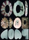
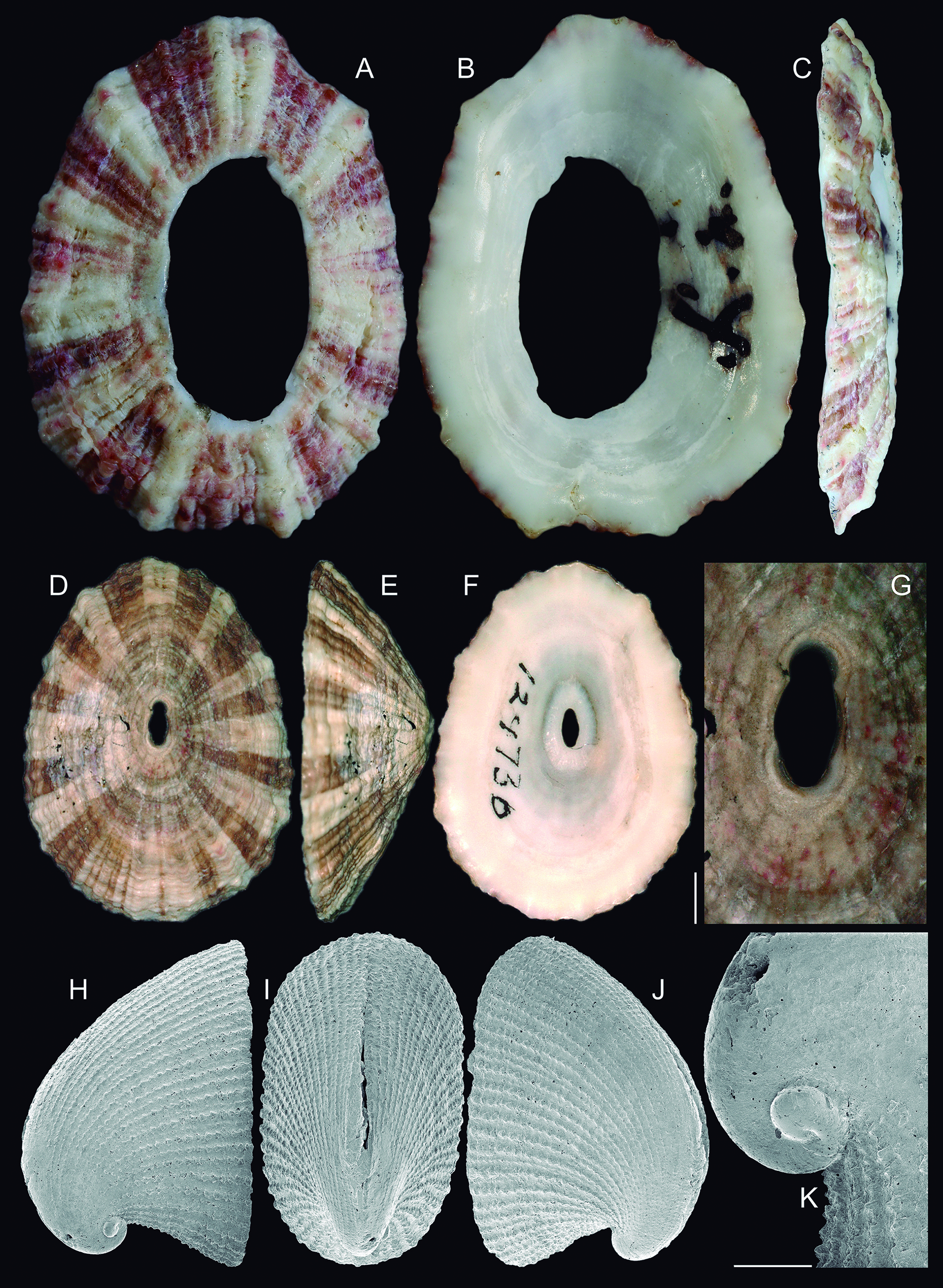
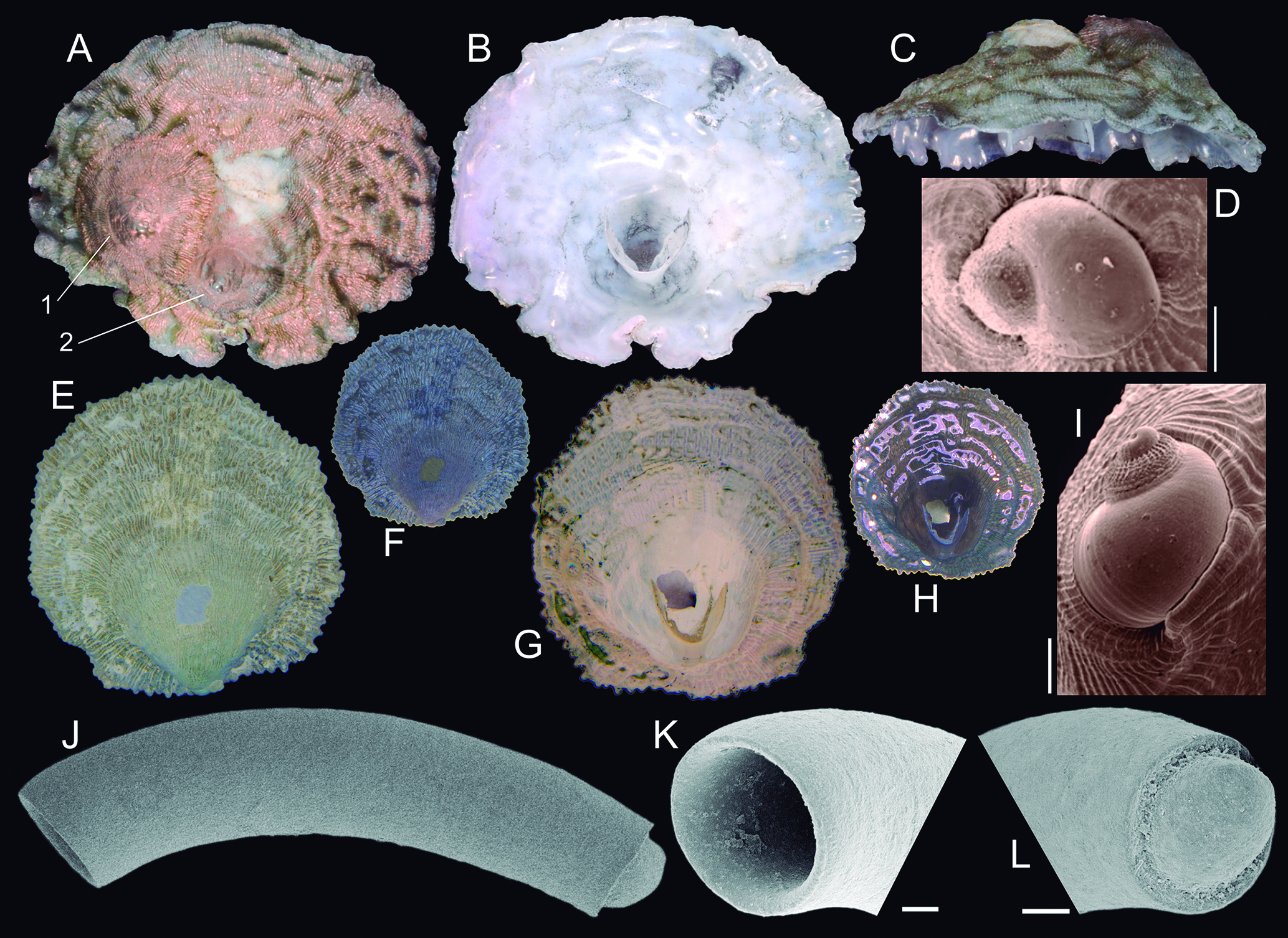
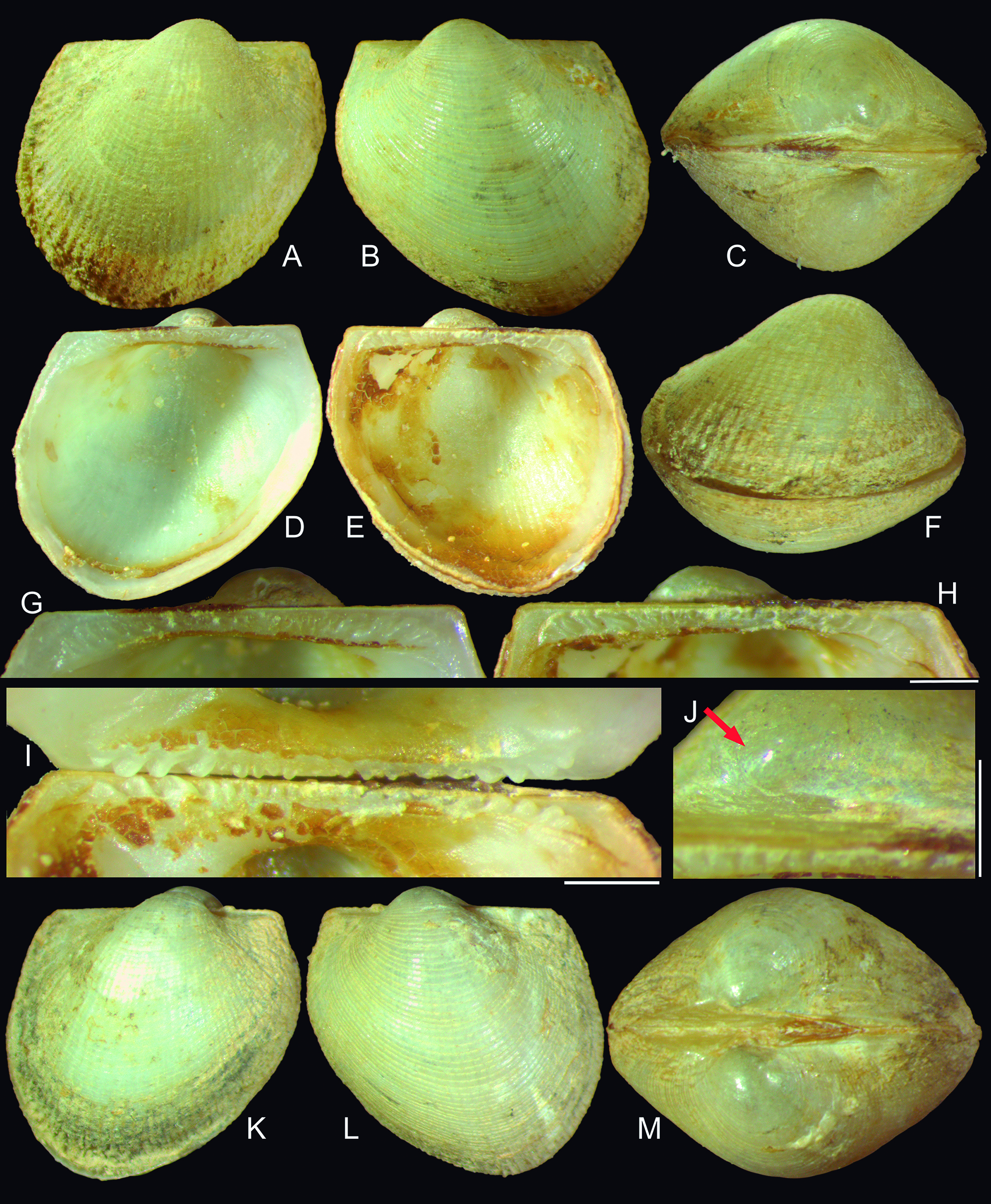
Fissurella itapema shell of types: (A C) holotype Senckenberg Mus. 3278 (L 13.3 mm), dorsal, ventral and left views (Courtesy Senckenberg Museum); (D G) holotype Fissurella clenchi, MCZ 124730 (courtesy MCZ); (D) dorsal view; (E) left view; (F) ventral view; (G) detail of apex, dorsal view, scale: = 1 mm; (H K) Cranopsis watsoni shell of lectotype USNM 95146 in SEM (L 3.1 mm) (courtesy USNM); (H) right view; (I) dorsal view; (J) left view; (K) detail of apex, right view, scale: = 200 µm.
Lucapina itapemaIhering, 1927: 102-103 (pl. 6, fig. 5-8); Rios, 1970: 20, 1975: 18, 1985: 17, 1994: 27, 2009: 35 (these in syn of F. rosea).
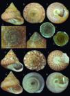
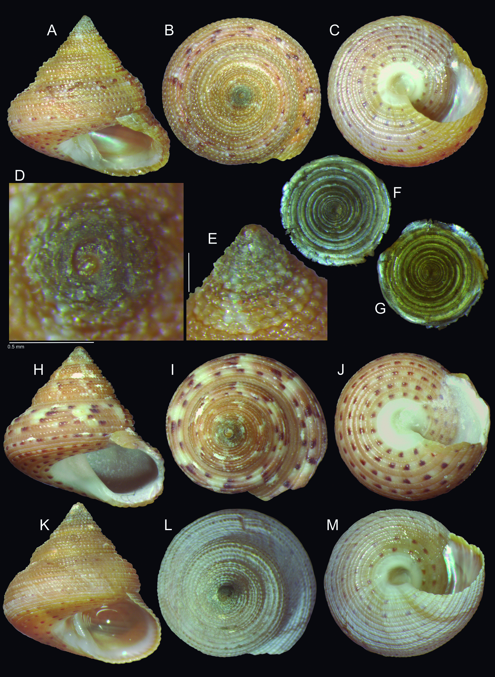
Calliostoma soror shells and operculum of type specimens: (A G) Holotype MZSP 121819 (W 8.3 mm); (A) frontal view; (B) apical view; (C) inferior view; (D) detail of apex, apical view, scale: = 0.5 mm; (E) same, profile; (F) operculum, holotype, outer view (W 3.5 mm); (G) same, inner view; (H J) paratype MZSP 121827 (W 9.9 mm), frontal, apical and inferior views; (K M) paratype MZSP 114401 (W 7.7 mm), frontal, apical and inferior views.
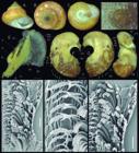
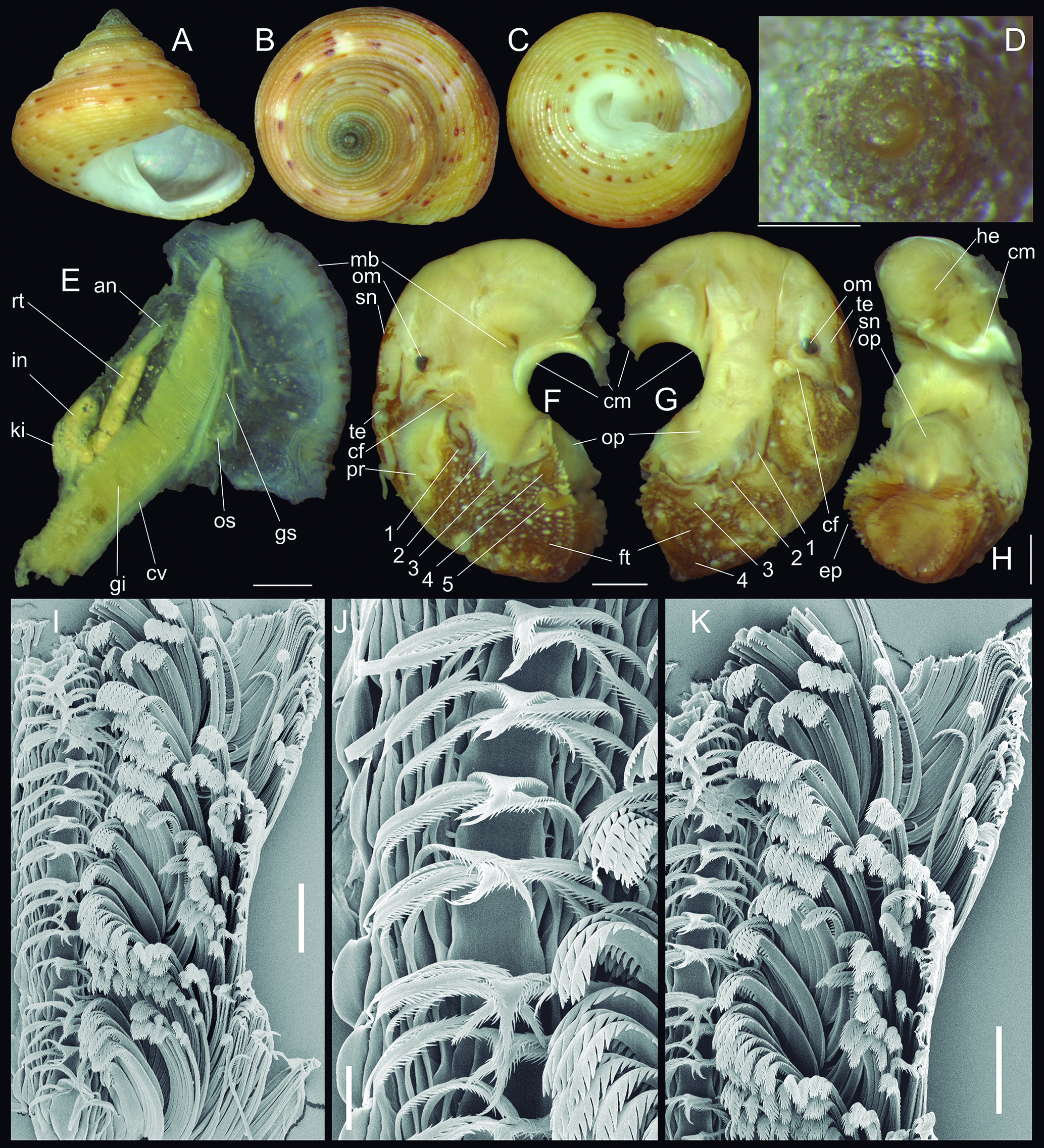
Calliostoma soror shells and anatomy of type specimens: (A C) paratype MZSP 104811 (W 10.5 mm), frontal, apical and inferior views; (D) detail of apex of paratype MZSP 114401, apical view, scale: = 0.5 mm; (E H) holotype details of anatomy, scales: = 1 mm; (E) pallial roof, ventral-inner view; (F) head-foot, left view; (G) same, right view; (H) same, ventral view; (I K) holotype radula in SEM; (I) detail of right side, scale: = 100 µm; (J) detail of central region, scale: = 30 µm; (K) detail of marginal region, scale: = 100 µm.
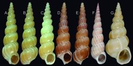
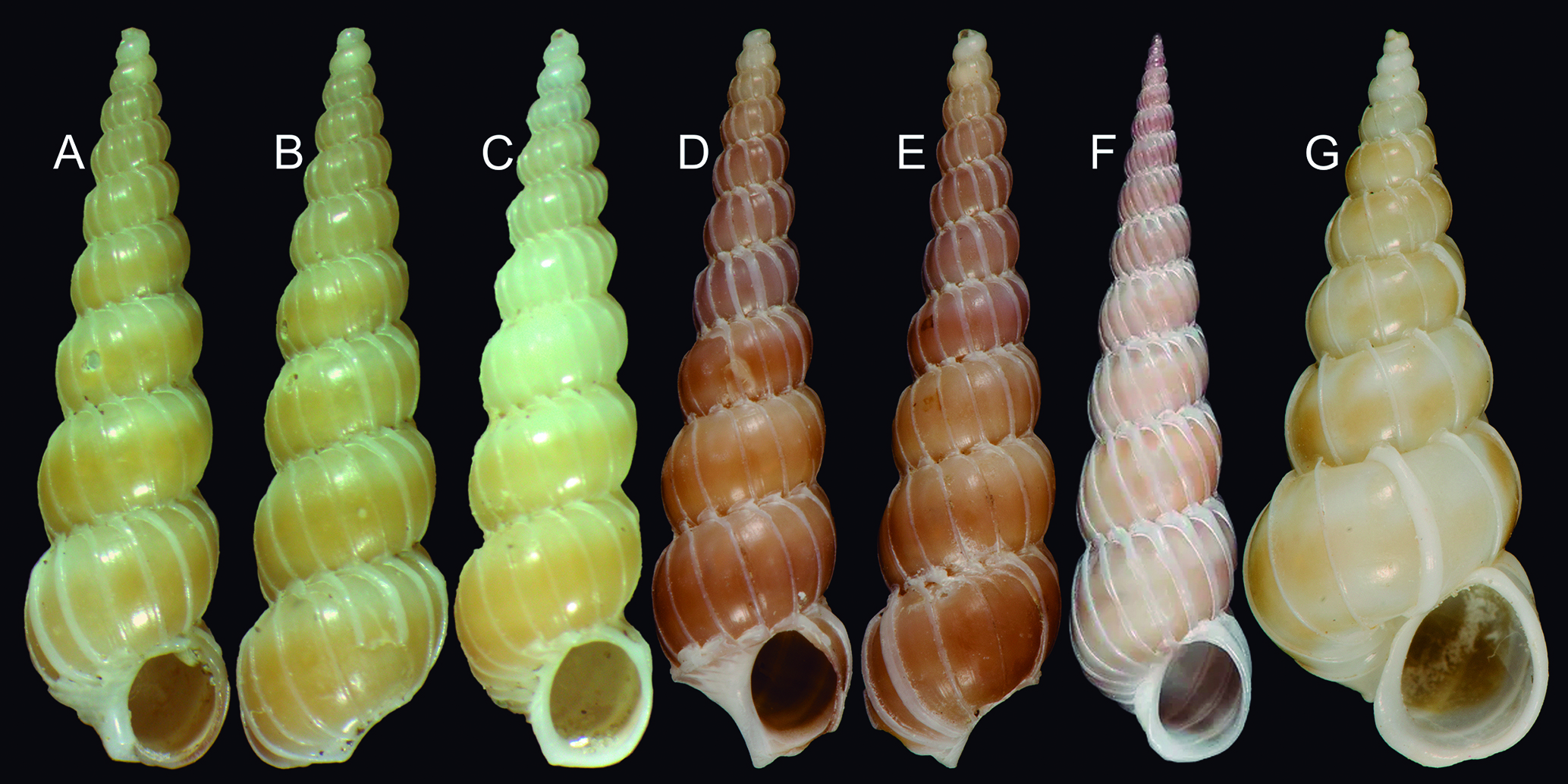
Depressiscala nautlae and D. nitidella shells: (A B) D. nautlae, MZSP 166500, from Ubatuba, São Paulo, Brazil (L 14.0 mm), frontal and dorsal views; (C) same, MZSP 165694, same locality (L 13.1 mm); (D E) Lectotype of Scala scipio USNM 10694 (L 14.9 mm); (F) D. nautlae (L 13.3 mm), ordinary specimen from Pompano Beach, Florida, ANSP 457201, frontal view; (G) Scala nitidella, lectotype USNM 83716 (L 13.1 mm), frontal view (USNM photos courtesy Ellen Strong).
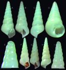
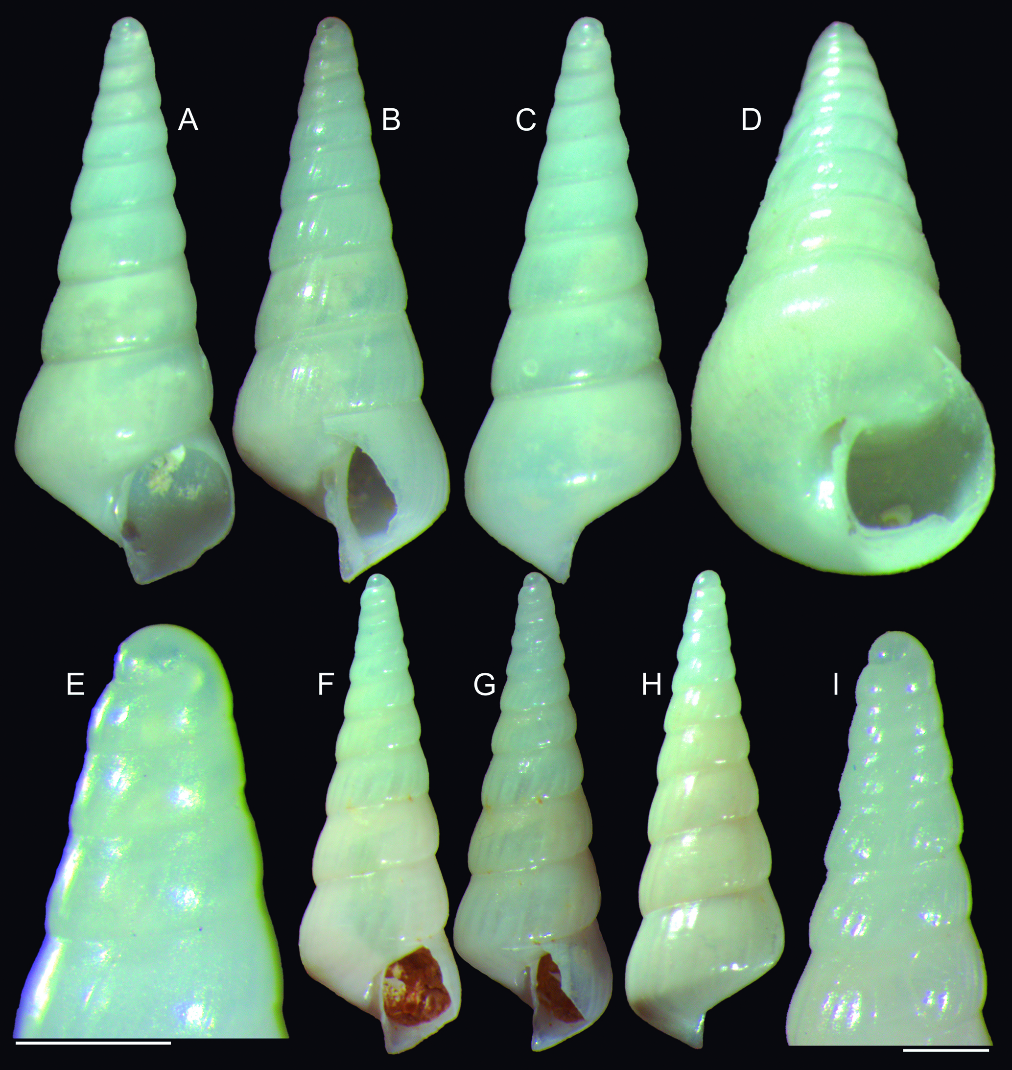
Abyssochrysos quasilissus type shells: (A E) holotype MZSP 117078 (L 5.6 mm); (A) frontal view; (B) right view; (C) dorsal view, (D) inferior-slightly frontal view; (E) detail of apex, profile; (F I) paratype MZSP 117074 (L 6.4 mm).
Fissurella (Cremides) clenchiFarfante, 1943: 11-12 (pl. 3, figs. 8-11) (new synonym); Métivier, 1970: 119, 1972: 406; Abbott, 1974: 28; Rios, 1975: 18 (pl. 4, fig. 34), 1985: 16 (pl. 8, fig. 63), 1994: 26 (pl. 7, fig. 61), 2009: 34 (fig. 70).
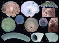
holotypes of Western Atlantic Cheilea species and Caecum jonesae: (A D) holotype (2) and paratype (1) of C. americana BMSM 17936 (courtesy J. Leal, BMSM) (W 19.4 mm); (A) dorsal view; (B) ventral view; (C) posterior view; (D) detail of protoconch; (E I) holotype of C. atlantica MNRJ 25542 (courtesy A. Pimenta, MNRJ) (W 7 mm); (E) shell digitally cleaned, dorsal view; (F) same, original photo; (G) shell digitally cleaned, ventral view; (H) same, original photo; (I) detail of protoconch; (J) Caecum jonesae, holotype MZSP 166590 (L 1.3 mm), left view; (K) same, detail of aperture, left-slightly anterior view; (L) same, detail of posterior region and mucro, posterior-slightly left view. Scales: (D, I) = 200 µm, (K L) = 50 µm.
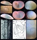
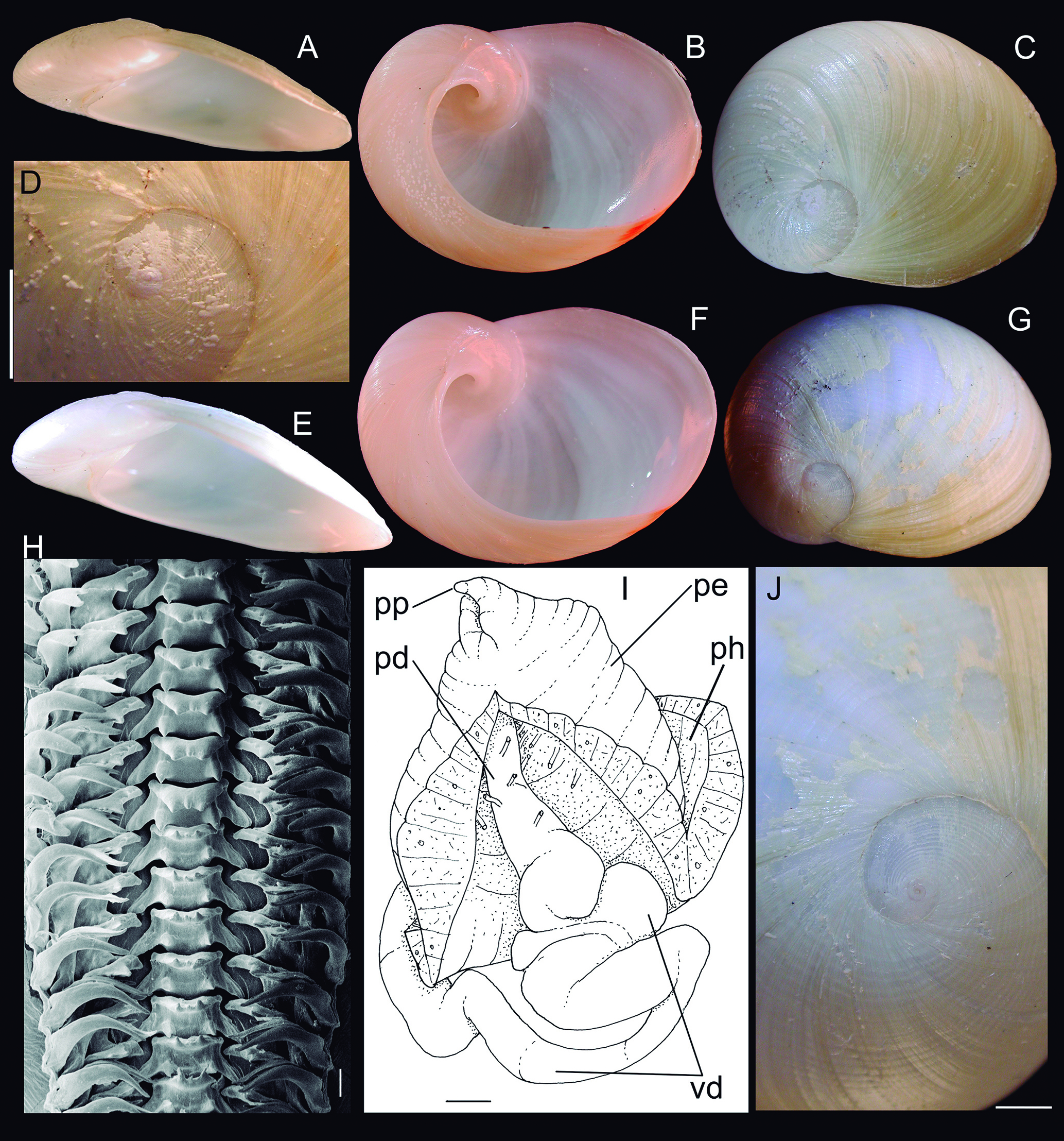
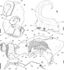
Sinum striotis shells, radula and penis of type specimens: (A D) Holotype MZSP 32260 (L 35.2 mm); (A) frontal view; (B) inferior view; (C) superior view; (D) detail of apex, scale: = 5 mm; (E G) paratype MZSP 32166 (L 36.0 mm), frontal, inferior and superior views; (H) radula in SEM, scale: = 100 µm; (I) drawing of dissected penis, dorsal view, scale: = 1 mm; (J) detail of Fig. G in apex region, scale: = 2 mm. Lettering: see next Figure.
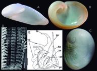
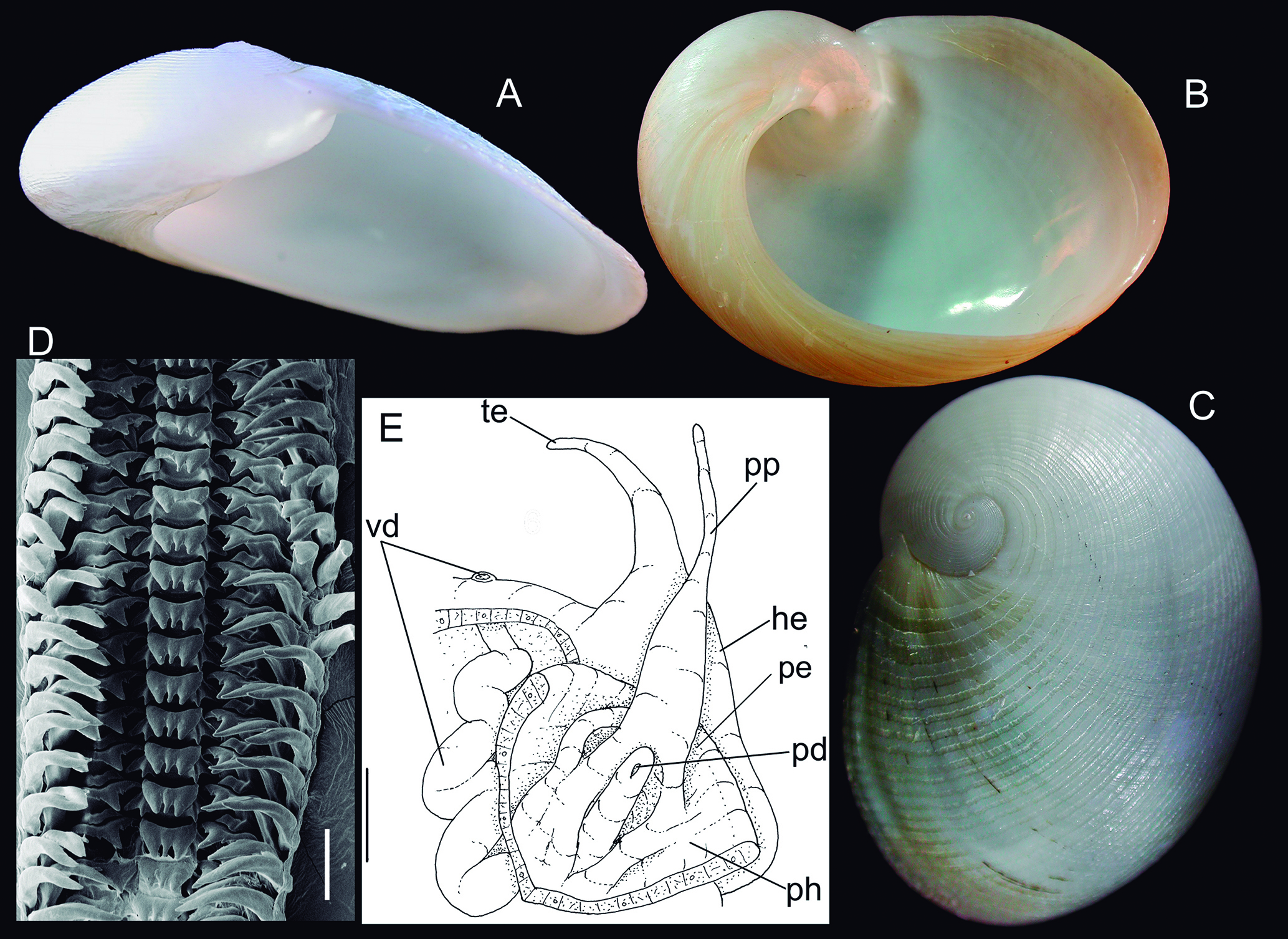
Sinum perspectivum shells, radula and penis of specimen USNM 801214 (from off Jacksonville, Florida, USA): (A C) shell (L 23.6 mm), frontal, inferior and superior views; (D) radula in SEM, scale: = 100 µm; (E) drawing of dissected penis, dorsal view, scale: = 1 mm. Lettering: he, head flap; pd, penis duct or aperture; ph, penis chamber; pp, penis papilla; te, cephalic tentacle; vd, vas deferens.
Lucapinella itapema:Morretes, 1949: 56.
Fissurella chenchi:Rios, 1970: 20 (pl. 2); Vermeij & Porter, 1971: 445, 447, 451; Calvo, 1987: 53 (fig. 17); Simone, 2008: 292, 303 MolluscaBase, 2023.
Fissurella rosea:Righi, 1963: 263-264; Rios, 1970: 20 (pl. 2), 1975: 18 (pl. 4, fig. 36), 1985: 16-17 (pl. 8, fig. 65), 1994: 27 (pl. 7, fig. 63), 2009: 35 (fig. 72); Métivier, 1972: 406 (part); Calvo, 1987: 53, 57 (fig. 19); Leal, 1991: 34-35 (part); Longo et al., 2014: 3, 8 (fig. 2N) (non Gmelin, 1789).
Types:F. itapema: holotype Senckenberg Mus. 3278 (Fig. 4A-C); F. clenchi: MCZ 124730 (Fig. 4D-G).
Type localities: Itapema, Santa Catarina, Brasilien (in holotype label); F. clenchi: Nova Almeida [-20.056568° -40.196843° (Datum: WGS84)], Espirito Santo, Brazil.
Remarks: In the time of description of Fissurella mesoatlantica Simone, 2008, endemic from São Pedro e São Paulo Archipelago, a remote Brazilian oceanic island, a comparison with coastal congener samples was performed, a study (Simone, 2008) that remains largely unpublished. It was observed that the entire Brazilian coast bear a single species of Fissurella, occurring intertidally. Some shells with a red tone were identified as F. rosea, while others as F. clenchi. This identification was mostly induced by some references (e.g., Rios, 1994, 2009), showing the color and details of the orifice as key characters. However, the Brazilian Fissurella species exhibit significant variability in shell features, including color and in orifice aspects; more trustable are the anatomical characteristics (Simone, 2008). These characteristics were primarily employed to distinguish the insular species. Fissurella rosea actually is a valid species, but does not occur in Brazil. It is a Caribbean species, with shell larger and much more sculptured, with scales and nodes more developed than the Brazilian population (Farfante, 1943). The southmost range of F. rosea appears to be Venezuela. Fissurella clenchi, on the other hand, seems to be endemic from Brazil, with confirmed north limit in Ceará and extending up to Rio Grande do Sul. Due to confusion with F. rosea in the southern Caribbean, the distinction between both species remains unclear, as some reports in French Guyana are documented (Farfante, 1943).
Lucapina itapema, formerly, was so far considered a synonym of F. rosea. The species was described based on a broken shell, in which the tip of it was amputated (Fig. 4A-C). Consequently, the shell took on the appearance of having a very wide orifice, simulating genera with this characteristic. This led to its first classification in Lucapina Gray in Sowerby, 1835 (Ihering, 1927) and a combination to Lucapinella Pilsbry, 1890 (Morretes, 1949) (in fact these genera do not have such large orifice, a more accurate classification would be Fissurellidea d’Orbigny, 1839). As L. itapema already sets up on F. rosea synonymy (see synonymic list above) the generic mistake and the shell breakage were noted by previous authors.
Despite Ihering (1927) not reporting a type locality in the original description of L. itapema, the specific epithet, the justifications of other species described with this same name in that paper, and the information of the holotype’s label, leave no doubt that the type locality is Itapema, a city of the coast of Santa Catarina, Brazil (~27°05′S 48°36′W). This locality is significantly distant from the true geographic distribution of F. rosea in the Caribbean. Conversely, L. itapema is, in fact, synonym of F. clenchi. However, as that name was described 16 years earlier, it takes precedence and must be considered valid. Despite being more comprehensively described, possessing complete types (Fig. 4D-G), and being more widely utilized, F. clenchi must be considered a synonym.
There is a prevailing belief among collectors and local taxonomists, as explained above, that, in Brazilian Fissurella, the reddish toned shells belong to F. rosea, while those with brownish are F. clenchi. Looking inside, the Brazilian Fissurella usually have a colorful halo surrounding the orifice. Sometimes it is red, thus implicating it is a presumed F. rosea, sometimes it is blue, thus implicating a F. clenchi. There are, however, a myriad of other variations, including brown, both (red and blue) and even absence (white - Fig. 4F). This demonstrates that the color is not a good taxonomical parameter for this group. Most possibly, the red tone was the responsible for considering L. itapema a F. rosea synonym. Observing both holotypes (L. itapema in Fig. 4A-C and F. clenchi in Fig. 4D-G), it is evident that they belong to the same species, as the sculpture and color pattern are similar, and they differ from any other regional species. The orifice conformation, highlighted by some authors as a distinguishing feature between F. rosea and F. clenchi (e.g., Farfante, 1943), is also variable in the local Fissurella. This variation results from the gradual reabsorption of the shell tip during growth, a process described by Simone (2008) and references therein, and it is not uniform. The edges can vary, e.g., the holotype itself, has the outer edge trilobed (Fig. 4D, G), but it is elliptic inside (Fig. 4F).
Genus Cranopsis A. Adams, 1860 Cranopsis watsoni (Dall, 1889) revalidated, new combination (Fig. 4H-K)
Puncturella (Cranopsis) granulata:Watson, 1883: 31 Dautzenberg & Fischer, 1896: 491, 1897: 180; Farfante, 1947: 124-126 (pl. 54, figs. 4-7); Rios, 1975: 14 (pl. 2, fig. 14), 1985: 12-13 (pl. 6, fig. 43), 1994: 24 (pl. 6, fig. 47), 2009: 29 (fig. 55) (non Seguenza, 1863).
Puncturella (Cranopsis) tuberculataWatson, 1883: 31 (invalid, see below).
Puncturella watsoniDall, 1889: 403-404.
Puncturella granulata:Poirier, 1954: 14; Abbott, 1974: 23 (sp. 79); Molluscabase, 2024 (non Seguenza, 1863).
Cranopsis granulata:Simone & Cunha, 2014: 452-453 (non Seguenza, 1863).
Type localities: Near Barbados, 100 fms. Sta. 20, 220 fms.; off Bahia Honda, Cuba; Off Yucatan, 200 fms. Focused to off Barbados, 100 fms by Farfante (1947) by lectotype designation. Puncturella tuberculata: Challenger sta. 24, north of Culebra Island; 18°38′30″N 65°5′30″W, 390 fms, off St. Thomas, N of Culebra Island, Danish W. Indies, 390 fms.
Remarks: The Atlantic Cranopsis watsoni has been considered synonymous with C. granulata. However, C. granulata was described by Seguenza (1863: 88) as a Miocene fossil from Messina, Italy. Despite age and geographic discrepancies, the Recent Atlantic specimens do not entirely match the original concept of the fossil C. granulata, particularly regarding the height of the shell, which was described as “poco elevata,” or low. In contrast, existing species are notable for having a tall, elevated shell (Fig. 4H, J). Therefore, the most appropriate taxonomic procedure is to consider C. granulata solely as a Miocene fossil species from the Italian region, and the Recent Atlantic populations as a distinct taxonomic entity.
Puncturella (Cranopsis) tuberculataWatson, 1883 should be an older name, but it was simply introduced as a synonym of P. granulata and was never mentioned again (Watson, 1883; MolluscaBase, 2024), rendering it a nomen nudum status and an unavailable name. The only available name for the Atlantic Recent species is Puncturella watsoni Dall, 1889, which fully meets the taxonomic requirements to be considered the valid name. It supersedes the previous concept known as C. granulata. Since P. tuberculata, despite being invalid, was clearly associated with Recent material, it is evidently a synonym of C. granulata.
The recent synonymy of the genus Cranopsis with Puncturella Lowe, 1827 (Cunha et al., 2019; MolluscaBase, 2024) is not adopted here, as the differentiation between both genera is deemed consistent, as reported by Simone & Cunha (2014).
Order Trochida Superfamily Trochoidea Family Calliostomatidae Genus Calliostoma Swainson, 1840 Calliostoma soror Simone & Dornellas, new species (Figs. 5-6) https://zoobank.org/CDE3E0AC-2CEB-47BA-998B-EEDB2896B840
Types: Holotype MZSP 121819, spm. Paratypes: MZSP 166230, 1 shell with same data as holotype, MZSP 114401, 1 shell from type locality (Abbate col., 15.vii.2013); BRAZIL. Espírito Santo; Trindade (Oceanic) Island, Enseada do Lixo (Paredão), 20°31′29.8″S 29°19′43.9″W, 15.4 m, MZSP 121827, 1 shell (J.B. Mendonça col., 04.xi.2014), Andradas, 20°28′47.69″S 29°18′24.03″W, MZSP 104811, 1 shell (C.H. Guimarães col., 23.i.2012), coast between Andradas and Tartagugas, 20°30′11.85″S 29°19′11.79″W, MZSP 136531, 1 spm (J.B. Mendonça col., 05.xi.2013).
Type locality: BRAZIL. Espírito Santo; Trindade Island, Farol, 20°29′52.3″S 29°19′15.6″W [Joel Braga Mendonça col., 08.iv.2014].
Diagnosis: Trindade species with ~10 mm of adult shell and suture well-marked. Sculptured by spiral cords composed of successive nodes; cords interspaces smooth, equivalent to cords width. Umbilicus mostly closed. Epipodium with papillated edge, and 5 tentacles in left side, with tentacle 2 bifid. Osphradium small. Radula with narrow and tall rachidian, with sharp pointed, curved tip; robust lateral teeth.
Description: Shell (Figs. 5A-E, H-M, 6A-D) about 10 mm; trochiform, as tall as wide. Walls thick, iridescent inside (Fig. 5A, K). Color light orange-brown; with small brown spots, as wide as spiral cords, relatively densely distributed in some specimens, joined with some axial white bands (Fig. 5H-J), similar pattern, but more scantly distributed in other specimens (Fig. 6A-C); white bands absent with scanty brown minute spots in other specimens (Fig. 5A-C), up to some specimens practically unicolor (Fig. 5K-M), with only some scanty spots in inferior surface; umbilicus always white (Figs. 5C, J, M, 6C). Protoconch usually dark brown, protruded, with 1 whorl, of ~0.5 mm; first 0.5 whorl smooth, last 0.5 whorl with strong, successive axial threads (Figs. 5D, E, 6D). Spire with angle ~70, teleoconch of ~7 convex whorls; suture relatively deep, performing angle ~150°. Sculpture of punctuated spiral cords, 7 in penultimate whorl; smooth interspaces, with same width as cords; each cord composed of successive, aligned hemispheric punctuations, very evident in most specimens (Fig. 5A-E, K-L), but in rare cases cords slightly smooth in last whorl, being punctuated in spire and in inferior surface (Fig. 6A-C); last whorl with ~25 spiral cords. Aperture rounded, occupying ~45% of shell width, ~36% of shell height; prosogyre, ~30° with longitudinal axis. Outer lip with cutting edge, undulating reflecting spiral sculpture. Inner lip with strong vertical rafter as edge, flanked by shallow (Fig. 5A, H) to relatively deep (Figs. 5K, 6A) furrow; remaining as smooth callus. Umbilicus usually totally occluded by callus (Fig. 5C, J) more rarely with shallow concavity (Fig. 5M), still rarer with deeper concavity (Fig. 6C).
Operculum (Fig. 5F, G): Rounded, highly multispiral, nucleus central; margin flexiclaudent, fragile; inner surface glossy. Head-foot of ~½ whorl (Fig. 6F-G), head as wide as foot; ommatophores (om) elongated, with dark eye at tip; cephalic tentacles (te) located just dorsal to ommatophores, tapering uniformly. Snout (sn) of ⅙ whorl. Foot large (~75% of head-foot volume), pigmented by light brown, with small white spots; cephalic flaps (cf) relatively small, simple; epipodium as short fold, with papillated edge (Fig. 6H: ep), bearing 4 right, and 5 left epipodial tentacles (Figs. 6F: 1-5, G: 1-4), each tentacle simple, tapering uniformly up to pointed tip, anterior tentacle as largest; second left tentacle small, white, bifid. Columellar muscle (cm) of 1.5 whorl, right posterior side slightly longer (Fig. 6H). Opercular pad (Fig. 6H: op) in middle level of dorsal foot surface.
Pallial cavity of ~⅕ whorl (Fig. 6E). Gill very elongated, posterior half of uniform width, anterior half tapering gradually up to bluntly pointed tip. Gill suspensory stalk (gs) with almost half of gill length. Osphradium (os) small, located in base of gill suspensory stalk. Rectum (rt) narrow, simple, full of fecal matter. Anus (an) siphoned (non-sessile), located posterior from mantle edge, close to right limit of mantle cavity.
Radula (Fig. 6I-K) about twice odontophore length. Rachidian occupying ~10% of radular ribbon width, ~twice longer than wide; stem rectangular; tip strongly curved inwards, tapering up to sharp pointed tip, having minute secondary cusps at both edges. Lateral teeth as 5 pairs (Fig. 6J), base wide, oval, ~⅓ of rachidian width; abrupt diminishing in long, flattened rod, as long as rachidian; narrow, arched inwards, tip sharp pointed; distal half of this rod with both edges bearing aligned small secondary cusps, each secondary cusp barb-like, minute, very narrow and pointed. Marginal teeth with ~35 pairs (Fig. 6I, K), mote central teeth ~twice longer than rachidian, ~⅓ its width; elongated, ~10 times longer than wide; basal 80% weakly arched, tip 20% strongly arched inwards, slightly broader, with both edged bearing 12-15 pairs of secondary cusps, each cusp triangular, ~3 times longer than wide in base, located close from each other; marginal row strongly arched, teeth gradually diminishing towards edges, more marginal teeth ~⅓ size of more central teeth; inner set of cusps slightly longer than outer set of cusps.
Etymology: The specific epithet is based on the Latin word soror, meaning sister, in allusion in being sister species of C. depictum Dall, 1927 from the mainland coast.
Distribution: Endemic of Brazilian oceanic islands of Trindade and Martin Vaz.
Habitat: on rocks, from intertidal up to ~10 m, usually found by diving.
Measurements (W, H in mm): Holotype MZSP 121819 (Fig. 5A-C): 8.3 by 8.3. Paratypes: MZSP 121827 (Fig. 5H-J): 9.9 by 10.0; 114401 (Fig. 5K-M): 7.7 by 7.9; 104811 (Fig. 6A-C): 10.5 by 9.3.
Additional material examined: BRAZIL. Espírito Santo; Trindade (Oceanic) Island, Ponta da Calheta, 20°30′18.72″S 29°18′31.67″W, MZSP 136733, 4 spm (J.B. Mendonça col., 25.vi.2015), 20°30′28.3″S 29°18′38.8″W, MZSP 104780, 4 shells (C.H. Guimarães col., 10.ii.2012), Tartaruga, 20°31′03.93″S 29°18′08.45″W, MZSP 104150, 1 shell (C.H. Guimarães col., 27.ii.2012), Andradas, 20°30′45.7″S 29°18′21.9″W, MZSP 140568, 6 shells (J.B. Mendonça col., 21.vii.2013), 20°28′47.69″S 29°18′24.03″W, MZSP 105364, 1 spm (06.ii.2012), Enseada do Lixo, 20°31′43.5″S 29°19′28.1″W, MZSP 105380, 1 shell (21.ix.2012), Farrilhões, 20°31′22.4″S 29°19′52.0″W, MZSP 115734, 2 spm (08.vii.2013), off E of, 20°30′15″S 29°18′22″W, 8 m, MZSP 101197, 1 shell (23.vii.2011); Ilha de Martin Vaz, West coast, 20°28′32.32″S 28°52′00.38″W (23.vii.2013), MZSP 115412, 4 shells, MZSP 115403, 1 spm.
Remarks: the present species I coauthored with Ana Paula S. Dornellas [Universidade Federal de Sergipe, E mail: dornellas.anapaula@gmail.com], a Vetigastropoda specialist. Calliostoma soror is only similar to C. depictum in the Brazilian region, as both species have deep suture, relatively small size, and the shell color pattern. Calliostoma soror possibly is derived from an isolated C. depictum population that reached the remote Trindade Island, or both species are derived from the same calliostomatids branch. Calliostoma depictum was recently reviewed, including anatomical aspects, which facilitates the present comparison (Dornellas & Simone, 2013). Only some details distinguish both species. Calliostoma soror has its shell with the spiral cords entirely composed of successive nodes, this does not occur in C. depictum, except for its first whorls, but the nodes fuse with each other, producing smooth spiral cords at least in the last two worlds (Dornellas & Simone, 2013); C. soror has punctuated spiral cords um to the last whorl. A single exception was found (Fig. 6A-C), but the smooth spiral cords are restricted to last whorl and to superior region only. The spiral arrangement of nodes in C. soror in first whorls are coarser and fewer than that of C. depictum, in the third whorl, for example, C. soror has 4 spiral cords, while C. depictum has 5. The suture of C. soror is usually shallower, while that of C. depictum usually is deeper. The umbilicus of C. soror is closed in most specimens (Fig. 5C, J), slightly opened umbilicus is exceptional; wile C. depictum the umbilicus is always deep. Related to the anatomy, C. soror differs in having 5 epipodial tentacles in left side, with the tentacle 2 bifid (Fig. 6F), while C. depictum has 4 epipodial tentacles in both sides. The epipodium edge is entirely papillated in C. soror (Fig. 6H: ep), while it is smooth in C. depictum. The projected portion of the gill and of the anus of C. soror (Fig. 6E: an, gs) are longer than those of C. depictum. The osphradium of C. soror (Fig. 6E: os) is very small if compared to that of C. depictum (Dornellas & Simone, 2013: fig. 79). The radula of C. soror (Fig. 6I-K) adds more distinctions if compared to that of C. depictum (Dornellas & Simone, 2013: figs. 48-50); the rachidian of C. soror is much narrower and longer, with a terminal tip much more pointed than that of the other species; the lateral teeth of C. soror are more robust, while those of C. depictum are almost filiform; the marginal teeth are similar in shape, but differ in number, being mush more abundant, ~50 pairs. There is no other Calliostoma species in which C. soror can be confused.
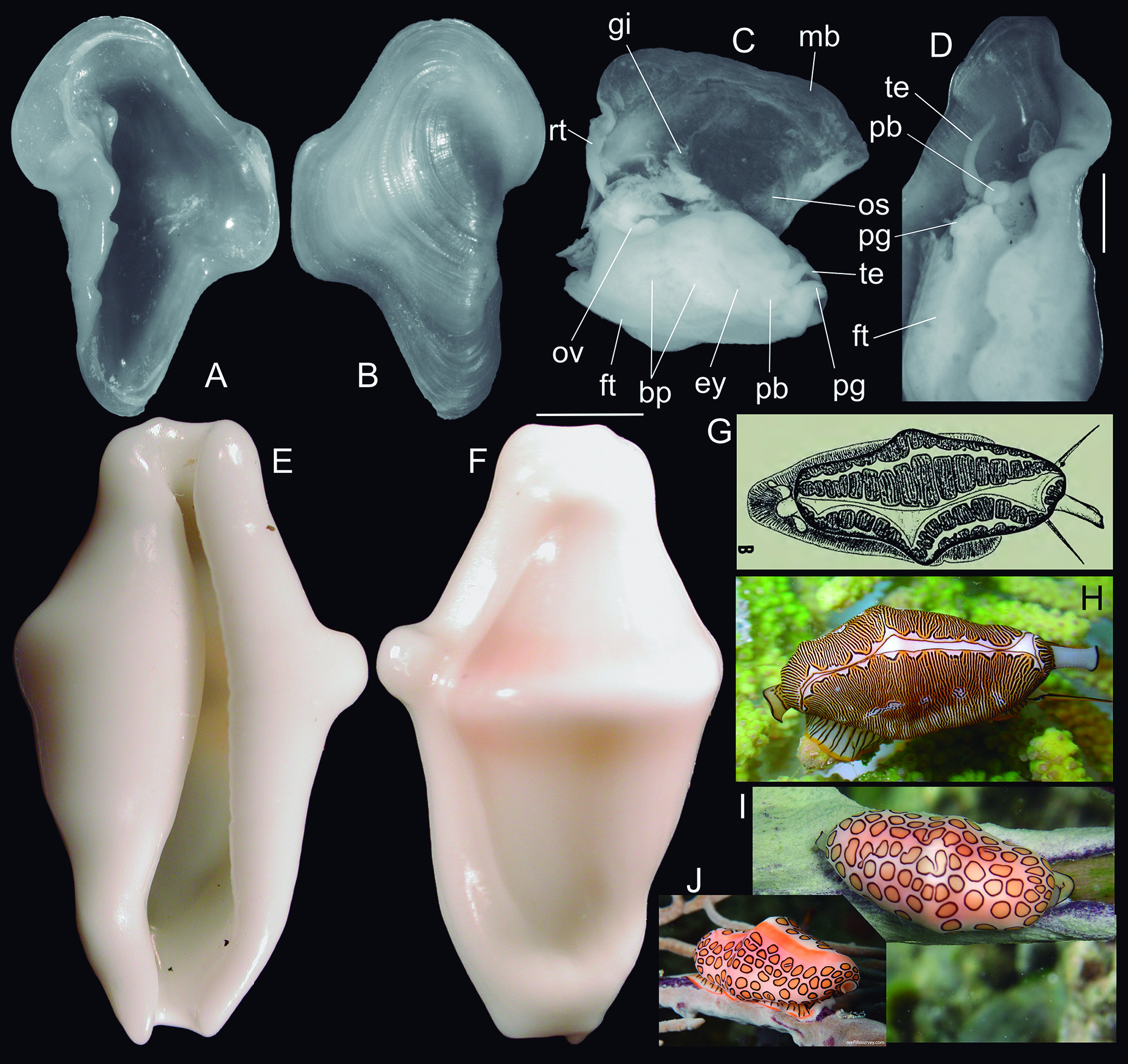
Pedicularia tibia and Cyphoma spp.: (A) replication of some images by Simone (2005) in higher resolution (in grayscale), shell, paratype MZSP 53683, frontal view (L 9.6 mm); (B) same, dorsal view; (C) paratype MZSP 53919 ♀, anterior view, shell extracted, mantle sectioned and deflected upwards; (D) paratype MZSP 53919 ♂, detail of anterior region, whole ventral view. Scales: = 2 mm. (E-F) Cyphoma macumba holotype USNM 780646 (L 22 mm), frontal and dorsal views; (G) replication of fig. 2B by Petuch (1979) showing color pattern of C. macumba; (H) living specimen of Pseudocyphoma intermedium, dorsal view from Guarapuá, Bahia, Brazil (courtesy Paulo Márcio S Costa) (L ~25 mm); (I) living specimen of C. gibbosum from Grand Cayman (courtesy Femorale by Marcus Coltro) (L ~25 mm). (J) living specimen of C. gibbosum from Bocas del Toro, Panama (courtesy Rick Stuart-Smith, Reef Life Survey) (L ~20 mm).
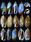
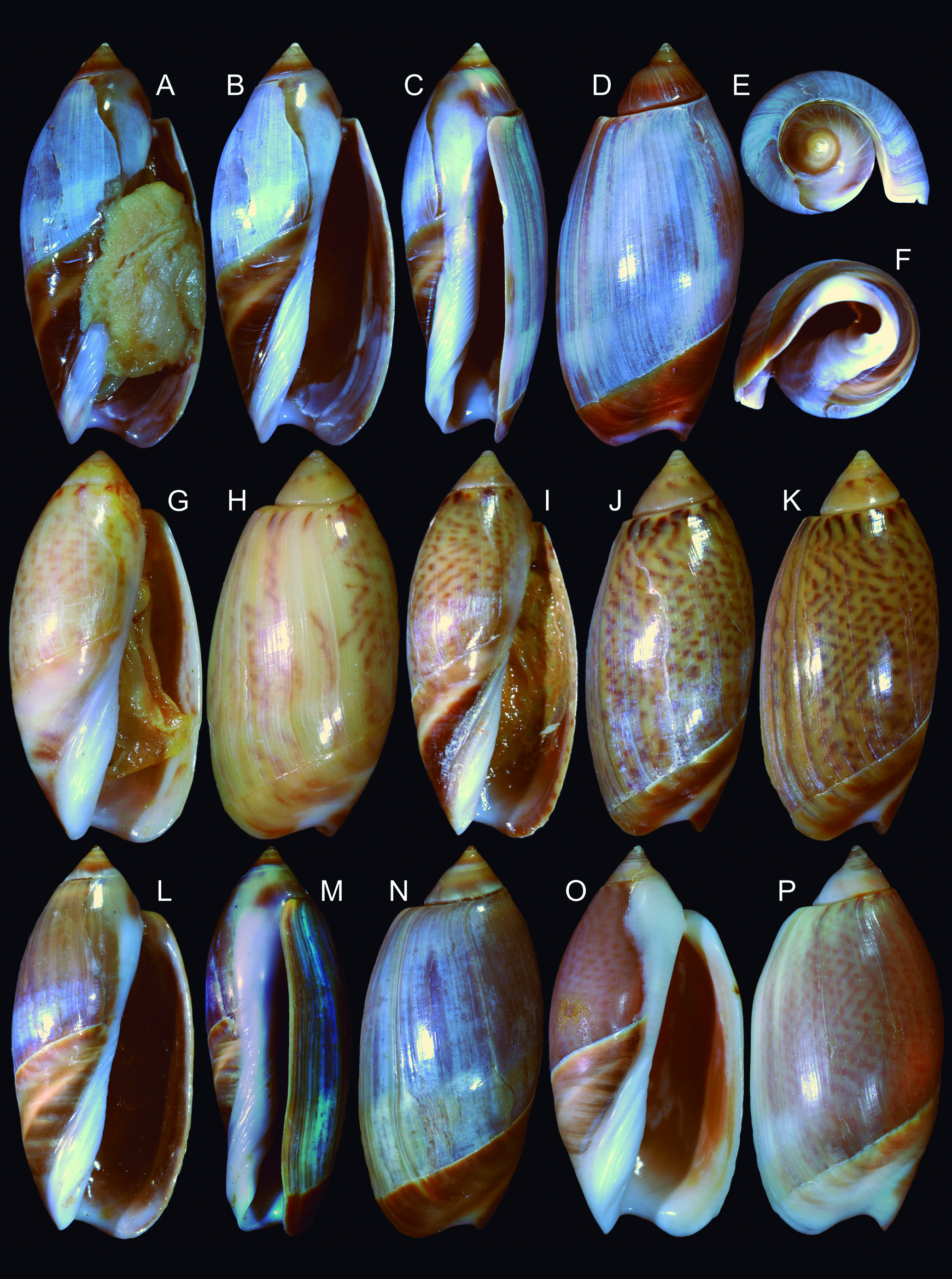
Agaronia sterica shells of some type specimens showing variation: (A-F) Holotype MZSP 166100 (L 48.2 mm); (A) frontal view with specimen still inside; (B) frontal view; (C) right view; (D) dorsal view; (E) apical view; (F) anterior view; (G-H) paratype MZSP 68586#1 (L 33.8 mm), frontal and dorsal views; (I-J) 68586#2 (L 34.8 mm), frontal and dorsal views; (K) paratype MZSP 69465 (L 40.0 mm), dorsal view; (L M) paratype MZSP 73374#1 (L 46.9 mm): frontal, right and dorsal views; (O P) paratype MZSP 51042 (L 49.3 mm), frontal and dorsal views.
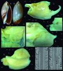
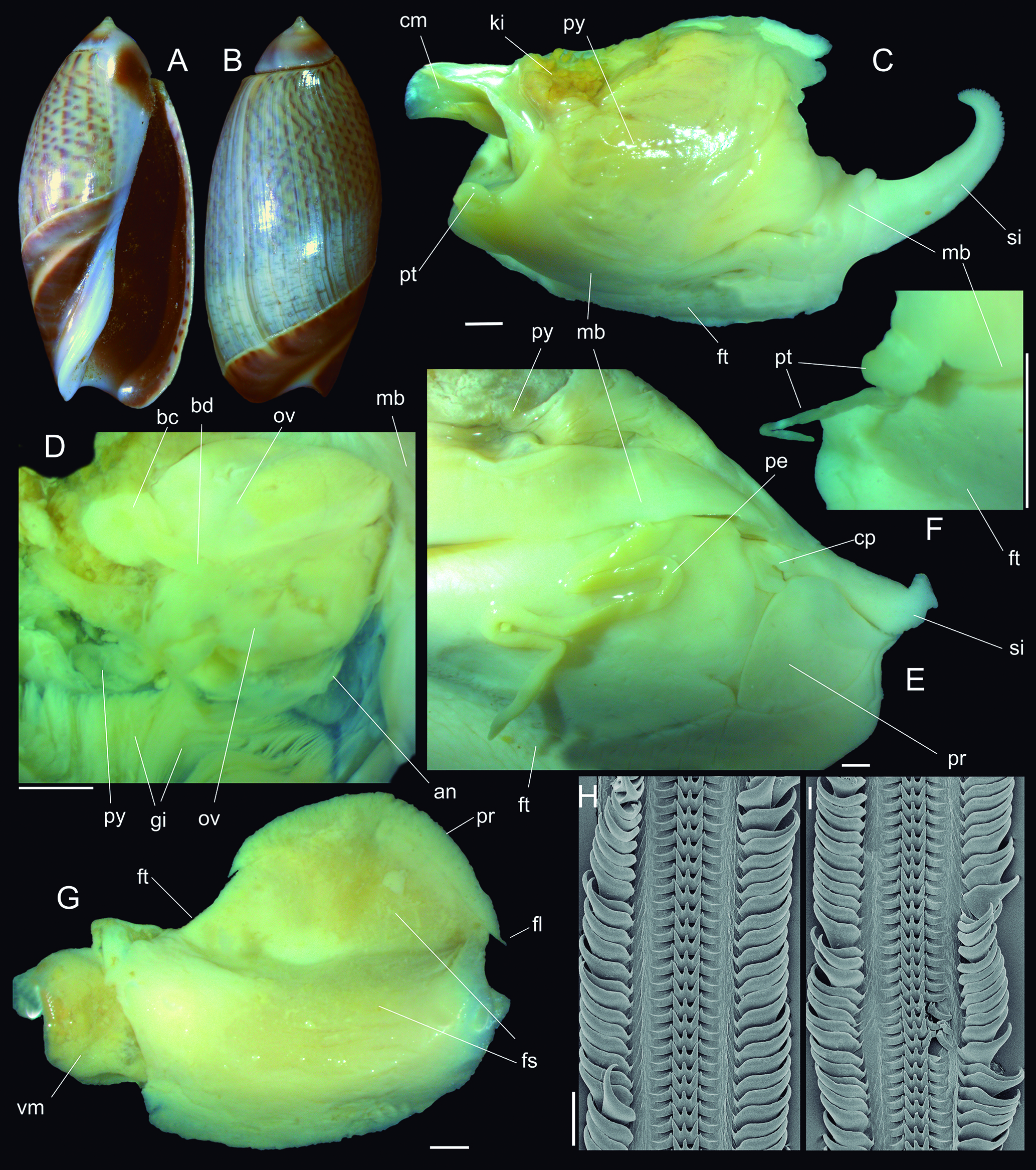
Agaronia sterica shell and anatomy of type specimens: (A-B) shell of paratype MZSP 73374#2 (L 31.0 mm), from Rio de Janeiro, frontal and dorsal views; (C) holotype head-foot and part of adjacent pallial region, dorsal-slightly right view; (D) same, right region of pallial cavity, ventral-inner view, specially showing pallial oviduct; (E) paratype MZSP 36498, anterior region of head-foot and adjacent pallial cavity, anterior-slightly right view, penis displaced from pallial cavity and deflected downwards; (F) same, detail of posterior region of mantel edge, showing pallial tentacle; (G) paratype MZSP 68586, entire specimen extracted from shell, ventral view, foot unfolded and expanded. Scales: = 2 mm. (H I) radulae in SEM, middle region (MZSP 69465), scale: = 100 µm. Lettering: an, anus; bc, bursa copulatrix; bd, bursa duct; cm, columellar muscle; cp, cephalic flaps; fl, foot lateral projections; fs, foot sole; ft, foot; gi, gill; ki, kidney; mb, mantle border; ov, pallial oviduct; pe, penis; pr, propodium; pt, pallial tentacle; py, pallial cavity, si, siphon; vm, visceral mass.
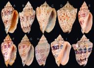
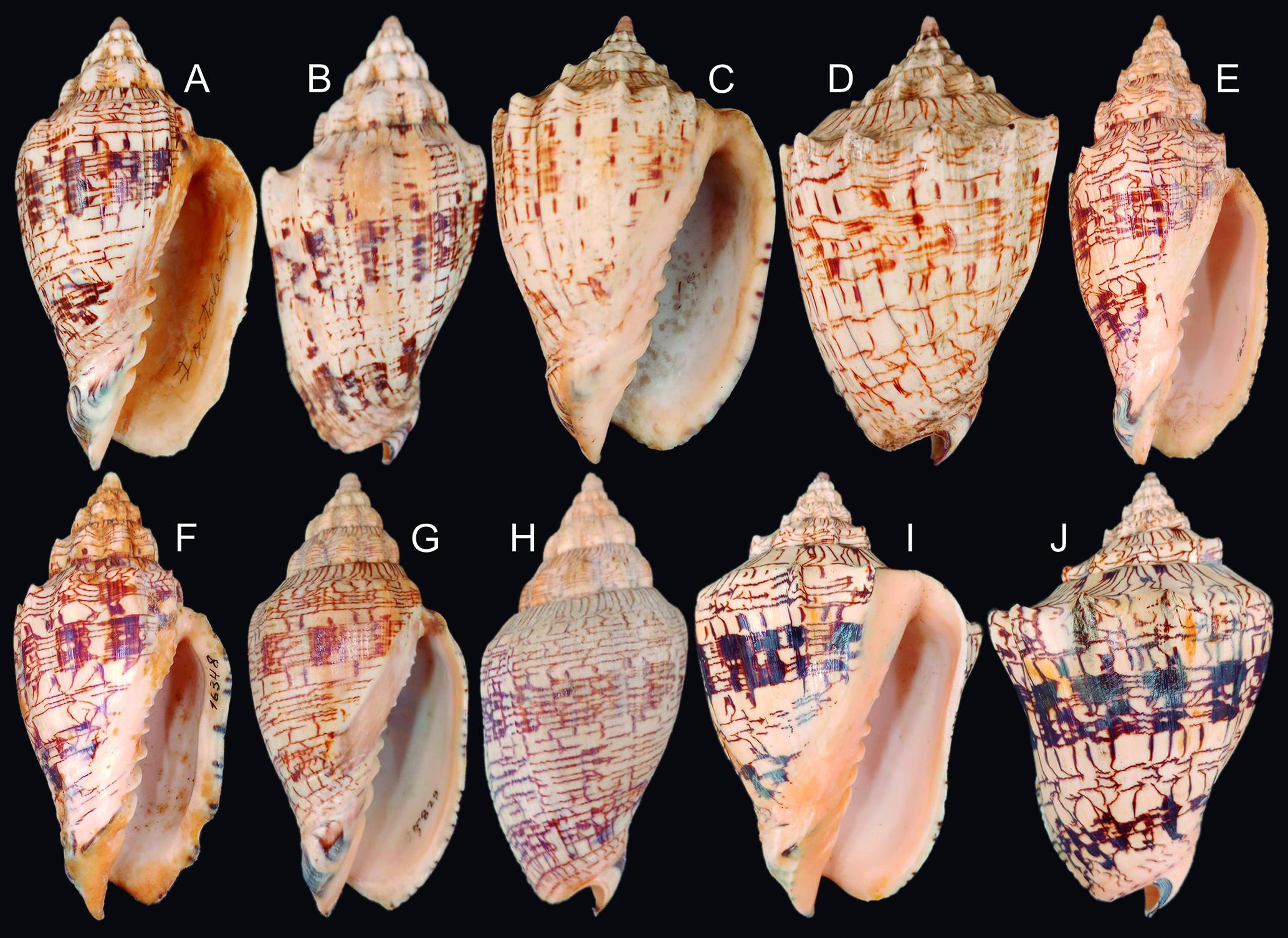
Voluta ebraea examples of shell variation in specimens from Piauí to Pernambuco (~700 km of Brazilian coast): (A-B) MZSP 61387 (L 134.2 mm), from Fortaleza, Ceará, frontal and dorsal views; (C-D) MZSP 51408 (L 120.2 mm), from Itamaracá, Pernambuco, frontal and dorsa views; (E) MZSP 16320 (L 167.8 mm), from Mundaú, Ceará, frontal view; (F) MZSP 16348 (L 144.9 mm), from Acaraú, Ceará, frontal view; (G-H) MZSP 35829 (L 118.5 mm), from Goiana, Pernambuco, frontal and dorsal views; (I-J) MZSP 159923 (L 181.4 mm), from Luis Correia, Piauí, frontal and dorsal views.
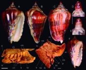
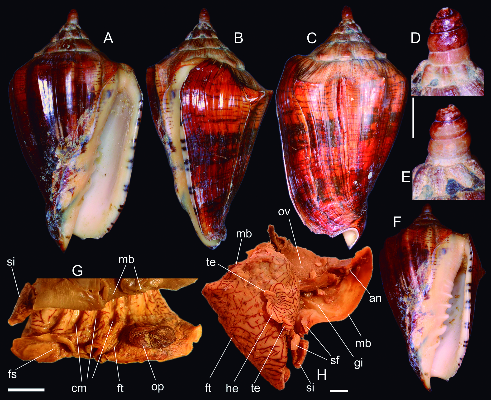
Voluta melodica holotype MZSP 135516 (L 116.9 mm): (A) shell, frontal view; (B) same, right view; (C) same, dorsal view; (D) detail of apex, dorsal view, scale: = 5 mm; (E) same, ventral view; (F) shell, frontal-slightly anterior view to show columellar folds; (G) extracted specimen. Detail of foot and adjacent region, right view, scale: = 10 mm; (H) same, head-foot in dorsal view, pallial cavity partially removed and deflected upwards, scale: = 10 mm. Lettering: an, anus; cm, marks of columellar folds; fs, foot sole; ft, foot; gi, gill; he, head flap; mb, mantle border; op, operculum; ov, pallial oviduct; sf, siphonal flaps; si, siphon; te, cephalic tentacle.
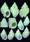
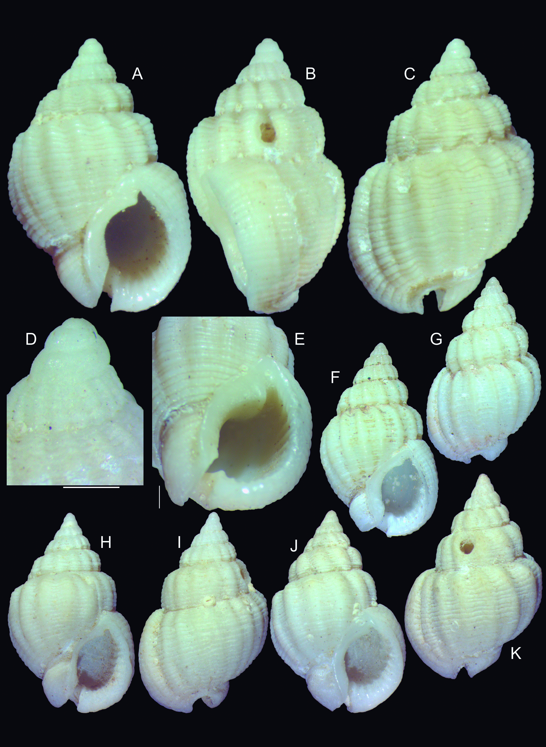
Phrontis rocas shell of types: (A-E) Holotype MZSP 166666 (L 10.5 mm); (A) frontal view; (B) right view; (C) dorsal view; (D) detail of apex, profile; (E) detail of peristome, frontal-slightly anterior view, both scales: = 1 mm; (F-G) paratype MZSP 166673#1 (L 10.6 mm), frontal and dorsal views; (H-I) paratype MZSP 166673#2 (L 11.6 mm), frontal and dorsal views; (J-K) paratype MZSP 62443#1 (L 12.1 mm), frontal and dorsal views.
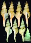
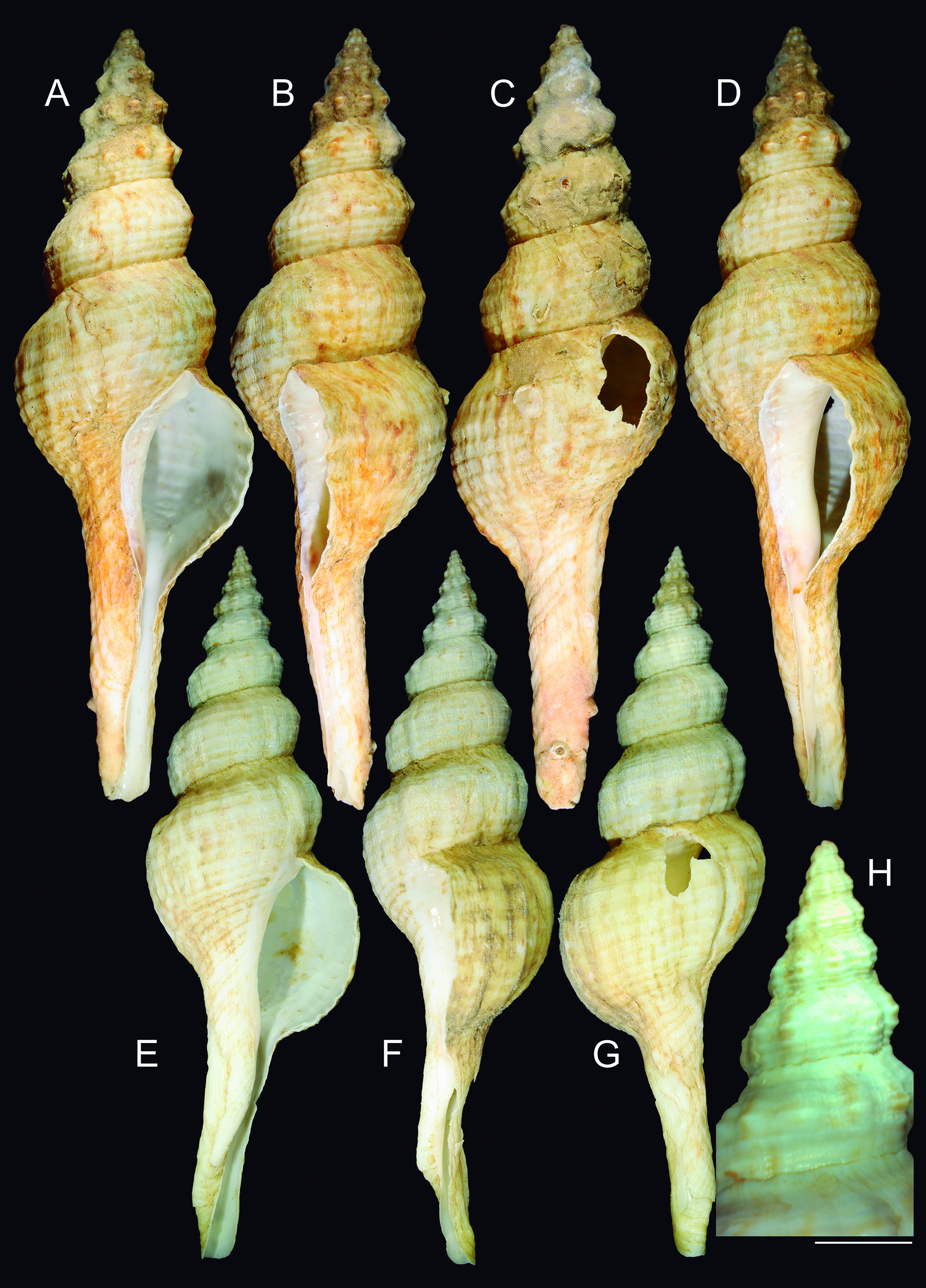
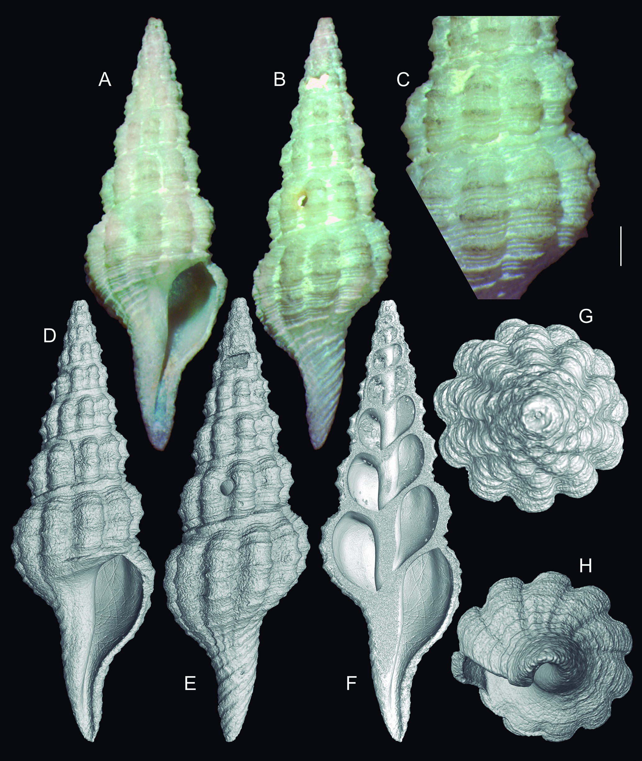
Goniofusus phoenix shells of types: (A-D) Holotype MNRJ 6259 (L 203.0 mm). (A) frontal view; (B) right view; (C) dorsal view; (D) right-slightly ventral view; (E-H) Paratype MNRJ 6258 (L 183.5 mm), (E) frontal view; (F) right view; (G) dorsal view; (H) detail of apical region, scale: = 5 mm.
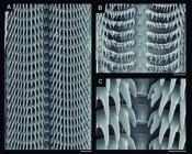
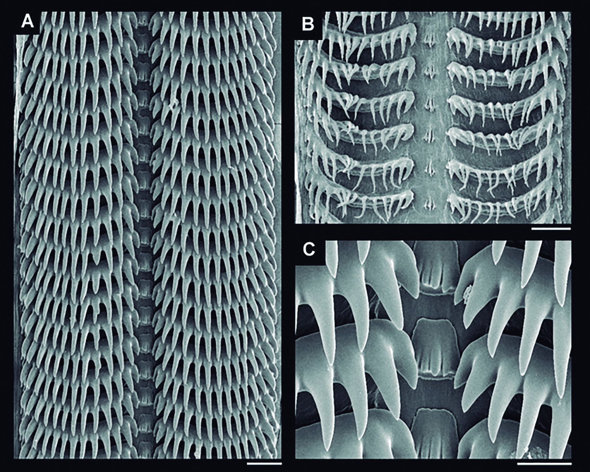
Goniofusus phoenix radula in SEM, Holotype MNRJ 6259: (A) wide view, scale: = 100 µm; (B) detail of posterior region, teeth in formation, scale: = 100 µm; (C) detail of central region, scale: = 20 µm.
Goniofusus phoenix anatomical drawings: (A) odontophore, ventral view, first layer of muscles and membranes removed, some muscles expanded, scale: = 1 mm; (B) mid-esophagus region, lateral view, salivary glands deflected, scale: = 5 mm; (C) central nervous system, dorsal view, scale: = 1 mm.
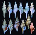
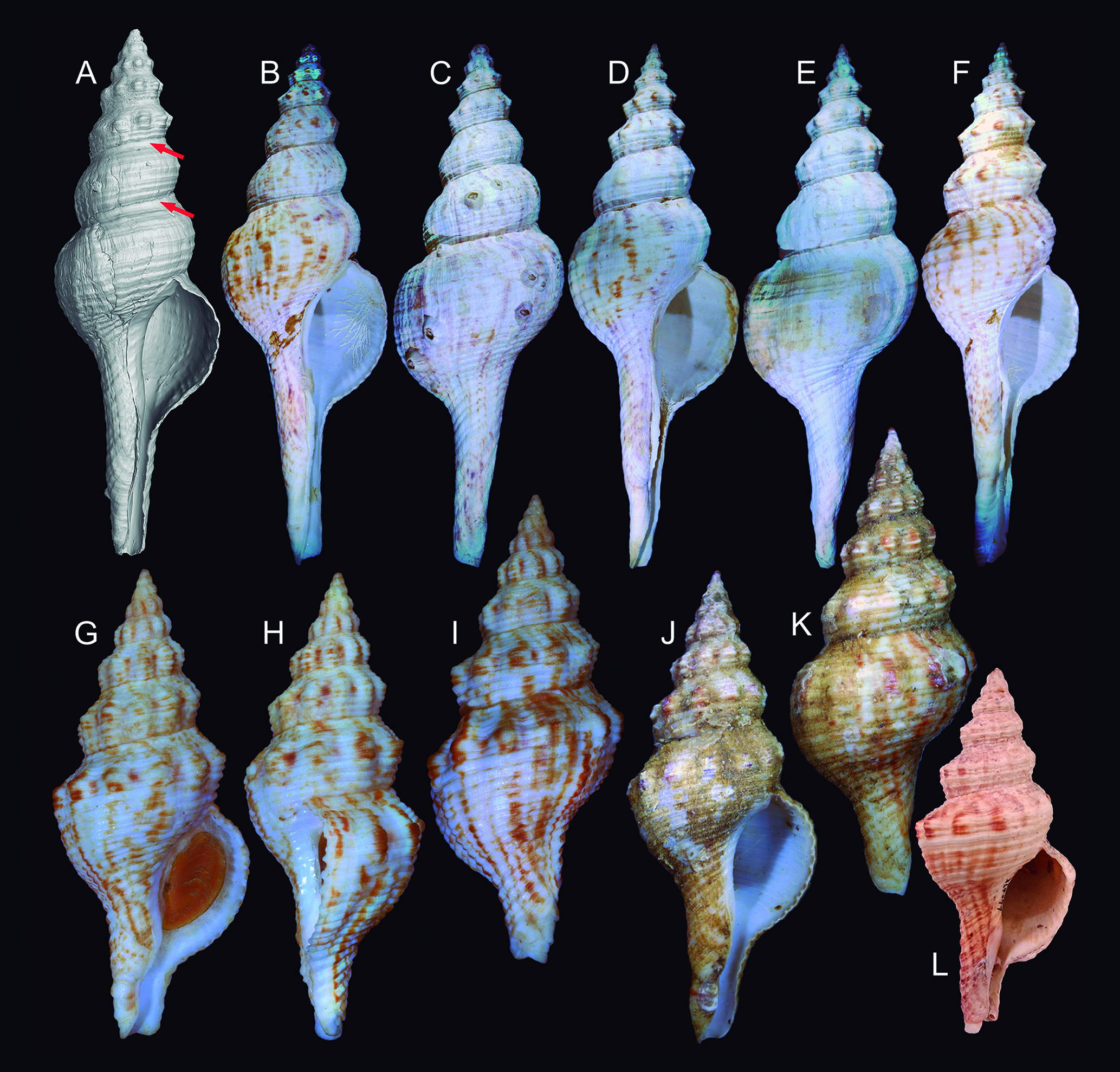
Shells of Goniofusus phoenix and G. damasoi: (A-F) G. phoenix; (A) Holotype MNRJ 6259, shell image in tomography (L 203.0 mm), arrows indicating subsutural groove; (B-F) paratypes MZSP 123143 from off Niterói, Rio de Janeiro; (B-C) #1, frontal and dorsal views (L 183.7 mm); (D-E) #2, frontal and dorsal views (L 185.0 mm); (F) #3, frontal view (L 161.9 mm); (G-L) G. damasoi; (G-I) Holotype MZSP 131311, frontal, right and dorsal views (L 67.6 mm); (J-K) specimen MZSP 130372, from São Paulo, frontal and dorsal views (L 76.4 mm); (L) shell USNM 364099 from Paquetá, Rio de Janeiro (L 76 mm).
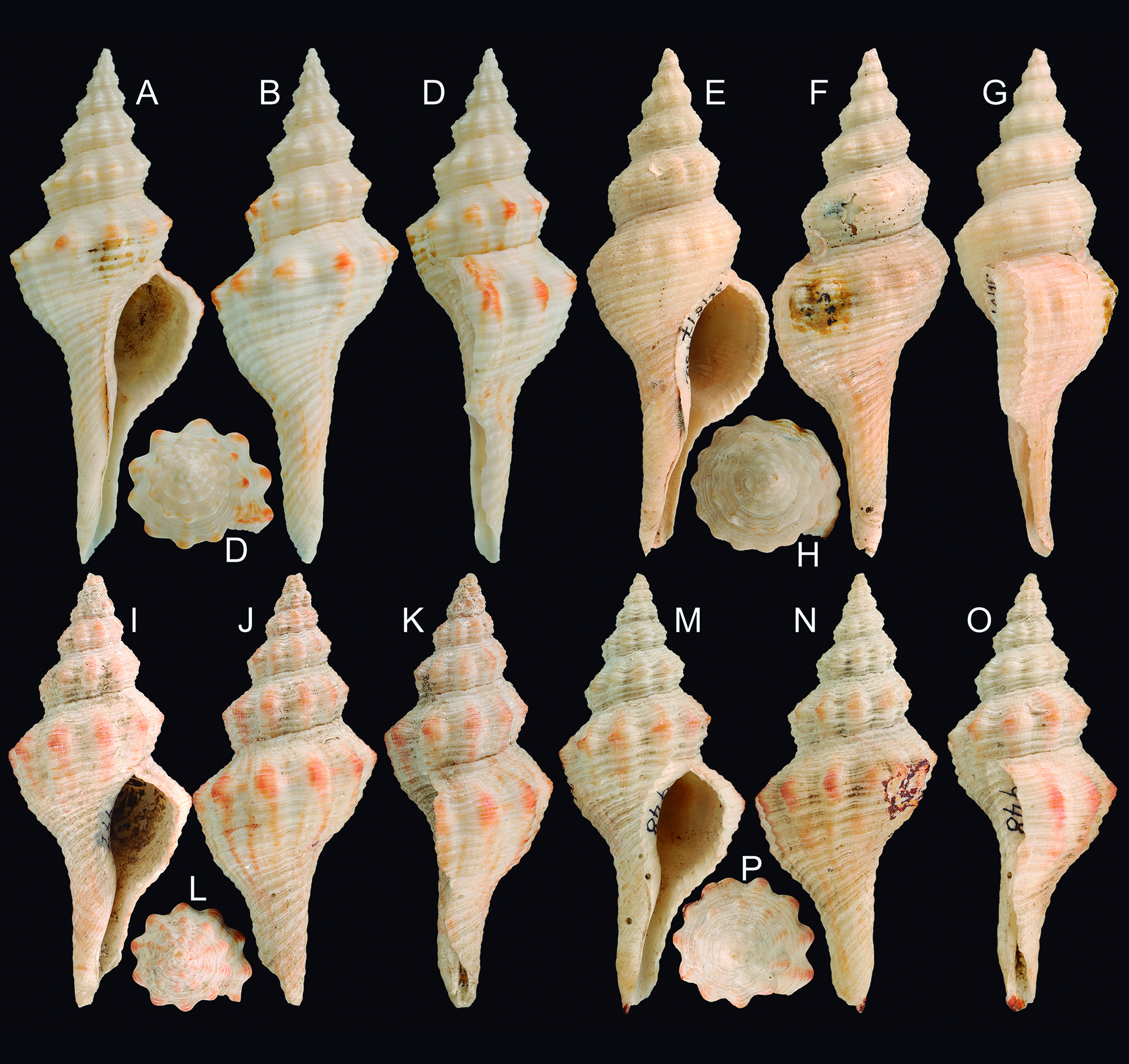
Goniofusus brasiliensis shells of MCZ types: (A-D) Holotype, MCZ 945 (L 80.2 mm), frontal, dorsal, right and apical views; (E-H) same for paratype MCZ 945a (L 80.0 mm); (I-L) same for paratype MCZ 947 (L 59.5 mm); (M-P) same for paratype MCZ 948 (L 50.9 mm) (all courtesy MCZ team, check acknowledgments).
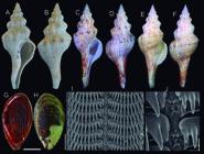
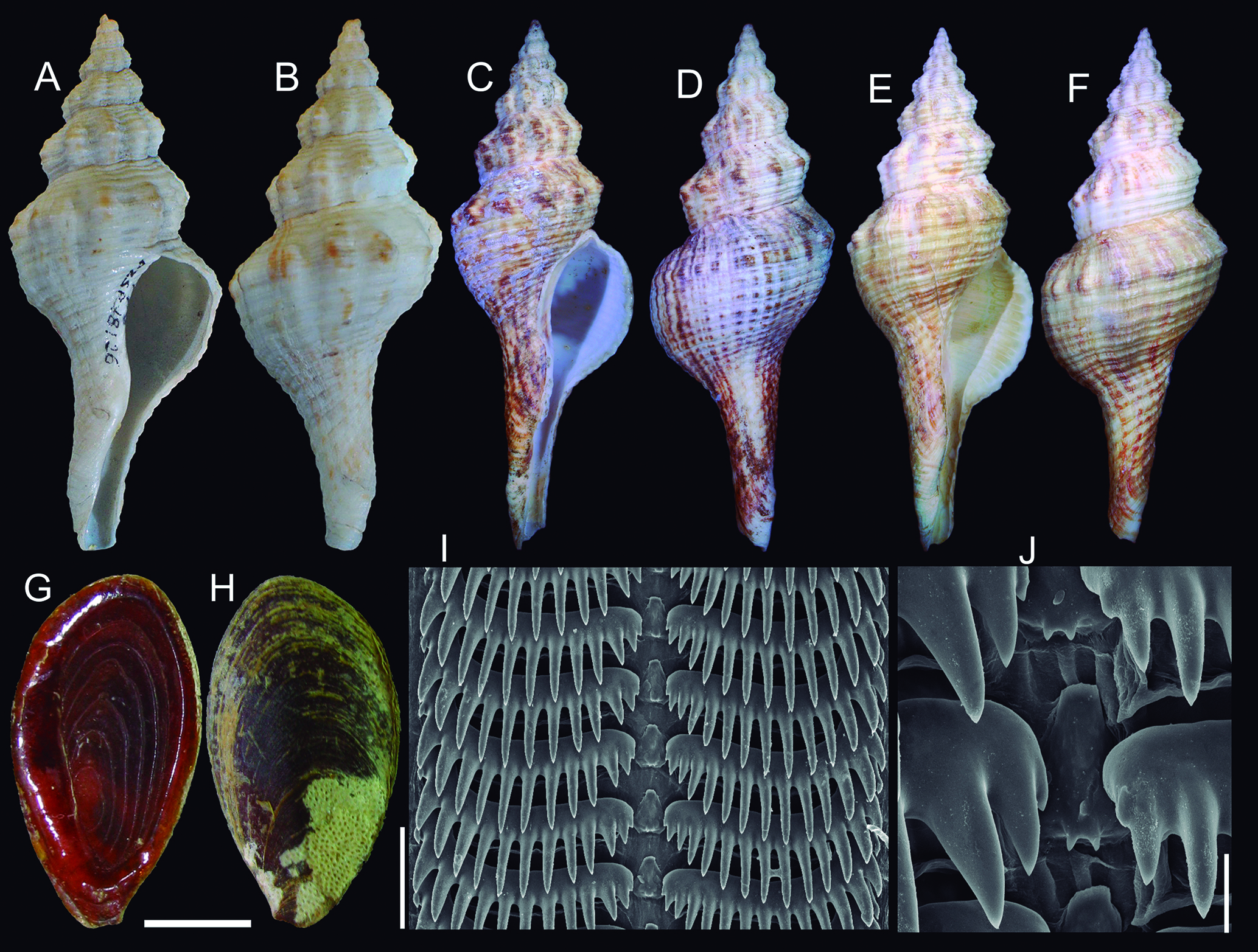
Goniofusus brasiliensis shells and other hard parts: (A) paratype MZSP 18126, frontal view (L 60.7 mm); (B) same, dorsal view; (C) shell MZSP 152767, from Santa Catarina (L 97.6 mm); (D) same, dorsal view; (E) shell MZSP 140957, from Rio de Janeiro, frontal view (L 88.1 mm); (F) same, dorsal view; (G-H) operculum, inner and outer views, scale: = 5 mm; (I) radula in SEM, panoramic view, scale: = 200 µm; (J) same, detail of central region, scale: = 20 µm.
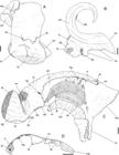
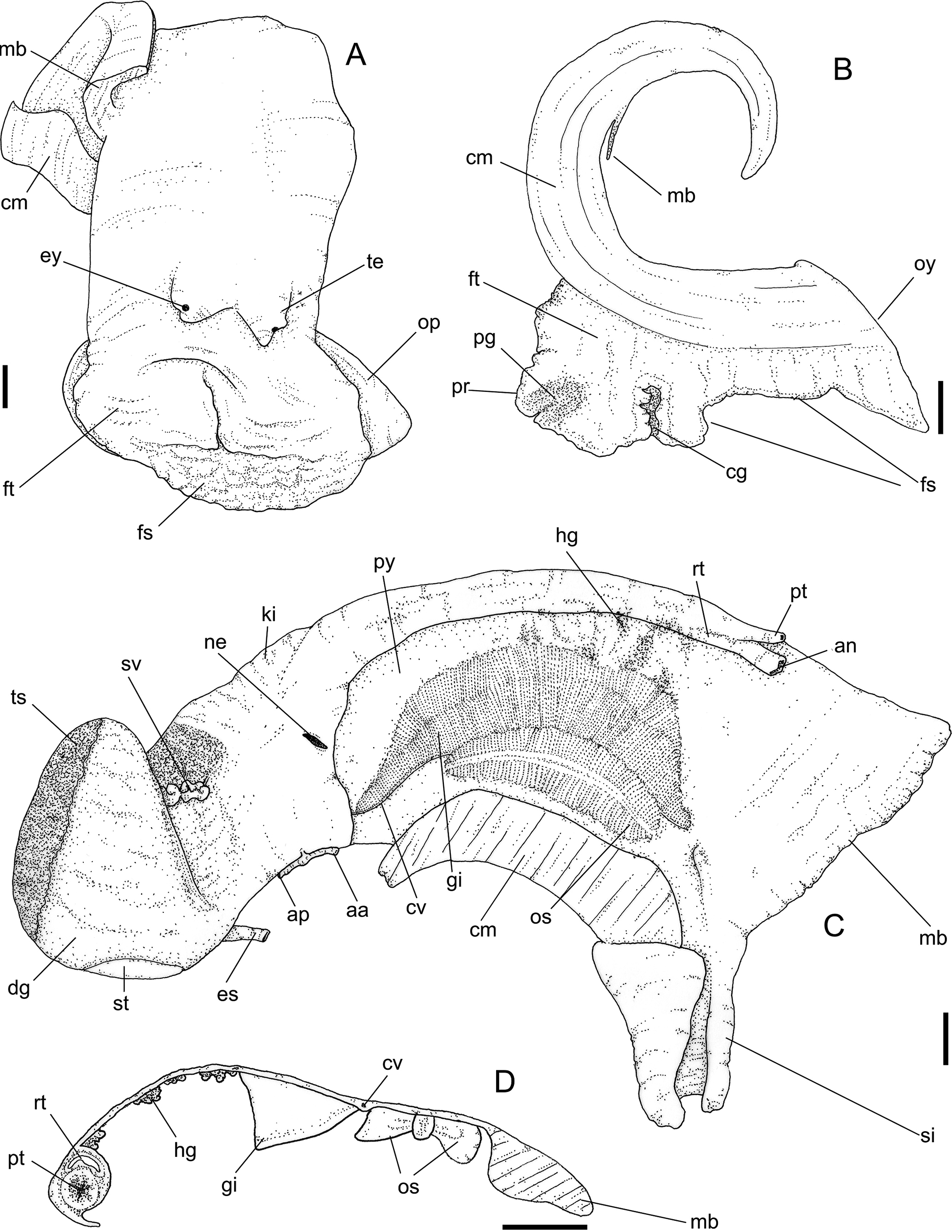
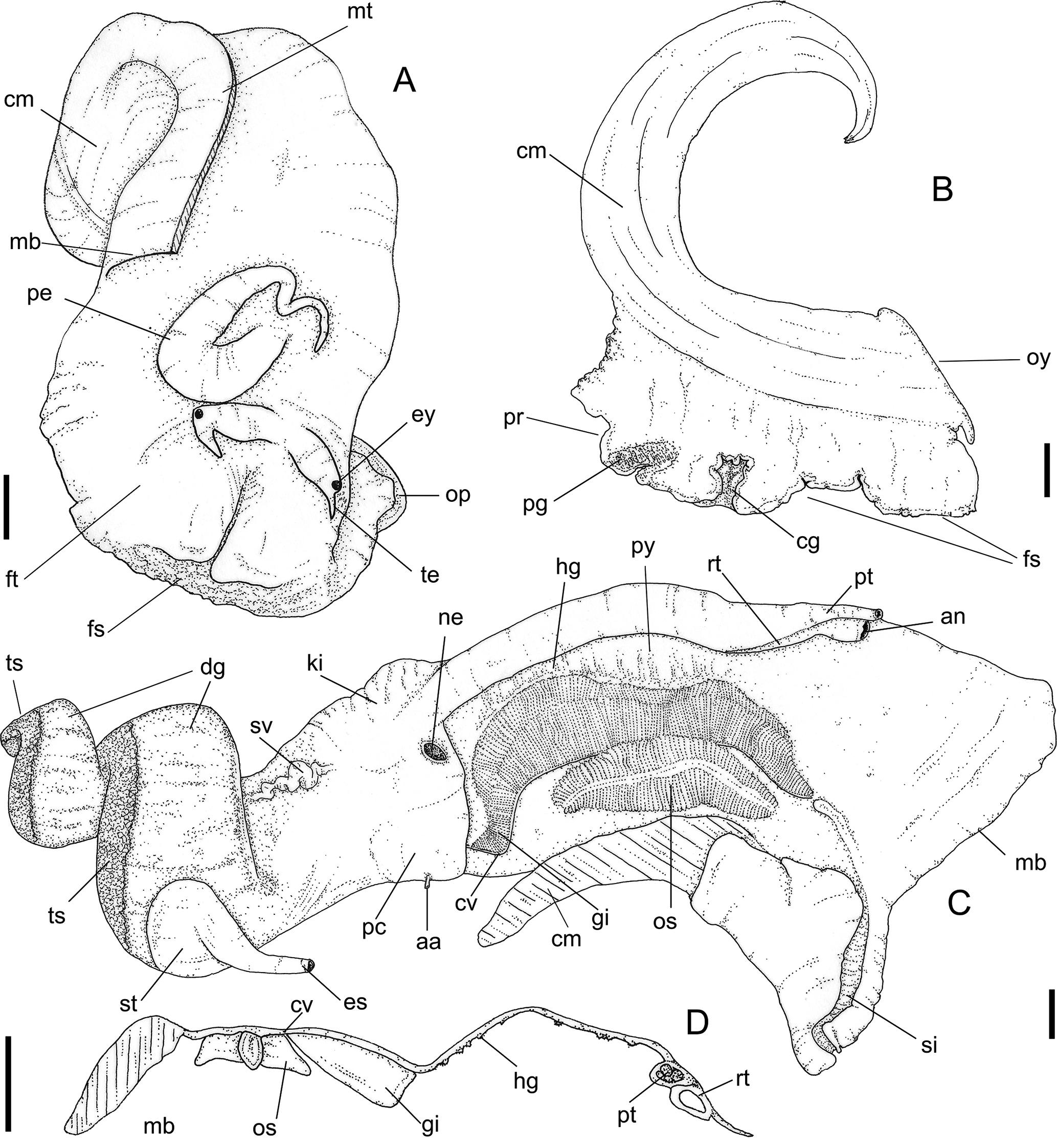
Goniofusus brasiliensis anatomical drawings: (A) head-foot, female, frontal view; (B) foot and columellar muscle, female, longitudinal section along medial line; (C) pallial cavity and partially uncoiled visceral mass, male, mostly ventral view; (D) transverse section of pallial roof in middle level of osphradium, male. Scales: = 2 mm (courtesy Diogo Couto, published under permission). Lettering: aa, anterior aorta; an, anus; ap, posterior aorta; cg, cement gland; cm, columellar muscle; cv, ctenidial vein; dg, digestive gland; es, esophagus; ey, eye; fs, foot sole; ft, foot; gi, gill; hg, hypobranchial gland; ki, kidney; mb, mantle border; ne, nephrostome; op, operculum; os, osphradium; oy, opercular pad; pg, pedal gland; pr, propodium; pt, prostate; py, pallial cavity; rt, rectum; si, siphon; st, stomach; sv, seminal vesicle; te, cephalic tentacle; ts, testis.
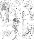
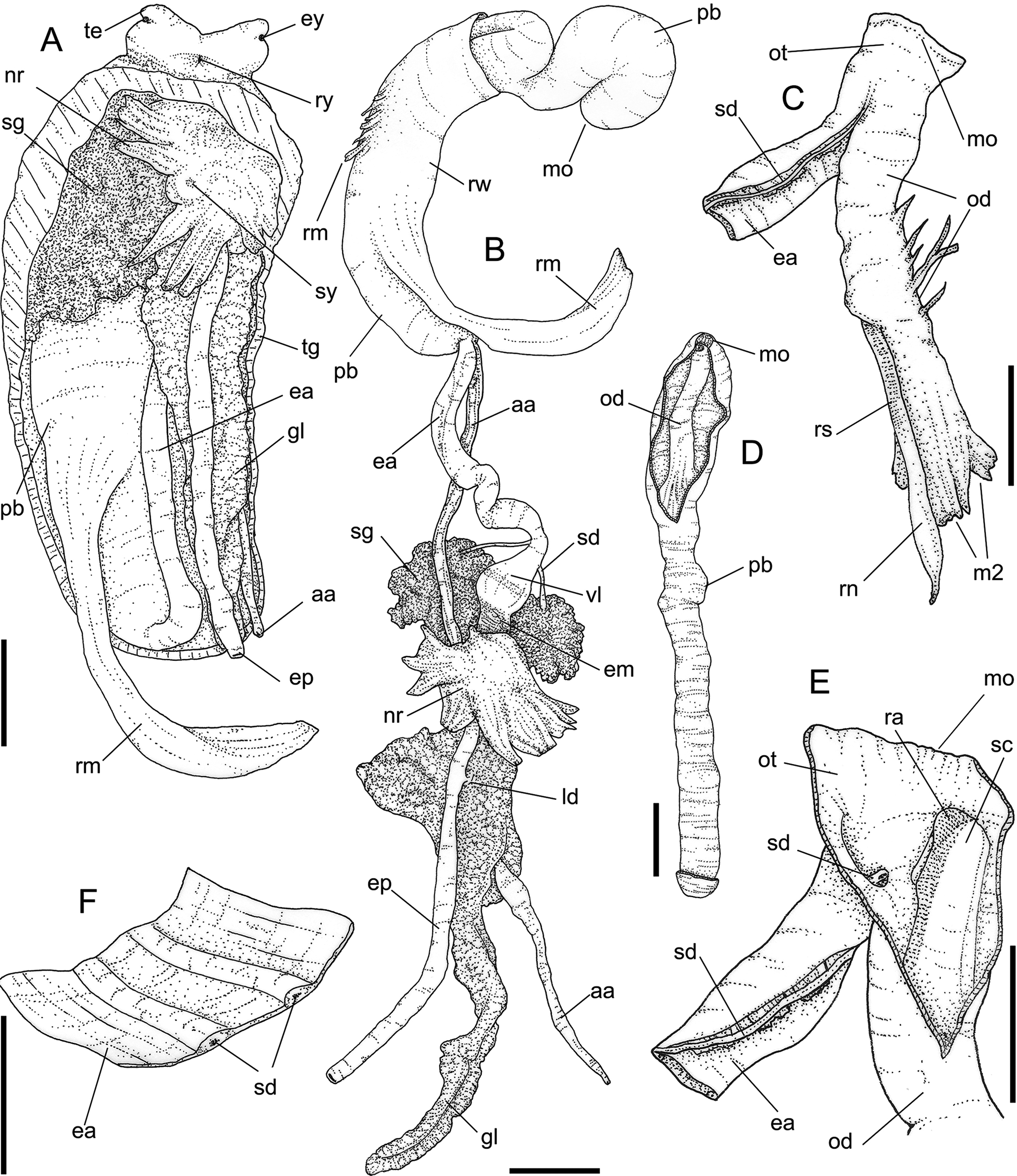
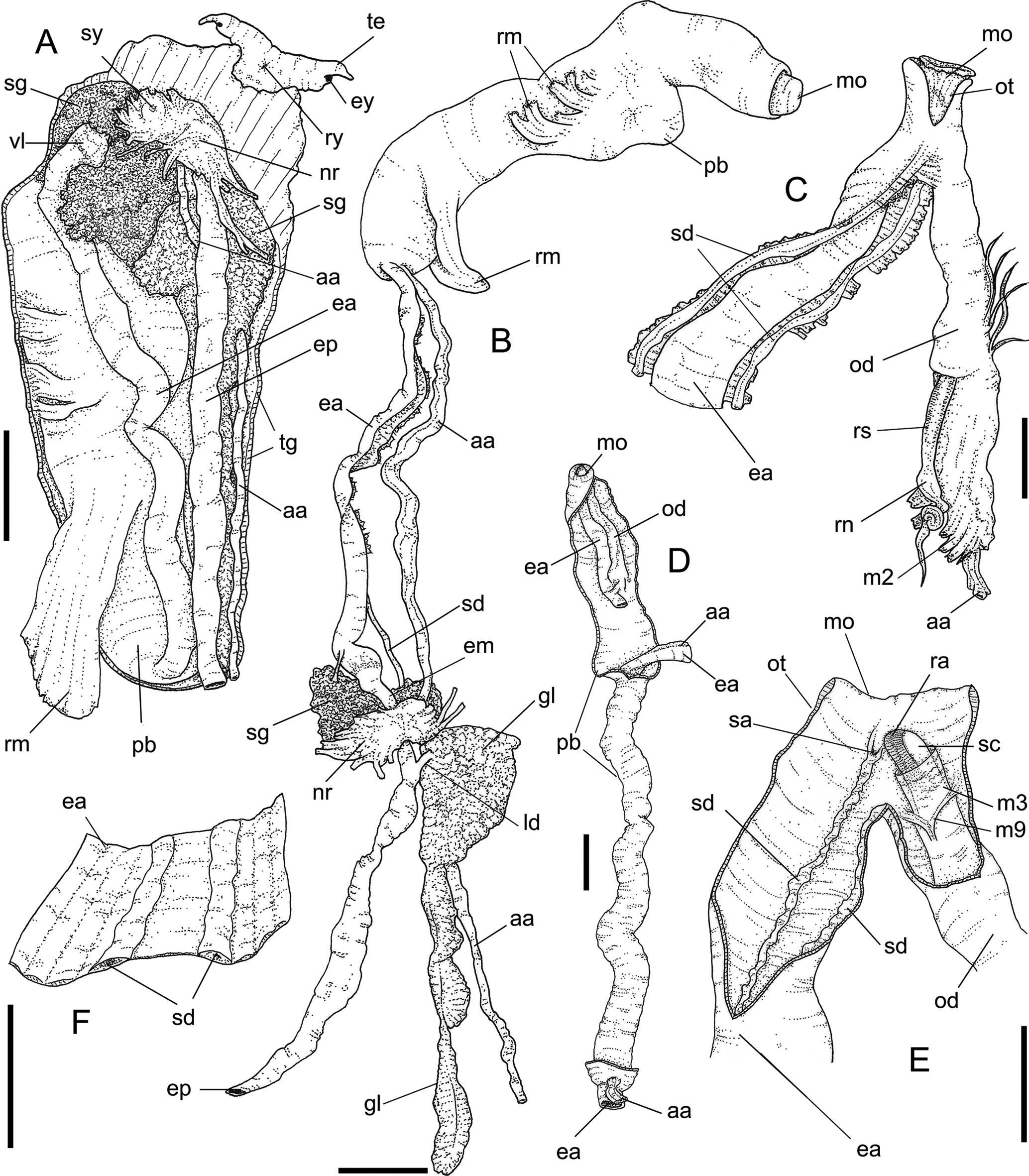
Goniofusus brasiliensis anatomical drawings: (A) Head, ventral view, foot and columellar muscle removed, scale: = 5 mm; (B) foregut uncoiled, mostly ventral view, scale: = 5 mm; (C) buccal mass, right view, esophagus deflected, scale: = 2 mm; (D) distal end of proboscis, dorsal view, distal region opened longitudinally, scale: = 2 mm; (E) buccal mass, right view, opened along right wall, scale: = 2 mm; (F) detail of opened dorsal wall of esophagus, scale: = 1 mm (courtesy Diogo Couto, published under permission). Lettering: aa, anterior aorta; ea, anterior esophagus; em, mid esophagus; ep, posterior esophagus ey, eye; gl, gland of Leiblein; ld, duct of gland of Leiblein; mo, mouth; nr, nerve ring; od, odontophore; ot, oral tube; pb, proboscis; ra, radula; rm, proboscis retractor muscles; rw, rhynchodeal wall; ry, rhynchostome; sc, subradular cartilage; sd, salivary duct; sg, salivary gland; sy, statocyst; te, tentacle; tg, integument; vl, valve of Leiblein.
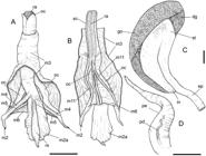
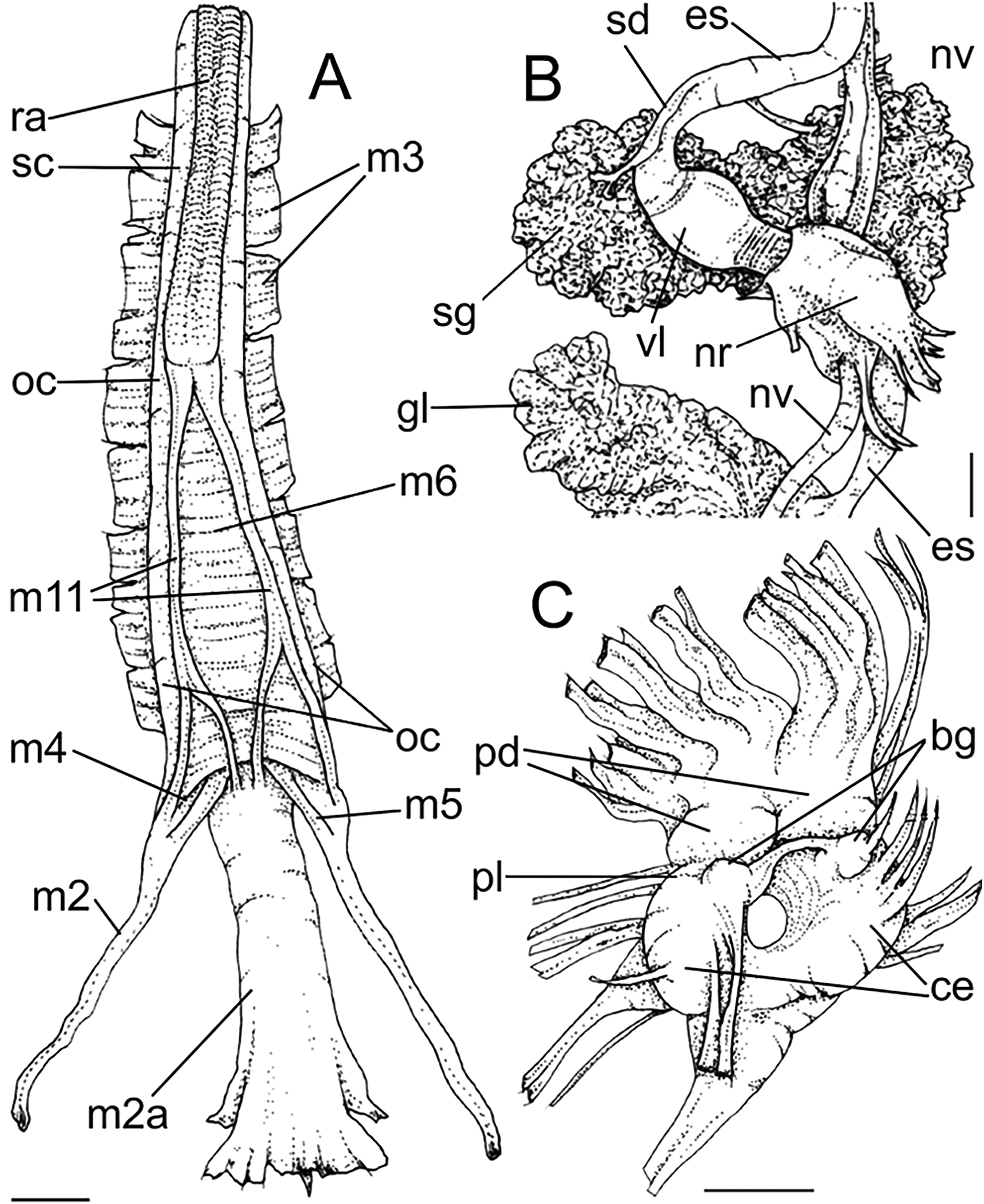
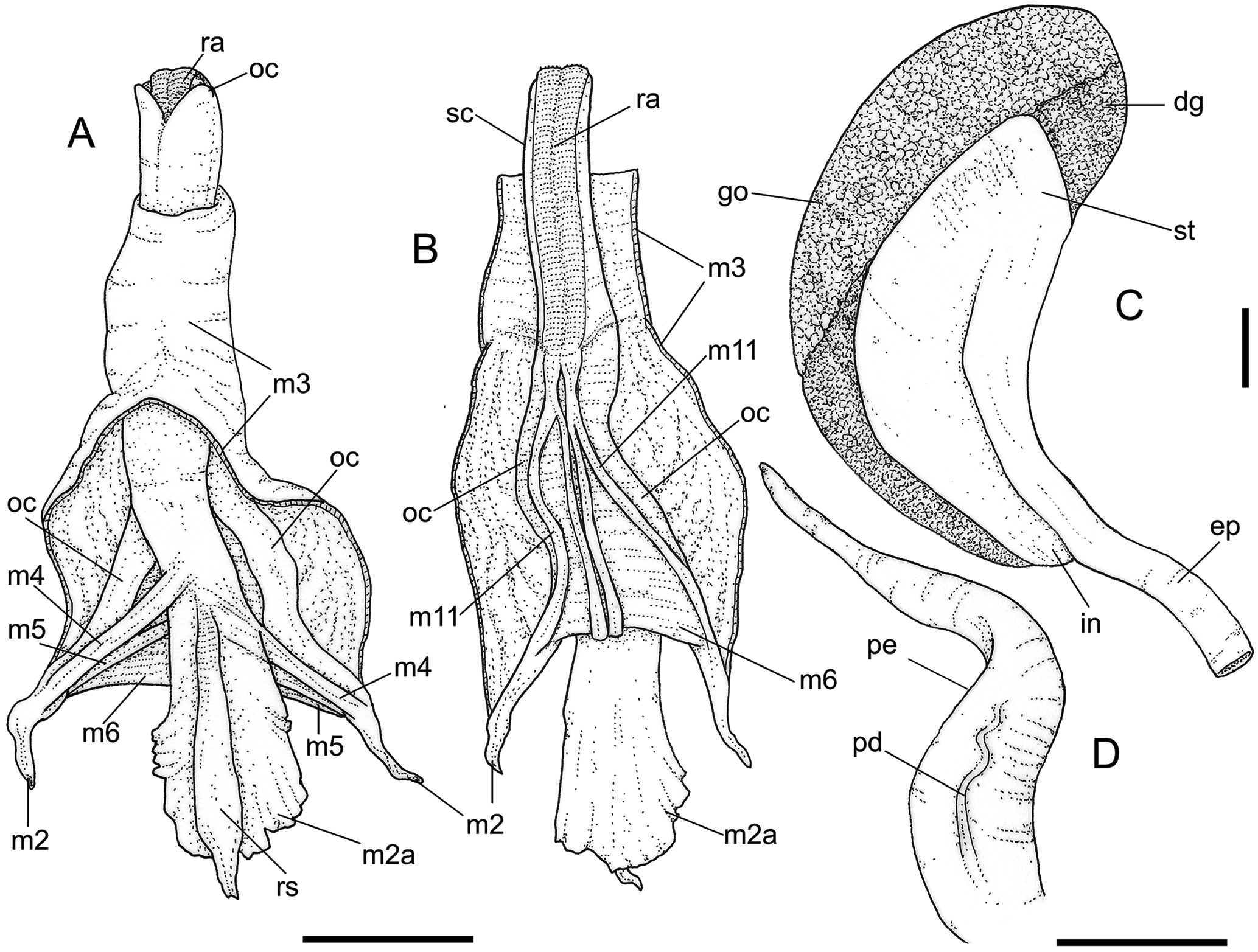
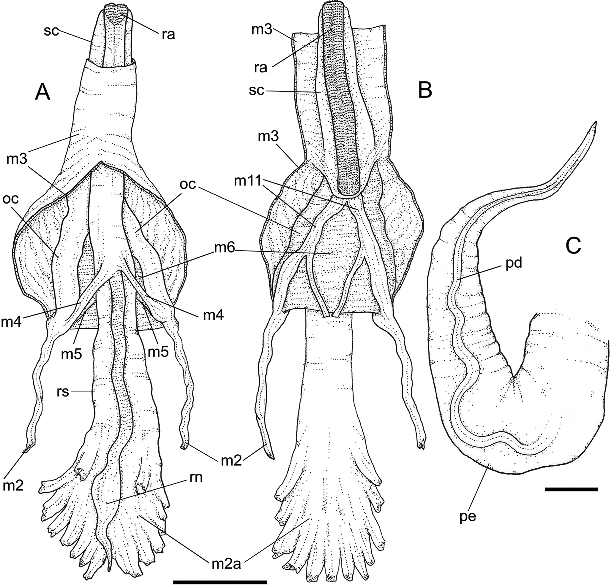
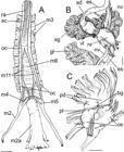
Goniofusus brasiliensis anatomical drawings: (A) Odontophore, dorsal view, posterior region opened longitudinally, with muscles expanded; (B) same, ventral view, superficial layer longitudinally sectioned; (C) stomach region, dorsal view. (D) penis, dorsal view. Scales: = 5 mm (courtesy Diogo Couto, published under permission). Lettering: dg, digestive gland; ep, posterior esophagus; go, gonad; in, intestine; m2 m11, intrinsic and extrinsic odontophore muscles; oc, odontophore cartilages; pe, penis; pd, penis duct; ra, radula; rs, radular sac; sc, subradular cartilage; st, stomach.
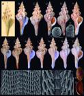
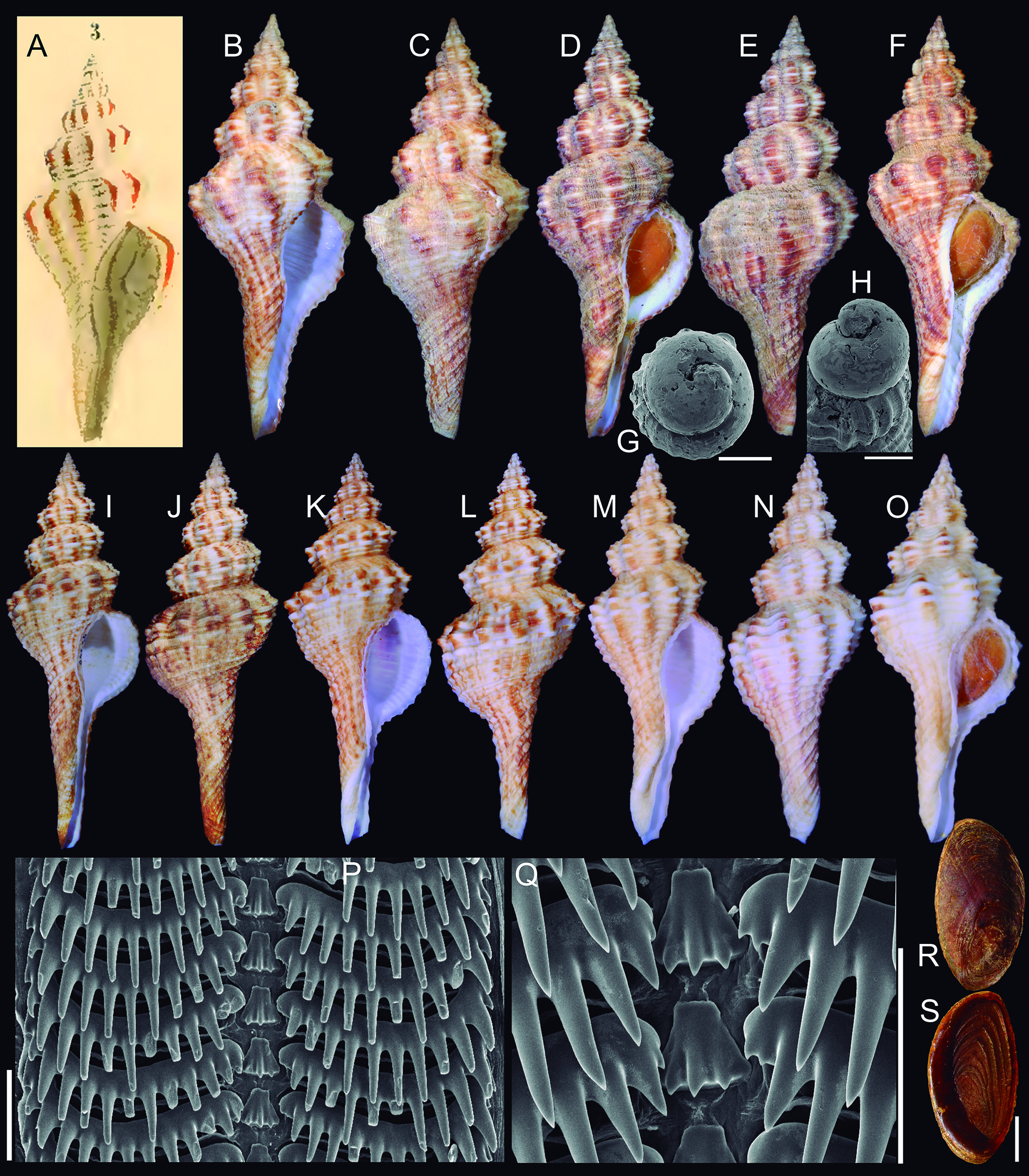
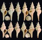
Goniofusus strigatus Shells and other hard parts: (A) Illustration by Philippi (1850: pl. 5, fig. 3), representing his Fusus strigatus. (B-F) 3 shells of G. strigatus MZSP 131745, from Bahia; (B) shell #1 similar to Philippi’s (1850) illustration, the carinate form, frontal view (L 69.4 mm); (C) same, dorsal view; (D) shell #2, the rounded form, frontal view (L 72.7 mm); (E) same, dorsal view; (F) shell #3, intermediary form, frontal view (L 64.1 mm); (G-H) apex in SEM, apical and profile, scale: = 1 mm; (I J) MZSP 55465, largest examined shell from Bahia (L 119.6 mm), frontal and dorsal views; (K-L) MZSP 159290, from Bahia (L 95.3 mm), frontal and dorsal views; (M-N) MZSP 71779, from Espírito Santo (L 77.4 mm), frontal and dorsal views; (O) MZSP 72753, from Rio de Janeiro (L 55.7 mm), frontal view. (P) radula in SEM, panoramic view, scale: = 200 µm; (Q) same, detail of central region; (R-S) operculum, outer and inner views, scale: = 5 mm.
Goniofusus strigatus anatomical drawings: (A) head-foot, male, frontal view; (B) foot and columellar muscle, female, longitudinal section along medial line; (C) pallial cavity and partially uncoiled visceral mass, male, mostly ventral view; (D) transverse section of pallial roof in middle level of osphradium, male. Scales: = 2 mm (courtesy Diogo Couto, published under permission). Lettering: aa, anterior aorta; an, anus; cg, cement gland; cm, columellar muscle; cv, ctenidial vein; dg, digestive gland; es, esophagus; ey, eye; fs, foot sole; ft, foot; gi, gill; hg, hypobranchial gland; ki, kidney; mb, mantle border; mt, mantle; ne, nephrostome; op, operculum; os, osphradium; oy, opercular pad; pe, penis; pg, pedal gland; pr, propodium; pt, prostate; py, pallial cavity; rt, rectum; si, siphon; st, stomach; sv, seminal vesicle; te, cephalic tentacle; ts, testis.
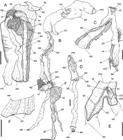
Goniofusus strigatus anatomical drawings: (A) Head, ventral view, foot and columellar muscle removed, scale: = 5 mm; (B) foregut uncoiled, mostly ventral view, scale: = 5 mm; (C) buccal mass, right view, esophagus deflected, scale: = 2 mm; (D) entire proboscis, dorsal view, distal region opened longitudinally, scale: = 2 mm; (E) buccal mass, right view, opened along right wall, Scale: = 2 mm; (F) detail of opened dorsal wall of esophagus, scale: = 1 mm (courtesy Diogo Couto, published under permission). Lettering: aa, anterior aorta; ea, anterior esophagus; em, mid esophagus; ep, posterior esophagus ey, eye; gl, gland of Leiblein; ld, duct of gland of Leiblein; m2, buccal mas retractor muscles; m3 m9, intrinsic odontophore muscles; mo, mouth; nr, nerve ring; od, odontophore; ot, oral tube; pb, proboscis; ra, radula; rm, proboscis retractor muscles; rw, rhynchodeal wall; ry, rhynchostome; sa, salivary aperture; sc, subradular cartilage; sd, salivary duct; sg, salivary gland; sy, statocyst; te, tentacle; tg, integument; vl, valve of Leiblein.
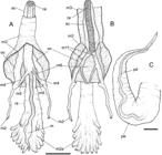
Goniofusus strigatus anatomical drawings: (A) Odontophore, dorsal view, superficial layer of muscles opened longitudinally, muscles expanded; (B) same, ventral view; (C) penis, dorsal view. Scales: = 2 mm (courtesy Diogo Couto, published under permission). Lettering: m2 m11, intrinsic and extrinsic odontophore muscles; oc, odontophore cartilages; pe, penis; pd, penis duct; ra, radula; rn, radular nucleus; rs, radular sac; sc, subradular cartilage.
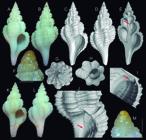
Shell of holotype MNHN of Dolicholatirus etherius (L 17.8 mm): (A-C) lightly photo; (A) frontal view; (B) dorsal view; (C) detail of sculpture of 3 last whorls, scale: = 1 mm.; (C H) tomography images (courtesy Daniel C. Cavallari); (D) frontal view; (E) dorsal view; (F) digital section in half, frontal view; (G) apical view; (H) anterior view.
Lightbournus rendatus type shells, photos and tomography images: (A-J) holotype MNHN (L 22.9 mm); (A) photo, frontal view; (B) same, dorsal view; (C) tomography, whole frontal view; (D) same, dorsal view; (E) digital section in half, frontal view, arrow shoring inferior furrow; (F) photo, detail of protoconch, profile, scale: = 0.5 mm; (G) tomography, apical view; (H) same, anterior view; (I) same, detail of superior region of aperture, frontal-slightly inferior view; (J) same, detail of inner lip, arrow showing inner lip folds; (K) paratype MZSP 105535, photo, frontal view (L 20.3 mm); (L) same, dorsal view; (M) same, detail of apex, profile, scale: = 0.5 mm.
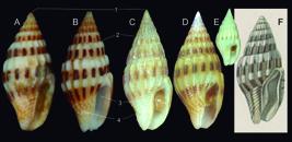
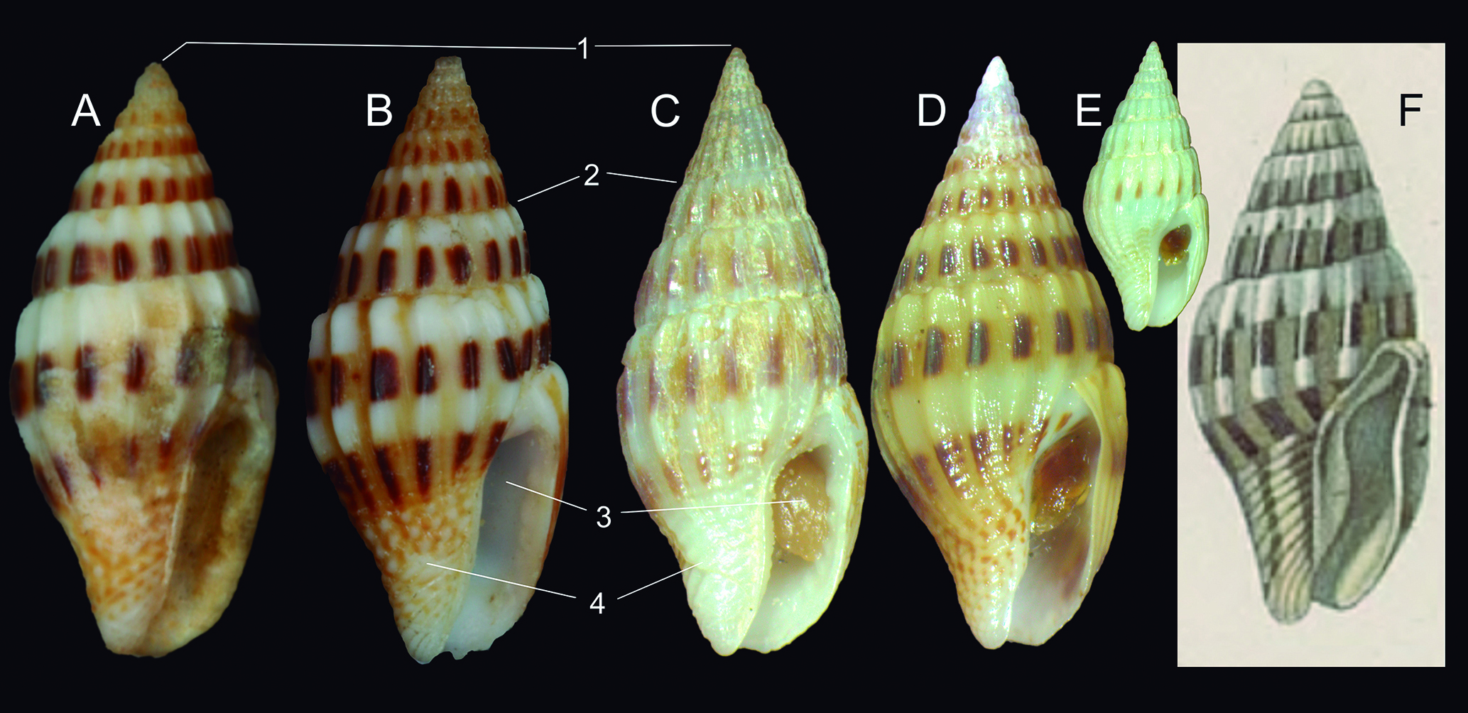
Anachis lyrata and A. veleda shells, frontal views: (A) L. lyrata syntype BMNH 1966320#1 (L 18.8 mm); (B) same spm#2 (L 19.0 mm); (C-E) typical Brazilian L. veleda; (C) MZSP 91070, from Guarapari, Espírito Santo (L 14.6 mm); (D) MZSP 107619, from São Sebastião, São Paulo (L 16.8 mm); (E) topotype MZSP 31255, from Boa Viagem Beach, Recife, Pernambuco (L 13.4 mm); (F) Replication of Duclos (1846, pl. 7, fig. 20), designed by Radwin (1977: 120) as lectopype of A. veleda.
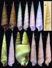
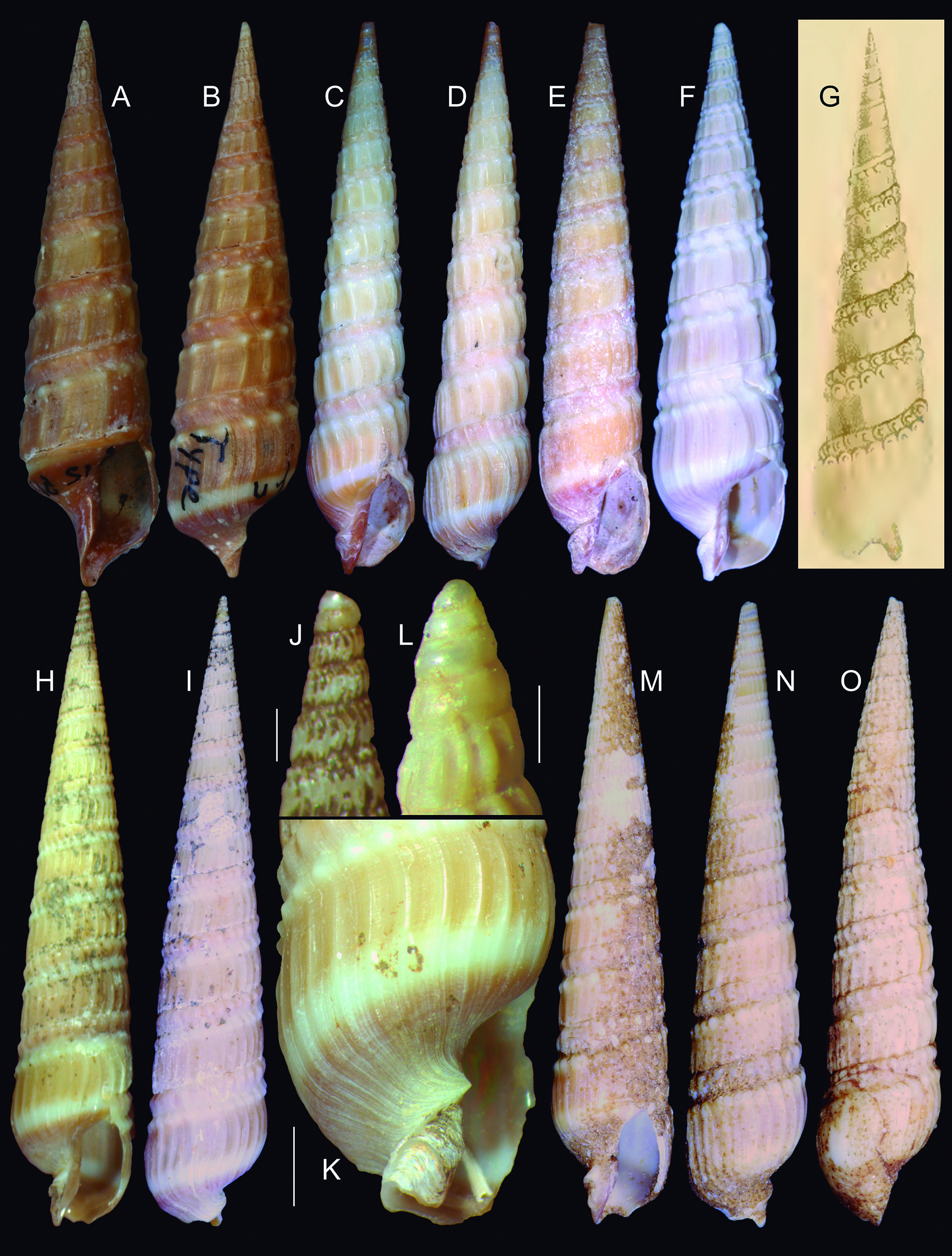
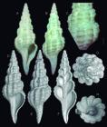
Terebra gemmulata and T. joculosa shell characters: (A-G) T. gemmulata; (A-B) lectotype GMNH 1152/76 (L 39.0 mm), frontal and dorsal views; (C-D) MZSP 10718#1 from Argentina (L 70.0 mm), frontal and dorsal views; (E) MZSP 10718#2 (L 56.2 mm); (F) MZSP 152322 from Espírito Santo, Brazil (L 41.1 mm); (G) Terebra patagonica, replication of d’Orbigny (1839) pl. 62, fig. 1; (H-O) Terebra joculosa shell of some types; (H-I) Holotype MZSP 166481 (L 37.8 mm), frontal and dorsal views; (J) same, detail of apex, profile, scale: = 0.5 mm; (K) same, detail of last whorl, right view, scale: = 2 mm; (L) Paratype 82756#5, young shell, detail of complete apex, profile, scale: = 0.5 mm; (M-N) paratype 166479 (L 45.4 mm), from Rio Grande do Sul, Brazil; (O) paratype MZSP 20569, from São Paulo, Brazil (L 45.5 mm).
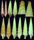
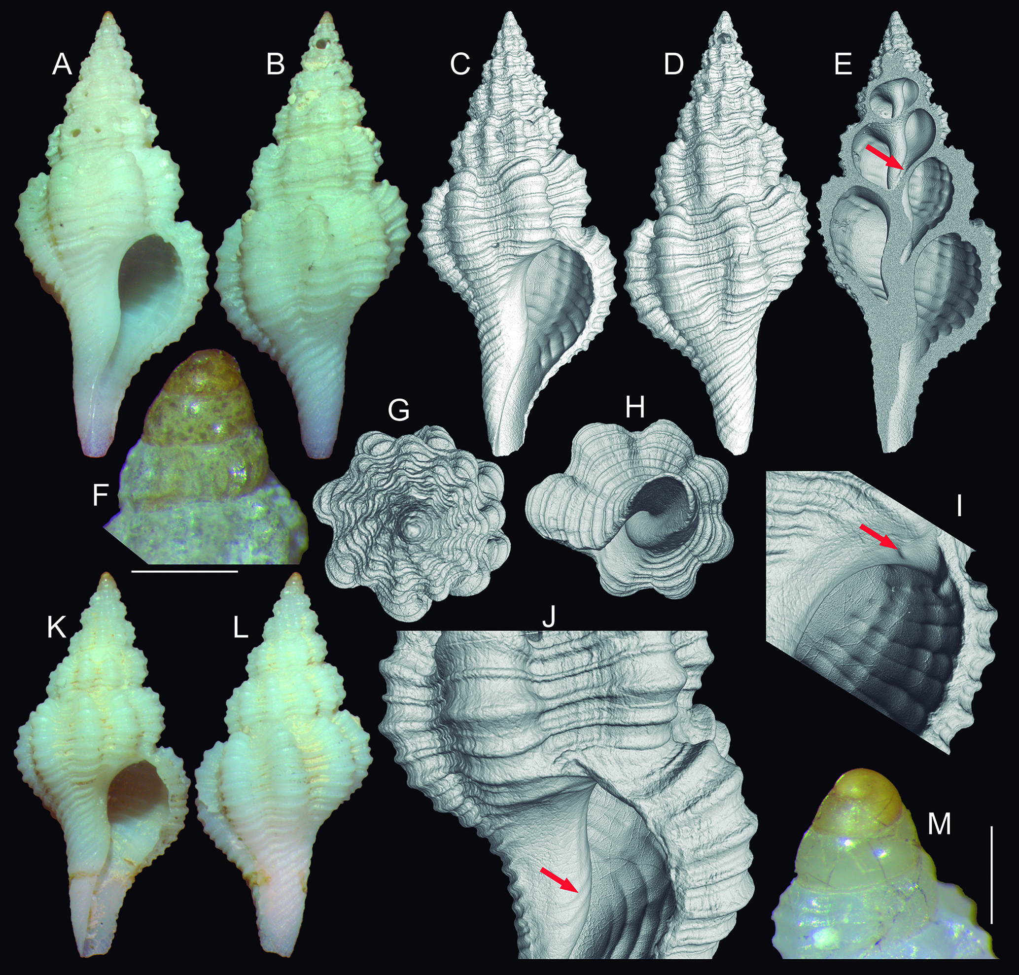
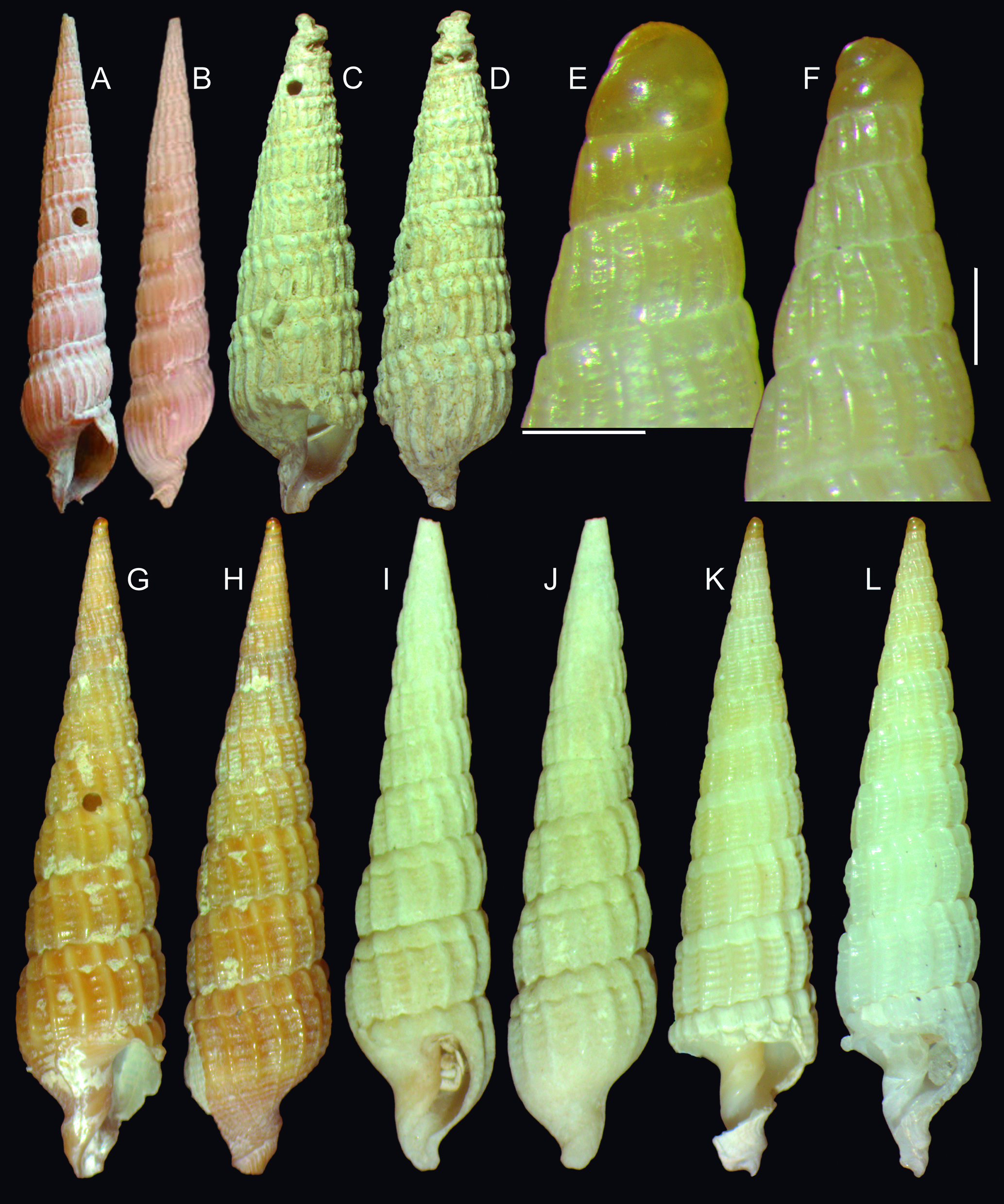
Shells of Terebra spp.: (A-B) T. chilensis, Holotype, MNHN IM 2000-2363 (L 39 mm), frontal and dorsal views; (C D) Terebra aff. joculosa MZSP 19326, from off Mandonado, Uruguay; (E-L) T. potiguar type shells; (E) paratype MZSP 93919#1, detail of apex, profile, scale: = 0.5 mm; (F) same for paratype MZSP 96986; (G-H) holotype MZSP 166494 (L 21.8 mm), frontal and dorsal views; (I-J) paratype 96919#2 (L 18.3 mm), frontal and dorsal views; (K) paratype 96919#1 (L 20.1 mm), dorsal view; (L) paratype MZSP 96986 (L 17.7 mm).
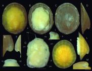
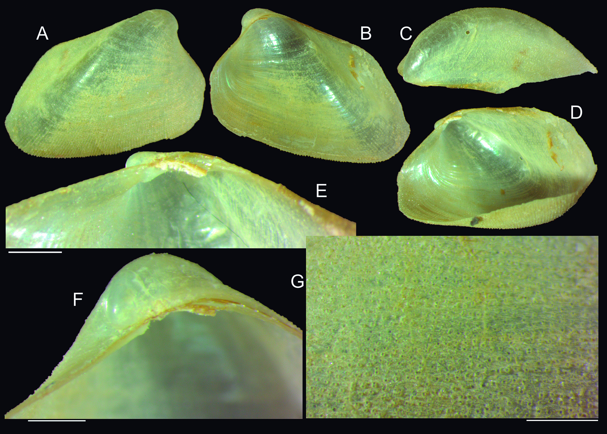
Trimusculus pifius shells and anatomy: (A-I) Holotype MZSP 165668; (A) whole dorsal view (L 4.9 mm); (B) same, ventral view; (C) shell only, dorsal view; (D) same, left view; (E) detail of apex, left view, scale: = 0.25 mm; (F) same, right view; (G) specimen extracted from shell, dorsal view, some structures seen be translucency, scale: = 1 mm; (H) detail of head, dorsal view, scale: = 0.25 mm; (I) same, right view; (K-M) paratype MZSP 165669 (L 3.8 mm); (K) whole dorsal view; (L) same, left view; (M) detail of apex, left view, scale: = 0.25 mm. Lettering: ey, eye; ft, foot; gi, gill; in, intestinal loop; mb, mantle border; mp, mantle papilla; sm, shell muscles.
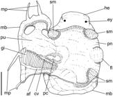
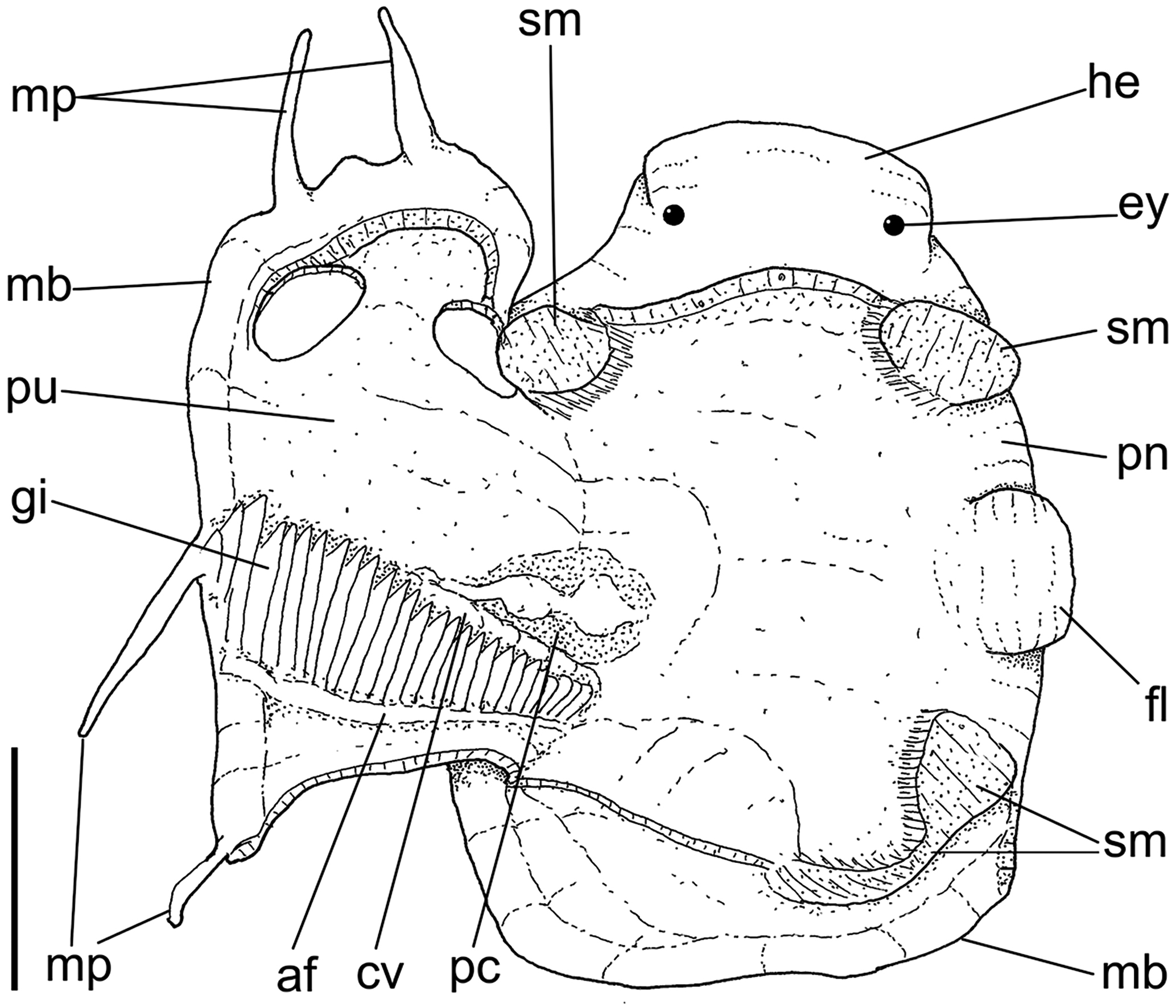
Trimusculus pifius anatomical drawing, specimen extracted from shell, dorsal view, pallial cavity with edges sectioned and deflected to left, exposing its inner structures; scale: = 1 mm. Lettering: af, afferent gill vessel; cv, ctenidial (efferent) vein; ey, eye; fl, anal flap; gi, gill; he, head; mb, mantle border; mp, mantle papilla; pc, pericardium; pn, pneumostome; pu, pulmonary (pallial) cavity; sm, shell muscles.
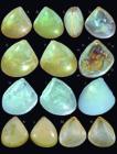
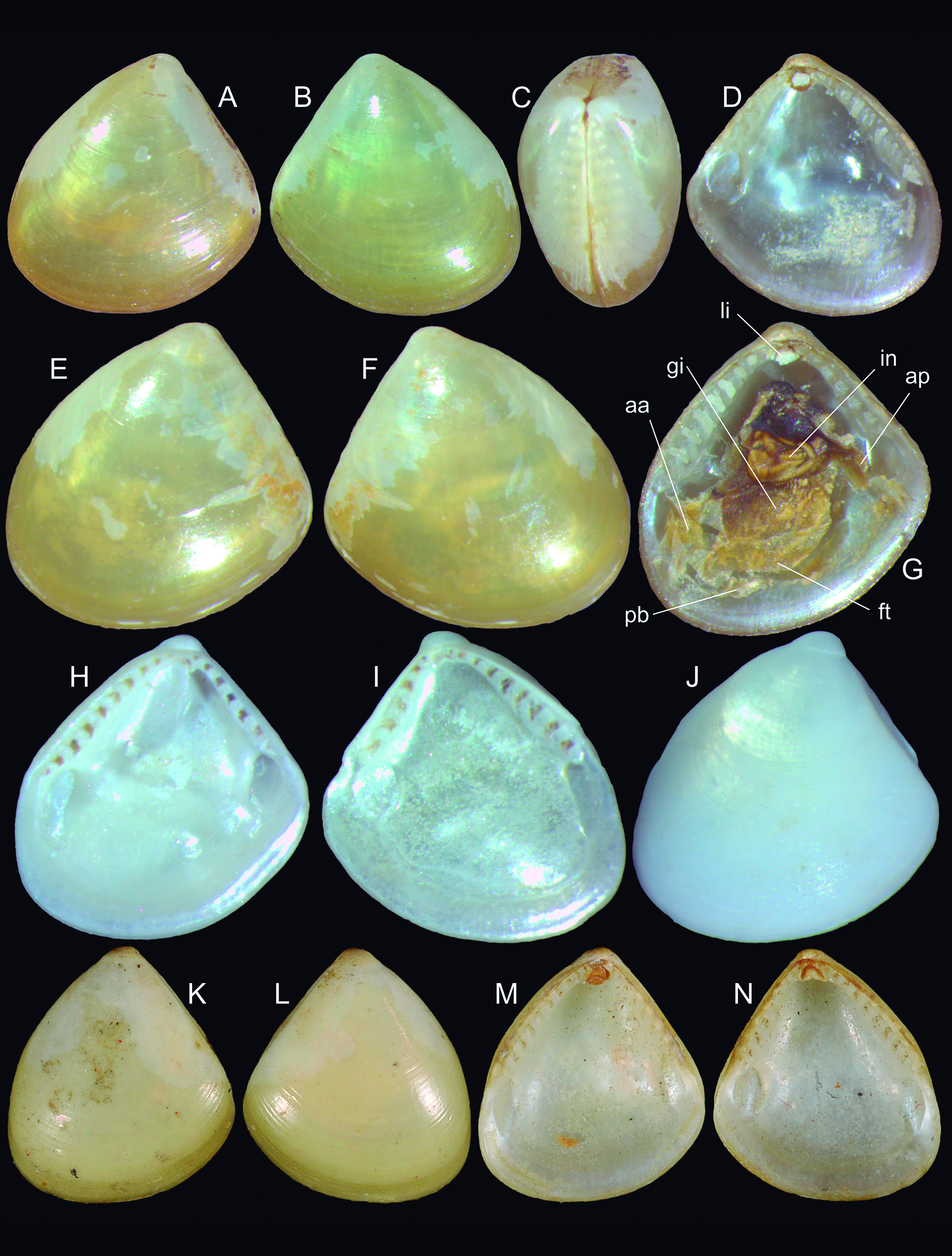
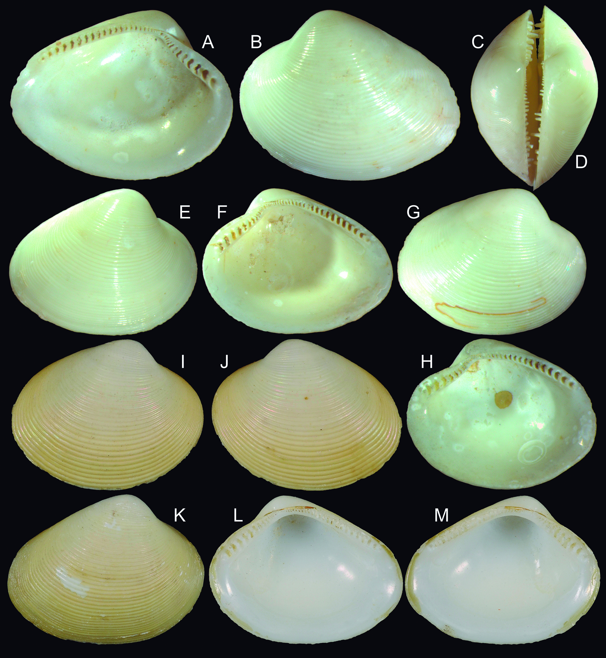
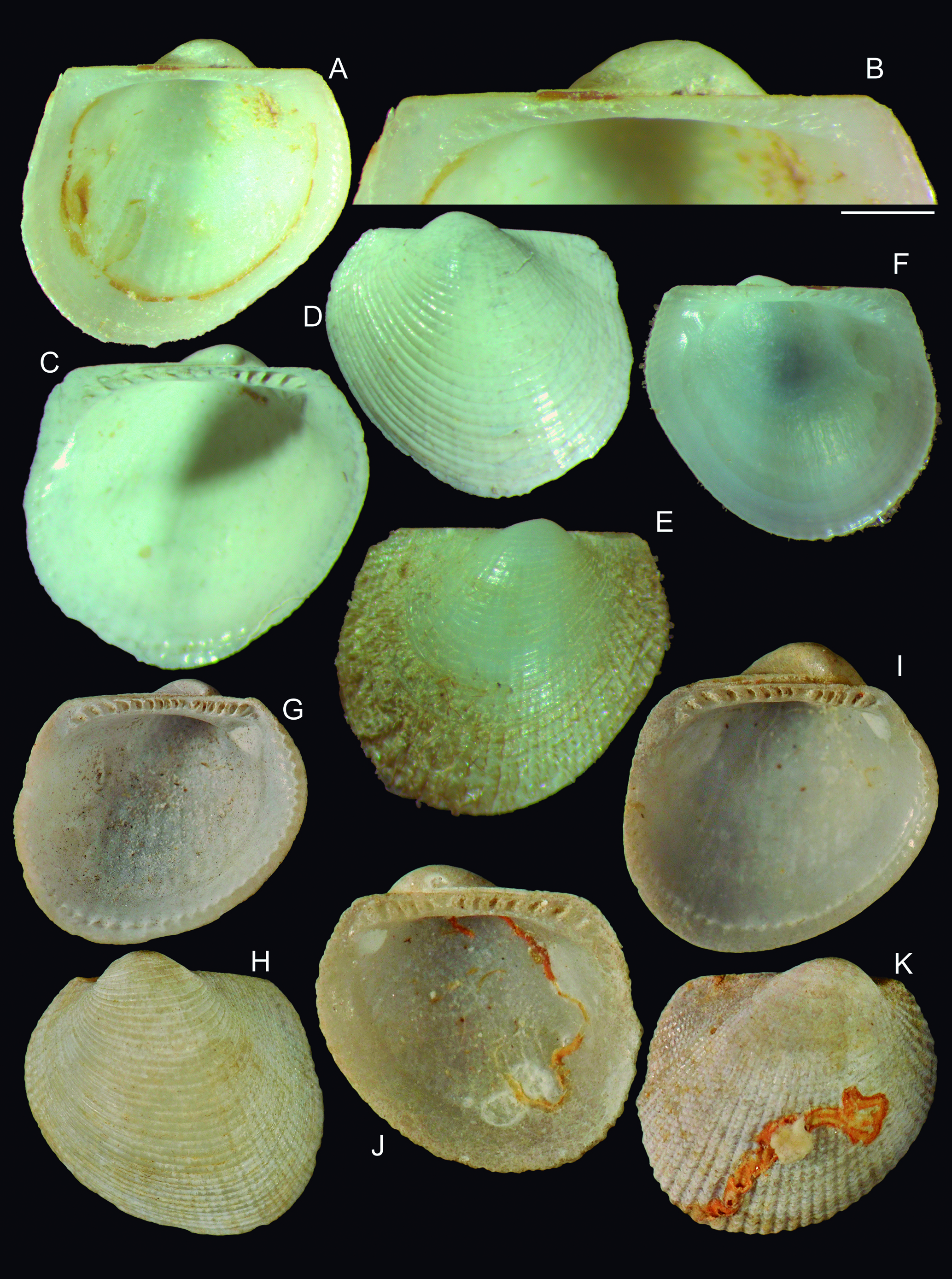
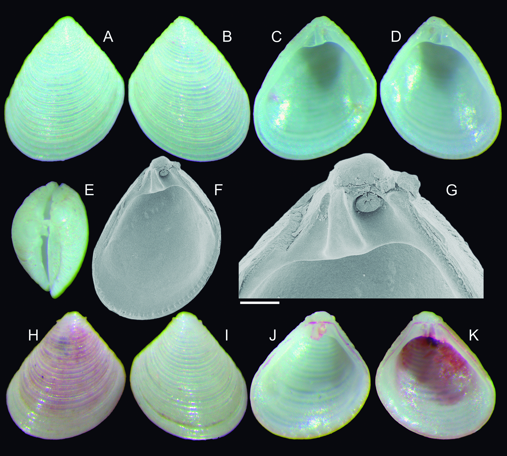
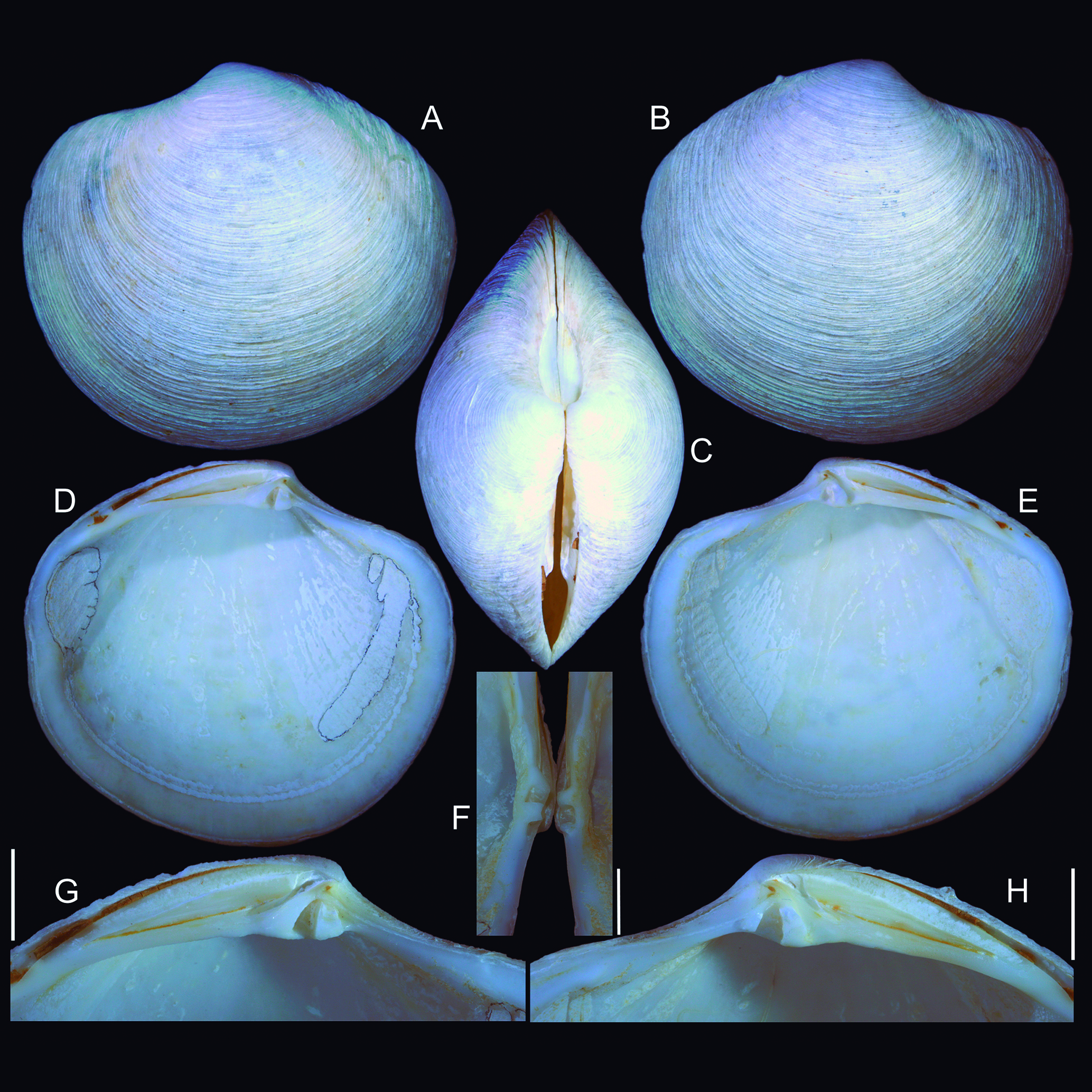
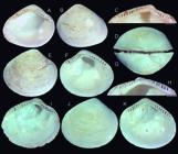
Shells of types of Brevinucula overa and B. verrillii: (A-C) B. overa, holotype MZSP 166720 (H 4.8 mm), left, right and dorsal views; (D) paratype MZSP 166721 (H 4.8 mm), left valve, inner view; (E) same, outer view; (F) right valve, outer view; (G) same, inner view, with mummified remains of soft parts; (H) paratype MZSP 166628#1 (H 4.5 mm), right valve, inner view; (I J) paratype MZSP 166628#2 (H 4.8 mm), inner and outer views; (K N) Holotype of Nucula trigona USNM 45752 (H 4.5 mm), renamed as B. verrillii (courtesy E. Strong, USNM); (K) left valve, outer view, (L) right valve, outer view; (M) left valve, inner view; (N) right valve, inner view. Lettering: aa, anterior adductor muscle; ap, posterior adductor muscle; ft, foot; gi, gill; in, intestine; li, ligament; pb, palp’s proboscis.
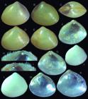
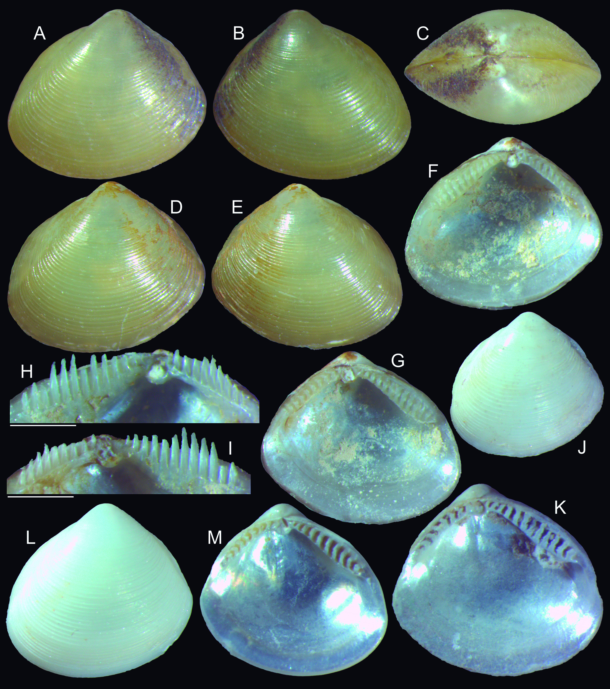
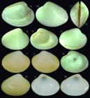
Ennucula ipepa shell of types: (A-C) holotype MZSP 166732 (L 4.3 mm), left, right and dorsal views; (D) paratype MZSP 166733 (L 5.1 mm), left valve, outer view; (E) same, right valve, outer view; (F) same, right valve, inner view; (G) same, left valve, inner view; (H) same, right valve, detail of hinge, inner-slightly ventral view (some teeth are broken); (I) same for left valve, scales: = 1 mm; (J-K) paratype MZSP 166629#1 (L 5.0 mm), left valve, outer and inner views; (L M) paratype MZSP 166629#2 (L 4.9 mm), left valve, outer and inner views.
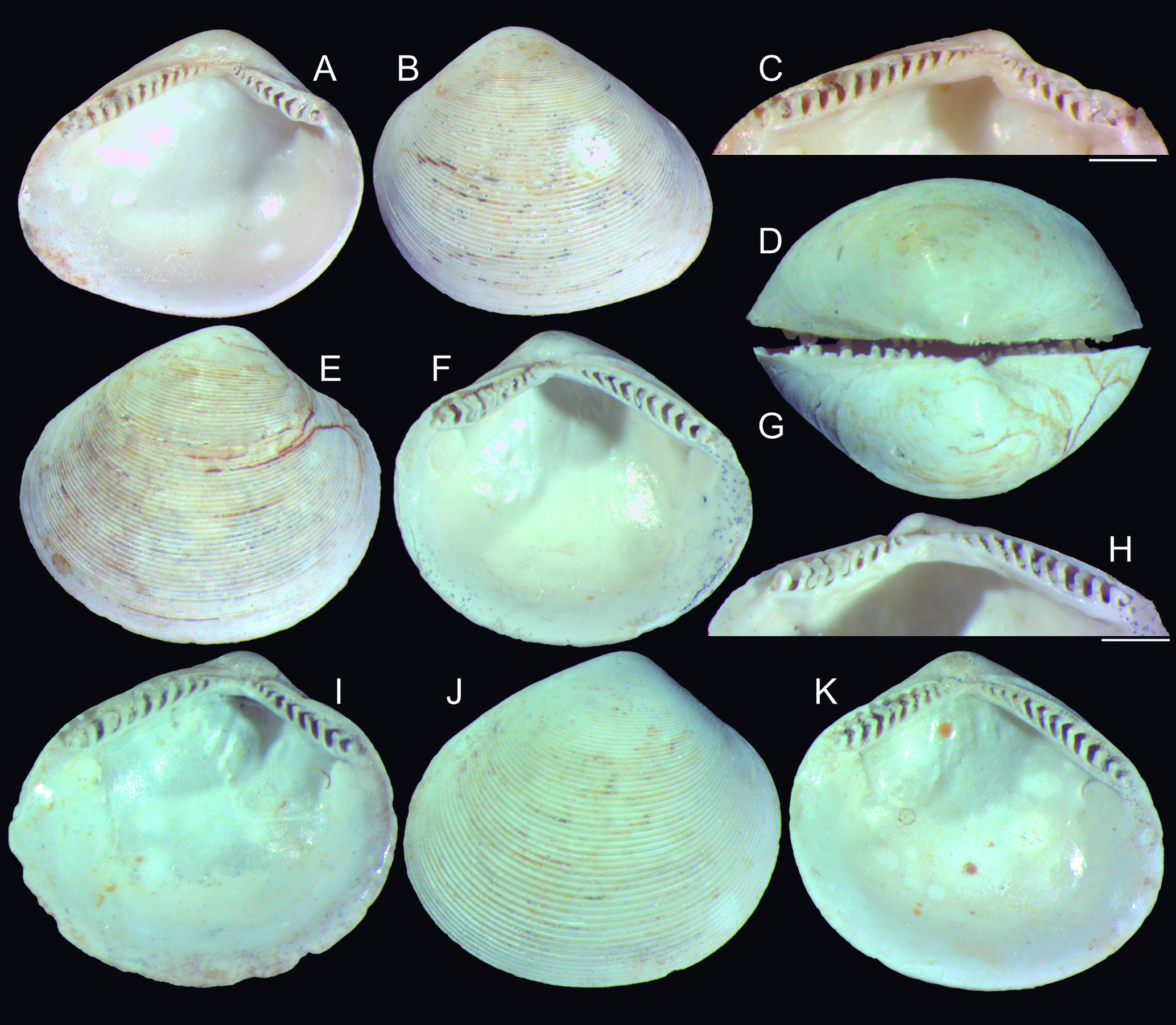
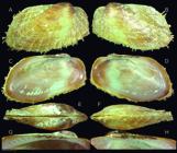
Tindaria ruru shell of types: (A) holotype MZSP 166688, left valve, inner view (L 6.4 mm); (B) same, outer view; (C) same, detail of hinge, inner-slightly ventral view, scale: = 1 mm; (D) same, dorsal view; (E) paratype MZSP 104207#1, right valve, outer view (L 6.9 mm); (F) same, inner view; (G) same, dorsal view; (H) same, detail of hinge, inner-slightly ventral view, scale: = 1 mm; (I) paratype MZSP 104207#2, left valve, inner view (L 5.9 mm); (J K) paratype MZSP 104207#3, right valve, outer view (L 5.8 mm).
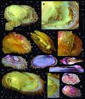
Tindaria cytherea shells: (A-C) MZSP 103165#, right valve 1 (L 10.4 mm), inner, outer and dorsal views; (D-F) MZSP 103165#2, left valve (L 12.0 mm), dorsal, outer and inner views; (G-H) MZSP 103165#3, left valve (L 8.8 mm); (I J) Syntype of Malletia amabilis USNM 94139 (L 12.1 mm), left and right views; (K M) Syntype of Malletia amabilis USNM 62653 (L 7.5 mm); (K) left valve, outer view; (L) same, inner view; (M) right valve, inner view. (I-M) courtesy E. Strong, USNM).
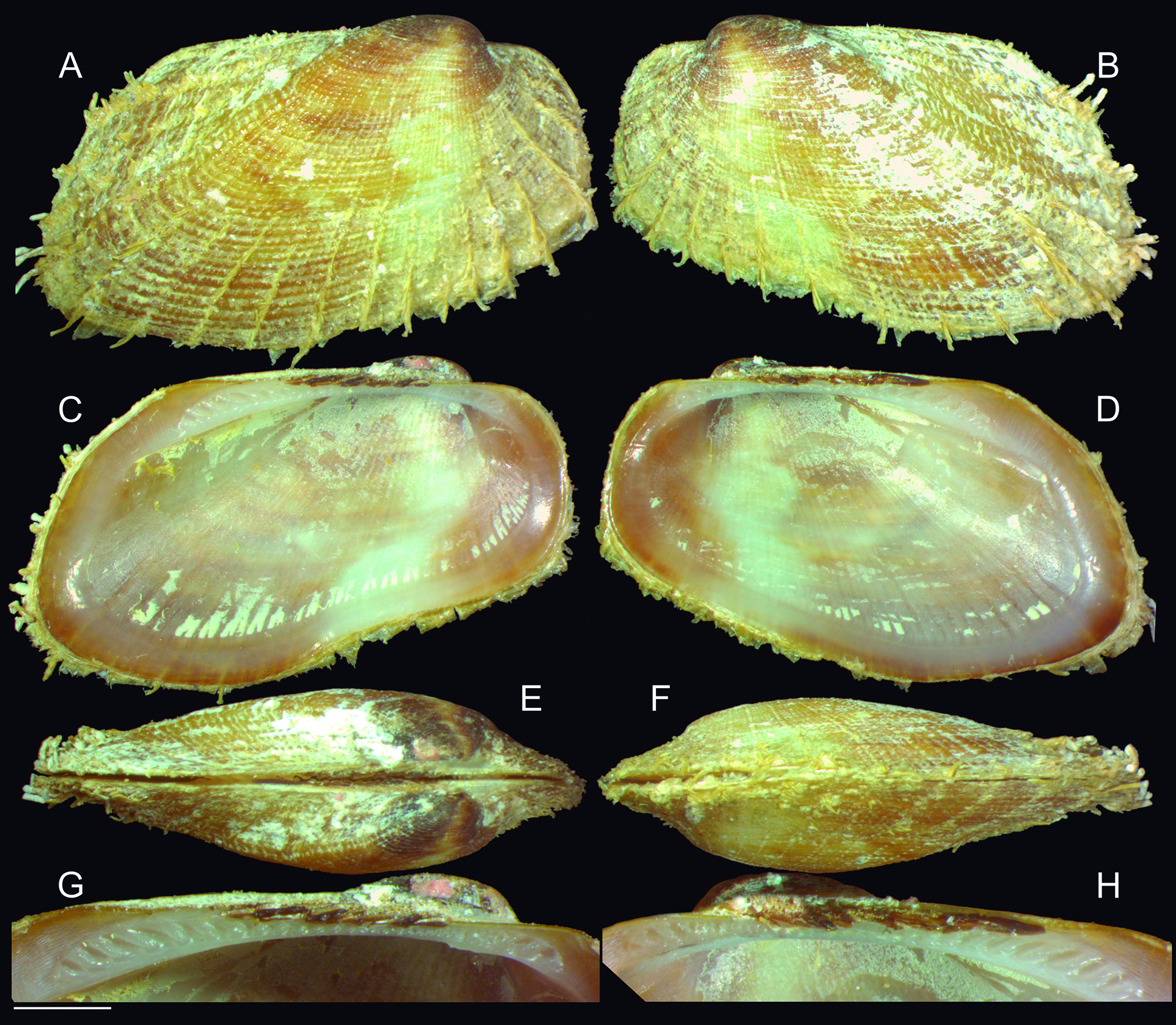
Barbatia pehenguis Holotype MZSP 165816, shell (L 19.0 mm): (A) right valve (RV), outer view; (B) left valve (LV), outer view; (C) LV, inner view; (D) RV, inner view; (E) whole dorsal view; (F) whole ventral view; (G) LV, detail of hinge, inner view, scale: = 2 mm; (H) same for RV.
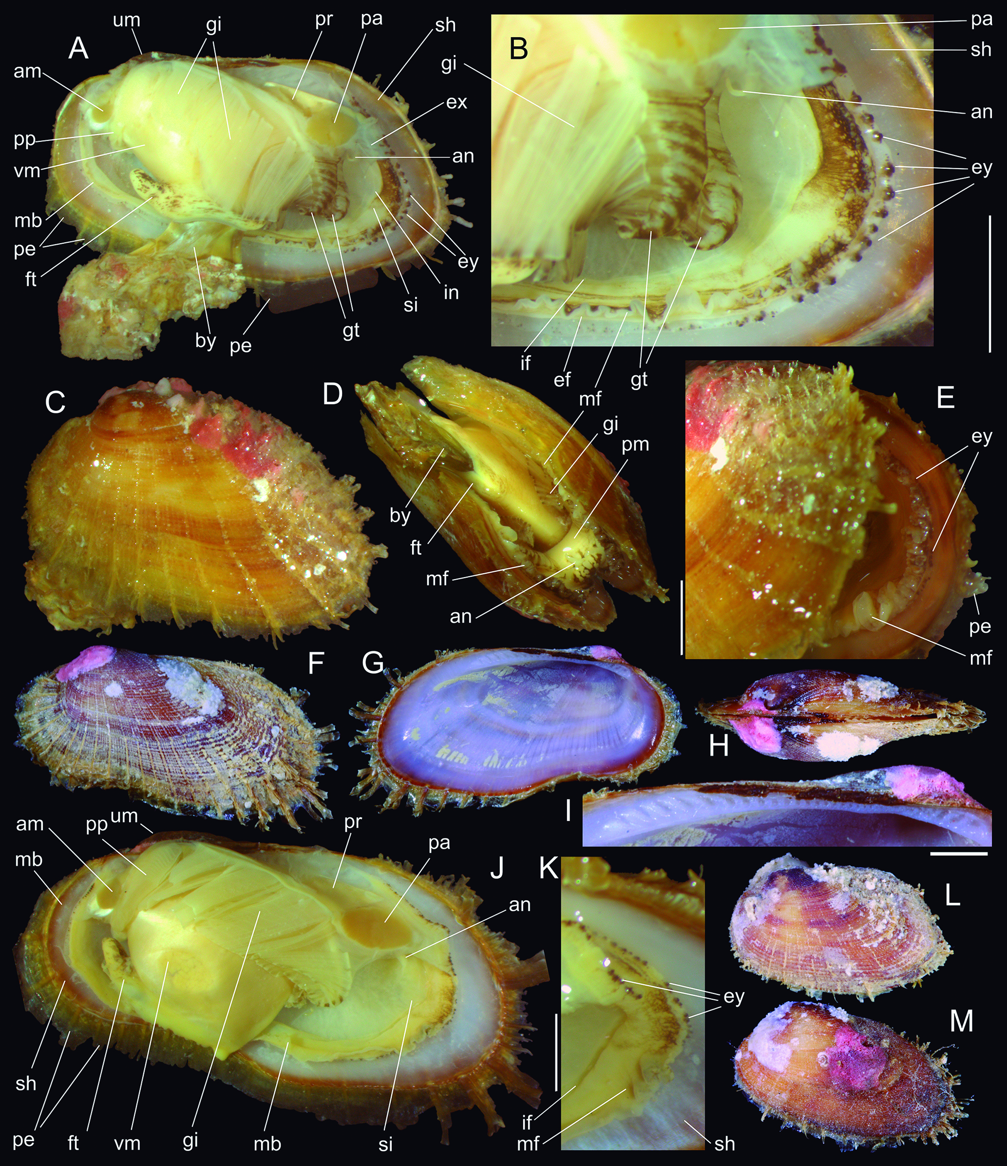
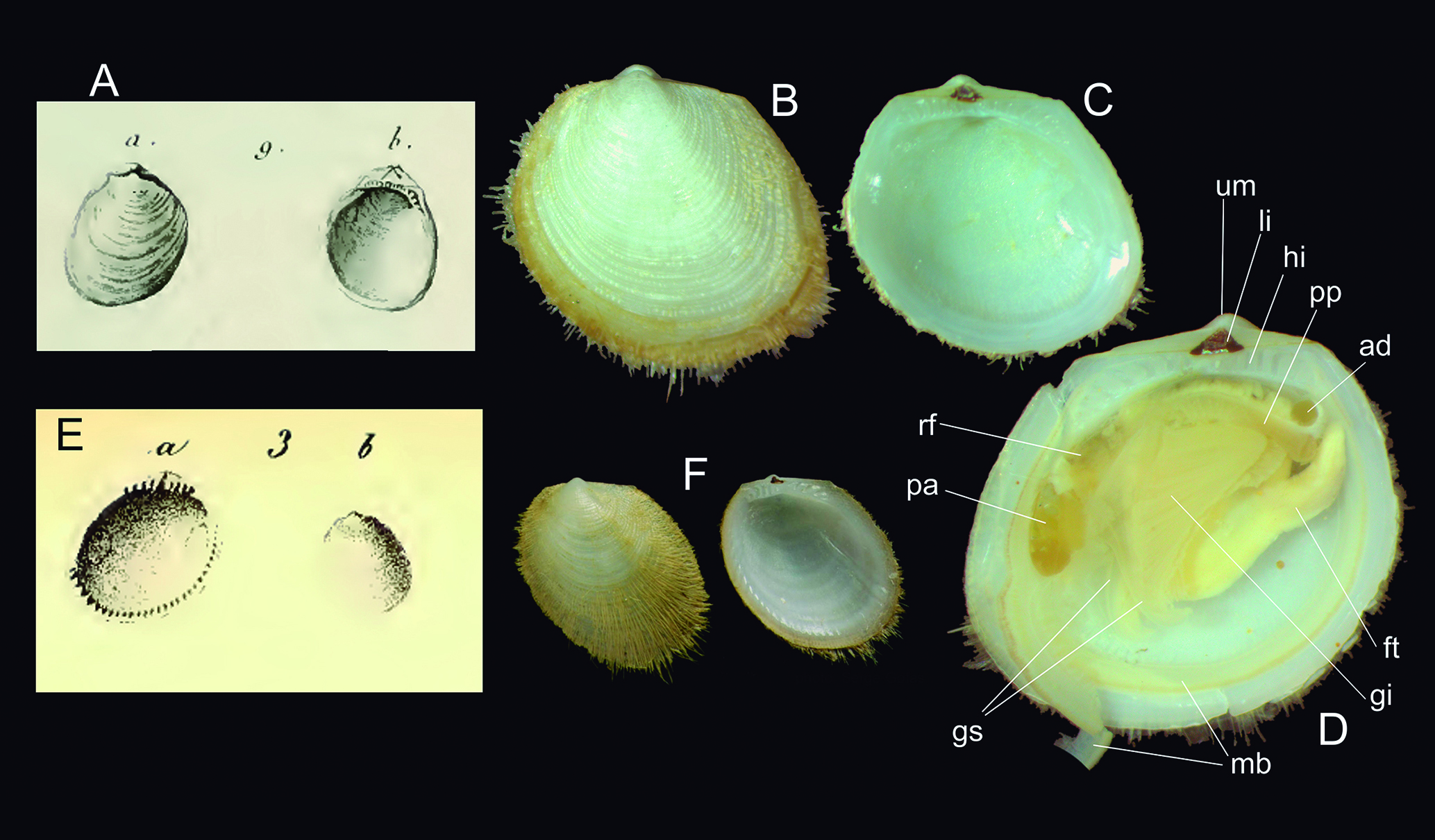
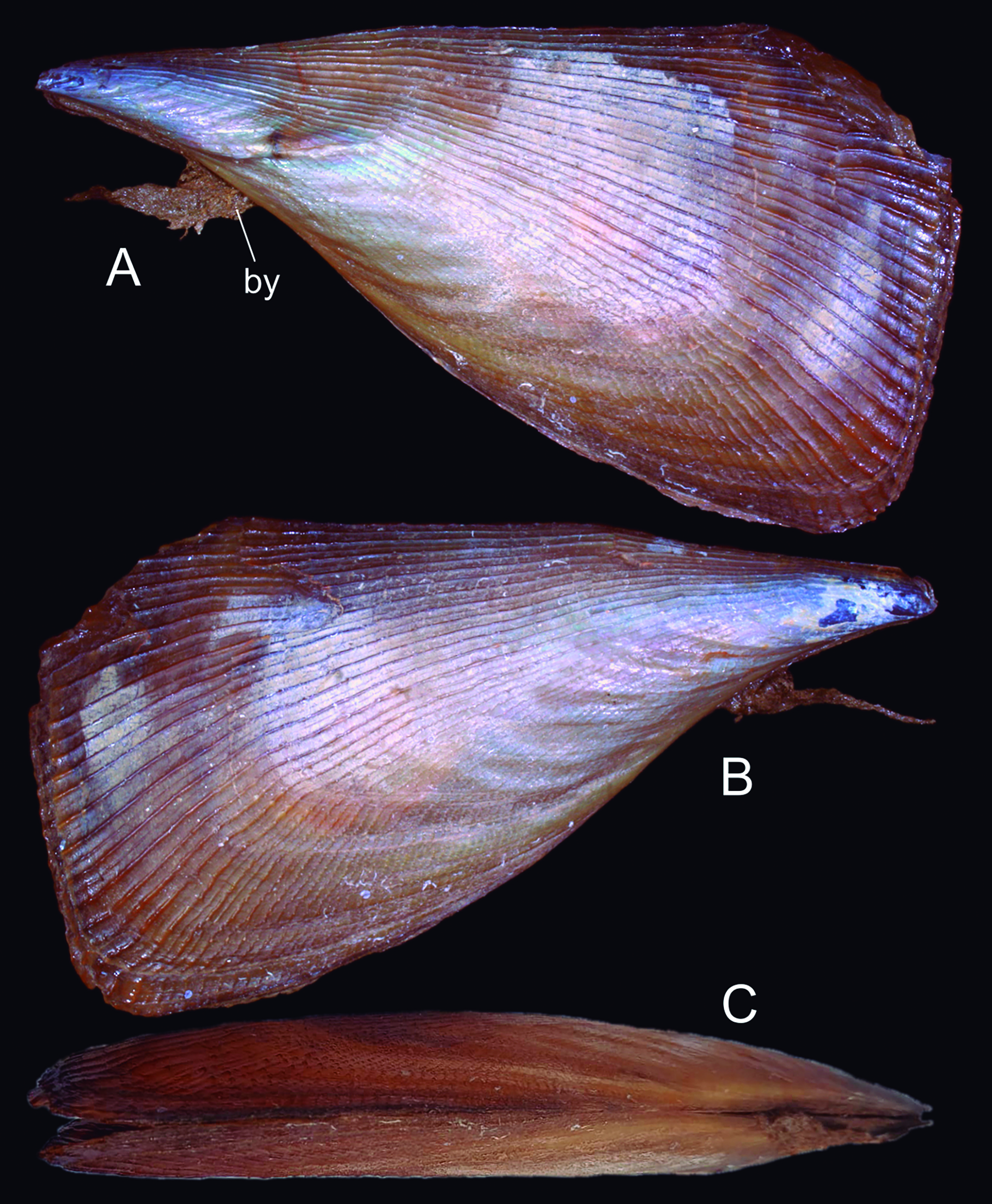
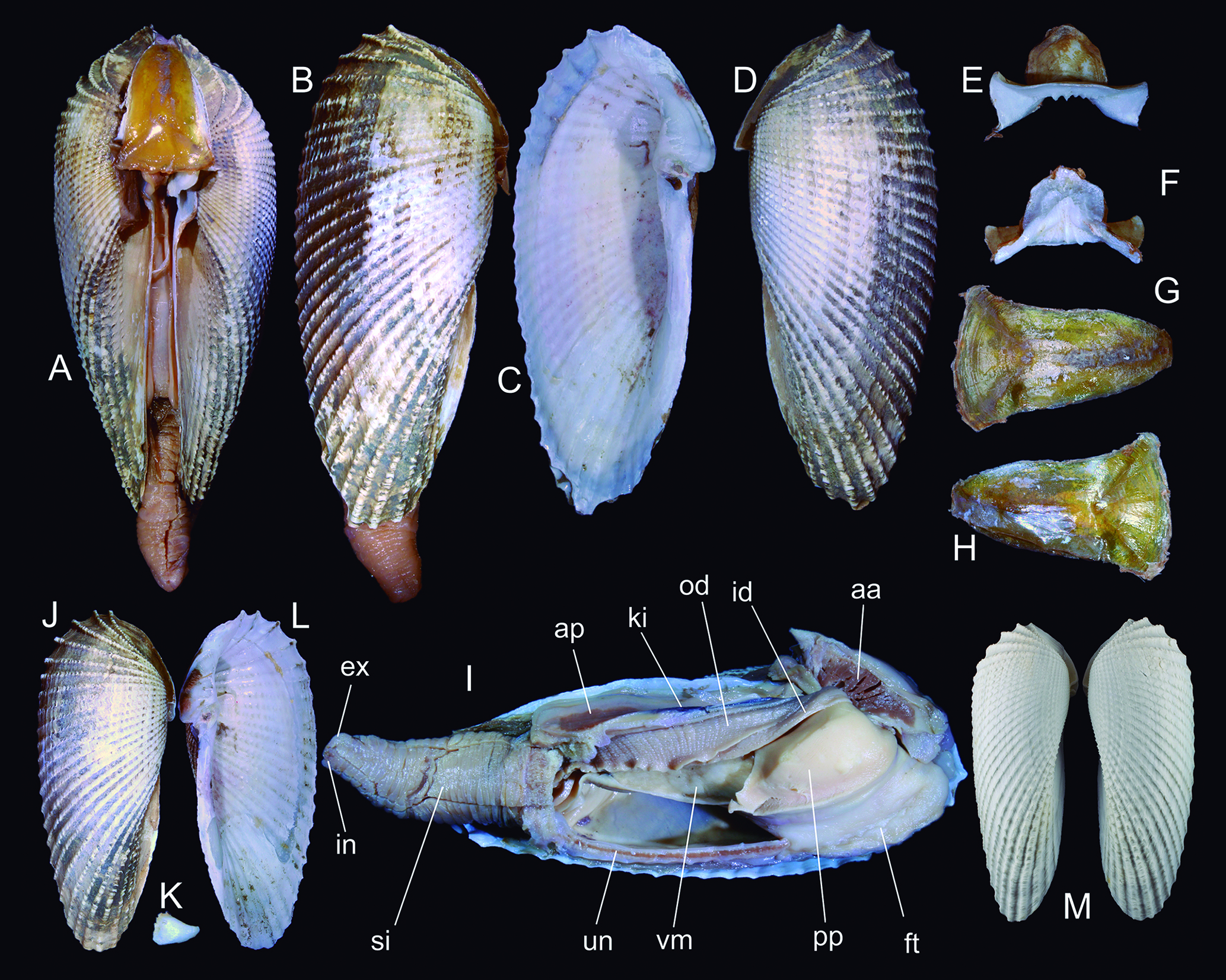
Barbatia pehenguis types, shells and anatomy: (A) Holotype (L 19.0 mm), left view, left valve and mantle lobe removed; (B) same, detail of posterior region; (C) paratype MZSP 31071 (L 18.6 mm), whole left view; (D) same, ventral view, ajar valves; (E) same, detail of posterior region, posterior-slightly left view; (F-K) paratype MZSP 146942 (largest specimen, L 28.0 mm), whole left view; (G) left valve, inner view; (H) dorsal view; (I) left valve, detail of hinge; (J) left view, left valve and mantle lobe removed; (K) detail of posterior region; (L) paratype MZSP 103275, left view (L 15.7 mm); (M) paratype MZSP 162535, left view (L 17.2). Lettering: am, anterior adductor muscle; an, anus; by byssus; ef, outer mangle fold; ex, excurrent canal; ey, eyes; ft, foot; gi, gill; gt, gill stalks; in incurrent canal; if, inner mantle fold; mb, mantle border; mf, middle mantle fold; pa, posterior adductor muscle; pe, periostracum; pp, palps; pr, posterior foot retractor muscle; sh, shell; si, siphonal flaps; um, umbo; vm, visceral mass.
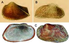
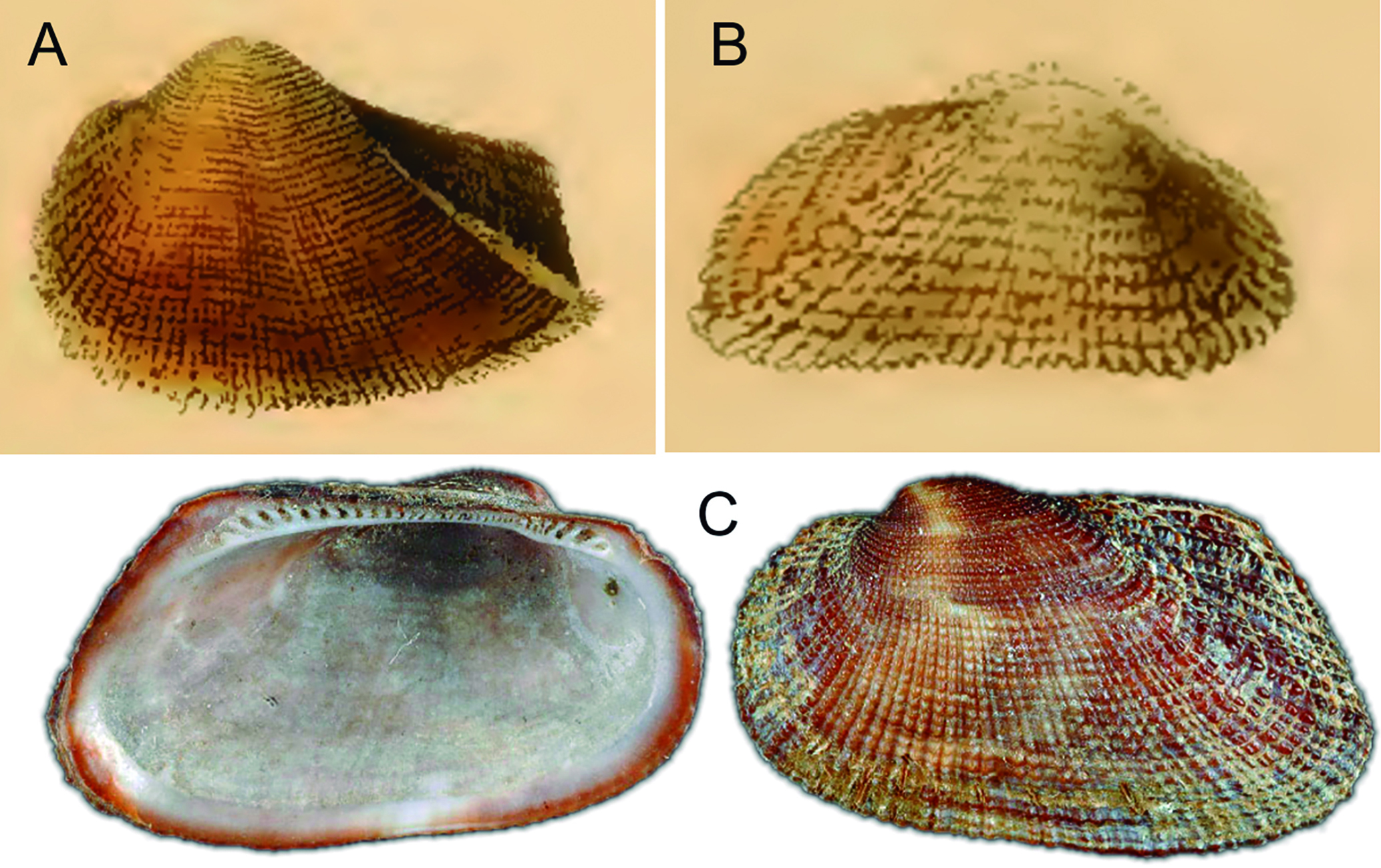
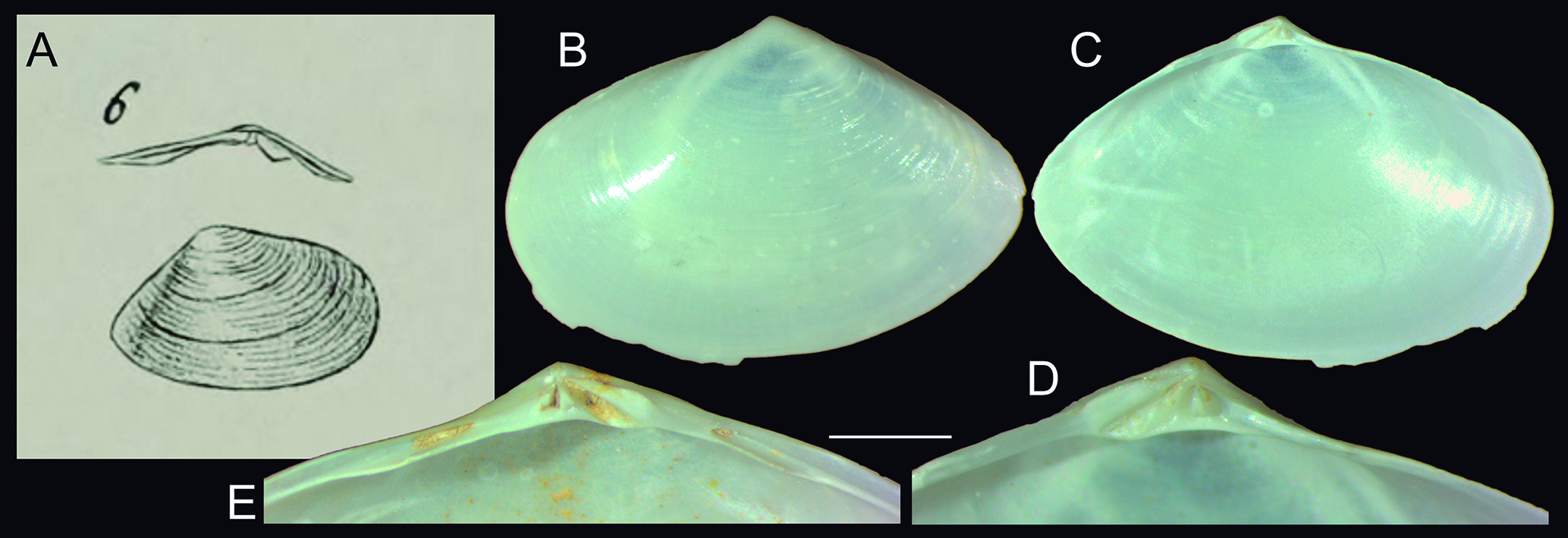
Shells of Barbatia cancellaria and Acar domingensis: (A) B. cancellata, illustration by Kobelt (1891, pl. 20, fig. 5); (B) A. domingensis, illustration by Kobelt (1891, pl. 47, fig. 5); (C) B. cancellaria, syntype MNHN IM 2000-24605 (L 21.4 mm) (courtesy MNHN).
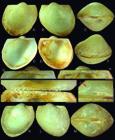
Bathyarca arcadia shell of types: (A-J) holotype MZSP 166512 (W 7.6 mm); (A) right valve, outer view; (B) left valve, outer view; (C) whole dorsal view; (D) left valve, inner view; (E) right valve, inner view; (F) ventral-sightly right view; (G) left valve, detail of hinge; (H) same for right valve; (I) semi-articulated hinge, ventral view, left valve up; (J) right valve, detail of umbo, left view, arrow showing prodissoconch; (K-M) paratype MZSP 166814#1 (W 7.4 mm), right, left and dorsal views. Scales: = 1 mm.
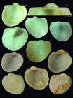
Bathyarca arcadia and B. glomerula shells of types: (A F) B. arcadia; (A) paratype 166514#2 (W 6.9 mm), left valve, inner view; (B) same, detail of hinge, scale: = 1 mm; (C) paratype MZSP 166516#1 (W 5.0 mm), left view, inner view; (D) same, outer view; (E) paratype MZSP 166516#2 (W 5.0 mm), right valve, outer view; (F) same, inner view; (G-K) B. glomerula syntypes USNM 63166 (courtesy Ellen Strong); (G) #2 (W 5.6 mm), left valve, inner view; (H) same, outer view; (I) #1 (W 5.3 mm), left valve, inner view; (J) #3 (W 4.1 mm)m right valve, inner view; (K) same, outer view.
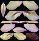
Bentharca celeris shell of types: (A-G) holotype MZSP 166556 (L 10.3 mm); (A) whole dorsal view; (B) right view; (C) left view; (D) left valve, inner view; (E) right valve, inner view; (F) left valve, detail of hinge, scale: = 1 mm; (G) same for right valve; (H-J) paratype MZSP 104210#2 (L 8.1 mm), right, dorsal and left views; (K-L) paratype MZSP 104210#3 (L 11.1 mm), left valve, inner and outer views; paratype MZSP 104139 (L 11.5 mm), left valve, outer views.
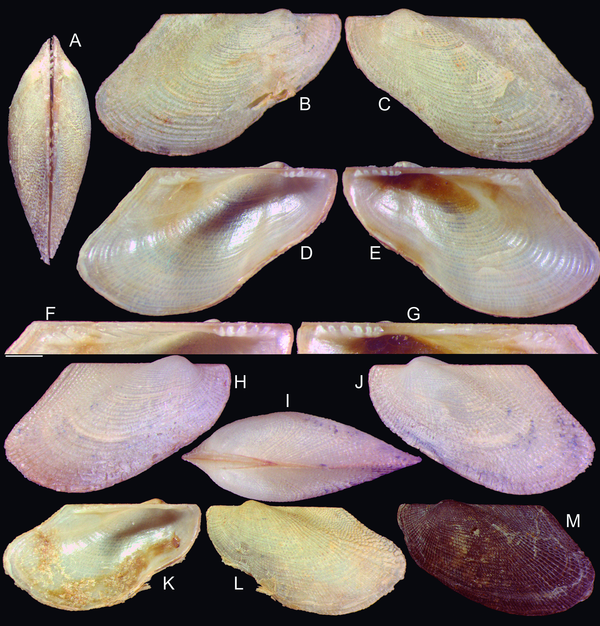
Reported species of Limopsidae: (A) replica of Arca aurita Brocchi, 1881 original image (pl. 11, sp. 9), a Pliocene Italian fossil; (B-D) L. paucidentata MZSP 156267 (W 10.4 mm); (B) fully left view; (C) right valve, inner view; (D) left valve containing soft parts, right view; (E) replica of Pectunculus minutus Philippi, 1836 original image (pl. 5, fig. 3), a Plio-Pleistocene Italian fossil; (F) Paracratis borealis from Josephine seamount (W 6.7 mm) (by Gofas Serge, MolluscaBase, reproduced under permission), left valve, outer view, and right valve, inner view.
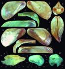
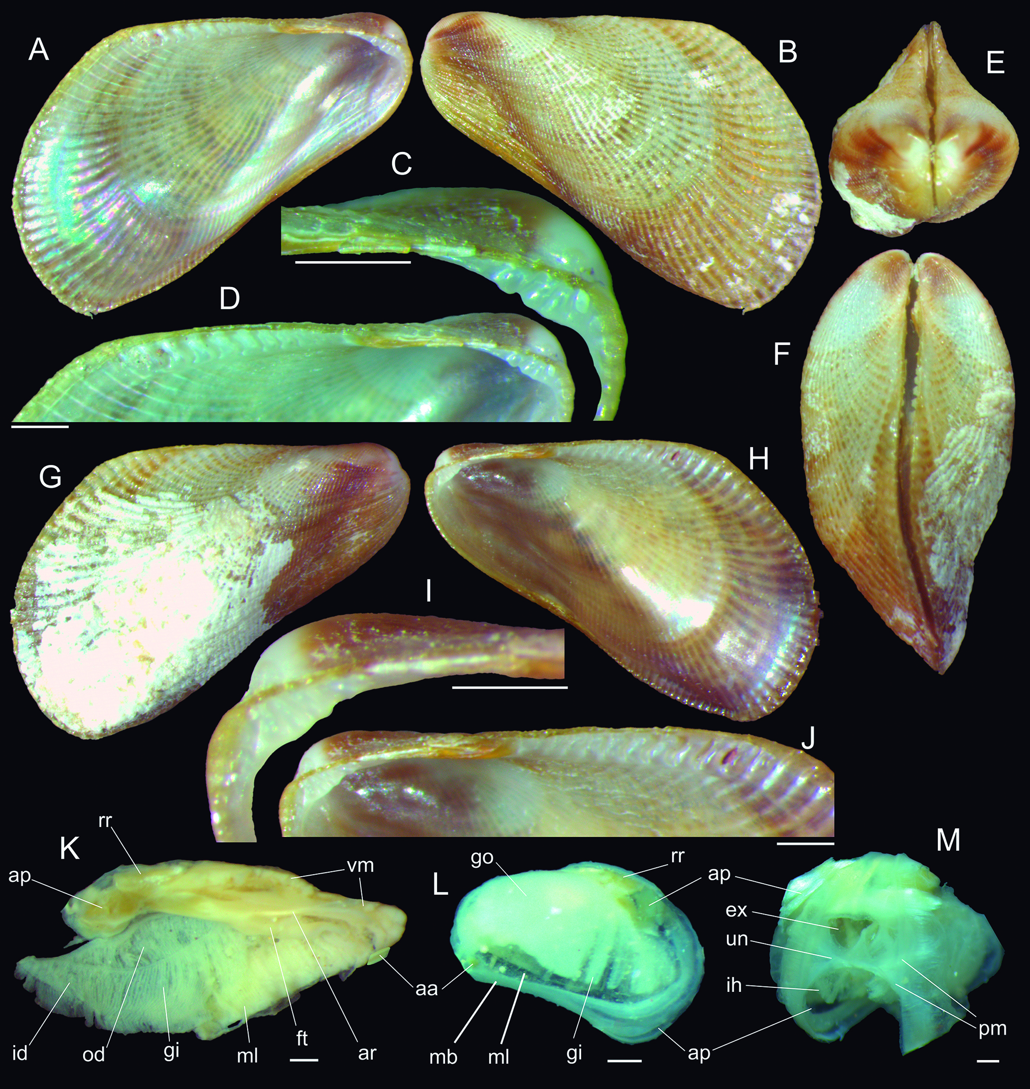
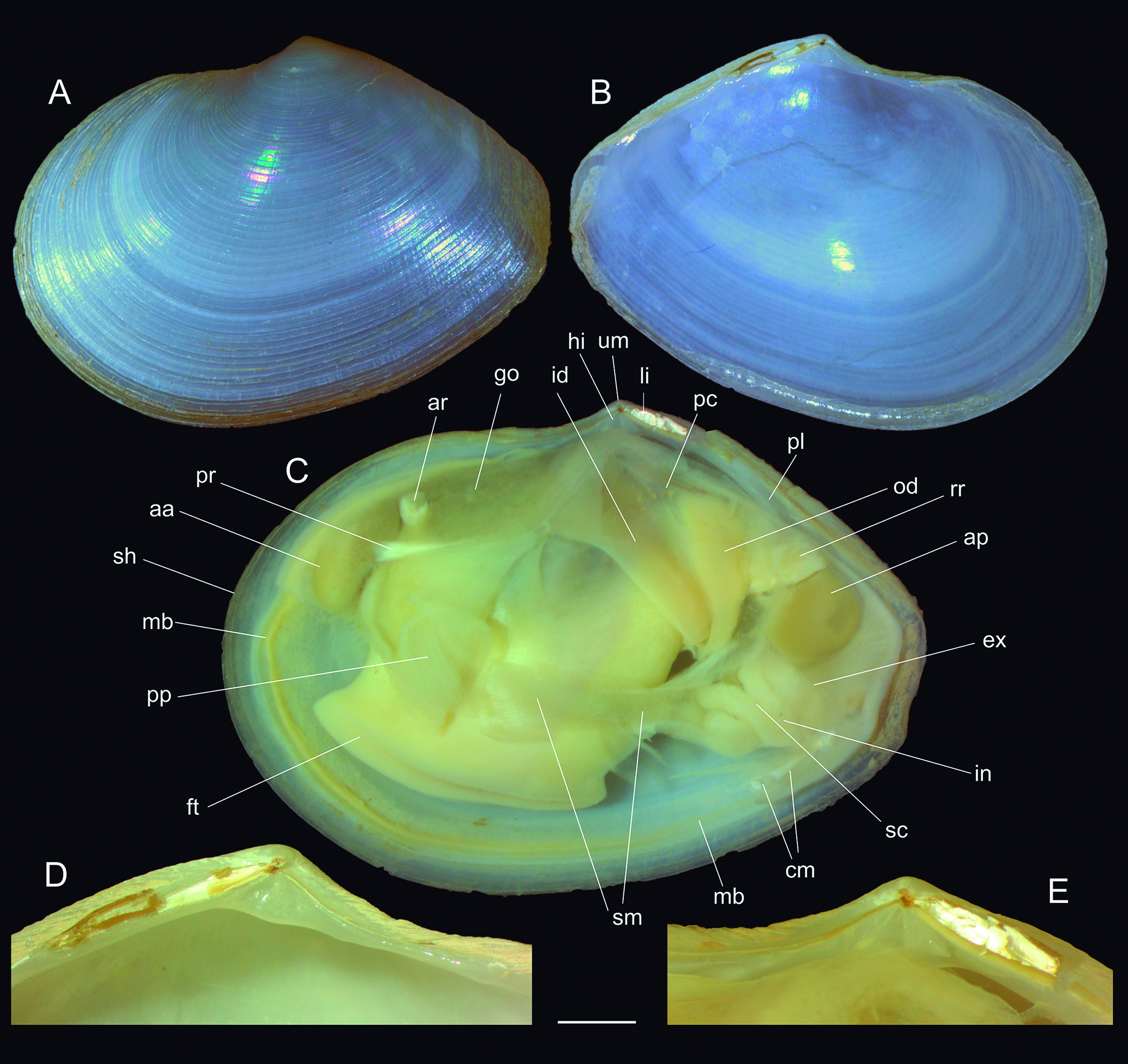
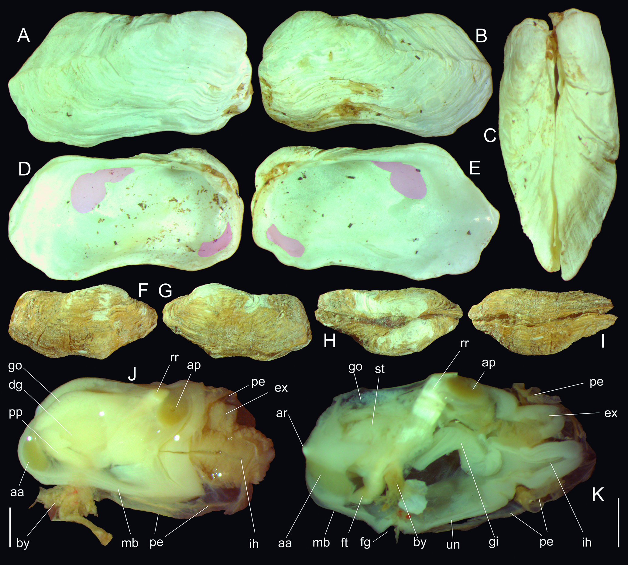
Brachidontes ynous Holotype MZSP 168671, shell and anatomy. (A) left valve, inner view (L 8.4 mm); (B) same, outer view; (C) same, detail of hinge; (D) same, detail of dorsal edge; (E) whole anterior view (W 4.8 mm); (F) whole dorsal view; (G) right valve, outer view; (H) same, inner view; (I) same, detail of hinge; (J) same, detail of dorsal edge; (K) holotype extracted specimen, right view, right mantle lobe, gill, and posterior region of mantle removed; (L M) soft parts of paratype 109572#1, just removed from shell, left and posterior views. Scales: = 0.5 mm. Lettering: ap, posterior adductor muscle; ar, anteriror foot retractor muscle; ex, excurrent aperture; ft, foot; id, inner demibranch; ih, incurrent aperture; gi, gill; go, gonad (in mantle lobe); ml, mantle lobe; od, outer demibranch; pm, pallial muscles; rr, posterior retractor muscle of foot; un, mantle fusion separating apertures; vm, visceral mass.
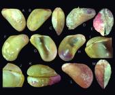
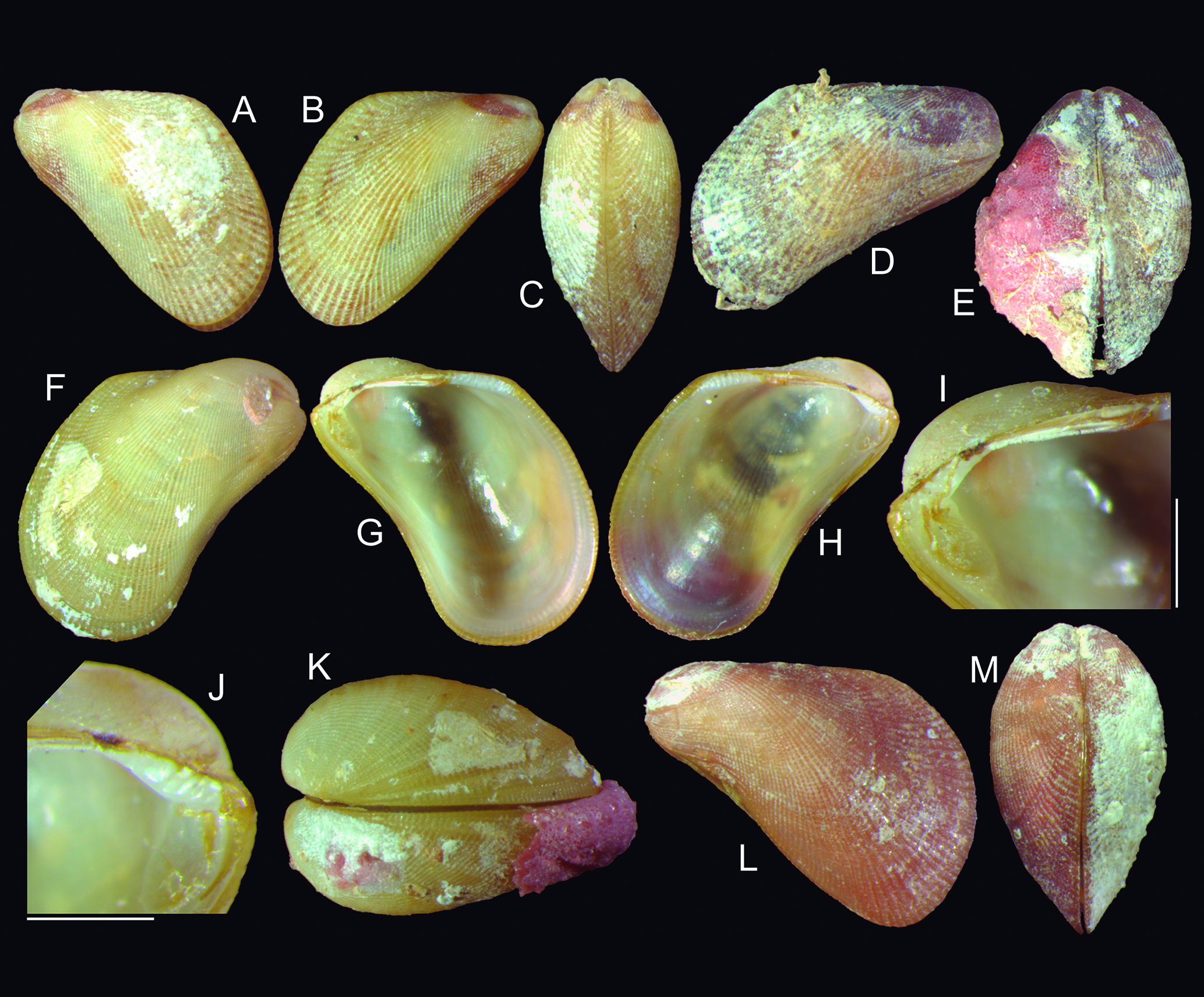
Brachidontes ynous paratypes shell. (A-C) MZSP 105355#1 (L 8.3 mm), left valve outer view, right valve outer view, and whole dorsal view; (D E) MZSP 105355#2 (L 6.3 mm), right valve outer view and whole dorsal view; (F-K) MZSP 109572#1 (L 7.6 mm); (F) right valve, outer view; (G) same, inner view; (H) left valve, inner view; (I) right valve, detail of hinge; (J) same for left valve; (K) whole dorsal view; (L M) MZSP 109572#2 (L 7.6 mm), left valve outer view, whole dorsal view. Scales: = 1 mm.
Subclass Caenogastropoda Epiathroidea Sorbeoconcha Superfamily Epitonioidea Family Epitoniidae Genus Depressiscala de Boury, 1909 Depressiscala nautlae (Mörch, 1875) (Fig. 7A-F)
Scala nautlaeMörch, 1875: 265.
Scala scipioDall, 1889: 27, 310.
Epitonium (Depressiscala) nautlae:Abbott, 1974: 122 (fig. 1271); Rios, 1975: 56 (pl. 15, fig. 217), 1994: 157 (pl. 52, fig. 743), 1994: 99 (pl. 32, fig. 400), 2009: 181 (fig. 439).
Depressiscala nautlae:Rosenberg et al., 2009: 641.
Type localities:S. nautlae: ad litus mexicanum inter Vera Cruz et Nautla. Scala scipio: Vera Cruz; sta. 2597, 20 miles SW by S from Cape Hattenas, N.C., 15 + 12 fms.
Remarks: Specimens found on the Brazilian coast with the shell characteristics shown in Fig. 7A-C were identified as both D. nautlae and D. niditella (Dall, 1889). A comparison to the lectotype of D. nitidella (USNM 83716) (Fig. 7G) reveals that they do not belong to that species, as the shells are much slenderer and lack an umbilicus. Conversely, their shells are virtually identical to the lectotype of Scala scipio (Fig. 7D, E), a species presently considered synonymous with D. nautlae.
The issue arises from the apparent existence of two different morphs under this epithet. One resembles the S. scipio model, with a deeper suture, more distant and narrower axial threads, and always with an amputated apex (Fig. 7A-E). The other morph, illustrated in Fig. 7F, features a shallower suture, taller and more closely located axial threads, and a complete apex. This latter morph is illustrated, for example, in Abbott (1974). Upon analyzing these specimens, the most parsimonious conclusion is that they belong to different species. However, it is important to determine whether the types of Mörch (1875) resemble them, as they have not been located or photographed to the best of our knowledge. This is an important issue for future surveys.
Material examined: USA. Florida; Pompano Beach, 52 m, ANSP 457201, 2 shells (Harriet, Robertson & Golberg col., 2004). BRAZIL. São Paulo; Ubatuba, praia Puruba, 23°21′09.2″S 44°55′54.3″W, MZSP 166500, 1 shell, 165694, 7 shells (D. Forcelli col., viii.2023). Plus 11 lots of MZSP collection from Piauí to São Paulo.
Genus Amaea H. & A. Adams, 1853 Amaea mitchelli (Dall, 1896)
Remarks: The species, with type locality Beach of Matagorda Island, Texas, is known as having geographic distribution from Texas up to Surinam (Turner, 1959). Recent dredges off Amazon river mouth, collected this species, being this an extension of its geographic distribution southwards, off Amapá coast, Brazil.
Material examined: BRAZIL. Amapá; off São João do Cassiporé, 03°49′08.5″N 50°02′50.3″W, 71 m, MZSP 140070, 4 spm (W.C. Santos col., o.t., 29.v.2018).
Superfamily Abyssochrysoidea Family Abyssochrysidae Genus Abyssochrysos Tomlin, 1927 Abyssochrysos quasilissus new species (Fig. 8) https://zoobank.org/8151116A-E473-48DB-92AB-0DB0F63F9038
Types: Holotype MZSP 117078, shell. Paratype: BRAZIL. Espírito Santo; slope off Itaúnas (R.V. Marión-Dufresne col., MD55), 18°59′S 37°48′W, 1,540-1,550 m, MZSP 117074, 1 shell (sta. DC70, 26.v.1987), 19°40.6′S 37°48.1′W, 790-940 m, MNHN IM 2000-39806, 2 shells (sta. CB77, 27.v.1987).
Type locality: BRAZIL. Espírito Santo; off Itaúnas, 18°59′S 37°48′W, 607-620 m [R.V. Marión-Dufresne col., MD55 sta. DC73, 27.v.1987].
Diagnosis: Shell from SE Brazilian coast, of ~6 mm, sculptured only by weak oblique striations.
Description: Shell ~6 mm; ~2.5 times longer than wide; spire angle ~24° (Fig. 8A-C, F-H). Color pure white. About 9 convex whorls, with wider region just above suture, causing suture implantation inferior to preceding whorl. Protoconch (Fig. 8E, I) of 1 whorl, width 0.9 mm, mamillated, blunt; limit with teleoconch unclear. Teleoconch surface glossy, shining; sculpture weak striations, like narrow undulations, ~25 in penultimate whorl, prosocline inclined at ~15° in relation to longitudinal shell axis (Fig. 8B, G, H); sculpture present since first teleoconch whorls. Aperture rounded (Fig. 8A, F, D), occupying ~24% of shell length, ~50% of shell width. Outer lip fragile, with cutting edge; inner lip with inferior ⅔ weakly concave, curved outside covering umbilicus, superior ⅓ as very thin callus attached to preceding whorl; inferior apertural region angulate, slightly turned to left. Umbilicus opened (Fig. 8D), with simple edges.
Etymology: The specific epithet is a conjunction of Latin prefix quasi, meaning like, almost; with the Greek word lissos meaning smooth, polished, an allusion to the almost smooth surface of the shell.
Distribution: Off Espírito Santo to Rio de Janeiro, Brazil.
Habitat: 607-1,550 m depth (dead shells only).
Measurements (in mm): Holotype MZSP 117078 (Fig. 8A-E): 5.6 by 2.2. Paratype: MZSP 117074 (Fig. 8F-I): 6.4 by 2.2.
Additional material examined: BRAZIL. Espírito Santo (R.V. Marión-Dufresne col. 1987); off Conceição da Barra, 18°59′S 37°50′W, 637 m, MZSP 168310, 1 shell (MD55 sta. CB76, 27.v). Rio de Janeiro; Saquarema, off Praia Seca, 23°54.2′S 42°10.5′W, 830 m, MNHN, 1 shell (sta. CB106, 02.vi).
Remarks:Abyssochrysos quasilissus shell differs from that of the single congener that occur in the Western Atlantic, A. brasilianus Bouchet, 1991, in being much smaller (A. brasilianus shell can reach ~30 mm, while it has ~6 mm), reflected even in the protoconch, in which A. quasilissus is less than half of that of A. brasilianus. Besides, A. quasilissus is sculptures by a delicate striation, while A. brasilianus has widely spaced axial threads, being taller in the inferior region of each whorl, giving to the shell a beautiful, ornamented aspect. Abyssochrysos quasilissus is, as the name suggests, more modestly ornamented.
Hypsogastropoda Divisio Rissooidea Superfamily Truncatelloidea Family Caecidae Genus Caecum Fleming, 1813 Caecum jonesae Swineen & Simone, new species (Fig. 9J-L) https://zoobank.org/AA5F927A-8504-45CB-A8CE-C67BEE25CC20
Types: Holotype MZSP 166590, shell. Paratypes: MZSP 166591, 3 shells from type locality.
Type locality: UNITED KINGTOM. Ultramarine Territory, Santa Helena island.
Diagnosis: Species from Santa Helena oceanic island with very narrow, slender shell; profile cylindric. Surface completely smooth. Mucro semispherical, highly protruded, occupying entirely inner area, with blunt dorsal beak.
Description: Adult shell of ~1.3 mm, slightly curved, ~6 times longer than wide; anterior aperture slightly larger than posterior end; profile rather cylindric (Fig. 9J). Surface smooth, lacking any kind of sculpture except for poorly visible growth lines (Fig. 9K). Posterior mucro semispherical, highly protruded, occupying entire inner area; flanked by relatively thick shell walls (Fig. 9L); bearing blunt dorsal, terminal beak. Aperture simple, peristome not deflected, rounded, weakly prosocline.
Etymology: The specific epithet is in honor to Kirsty Jones, an honorable temporary resident in St. Helena who sent sediment to be sorted and contributed to the discover of the presently described species.
Distribution: Santa Helena oceanic island.
Habitat: Collected sorting sediment.
Measurements (in mm): Holotype MZSP 166590 (Fig. 9J-L): 1.3 by 0.2.
Remarks:Caecum jonesae, coauthored by Frank Swinnen [Reseach Associate of Royal Belgian Institute of Natural Sciences, D.O. Taxonomy & Phylogeny, Vautier Straat 29, 1000 Brussels, Belgium & Estação de Biologia Marinha and Museu Municipal do Funchal, Madeira, Cais do Carvão Promenade da Orla Maritima do Funchal, Portugal. E mail: f.swinnen.lommel@telenet.be]. It belongs to the subgenus Brochina Gray, 1857, characterized by its spheric, protruded mucro and smooth, unsculptured shell. It shares similarities with Caecum antillarum Carpenter, 1858, a prevalent species along the Western Atlantic coast from Florida to Uruguay (Gomes & Absalão, 1996; Absalão & Gomes, 2001). However, it differs by having a rounded mucro and a narrower, less arched shell.
Compared to C. strigosum Folin, 1868, found from Florida to Rio de Janeiro, C. jonesae exhibits a proportionally smaller mucro, less rounded and not closely inserted to the shell edge, and a slender shell with more pronounced growth lines. Similarly, it differs from C. striatum Folin, 1868, occurring from Pernambuco to Bahia, due to its smaller mucro, narrower shell, and absence of striate sculpture on its surface.
Distinguishing characteristics from other related species include its rounded mucro and lack of striate sculpture, as opposed to C. achirona Folin, 1867, from Maranhão to Uruguay, and its shorter mucro and cylindrical shell compared to C. someri (Folin, 1867), found from Amapá to Rio de Janeiro.
Further distinctions arise from its protruded mucro, cylindrical shell, and simple aperture, setting it apart from C. circumvolutum Folin, 1867, found from Florida to Pernambuco, and C. johnsoni Winkley, 1908, characterized by a narrower mucro, slender shell, and simple aperture.
In comparison to species along the African coast, C. jonesae differs in its rounded mucro and simple aperture, contrasting with the deflected aperture seen in C. morganVannozzi, Pizzini & Raines, 2015, C. austrafricanum Vannozzi, Pizzini & Raines, 2015, and C. knysnaense Vannozzi, Pizzini & Raines, 2015. Additionally, it exhibits a taller and rounded mucro, slenderer shell, and simple aperture distinct from C. intortum Vannozzi, Pizzini & Raines, 2015, and C. incisum Vannozzi, Pizzini & Raines, 2015.
Differentiating from C. maraisi Vannozzi, Pizzini & Raines, 2015, C. jonesae features a more rounded mucro and less curved shell, while it deviates from C. leilae Vannozzi, Pizzini & Raines, 2015, in its less curved shell and less pronounced growth lines. Furthermore, it contrasts with C. glabrum (Montagu, 1803) in its taller mucro, longer and slenderer shell, and simple aperture, and with C. variegatum Folin, 1867, due to its rounded mucro and elongated, slenderer shell. Lastly, compared to C. subquadratum Carpenter, 1859, C. jonesae presents a taller, rounded mucro, and a cylindrical shell.
Strombogastropoda Rhynchogastropoda Superfamily Calyptraeoidea Family Trichotropidae
Remarks: The family Trichotropidae has been recently considered synonymous with Capulidae (e.g., Bouchet & Rocroi, 2005; MolluscaBase, 2023). However, this contradicts the results of phylogenetic studies based on a phenotypic database (Simone, 2002, 2011), which show that Trichotropidae and Capulidae are distinct branches. Reuniting them would create a paraphyletic arrangement, so both families are maintained separately.
Conversely, at the superfamily level, Capuloidea, Calyptraeoidea, Hipponicoidea, and Vanikoroidea are considered different taxa (MolluscaBase, 2023). However, the same published studies (Simone, 2002, 2011) demonstrate they are actually mixed in the same branch, supported by 27 (Simone, 2002) or 21 (Simone, 2011) synapomorphies. They are, thus, considered subdivisions of the same branch, simply named Calyptraeoidea.
Family Hipponicidae Genus Cheilea Modeer, 1793
Cheilea equestris (Linné, 1758)
Cheilea atlantica Rolán, Leal & Fernández, 2014 (Fig. 9E-I)
Cheilea americana Rolán, Redfern & Fernández, 2014 (Fig. 9A-D)
Remarks:Simone (2002: 90-96, figs. 42, 43, 87-89, 348-369) described anatomically what in that time was identified Cheilea equestris in Brazilian coast. The species had as type locality “O[ceano] Indico” (Linné, 1758: 780), but has been considered of worldwide occurrence, including both coasts of Atlantic Ocean. In Simone (2002: 95) the possibility of the thus far concept of C. equestris should be a complex of species with more restricted distribution was suggested. However, the paper adopted a more conservative approach due to (1) a lack of material from other regions for comparative studies and (2) a list of 8 synonyms, the types, and other data of which need to be surveyed for possible revalidations. Considering these factors, albeit partially, challenges could be raised against the notion of a globally distributed C. equestris.
Despite these precautionary steps, Rolán & Fernandez (2014) did not consider them, neither various studies on the genus and species (including Simone, 2002), along with all known synonyms (MolluscaBase, 2023). This oversight created an opportune scenario for describing three new species in the Atlantic and designating a lectotype for C. equestris from a specimen held by the Linnean Society of London, UK.
The issue is that there are synonyms of C. equestris with a potential for revalidation, and they have a type locality in the Atlantic. The ideal way should be to survey data and specimens (type and types) of all of them. There is the possibility that those newly described species be synonyms of some revalidated synonym.
These are the C. equestris synonyms with (or potentially with) Atlantic type locality.
-
1) Calyptraea aculeata Reeve, 1858: pl. 8, fig. 31a b, Honduras.
-
2) Calyptraea chlorina Gould, 1846: pg. 161, Port Praya, Cape de Verds.
-
3) Mitrularia neptuni Schumacher, 1817: 183 - locality not stated (based on Martini, 1769, pl. 13, fig. 119 120).
-
4) Patella neptuni Dillwyn, 1817: 1016, St. Domingo and Falkland Islands.
-
5) Calyptraea planulata C.B. Adams, 1852: 447, I. of Muerte, Panama.
-
6) Calyptraea stella Reeve, 1858: pl. 6, fig. 24, Belize, Honduras.
-
7) Calyptra tubifera Gray, 1868: 748, Honduras.
-
8) Calyptraea umbo Reeve, 1858: pl. 8, species 33, Honduras.
None of the species listed above share the same type locality as the Atlantic species mentioned in Rolán & Fernandez (2014), namely C. striata Usticke, 1959 (St. Croix), C. americana (Bahamas) (Fig. 9A-D), and C. atlantica (Montague seamount, Brazil) (Fig. 9E-I). Except for C. africana Rolán & Fernández, 2014: Sal Is., Cape Verde, which has a close locality as C. chlorina.
While these taxonomic issues remain unresolved, what was previously referred to as C. equestris on the Atlantic coast of South America will now be designated as C. americana and C. atlantica. However, it is crucial to note that these species may potentially be synonyms of some of the previously listed synonyms, contingent on a future taxonomic revision.
Both species (C. americana and C. atlantica) are primarily distinguishable by their protoconch. The protoconch of C. americana is paucispiral (Fig. 9D), whereas that of C. atlantica is multispiral (Fig. 9I). Upon examination of the MZSP collection, which contains abundant samples of Cheilea from the NE Brazilian coast and Fernando de Noronha oceanic island, all specimens correspond to C. americana. This conclusion includes the samples that resulted the description by Simone (2002). Conversely, the description (including anatomy) provided by Simone (2002) should be reassigned to what is presently referred to as C. americana. Cheilea atlantica, for now, retains to the material studied in the original paper by Rolán & Fernandez (2014). The examined holotype is metalized (Fig. 9F, H), and a digitally cleaned version is presented here (Fig. 9E, G).
Adenogastropoda Superfamily Naticoidea Family Naticidae Genus Naticarius Duméril, 1805 Naticarius cayennensis (Récluz, 1850)
Natica cayennensisRécluz, 1850: 383 (pl. 14, fig. 6); Díaz & Puyana, 1994: 163 (fig. 596).
Natica (Glyphepithema) cayennensis:Warmke & Abbott, 1961: 96-97 (pl. 3e, 17f); Rios, 1970: 65-66, 1975: 68 (pl. 19, fig. 274), 1985: 68 (pl. 24, fig. 300), 1994: 81 (pl. 27 fig. 314), 2009: 145 (fig. 345); Abbott, 1974: 159 (fig. 1717).
Naticarius cayennensis:Simone, 2011: 269, 276, 283, 290, 297, 304 (figs. 15, 20).
Stigmaulax cayennensis:Rosenberg et al., 2009: 638; MolluscaBase, 2023.
Type locality: Cayenne [French Guiana].
Remarks: Recently, Naticarius cayennensis has been reclassified into the genus Stigmaulax Mörch, 1852, and formerly into the subgenus Glyphepithema Rehder, 1943 (refer to synonymy above). However, phylogenetic studies, partially published in Simone (2011), reveal that N. cayennensis is the sister taxon of Naticarius canrena (Linné, 1758), the type species of this genus. This close relationship is based on anatomical similarities, as well as attributes of the shell, radula, and operculum. These factors provide justification for the suggested combination herein.
Genus Falsilunatia Powell, 1951 Falsilunatia limbata (d’Orbigny, 1837) new combination
Natica limbata d’Orbigny, 1837: 402 (pl. 57, figs. 7-9).
Type locality: Côtes de la Patagonie; Balda de San-Blas et à l’embouchure du Rio Negro.
Remarks: By the absence of umbilical fold (check Pastorino, 2005: figs. 15-26), the best generic attribution for N. limbata is in Falsilunatia. This is the main reason for the suggestion of the present new combination.
Genus Polinices Montfort, 1810 Polinices hepaticus (Röding, 1798)
Natica castaneaKing, 1832: 345 (non Lamarck, 1822). New synonym.
Remarks:N. castaneaKing, 1832, is a nomen oblitum, having never been referenced since its initial description. Its designated type locality is ‘ad Brasiliae oras, circa Santos.’ However, it is pre-occupied by N. castanea Lamarck, 1822. Despite the absence of figures and the lack of a discovered type specimen, the succinct original description contains information strongly indicating its placement within the P. hepaticus synonymy. It stands as the fifth nominal synonym of the species (MolluscaBase, 2023).
Genus Sinum Röding, 1798 Sinum striotis new species (Fig. 10) https://zoobank.org/A7F9DB82-3787-443B-AADE-21530989400B
Sinum sp. Brazil: Simone, 2011: 168, 214, appendix 2.
Types: Holotype MZSP 32260, ♀. Paratype: BRAZIL. São Paulo; Santos, off, 24°25′41″S 45°54′14″W, 50-60 m, MZSP 32166, 1♂ (fishermen, o.t., iv.1999).
Type locality: BRAZIL. São Paulo; between Queimada Grande Island and Lage de Santos Island, 24°25′10″S 46°24′06″W, 30-35 m [fishermen, o.t., xii.1998].
Diagnosis: Brazilian species with more flattened shell. Sculpture delicate, with narrow and uniform lines, gradually increasing from periphery up to suture. Radula rachidian not so inclined inwards, central cusp much smaller, and all cusps blunt. Penis retractile, but robust, with thick muscular walls, with small papilla at tip.
Description: Shell (Fig. 10 G) of ~40 mm, highly discoid, aperture very ample. Color white, periostracum light yellow to beige, deciduous in older portions (Fig. 10C, G). Outline elliptic, ~1.4× longer than wide; flattened, ~2.6 times longer than tall; up to 3.5 whorls. Spire plane to weakly convex, occupying ~¼ of dorsal surface (Fig. 10C, G). Protoconch of 1 whorl, smooth, planar, simple (Fig. 10D, J), of ~1 mm; located in middle of right-posterior quadrant of dorsal surface. Penultimate whorl sculptured by 22-24 spiral low, delicate, uniform lines, interspaces equivalent to their width; spiral lines uniformly growing up to last whorl, covering entire dorsal surface; ventral surface only possessing well-marked growth lines, commarginal with peristome inner lip. Suture planar, but visible. Periphery with rounded edges (Fig. 10A, E). Aperture occupying ~60% of inferior region, glossy, outer lip simple, amply convex; inner lip also simple, symmetrically concave to outer lip (Fig. 10B, F). Callus relatively well-developed, white, planar, with inferior expansion running along ~20% of inner lip, tapering up to fusing with inner lip (Fig. 10B, F), posterior edge forming narrow umbilicus.
Radula (Fig. 10H) with rachidian occupying ~⅓ of radular ribbon width, ~3× wider than long, central half rectangular, with distal edge possessing trapezoidal, thick projection, strongly curved inwards; in its tip 3 blunt cusps, being central cusp smaller than lateral cusps; rachidian central half still having pair of basal, lateral, blunt cusps articulating with neighbor tooth; rachidial lateral ¼ of each side as oblique projections. Lateral pair of teeth slightly narrower than rachidian, base flattened, twice longer than wide; tip relatively thick, with internal expansion; distal edge with blunt central larger cusp and 2 smaller cusps in each side. Marginal teeth elongated slightly curved inwards, mainly close to tip; both occupying ~⅕ of rachidian width in each side, inner and outer marginal similar with each other, inner with about double of outer teeth width, more flattened, and with subterminal small cusp in inner edge (absent in outer marginal); both with blunt tip.
Penis (Fig. 10I) totally retractile inside penial cavity (ph). Vas deferens (vd) thick, convolute, running inside haemocoel, opening in penis tip. Penis broad, short, with thick-muscular walls. Tip with transverse aperture, flanked by papilla (pp) in a side.
Etymology: The specific epithet is based on the Latin word striatis, meaning stria, and otis, meaning ear; being the shell looking like a striated ear.
Distribution: Amapá to Santa Catarina, Brazil.
Habitat: Sandy bottoms, 5-25 m.
Measurements (L, W, H in mm): Holotype MZSP 32260 (Fig. 10A-C): 35.2 by 35.3 by 14.4. Paratype: MZSP 32166 (Fig. 10E-G): 36.0 by 29.1 by 16.3.
Additional material examined: Types. BRAZIL. Amapá; off Cabo Orange, 3.62°N 49.95°W, 73 m, MZSP 135565, 1♀ (Romão Jr. col., o.t., 06.ix.2016). Pernambuco; Suape, 08°17′S 34°56′W, MZSP 38598, 1 shell (Montouchet col., 04.ix.1970). Alagoas; Paripueira, praia de Paripueira, 09°28′00.66″S 35°32′36.22″W (P.S. Cardoso col.), MZSP 33625, 1 shell (xi.1968), MZSP 45127, 1 shell. Bahia; Salvador, 12°S 38°W, MZSP 93977, 2 spm (G.P. Oliveira col). Espírito Santo; Guarapari, Canal, 20°39′53″S 40°28′48″W, MZSP 43198, 1 spm (C. Lyra col., viii.2004). São Paulo; Santos, Laje de Santos, 24°19′S 46°11′W, MZSP 52761, 3 shells (M. Ribas col., i.1957). Paraná; Guaratuba, praia de Guaratuba, 25°54′11.81″S 48°34′06.41″W, MZSP 52764, 1 shell (Morretes col., 1927). Plus 48 lots not listed here.
Material examined of S. perpectivum: United States of America. Florida; Jacksonville, 30 miles off, 30°23′N 81°15′W, 15 m, USNM 801214, 1♂ (R.V. Pierce col., o.t., 28.ii.1977); Tampa, Eastern Shore, 27°57′31.41″N 82°33′14.01″W, MZSP 32267, 2♂, 3♀ (06.ii.2000, A. Vik col.); Palm Beach, off, 26°42′15″N 80°00′20″W, 50-60 m, MZSP 165498, 2 shells (Femorale; M. Glickstein col., 1981); St. Petersburg, Skyway Bridge Park, 24°42′07.2″N 82°40′37.1″W, MZSP 68884, 1 shell (Femorale, B. Lipe col., v.1995); Indian River County, Sebastian Inlet, 27°50′N 80°26′W, MZSP 120021, 2 shells (29.i.1994). Texas; Quintana, Quintana Jetty, 28°56′N 95°18′W, MZSP 143194, 1 shell.
Remarks:Sinum striotis has thus far been identified as S. perspectivum (Say, 1831) in Brazilian waters. In a broader study on several naticoideans, partially published in Simone (2011), various details indicated that the Brazilian population differs from those in northern waters, mainly in Florida and the Caribbean. Despite several structures presenting interesting differences, three of them have been selected here for the basis of the species’ description: the shell, the radula, and the penis. The complete study will be published in the near future.
The shell of S. striotis is slightly more flattened than that of S. perspectivum (Fig. 11); the length-to-height ratio of the former is ~3.3 (Fig. 10A, E), while that of the latter is ~2.7 (Fig. 11A). The sculpture of S. striotis is much more delicate, with narrower and more uniform lines (Fig. 10C, G), while that of S. perspectivum is coarser and more evident. The cords are unequal, with those closer to the suture broader than those closer to the periphery (Fig. 11C); the interspaces are equivalent to the lines’ width in S. striotis, while they are ~⅓ of the lines’ width in S. perspectivum, except for those closer to the suture, which have wider interspaces (Fig. 11C).
The radula of S. striotis (Fig. 10H) also exhibits some differences from that of S. perspectivum (Fig. 11D). The cutting edge of the rachidian is not as inclined inwards as that of S. perspectivum, and the central cusp is half the size of the lateral cusps, while those cusps in S. perspectivum are of similar size. All rachidian cusps, including the pair of basal cusps, are much more sharply pointed in S. perspectivum than those in S. striotis. The pair of lateral teeth has its terminal cusp much shorter and blunter in S. striotis, while the cusps are more elongated and sharply pointed in S. perspectivum.
The penis of the studied Sinum, as well as some other naticids (Simone, 2011), is retractile, i.e., it can be withdrawn inside a cavity (ph), disappearing from the surface. This is a unique condition in caenogastropods, resembling the condition of the heterobranchs. The penis of both species is the most distinctive structure. That of S. striotis (Fig. 10I) is a robust conic structure, with thick muscular walls, and a small papilla flanking one side of the penis aperture at the penis tip. The penis of S. perspectivum, on the other hand (Fig. 11E), is relatively short and cup-shaped (pe). In its central region, a very large papilla (pp) is present, with the vas deferens opening as a secondary papilla (pd) at its wide base. A secondary fold also surrounds the papilla base, protecting it from the penis walls.
As mentioned earlier, other phenotypic distinctions were also found, but those of these three structures are convincing evidence of the specific differences. The new S. striotis is not merely the southern occurrence of S. perspectivum but a distinct entity, apparently having a geographic range from Rio Grande do Norte to Santa Catarina, Brazil, while S. perspectivum occurs from North Carolina, USA, up to Venezuela (Díaz & Puyana, 1994).
Siphonogastropoda Superfamily Cypraeoidea Family Pediculariidae Pedicularia tibia Simone, 2005 revalidated (Fig. 12A-D)
Pedicularia tibiaSimone, 2005: 8-11 (figs. 13-27), 2019a (fig. 3); Lorenz & Fehse, 2009: 137 (in syn. of P. decussata); Rios, 2009: 140; Dornellas & Simone, 2011: 40; Simone & Cunha, 2012: 752 (fig. 3I-L); Molluscabase, 2023 (in syn. of P. decussata).
Type locality: BRAZIL. Ceará; Canopus Bank, off Fortaleza, 02°14′25″S 38°22′50″W; 60 m.
Remarks:Pedicularia tibia (Fig. 12A, B) has been considered a synonym of P. decussata Gould, 1855 by some authors (Lorenz & Fehse, 2009). However, in the original description (Simone, 2005), a significant number of conchological differences were indicated to support the separation of both species. This set of differences was maintained even with the collection of hundreds of specimens (MZSP collection), despite the plasticity of both species’ shells. Additionally, at least two notable anatomical differences were pointed out, as P. decussata was anatomically studied by Simone (2004), and P. tibia could be compared. Pedicularia tibia has no clear siphon (Fig. 12C, D), while the siphon is well developed in P. decussata. The osphradium (Fig. 12C: os) has several well-developed filaments on both sides, while the osphradium of P. decussata is practically monopectinate.
Geographically, P. decussata occurs from Florida to Barbados, being restricted to northern Caribbean waters. In contrast, P. tibia occurs farther south, from Ceará to Espírito Santo, Brazil. Both species inhabit deep waters and hydrocoral environments. There is an approximate gap of 2,100 kilometers between these regions.
Geographically, P. decussata occurs from Florida to Barbados, being restricted to northern Caribbean waters. While P. tibia occurs far south, from Ceará to Espírito Santo, Brazil. Both species live in deep waters and on hydrocoral. There is a gap between these regions of ~2,100 km.
Based on the conchological and anatomical differences, as well as the geographic separation, both P. decussata and P. tibia are considered distinct entities.
Family Olulidae Genus Pseudocyphoma Cate, 1973 Pseudocyphoma intermedium (Sowerby I, 1828) (Fig. 12E-G)
Ovulum intermediumSowerby I, 1828: 158.
Cyphoma macumbaPetuch, 1979: 515-517 (figs. 1C-D, 2B-C); new synonym.
Type locality: Not stated. Cyphoma macumba: Off Parcel das Paredes, Abrolhos Reef Complex, Bahia State, Brazil (17°47′S 39°1′W) 2 m.
Remarks: In the paper by Simone (2004), which anatomically reviewed Western Atlantic Cyphoma Röding, 1798, a notable degree of shell variation was observed in C. intermedium. The anatomical features and its placement in the cladogram suggested a distinct genus - Pseudocyphoma - with C. intermedium as its type species. This particular species is easily identifiable by the relatively pointed ends of its shell, distinguishing it from others where the ends are more rounded. However, there are variants of P. intermedium with shells exhibiting rounded extremities, complicating the identification of Western Atlantic Cyphoma species based solely on the shell.
A more reliable method for identifying Western Atlantic Cyphoma species is through the examination of mantle lobes color patterns, as detected by Simone (2004). The mantle lobes of C. macumba, as illustrated by its describer (Fig. 12G), exhibit a pattern similar to that of P. intermedium (Fig. 12H). At the time of Simone’s (2004) study, it was not considered a synonym for P. intermedium due to the robustness of its shell, which resembled that of a C. gibbosum-C. signatum shell.
Despite Simone’s (2004) documentation of the high degree of variation in P. intermedium, as depicted in color - pl. 88 (2004: 14), specimens resembling the robust conformation of the C. macumba holotype (Fig. 12E F) had not been collected at that time. Presently, such robust conformations have been found in other specimens along the Brazilian coast, suggesting a gerontic stage and supporting the synonymy with P. intermedium.
However, upon consulting more recent literature, it has been established that C. macumba is now synonymized with C. gibbosum (Linné, 1758) (Reijnen & Meij, 2017; MolluscaBase, 2023). The color pattern on the mantle lobes of C. gibbosum differs significantly, characterized by orange bubbles on a white base (Fig. 12I, J; Simone, 2004: fig. 311). In contrast, P. intermedium displays a distinct pattern featuring rectangular sets of dark transverse stripes (Fig. 12G, H; Simone, 2004: fig. 323). This crucial distinction in mantle lobe color patterns leaves no doubt that C. macumba is indeed a synonym of P. intermedium.
Peogastropoda Superfamily Tonnoidea Family Cassidae Genus Semicassis Mörch, 1852 Semicassis iheringi (Carcelles, 1953) revalidated, new stratum
Remarks: The species has been previously considered both a subspecies of Semicassis labiata (Perry, 1811) or a synonym of S. labiata zeylanica (Lamarck, 1822), taxa described in the Indo-Pacific region. In an ongoing study on Tonnoidea, samples of Semicassis from Brazil, Australia, and New Zealand were compared, revealing significant anatomical differences among them. These differences support the separation of these populations at the species level, with the Brazilian population identified as S. iheringi, and those in the Southwest Pacific region identified as S. zeylanica and S. labiata. While all the data are still being prepared for publication, this preliminary evidence suggests that S. iheringi should be treated as a separate and valid species from now on.
Order Neogastropoda Suborder Stenoglossa Superfamily Muricoidea Family Muricidae Genus Coronium Simone, 1996
Remarks: The genus Coronium was initially described to include three Brazilian species (Simone, 1996). Presently, it encompasses six species (MolluscaBase, 2024). This remark is in respect the gender attribution of this genus, originally described as “masculine” (Simone, 1996: 47), but it should actually be “neuter.” This correction is necessary as its type species, Coronium coronatum (Penna N. & Leme, 1978), along with the remaining species, were always treated as neuter. This error is rectified here.
Superfamily Olivoidea Family Olividae Genus Agaronia Gray, 1839 Agaronia sterica new species (Figs. 13-14) https://zoobank.org/31899908-C9BC-451E-AAF2-F6D62F828E37
Agaronia steeriae:Morretes, 1949: 101; Rios, 1970: 102 (pl. 32); Teso & Pastorino, 2011: 24 (in syn of O. carcellesi) (non Reeve, 1850).
Olivancillaria steeriae:Rios, 1975: 110 (pl. 32, fig. 465), 1985: 110 (in syn of O. vesica), 1994: 142 (in syn of O. vesica), 2009: 270 (in syn of O. vesica); Oliveira et al., 1981: 223; Teso & Pastorino, 2011: 24 (in syn of O. carcellesi) (non Reeve, 1850).
Olivancillaria cf. carcellesi:Teso & Pastorino, 2011: 25 (fig. 15G, H, J) (non Klappenbach, 1965).
Types: Holotype MZSP 166100, ♀. Paratypes: MZSP 36498, 1♂ from type locality. BRAZIL. Espírito Santo; Vitória, Praia da Guarderia (Praia Comprida), 20°18′24″S 40°17′07″W, MZSP 51042, 3 shells (Castro col., o.t., xii.1974); Guarapari, off, 20°54′12″S 40°07′55″W, 50-60 m, MZSP 69465, 2 spm (Femorale #18772, o.t., vii.2005). Rio de Janeiro; Campos dos Goytacazes, off Cabo de São Tomé, 22°05′02″S 40°45′10″W, 30-40 m, MZSP 73374, 3 shells (Femorale, o.t., ix.2000); Cabo Frio, off, 22°55′30″S 41°57′44″W, 30-40 m, MZSP 68586, 15 spm (Femorale #21803, o.t., ix.2005).
Type locality: BRAZIL. Espírito Santo; Meaípe, off Meaípe, 20°46′32″S 40°31′35″W, 20-25 m [o.t., x.1993].
Diagnosis: SE Brazilian species generally with 35-40 mm, slender. Color uniform brownish grey to spotted brown on beige base. Spire short, bluntly angled. Aperture narrow, with narrow callus; inferior half of inner lip weakly folded obliquely. Radula with rachidian wide, with same-sized central cusps.
Description: Shell (Figs. 13, 14A-B) up to 50 mm. Fusiform, lateralized; ~2.0-2.2 times longer than wide; spire~20% of total length, last whorl ~93% of total length. Spire angle ~70°. Spire up to 4 plane whorls; suture pane, only seen by narrow furrow. Protoconch of ~2 smooth, glossy whorls, ~1.8 mm, white (Fig. 13E); profile blunt (Fig. 13G-K), slightly pointed (Fig. 13A-D, L-M) or mamillated (Figs. 13O-P, 14A-B). Shell surface smooth, glossy, weak growth lines. Color siphonal slope always brown, with narrow (Fig. 13J, K), to wide middle spiral white band (Fig. 13H, P), sometimes absent (Fig. 13D); remaining body whorl variating from uniform gray (Fig. 13B-D), predominantly gray with some beige (Fig. 13L, N), basic light beige with brown drizzle denser (Figs. 13J, K, P, 14B), or sparser (Fig. 13H); spire variating from brown (Fig. 13D), beige (Fig. 13G-K), light beige (Fig. 13P), to bicolor (Figs. 13N, 14B). Aperture elongated, ~4 times longer than wide; superior end continuing with sutural narrow furrow. Canal widely opened, of ~40% of last whorl width. Outer lip simply arched. Inner lip white, weakly concave, almost straight; callus relatively thick in larger specimens (Figs. 13A, B, O, 14A), located in superior region, sometimes covering part of spire; sometimes white (Fig. 13O), sometimes with brown spot (Fig. 13S); callus narrowing in middle level of inner lip; inferior half of inner lip with set of ~10 oblique narrow, relatively uniform folds, located close from each other (Fig. 13C, M); superior fold usually larger than others, groove as inferior limit of this set of folds, ending in anterior blunt beak (Figs. 13B, C, G, I, L, M, O, 14A). No umbilicus. No operculum.
Head-foot entirely retracted inside shell (Fig. 13A, G, I). Head small, marked by small pair of cephalic flaps (Fig. 14E: cp); eyes and tentacles absent. Foot very wide and ample (Fig. 14G: ft); anterior edge covered by well-developed propodium (Fig. 14E, G: pr), laterally expanding in pair of small, pointed lateral projections (Fig. 14G: fl). Mandle edge relatively thick, unpigmented (Fig. 14C, E: mb); pallial tentacle (Fig. 14E, F: pt) located in posterior end of mantle edge; basal region broad, tapering gradually up to pointed tip. Pallial cavity rather triangular, lateral positioned, gill (Fig. 14D: gi) and osphradium located in left side; wide pallial gonoducts in right side). Rhynchostome pore-like, located between and ventral to cephalic flaps.
Radula (Fig. 13H-I) rachidian ~40% of radular width; ~3 times wider than long; base chevron-like, posterior arched, concave; cutting edge with 3 central, tall cusps, divergently positioned, median cusp slightly smaller than lateral cusps; single very small cusps in base of lateral cusps; remaining marginal rachidian third as flattened, simple weakly arched rod. Lateral teeth with wide base (~50% of rachidian width), abruptly tapering up to hook-like terminal tip, tuned inside.
Pallial oviduct occupying ~⅓ of pallial cavity, rounded; displacing rectum and anus to left, anus aperture away from right corner (Fig. 14D: an). Bursa copulatrix spheric, located in middle level of pallial oviduct posterior edge (Fig. 14D: bc); bursa duct (bd) wide, running transversely along oviduct ventral wall dividing oviduct approximately in half, up to region close to anus. Penis filiform, very elongated (Fig. 14E: pe), inserted at some distance posterior and at right from cephalic flaps, tapering very gradually; tip bluntly pointed; penis duct entirely closed (tubular), opening at tip.
Etymology: The specific epithet is the nickname of the species when the Brazilian population was called “steeriae”, a joking related to the name hysterical, related to the confusion with the African species, and the lability of shell colors.
Distribution: Espírito Santo and Rio de Janeiro coasts.
Habitat: Collected by otter trawl, 10-70 m.
Measurements (L, W in mm): Holotype (Fig. 14A-F): 48.2 by 22.0. Paratypes: MZSP 73374#2 (Fig. 14A-B): 31.0 by 13.9; MZSP 68586#1 (Fig. 13G-H): 33.8 by 16.2; #2 (Fig. 13I-J): 34.8 by 16.0; MZSP 69465 (Fig. 13K): 40.0 by 18.2; MZSP 73374 (Fig. 13L-N): 46.9 by 21.9; MZSP 51042 (Fig. 13O-P): 49.3 by 24.6.
Additional material examined: BRAZIL. Espírito Santo; off, MZSP 51022, 1 shell; Serra, Praia de Carapebus, 20°17′08″S 40°12′23″W, MZSP 33605, 10 shells (L. Toffalini col., o.t., xii.1968); Vitória, off, 50-60 m, 20°31′55″S 39°55′42″W, MZSP 73677, 6 shells (Femorale #18772, o.t., xii.2003), MZSP 74647, 1 shell (Femorale #18772, o.t., xii.2006); Guarapari, 20°39′28″S 40°30′39″W. MZSP 139683, 2 shells (Adm. Aratanha col.), off, 20°54′12″S 40°07′55″W, 50-60 m, MZSP 73477, 3 shells (Femorale #1079, o.t., vii.2000). Rio de Janeiro; Campos dos Goytacazes, off Cabo de São Tomé, 22°05′02″S 40°45′10″W, 30-40 m, MZSP 73780, 2 shells (Femorale, o.t., vi.2001), 20-30 m, MZSP 65711, 2 shells (Femorale #21800, o.t., ix.2000); Cabo Frio, off, 23°36′S 41°23′W, MZSP 153282. 1 spm (Femorale, o.t., 2010), 60-70 m, MZSP 73678, 1 shell (Femorale #9082, o.t., v.1984).
Remarks:Agaronia sterica had previously been identified as A. steeriae (Reeve, 1850) on the Brazilian coast, a species with its type locality in the ‘Republic of Gambia’ (Africa). However, the specimens identified as such in the literature and collections vary, resembling some variants of Olivancillaria vesica (Gmelin, 1791), particularly in populations from southern Brazil, which exhibit mottled coloration. Additionally, they include young specimens of A. travassosi Morretes, 1938, and O. carcellesi Klappenbach, 1965. There is also a population with slender shells collected off the coasts of Espírito Santo and Rio de Janeiro using otter trawl in relatively deep waters. The mystery began to unravel upon examination of the type specimen of African Oliva steeriae in London (NHMUK 1892.9.24.19), as illustrated by Teso & Pastorino (2011: fig. 14C D), revealing intriguing differences from any Brazilian specimens. The shell features reveal a much shorter and more acuminate spire, especially in its first whorls, which abruptly enlarges only in the last whorl. The superior callus is very narrow, and the folds of the inferior half of the inner lip are longitudinal rather than oblique. The aperture is narrower, with the inner lip completely straight. The protoconch is highly reduced, resulting in a very pointed shell beak. These differences render the occurrence of A. steeriae on the Brazilian coast highly improbable.
Interestingly, most of olivid species, even with uniform color, has some yellow-orange variants, as well as mottled variants. As the first report of the African A. steeriae in Brazil was in Morretes (1949) from Guaratuba, Paraná, South Brazil, mostly possibly this could be a mottled specimen of Olivancillaria vesica or O. carcellesi, common species in that region. The population from Espírito Santo-Rio de Janeiro (SE Brazilian coast), with slender shell, however, still remains unnominated, ant it is here formally described.
The shell of Agaronia sterica can be distinguished from A. travassosi in being smaller, as the largest specimen has ~50 mm (Fig. 13O-P), while A. travassosi easily reaches 100 mm; in having much shorter spire, ~20% of total length, while that species has ~40% of spire; a spire angle of ~70°, while A. travassosi has a much more acuminate spire, of ~40°; a posterior callus more developed in larger specimens (Fig. 13B, O), while A. travassosi it is always narrow; a slender outline (~2.0-2.2 times longer than wide), while that species is almost 3.0; and the inferior half of the inner lip is much less folded. Related to the radula, that of A. sterica has a wider rachidian (Fig. 14H-I), with the 3 central cusps approximately of the same size (in contrast to A. travassosi where the central cusp is much smaller), and the secondary pair of small cusp is more individualized; the lateral tooth has a cusp more acuminated than that of A. travassosi (Calvo, 1987), which is more triangular.
The generic attribution to Agaronia sterica, instead of Olivancillaria d’Orbigny, 1840, is mainly based on Teso & Pastorino (2011). The main characteristics are the shell callus does not covering the spire; the acuminated spire; the foot entirely retracting inside the shell (Fig. 13A, G, I) (while Olivancillaria species the foot remains partially outside); and the penis duct completely closed (tubular) (Fig. 14E: pe) (while that of Olivancillaria is completely open).
There are no other Agaronia species found along the Brazilian coast. Agaronia sterica appears to be endemic to the states of Espírito Santo and Rio de Janeiro, typically collected using otter trawl at depths ranging from 10 to 70 m, with a higher prevalence around 30 m. Agaronia travassosi also shares this northern limit in these states but boasts a broader distribution, extending further south to Santa Catarina. Additionally, it inhabits slightly deeper waters, ranging from 15 to 75 m, with peak abundance observed between 40-50 m.
Superfamily Volutoidea Family Volutidae Genus Voluta Linné, 1758 Voluta ebraea Linné, 1758 (Fig. 15)
Voluta ebraeaLinné, 1758: 733.
Voluta theveniniCossignani & Allary, 2024: 24-26; Crabos, 2024: 37-38 (fig. 1), new synonym.
Type localities: In O[ceano] Asiatico. Voluta thevenini: Parigi, Camocim, Ceará, Brazil.
Remarks:Voluta ebraea is a distinctive Brazilian species endemic to the Northeastern coast. Its beautifully ornamented shell and large size make it a sought-after item for collections, decoration, and crafts. However, its popularity among collectors has contributed to its status as an endangered species, leading to protection measures in most coastal states where it is found.
The shell of V. ebraea exhibits considerable variation, particularly in the pigmentation mosaic, sculpture, presence and intensity of axial undulations, and the intensity of the shoulder. Some specimens display a rounded periphery (e.g., Fig. 15G-H), while others have an almost carinate shape, bearing nearly regular spines (Fig. 15A-D) or prominent nodes (Fig. 15J). The spire can range from low (Fig. 15C, D). medium (Fig. 15I, J), high (Fig. 15A, B, F-H), to very high (Fig. 15E). The outline varies from wide (Fig. 15D, D, I, J) up to narrow and elongated (Fig. 15A, E, F). The color mosaic can vary from faint (Fig. 15D, H) to dense (Fig. 15J), among other features.
While it is challenging to synonymize a recently described species like V. thevenini, it bears a clear resemblance to a variation of V. ebraea, as seen in Fig. 15I-J. Interestingly, no specific variation model can be associated with a particular geographic region; rather, several morphological models coexist in the same area. This suggests that shell variation is more likely influenced by genetic factors rather than environmental ones. Consequently, V. ebraea cannot be subdivided into subspecies. However, thevenini can be considered a “form” (Fig. 15I-J), similar to how the shell in Fig. 15C-D can be referred to as deltoidea, Fig. 15E as form elongata, Fig. 15G-H as form glabra, and the remaining specimens (Fig. 15A-B, F) as form typica. These forms carry no taxonomic significance but are relevant in collecting circles. The synonymy of V. thevenini, suggested by Crabos (2024), is formalized herein.
Material examined: BRAZIL. Piauí; Luis Correia, off Delta do Parnaíba River, 02°48′13″S 41°36′38″W, MZSP 159923, 1 shell (W. Vailant col., iv.2021). Ceará; Fortaleza, 03°36′S 38°23′W, MZSP 61387, 1 shell (o.t); Trairi, Mundaú Beach, 03°10′56.45″S 39°22′40.22″W, MZSP 16320, 1 shell (Tanimoto col., 26.vii.1965); Acaraú, 02°53′47″S 40°07′08″W, MZSP 16348, 1 shell. Pernambuco; Itamaracá, 07°44′53.08″S 34°49′50.28″W, MZSP 51408, 1 shell (Expedition ABC MZUSP. 06.v.1974); Goiana, Ponta de Pedras, 07°37′49.05″S 34°48′30.27″W, MZSP 35829, 2 shells (Montouchet col., 30.viii.1970). Plus 98 MZSP lots not listed herein.
Voluta melodica new species (Fig. 16) https://zoobank.org/83FC8475-C98E-4F50-9EFB-D8FBBE6CA435
Type: Holotype MZSP 135516 ♀.
Type locality: BRAZIL. Amapá; off Amazon river mouth, 1.43°N 47.88″W, 46 m [M. Muniz leg., o.t., 01.iii.2017].
Diagnosis: N Brazilian deepwater species with shape conic; color mosaic of V. ebraea model. Protoconch highly protruded, rather cylindric, of ~3.5 whorls. Spire with plane whorls profile.
Description: Shell about 120 mm, outline rather conic; ~twice longer than wide. Basic color beige, with mosaic of interrupted, not-aligned spiral lines, with narrow spiral areas of absence, and wide spiral irregular areas of coalescence in 2 equidistant areas in last whorl (Fig. 16B, C) (V. ebraea model). Periostracum dark brown, particularly thick near outer lip and in ventral region of last whorl (Fig. 16A, B), translucent in other areas. Protoconch highly protruded, rather cylindric, of ~3.5 whorls, length ~8 mm, maximum width ~5 mm (Fig. 16D, E), apex rounded, remaining whorls of similar size; occupying ~6.5% of shell length, ~8% of shell width; transition with teleoconch unclear. Spire ~30% of shell length. Teleoconch of ~5 whorls; 3 first whorls with plane profile, bearing regular axial threads; penultimate whorl with narrow shoulder, sculptured by 10 regular axial undulations bearing small beak in apex of shoulder. Last whorl also with 10 narrow axial undulations bearing small beak in apex of shoulder; beak located preceding superior ¼ of last whorl length. Aperture narrow, ~4 times longer than wide, occupying ~63% of shell length, ~23 of shell width, slightly broader anteriorly than posteriorly. Peristome glossy, pale beige. Outer lip very thick, simple, slightly projected outside, with 7 pairs of equidistant, transverse, dark spots. Inner lip straight, plane callus extending little beyond aperture; inferior ⅔ with 6 well-developed, oblique columellar folds (Fig. 16A, F), penultimate fold as broader, taller fold; superior fold as lowest, double, as inferior end of series of low, weak parallel folds in superior ⅓; folds ending at some distance from callus edge, forming smooth, pointed right peri-siphonal edge. Peristome superior region dorsally arched as weak anal canal (Fig. 16B). Canal short, strongly turned to right (Fig. 16C), ~12% of shell length, ~30% of shell width. No umbilicus.
Head-foot (Fig. 16G, H) of almost 1 whorl; color beige, pigmented by mosaic of irregular, brown lines, like random scribbles in exposed areas, except for foot sole. Head (he) long, flap-like, ~¼ of foot width; pair of flattened cephalic tentacles (te) in each side. Foot (ft) very wide, with marks of shell columellar folds (cm). Operculum (op) small, almost vestigial, elliptical, ~twice longer than wide, rather deformed, nucleus subterminal.
Pallial cavity of ~1 whorl, wide, triangular. Mantle edge (mb) simple, thick, unpigmented. Siphon (si) well-developed, with similar pigment as foot; its base flanked by pair of siphonal flaps (sf). Gill (gi) low and wide. Pallial oviduct (ov) very wide, solid.
Etymology: The specific epithet is derived from the Latin word melodiam, meaning melody, in allusion to the shell and head-foot pigmentation, looking like musical scores.
Distribution: Only known from type locality.
Habitat: Collected in sandy-mud, 46 m, o.t., off Amazon River mouth.
Measurements (in mm): 116.9 by 65.1.
Remarks: The shell of Voluta melodica exhibits an intriguing amalgamation of traits from two distinct species. It bears the outline and sculpture characteristic of the Caribbean species V. virescens Lightfoot, 1786, while displaying the size, robustness, and mosaic pigmentation reminiscent of the Brazilian species V. ebraea. One distinctive feature of V. melodica is its protruding protoconch, comprised of approximately 3.5 similarly sized whorls, a characteristic not observed in any other congeners. In contrast, other species possess a wider, 2 whorl protoconch that harmoniously integrates into the spire.
It also distinguishes from V. ebraea by its planar spire whorls, whereas V. ebraea exhibits shouldered whorls throughout. Furthermore, it deviates from V. virescens due to its pronounced columellar folds. No other Caribbean species necessitates comparison with V. melodica. The dark reddish-brown periostracum observed in V. melodica is typical of mollusks found near river mouths along the Brazilian coast, likely attributed to iron accumulation transported by river currents.
Superfamily Buccinoidea Family Nassariidae Genus Phrontis H. & A. Adams, 1853 Phrontis rocas Simone & Abbate, new species (Fig. 17) https://zoobank.org/C18E0627-850C-4C55-9021-B2D72F09A068
Types: Holotype MZSP 166666, shell. Paratypes: MZSP 166673, 2 shells, MZSP 62443, 14 shells from type locality.
Type locality: BRAZIL. Rio Grande do Norte; Atol das Rocas, 03°51′28.05″S 33°47′58.9″W, 1 m [E.C. Rios leg., L.J. Barcellos col., i.1982].
Diagnosis: Species from Atol das Rocas with shell of relatively large size, walls thick. Color whitish to light beige, monochromatic. Protoconch blunt, of ~2 smooth whorls. Teleoconch whorls shouldered; sculptured by uniform, tall, narrow axial threads, and numerous narrow spiral cords, located close from each other. Aperture slightly shorter than spire. Peristome thick deflected; outer lip with inner liration; callus folded, but restricted to peristome; with large spiral superior fold.
Description: Shell of ~11 mm, fusiform, walls thick. Color monochromatic, whitish to light beige. Weakly elongated, ~1.5-1.7× longer than wide. Protoconch mamillated, blunt, of ~2 smooth, rounded whorls (Fig. 17D); width ~1.4 mm; transition with teleoconch unclear. Teleoconch of 5-6 whorls, each whorl highly convex, bluntly shouldered; sculptured by uniformly distributed, tall, narrow axial threads, ~13 in penultimate whorl; also numerous narrow spiral cords, located close from each other, from suture to suture, covering interspaces between axial threads and part of threads (Fig. 17F-G, H-I), or entire surface (Fig. 17A-C, J-K); interspaces between spiral cords equivalent to ⅓ their width (Fig. 17A-C) of slightly more spaced (Fig. 17F-G), ~12 in penultimate whorl. Sculpture of last whorl similar to preceding whorls, with axial threads and spiral cords reaching deep peripheric siphonal sulcus; peristomal thread similar to preceding threads, but clearly larger. Peristome white, thick, rounded; occupying ~45% of shell length, ~55% of shell width (Fig. 17A, E, F, H, J). Outer lip rounded, thick. Anal notch clear, narrow; remaining inner surface with 12-13 spiral liration, with longitudinal, well-spaced, narrow folds, ending at some distance from lip edge. Inner lip as rounded, concave, uniform callus, ventral edge slightly elevated, but extending little beyond peristome area; large spiral superior fold flanking anal notch (Fig. 17E). ending at some distance from callus edge; remaining callus smooth or with low transverse undulations. Siphonal canal short, curved, protruding in right side of anterior half of inner lip, flanked externally by siphonal sulcus; canal narrow, slightly opened and expanded anteriorly. Umbilicus only formed by umbilical sulcus.
Etymology: The specific epithet is derived from the name of its place of occurrence: Atol das Rocas.
Distribution: Endemic from Atol das Rocas.
Habitat: In tide pools up to few m depth, all collected shells with pagurus.
Measurements (in mm): Holotype (Fig. 17A-E): 10.5 by 7.2. Paratypes: MZSP 166673#1 (Fig. 17F-G): 10.6 by 6.3; #2 (Fig. 17H-I): 11.6 by 7.5; MZSP 62443#1 (Fig. 17J-K): 12.1 by 8.0.
Additional material examined: BRAZIL. Rio Grande do Norte; Atol das Rocas, 03°52′S 33°48′W, MZSP 62446, 5 shells (Equipe MORG col., ii.1977), MZSP 49158, 5 shells (N. Menezes col., ii.1972), MZSP 81785, 14 shells (Simone col., i.1995), Cemitério Pool, 03°51′48.24″S 33°48′50.92″W, MZSP 146887, 2 shells (H. Galvão Fo. col., 10.x.2014), Podes Crer pool, 03°52′20.3″S 33°48′45.82″W, MZSP 146879, 1 shell (H. Galvão Fo. col., 13.ix.2014).
Remarks:Phrontis rocas, coauthored by Daniel Abbate, MZSP, clearly belongs to the “complex Nassarius albus”, as such it has been identified so far. Nassarius albus nowadays Phrontis alba (Say, 1826) (Galindo et al., 2016; MolluscaBase, 2024) is the main issue of a project that has proven to represent a set of several species with similar characterized shells. The set of shell characters reported in the above diagnosis of P. rocas has been shown to be exclusive of that population, being easy to be convincing of being a different entity. The relatively large size, thick walls, and the shell sculpture, with narrow spiral cords covering entirely the shell surface, including the uniform axial threads; the shouldered whorls; and the obese outline, are a set of features that makes easily to recognize it from any other congeners. The coastal populations usually have shells of smaller size, not shouldered, and the spiral cords are more spaced.
Family Fasciolariidae Genus Goniofusus Vermeij & Snyder, 2018 Goniofusus phoenix Simone & Couto, new species (Figs. 18-20, 21A-F) https://zoobank.org/A18ECE67-6695-4B3B-B562-2173CDEC2FBB
Fusinus sp.: Couto & Simone, 2019: 4, 43-44 (figs. 1G, 32B, 49B, 63A).
Types: Holotype MNRJ 6259, specimen. Paratype: BRAZIL. Rio de Janeiro; Macaé, off Paulista beach, 22°59′S 41°13′W, 70 m, MNRJ 6258, 1 specimen (boat Riobaldo col., 10/v/1974); Niterói, off, 23°27′S 42°49′W, 100-120 m, MZSP 123143, 10 shells (Femorale #38770, o.t., i.2006).
Type locality: BRAZIL. Rio de Janeiro; Macaé, off Paulista beach, 23°02′S 41°17′W, 78 m [boat Riobaldo col., 10/v/1974].
Diagnosis: Shell large (~200 mm). Spire elongated, ~30° of angle; axial sculpture in first 5-6 whorls only, remaining only low spiral cords; last whorls rounded. Presence of wide subsutural groove.
Description: Shell about 200 mm. Elongated, ~3.5 times longer than wide; spire~45% of total length, last whorl ~55% of total length. Spire angle ~30°. Spire up to 9 convex whorls; suture well-marked, with angle ~130°. Protoconch not preserved (eroded). Sculpture 6-7 low spiral cords, of rounded profile, interspaces equivalent to their width; in 5-6 first whorls additionally possessing strong, wide axial threads (12 in 5th whorl), being taller at middle, in which spiral cords forming elongated nodes and stubby beaks; axial threads disappearing in penultimate whorl, remaining only spiral sculpture up to last whorl; additionally, subsutural wide groove present since first whorls, being more developed in 3 last whorls (Fig. 21A: arrows). Last whorl with ~10 spiral cords similar to those of preceding whorls; same sculpture along siphon, as oblique cords. Some axial undulations in last whorl as scars of old apertures (Fig. 18D, F). Aperture elliptic, ~twice longer than wide; weak anal notch corresponded to subsutural groove. Canal relatively cylindric, with 30% of shell length, and ~30% of last whorl width in its base. Outer lip simply arched, weakly sinuous because of spiral sculpture; inner lip weakly concave, callus narrow, thin, not exceeding apertural limit. Umbilicus small, at siphon base.
Operculum: corneous, dark brown. Elliptic, with inferior terminal nucleus, on terminal beak.
Head ~half of head-foot width; cephalic tentacles less than ⅔ of head width. Gill slightly narrower than osphradium. Osphradium slightly asymmetric. Kidney with pipped him in nephrostome. Rhynchostome edge labiate. Odontophore elongated, with typical buccinoidean organization (e.g.,Pastorino & Simone, 2021), except for m2a narrow, inserted in posterior end of cartilages; m2a as single, broad medial bundle (Fig. 20A); pairs m4 and m5 very narrow; pair m11 y shaped, with double origins, in m2 level entering into odontophore, and in posterior end of cartilages, by side of m4 origin (Fig. 20A). Odontophore cartilages narrow, ~30 times longer than wide, ~⅓ fused with each other in anterior region.
Radula (Fig. 19) rachidian ~5% of radular width; ~1.5 times longer than wide; base trapezoid, posterior edge ~1.5 times wider than anterior edge; 3 cusps projecting over posterior edge, longitudinally elongated, appearing gradually in middle tooth level, median cusp usually slightly larger than remaining cusps. Lateral teeth symmetric, slightly arched (concavity posterior, smooth) (Fig. 19A); cutting (anterior) edge with 14 elongated, slender cusps; each cusp slightly longer than base, tapering gradually up to sharp pointed tip; length of cusps relatively uniform, except for those of both ends, being successively smaller towards ends (Fig. 19C). Radula in region in formation already with developed cusps (Fig. 19B).
Mid esophagus with well-developed valve of Leiblein (Fig. 20B: vl). Pair of salivary gland ducts (sd) running attached to esophageal wall short distance anterior to valve. Gland of Leiblein (gl) totally glandular, of medium size. Nerve ring (Fig. 20C) with pedal ganglia (pd) less than half of its length. Pair of buccal ganglia (bg) ~⅕ of pedal ganglia’s size, located attached to nerve ring, with long commissure. Pair of cerebral (ce) and pleural (pl) ganglia forming single arched mass of equivalent size of pedal ganglia, located very close to them, connectives not visible.
Etymology: The specific epithet is based on the legendary bird phoenix, the bird that rises from its own ashes, an allusion of the survival of both samples from the terrible fire at the National Museum (September 2, 2018).
Distribution: Espírito Santo and Rio de Janeiro coasts.
Habitat: Collected by otter trawl, 20-120 m.
Measurements (L, W in mm): Holotype (Figs. 18A-D, 19A): 203.0 by 57.5. Paratype: MNRJ 6258 (Fig. 18E-H): 183.5 by 48.9; MZSP 123143; #1: 183.7 by 56.9; #2 (Fig. 21C-D): 185.0 by 61.2; #3: 161.9 by 49.4.
Additional material examined: BRAZIL. Espírito Santo; Conceição da Barra, off, 18°41′S 38°31′W, 40-60 m, MZSP 1142, 1 shell (Femorale #50749, vii.2008). Rio de Janeiro; Cabo Frio, off, 23°16′31″S 41°13′34″W, 100-120 m, MZSP 72863, 2 shells (Femorale #952, v.2005), 22°45′30″S 42°01′01″W, 60-80 m (Femorale #164478, o.t., viii.2015), 23°00′S 41°28′W, 80 m, MZSP 100304, 1 shell (Mar a Mar #665). Niterói, off, 30-40 m, 23°04′12″S 43°06′140″W, MZSP 124298, 2 shells (Femorale #67500, o.t., viii.2009), MZSP 130185, 2 shells (Femorale #91852, o.t., viii.2011), MZSP 72262, 3 shells (Femorale #953, o.t., ix.2004), 23°27′S 42°49′W, 100-120 m, MZSP 70494, 1 spm (Femorale #38770, o.t., i.2006), MZSP 66885, 2 shells (Femorale #38769, o.t., i.2006). Plus 4 lots (2 spm, 5 shells) not listed herein.
Remarks: This species is described in partnership with Diogo Ribeiro Couto. We studied together some Fasciolariidae, and published phylogenetic papers both, with morphological base (Couto & Simone, 2019), and using molecular approaches (Couto et al., 2016). In the former paper, among the several analyzed taxa, there is a “Fusinus sp.”, a figured unknown species. As that species still remains undescribed, that gap is here fulfilled. Additional discussion below.
Goniofusus damasoiPetuch & Berschauer, 2016 new combination (Fig. 21G-L)
Fusinus marmoratus:Rios, 1985: 108 (pl. 37, fig. 477), 1975: 105 (pl. 30, fig. 446) (non Philippi, 1846).
Fusinus damasoiPetuch & Berschauer, 2016: 262-263 (fig. 3A D); MolluscaBase, 2023.
Type: Holotype MZSP 131311 (Fig. 21G-I).
Type locality: BRAZIL. Rio de Janeiro; off Arraial do Cabo, 45 m [22°58′S 42°02′W].
Diagnosis: Shell ~75 mm. Spire wide, ~45° of angle; axial sculpture reaching last whorl, sometimes aperture; last whorl weakly shouldered. Canal short, shorter than 20% of total length.
Description: Proper description in Petuch & Berchauer (2016: 262). Complement: spire angle ~45°. Canal from 15% (Fig. 21G-I) to 20% (Fig. 21J, K) of total shell length.
Distribution: Rio de Janeiro to São Paulo coasts.
Habitat: 20-140 m, sand and sandy-mud, sometimes inside submerged caves.
Measurements:G. damasoi holotype (Fig. 21G-I): 67.6 by 28.4; MZSP 130372 (Fig. 21J K): 76.4 by 30.6; USNM 364099 (Fig. 21L): 76.0 by 32.0.
Material examined: Holotype. BRAZIL. Rio de Janeiro; Arraial do Cabo (P. Gonçalves col.), off, 40-45 m, ~22°58′S 42°02′W, MZSP 71604, 26 shells (Femorale #43262, ix.2006), MZSP 19931, 7 shells (Femorale #149771, v.2005), MZSP 73352, 11 shells (Femorale #26346, ix.2002); MZSP 130379, 15 spm (Femorale #75974, i.2010); Cabo Frio, off, 23°16′31″S 41°13′34″W, 100-120 m (fishermen col.), MZSP 130364, 3 spm (Femorale #51189, viii.2007), MZSP 130368, 3 spm (Femorale #4007, vii.2007); Niterói, off, 26°06′S 42°58′W, 130-140 m, MZSP 130371, 5 spm (Femorale #4608, xi.2007). São Paulo; Ilhabela, off Búzios Island, 23°47′45″S 45°06′22″W, 20 m, MZSP 130372, 10 spm (Femorale #18655, ii.2007).
Remarks: See below.
Goniofusus brasiliensis (Grabau, 1904) (Figs. 22-26)
Fusus brasiliensisGrabau, 1904: 65, 66-68 (pl. 4 figs. 1-4); Lyons & Snyder, 2019: 16, 21, 34.
Fusinus brasiliensis:Rios, 1970: 97 (pl. 29), 1975: 104 (pl. 30, fig. 442), 1985: 108 (pl. 37, fig. 474); Dornellas & Simone, 2011: 8 (figs. 146-147); Couto et al., 2016: 311; Petuch & Berschauer, 2016: 262, 263; Lyons & Snyder, 2019: 20.
Fusinus marmoratus:Morretes, 1949: 100; Rios, 1970: 97 (pl. 28), 2009 (fig.); Couto & Simone, 2019: 43-44 (figs. 1H, 16B, 24A, 47C) (non Philippi, 1851).
Goniofusus brasiliensis:Vermeij & Snyder, 2018: 62-63, 67; MolluscaBase, 2023.
Types: Holotype MCZ 945. Paratypes: BRAZIL*, MCZ 950, 945a, 946, 947, 948, 949, 950a, MZSP 18126. Rio de Janeiro; off Cabo Frio, 35 fms, MCZ 961. (* inferred by the name).
Type locality: Brazil*.
Diagnosis: Shell up to 100 mm. Spire wide, ~43° of angle; axial sculpture usually ending in penultimate whorl. Whorls shouldered, with weak shoulder in last whorl.
Differential redescription: Shell about 90 mm. Elongated, ~2.7 times longer than wide; spire~41% of total length, last whorl ~53% of total length. Spire angle ~43°. Spire up to 9 convex whorls; suture well-marked, with angle ~110°. Protoconch not preserved (eroded). Sculpture 9-10 low spiral cords, of rounded profile, interspaces equivalent to their width; additionally strong, wide axial undulations (~12 in penultimate whorl), being taller at middle, in which spiral cords forming blunt beaks; axial undulations becoming weak in some specimens (Fig. 23C, D), very strong in others (Figs. 22A, B, I, J, N, O, 23A, B). Transverse section circular (Fig. 22D, H, L, P). Last whorl with ~15 spiral cords similar to those of preceding whorls; same sculpture along siphon, as oblique cords (Figs. 22E, I, M, 23D, F). Some axial undulations in last whorl as scars of old apertures (Figs. 22D, G, 23D, F). Aperture elliptic, ~1.5 limes longer than wide; anal notch almost absent (Figs. 22A, E, I, M, 23A, C, E). Canal relatively cylindric, straight, with 30% of shell length, and ~30% of last whorl width in its base. Outer lip simply arched, weakly sinuous because of spiral sculpture; inner lip weakly concave, callus narrow, thin, not exceeding apertural limit. Umbilicus closed (Figs. 22I, M, 23A, C) to very narrowly opened (Fig. 22A, E).
Operculum (Fig. 23G, H): corneous, dark brown. Elliptic, with inferior terminal nucleus, on terminal beak. Inner scar occupying ~85% of inner area, with well-marked concentric, equidistant lines initiated from middle level of outer edge. Outer and interior edges thicker than remaining edges.
Head ~half of head-foot width (Fig. 24A); cephalic tentacles (te) rather triangular, less than ⅔ of head width, distal end clearly narrower than basal region. Head flap-like (Fig. 25A). Females with well-developed, deep cement gland (Fig. 24B: cg). Siphon stubby, very muscular (Fig. 24C: si). Gill (Fig. 24C: gi) slightly broader than osphradium (os), ~30% longer, with isosceles filament (Fig. 24D). Osphradium symmetric (Fig. 24D: os). Proboscis very long, slender (Fig. 25B, D: pb). Rhynchostome as small pore (Fig. 25A: ry). Odontophore elongated, similar to preceding species (Fig. 26A, B), except for bifurcation of m11 being more anterior (Fig. 26B: m11).
Radula (Fig. 23I, J) similar to that of G. phoenix, distinctions following. Rachidian having only pair of terminal, small cusps, and broad basal reinforcement. Lateral teeth slightly less arched, having less - 11-12 - cusps.
Mid esophagus with well-developed valve of Leiblein (Fig. 25B: vl). Pair of salivary gland ducts running attached to esophageal wall short distance anterior to valve (Fig. 25B, C, F: sd). Salivary aperture in postero-lateral region of dorsal surface of buccal cavity (Fig. 25E: sd). Gland of Leiblein (gl) totally glandular, of medium size, broad anteriorly, filiform posteriorly; its duct (ld) narrow. Stomach as simple curve (Fig. 26C: st), not bulged. Penis with base wide, tapering gradually up to pointed tip (Fig. 26D), beading small terminal papilla; penis duct (pd) totally closed (tubular). Nerve ring (Fig. 26B: nr) similar to preceding species.
Distribution: Espírito Santo to Santa Catarina coasts.
Habitat: Intertidal up to 40 m, sand among rocks.
Measurements (in mm): Holotype MCZ 945 (Fig. 22A-D): 80.2 by 30.4. Paratypes: MCZ 945a (Fig. 22E-H): 80.0 by 28.0; MCZ 947 (Fig. 22I-L): 59.5 by 25.5; MCZ 948 (Fig. 22M-P): 50.9 by 22.3. Paratype: MZSP 18126: 60.7 by 23.8 (Fig. 21J-K); MZSP 152767: 97.6 by 33.3 (Fig. 23L-M); MZSP 140957 (Fig. 23N-O): 88.1 by 30.4.
Material examined: BRAZIL. Espírito Santo; Vitória, Santa Cruz, Praia do Laboratório, MZSP 26945, 1 spm (Luluca col., 20.vii.1970), off, 20°20′S 40′09″W, 30-40 m, MZSP 73350, 1 shell (Femorale #25239, v.1998), MZSP 70512, 8 spm, 20 shells (o.t., xii.2003); Guarapari, 20°44′S 40°26′W, infratidal (Coltro col., 11.xii.2003), MZSP 39456, 20 spm, MZSP 39454, 6 spm, off, 20-25 m, 20°36′47″S 40°20′28″W, MZSP 163556, 1 shell (Femorale, Carlinhos col., 14.ix.2023). Rio de Janeiro; Campos dos Goytacazes, Cabo de São Tomé, 21°58′53.73″S 40°58′30.75″W, MZSP 57031, 3 spm (boat Muriaé II col., xi.1994). São Paulo; Ubatuba, Enseada, 23°29′13″S 45°04′14″W, MZSP 32991, 3 spm (J.V. Cobo col., 16.vii.2000); São Sebastião MZSP 524, 10 shells (1898), W side of Araçá, 23°49′07.39″S 45°24′17.1″W, MZSP 95006, 2 shells (R.C. Marques et al. col., v.2010). Ilhabela, praia da Vila, 23°46′44″S 45°21′32″W, MZSP 41748, 1 spm (Simone et al. col., 05.v.2004), MZSP 63633, 1 spm (Simone col., 25.ix.1988), Saco do Poço, 23°45′12.41″S 45°15′25.02″W, MZSP 112241, 1 spm (20.i.2013), off Ilha Vitória, 23°47′09″S 45°00′03″W, 36-40 m, MZSP 56880, 1 spm (C.M. Cunha col., vi.1999); Santos, Barra de Santos, 30-40 m, MZSP 32208, 1 spm (o.t., i.2000). Santa Catarina; Bombinhas, Praia do Trapiche, 3 m, 27°08′50.52″S 48°28′57.79″W, MZSP 34564, 1 spm (Simone col., 20.ii.2002). Plus 89 lots not listed herein.
Remarks: See below.
Goniofusus strigatus (Philippi, 1850) (Figs. 27-30)
Fusus strigatusPhilippi, 1850: 116-117 (pl. 5, fig. 3); Lyons & Snyder, 2019: 15-17, 34.
Fusinus brasiliensis:Morretes, 1949: 100; Rios, 2009: 249 (fig.); Couto & Simone, 2019: 43-44 (figs. 1F, 9C, 11B, 13B, 30C, 31C) (non Grabau, 1904).
Fusinus strigatus:Rios, 1970: 97 (pl. 29), 1975: 105 (pl. 30, fig. 447), 1985: 108 (pl. 37, fig. 478), 2009: 251 (fig.); Petuch & Berschauer, 2016: 262; Lyons & Snyder, 2019: 20.
Fusinus marmoratus:Petuch & Berschauer, 2016: 262 (non Philippi, 1851).
Goniofusus strigatus:Vermeij & Snyder, 2018: 62-63; MolluscaBase, 2023.
Type: Unknown.
Type locality: Not stated.
Diagnosis: Shell usually of ~70 mm. Spire narrow, ~40°; axial sculpture reaching last whorl in same intensity as in spire. Specimens with angulated whorls, rounded whorls, and intermediary whorls in approximately in same records. Sculpture more intense and uniform.
Differential redescription: Shell about 70 mm. Elongated, ~3 times longer than wide; spire~41% of total length, last whorl ~56% of total length. Spire angle ~40°. Spire up to 9 convex whorls; suture well-marked, with angle ~90°. Protoconch (Fig. 27G-H) of 2 whorls, first whorl smooth, bulged, second whorl with same size as preceding whorl, with gradual appearing of axial riblets in last half whorl, each riblets interspaced by equivalent width as their width, interspaces fulfilled by minute spiral striae. Transition protoconch-teleoconch abrupt, orthocline (Fig. 27H). Sculpture 9-10 low spiral cords, of rounded profile, interspaces equivalent to half their width; additionally strong, wide axial undulations (10 in penultimate whorl), being taller at middle, in which spiral cords may be rounded (Fig. 27D-E), up to forming blunt beaks (Fig. 27A-C), with some intermediary specimens (Fig. 27F); axial undulations persisting up to aperture. Last whorl with ~12 spiral cords similar to those of preceding whorls; same sculpture along siphon, as oblique cords (Fig. 27C, E); associated 9-10 string axial undulations similar to those of spire. Aperture elliptic, ~1.6 limes longer than wide; anal notch almost absent. Canal relatively cylindric, straight, with 30% of shell length, and ~30% of last whorl width in its base. Outer lip simply arched or angular, weakly sinuous because of spiral sculpture; inner lip weakly concave, callus narrow, thin, not exceeding apertural limit. Umbilicus very narrow in siphon base.
Operculum (Fig. 27K, L): Similar to preceding species. Differing in being lighter brown (Fig. 27D, F). Nucleus more centrally positioned. Concentric lines in scar more numerous (Fig. 27L).
Head ~half of head-foot width (Fig. 28A); cephalic tentacles (te) more elongated. Head flap-like (Fig. 29A). Females with more widely opened cement gland (Fig. 27B: cg). Siphon stubby, very muscular (Fig. 28C: si), with strong elongated base anterior to gill. Gill (Fig. 28C: gi) slightly broader than osphradium (os), ~40% longer, with right-triangle-shaped filament (Fig. 28D). Osphradium symmetric (Fig. 28D: os), with pointed filaments. Proboscis long, slightly broader (Fig. 29A, B, D: pb). Rhynchostome as small pore (Fig. 29A: ry). Odontophore elongated, similar to preceding species (Fig. 29A, B), except for bifurcation of m11 being in its middle level (Fig. 30B: m11), and longer exposition of radular sac along m2a (Fig. 30A).
Radula (Fig. 27I, J) similar to that of G. phoenix, distinctions following. Rachidian having 3 terminal, larger, more spaced cusps, and narrower basal reinforcement. Lateral teeth slightly more arched, having less - 11 - cusps, being middle cusps more elongated.
Mid esophagus with valve of Leiblein proportionally smaller (Fig. 29B). Pair of salivary gland ducts running attached to esophageal wall short distance anterior to valve (Fig. 29B, C, F: sd). Salivary aperture much more anteriorized in dorsal surface of buccal cavity (Fig. 29E: sa). Gland of Leiblein (Fig. 29B: gl) of medium size, broad anteriorly, abruptly filiform posteriorly; its duct (ld) broader. Penis relatively small (Fig. 28A: pe), with base wide, tapering gradually up to pointed tip (Fig. 30C), lacking terminal papilla; penis duct (pd) totally closed (tubular). Nerve ring (Fig. 29B: nr) similar to preceding species.
Distribution: Alagoas to north Rio de Janeiro coasts.
Habitat: Intertidal to 50 m, sand among rocks.
Measurements:G. strigatus: MZSP 131745; #1 (Fig. 27B-C): 69.4 by 22.9; #2 (Fig. 27D-E): 72.7 by 27.5; #3 (Fig. 27F): 64.1 by 23.5; MZSP 55465 (Fig. 27I-J): 119.6 by 41.7; MZSP 159290 (Fig. 27K-L): 95.3 by 35.7; MZSP 71779 (Fig. 27M-N): 77.4 by 29.2.
Material examined: BRAZIL. Alagoas; Maceió, Praia de Pajuçara, 09°40′11.99″S 35°42′42″W, MZSP 79924, 1 shell (Simone col., vii.1989). Bahia, MZSP 1109, 1 shell (1904; id. as F. closter by W. Dall; as F. strigatus by Ihering); Salvador, Farol da Barra, 15 m, 13°00′37.44″S 38°31′32.69″W, MZSP 26972, 1 spm (B. Linhares col., 1983), Bonfim, 2 m 12°55′16.5″S 38°30′35.35″W, MZSP 79925. 4 shells (Simone col., 24.ix.1990); Itaparica, 12°52′43.11″S 38°41′02.09″W, MZSP 55465, 1 shell (Floriano col., ix.1990, Tarasconi leg), MZSP 27303, 1 shell (subtidal, G.S.P. Oliveira col., 1981); Alcobaça, off, 10-20 m, 17°30′14″S 38°48′51″W, MZSP 159290, 20 shells (Femorale, W. Vailant col., 2017), Coroa Vermelha, 15-20 m, 16°18′00″S 38°54′36″W, MSP 164984, 3 shells (Femorale, A. Bianchi col., 28.vi.2018). Espírito Santo; Conceição da Barra, off, 40-50 m, 18°39′S 39°02′W, MZSP 71779, 1 shell (Femorale #19012, o.t., viii.2006), MZSP 163156, 12 shells (Femorale #42796, o.t., viii.2006); off Vitória, 30-40 m, 20°20′S 40°09′S, MZSP 65667, 2 shells (Femorale #3455, o.t., iv.2002), MZSP 65668, 2 shells (Femorale #19012, o.t., viii.2000), MZSP 73351, 1 shell (Femorale #33455, o.t., vi.1998); Guarapari, off, 20°39′S 40°30′W, MZSP 91075, 14 spm (Femorale, o.t., 2009), MZSP 162696, 6 shells (Femorale, Coltro col., xii.2022), off Praia dos Adventistas, 10 m, 20°38′26″S 40°27′50″W, MSP 164419, 4 spm (Femorale, A. Bianchi col., 28.x.2014). Rio de Janeiro; Cabo Frio, 23°36′S 41°23″W, MZSP 35159, 3 spm (P. Gonçalves col., v.2002), MZSP 72753, 1 shell (Femorale #25238, o.t., i.2001). Pus 51 lots not listed herein.
Remarks:G. strigatus was originally described in the invalid genus Fusus Bruguière, 1789, without a specified type locality. To date, no type specimen has been located. The single available image (Fig. 27A) is somewhat deficient in details, raising the possibility of considering it as a nomen dubium. Furthermore, the species has not been adequately redescribed in any subsequent reviews.
As indicated in the generic discussion below, the species was only referenced as part of the ‘Fusinus marmoratus-strigatus-brasiliensis species group of central Brazil’ (Lyons & Snyder, 2019), and as a component of the genus Goniofusus by its creators (Vermeij & Snyder, 2018). However, the original description (Philippi, 1850: 116-117) provides rich details, allowing for a clear differentiation from closely related species and a relatively secure redefinition, presented herein.
Philippi (1850) explicitly described the spiral and axial sculpture, as well as the color, defined as white with brown bands mainly on the axial sculpture. Examination of the provided illustration (Fig. 27A) reveals that the specimen exhibited a peripheric angulate middle shoulder in each whorl. Approximately 30% of G. strigatus specimens exhibit this conformation (Fig. 27B, C, K, L); another ~40% have a more rounded shape of the whorls (Fig. 27D, E, M, N); the remaining (~30%) are intermediary specimens, in which the peripheric shoulder is weak (Fig. 27F, I, J, O). This represents an interesting range of variation within the species, ranging from specimens with pointed axial undulations to those with rounded undulations (slightly more common), with an equal number of intermediary specimens. As observed, Philippi (1850) dealt with a carinated specimen.
Discussion of Goniofusus studied herein
Two of the four fasciolariid species mentioned earlier were initially classified as Goniofusus by their own authors (Vermeij & Snyder, 2018): G. brasiliensis and G. strigatus. Goniofusus damasoi, described two years earlier (Petuch & Berschauer, 2016), was previously categorized as Fusinus. However, as it also aligns with the genus concept, a new combination is proposed here. The recently introduced G. phoenix completes the quartet of species found along the Brazilian coast. All of them were mentioned in the literature as F. marmoratus (Philippi, 1846) presently considered synonym of F. verrucosus (Gmelin, 1791) an Indo-Pacific species (Lyons & Snyder, 2019: 20).
Key shell characteristics of Goniofusus include a widely open canal, angular spiral cords, short axial ribs typically forming spirally elongate tubercles on a well-defined peripheral cord, and an inner surface of the outer lip with lirae. This distinctive sculpture is more pronounced in the initial teleoconch whorls of all four studied species. However, among these species, only G. strigatus (Fig. 27B-O) maintains this sculpture up to the last whorl. In the other three species, the sculpture gradually weakens after 4-5 whorls. Sometimes, a faint axial sculpture persists (Figs. 21G-I, L, 23A-B, F), or it disappears entirely, leaving only low spiral sculpture (Figs. 18, 21A-E, J-K, 23C-D).
The size is also a distinctive feature. Among specimens with approximately the same number of whorls (around 9), G. phoenix stands out as the largest, easily reaching 200 mm. In contrast, G. strigatus is typically the smallest, usually measuring less than 70 mm, with a rare giant specimen reaching 112 mm (Fig. 27I-J). G. damasoi generally has a size of around 75 mm, while G. brasiliensis reaches about 100 mm.
The most straightforward way to distinguish G. phoenix, aside from its larger size, is the presence of a subsutural wide groove (Figs. 18, 21A: arrows, B F), resulting in a more elongated shape of the spire. Additionally, in adult specimens, it is the species with the smoothest, rounded last whorl, featuring only low, uniform spiral cords (Figs. 18C, G, 21C, E).
On the other hand, G. damasoi is easily distinguishable by the shortness of its canal, which comprises 15% (Fig. 21G-I) to 20% (Fig. 21J, K) of the total shell length, contrasting with the ~30% canal proportion of the other species. The spire angulation also differs among the four species: G. phoenix has the most acute angle, approximately 30°; G. damasoi has the bluntest angle, around 45°; closely followed by G. brasiliensis, with an angle of approximately 43°; G. strigatus is intermediary, with a spire angle of around 40°.
In terms of anatomical features, nothing can currently be compared to G. damasoi, as its anatomy remains unpublished. The above descriptions have already been conducted in a comparative manner. The main distinctions are as follows: the radular rachidian of G. phoenix has four unequal cusps and a broad base (Fig. 19); that of G. brasiliensis is narrower, with only a single pair of small, similar-sized cusps and a narrow base, also featuring a medial, subterminal cusp (Fig. 23I-J); while that of G. strigatus is relatively similar to the last species but has only three terminal, equidistant cusps (Fig. 27P-Q). The radular lateral teeth have more cusps in G. phoenix than in the other two species; this tooth is straighter, at least in its medial region, in G. brasiliensis than in the other two species; and this tooth also has longer and narrower cusps in G. strigatus than those of the other two species. Interestingly, the odontophore ventral tensor muscle of the radula (m11) is bifid in examined fasciolariids, but this bifurcation is very posterior in G. phoenix (Fig. 20A), very anterior in G. strigatus (Fig. 26B), and intermediary in G. brasiliensis (Fig. 30B). The penis of G. strigatus has a small terminal papilla (Fig. 26D), which is absent in F. brasiliensis (Fig. 30C).
Related to the distribution, G. phoenix occurs only from Espírito Santo to Rio de Janeiro. Goniofusus damasoi occurs only from Rio de Janeiro to São Paulo. The other 2 species are more wide-ranging, G. brasiliensis from Espírito Santo to Santa Catarina, while G. strigatus from Alagoas to Rio de Janeiro.
Related to bathymetry, G. phoenix has the range of 20-120 m, G. damasoi of 20-140 m, being both the species that live deeper, especially the former species (very rarely shallower than 80 m). The other 2 species present following the ranges: G. brasiliensis 0-40 m, G. strigatus 0-50 m, occurring intertidally.
Genus Leucozonia Gray, 1847 Leucozonia nassa (Gmelin, 1791)
Murex nassaGmelin, 1791: 3551.
Pleuroploca granulilabrisVermeij & Snyder, 2003 (new synonym).
Leucozonia granulilabris:Snyder et al., 2012: 58; MolluscaBase, 2023.
Type locality: Not stated. Pleuroploca granulilabris: off Rio de Janeiro, Brazil, 70 m.
Remarks:L. nassa exhibits a remarkable geographic distribution, spanning marine coastal environments from North Carolina, USA, to Santa Catarina, Brazil, without any discernible gaps. It is also found in all oceanic islands. Due to its wide range, L. nassa displays several regional variants, leading to the detection of 14 nominal synonyms. This study proposes a 15th synonym, Pleuroploca granulilabris. Leucozonia nassa has an interesting post-larval life cycle (personal observations), the post-larval life cycle begins in intertidal algae habitats when the shell size is only a few millimeters. As it grows, L. nassa transitions into an intertidal predator within the first few centimeters of shell development. Individuals exceeding ~4 cm in shell size venture into deeper, infratidal environments, and even venture out from the rocky or reef bottoms. Pleuroploca granulilabris is described based on an individual in this gerontic stage. Notably, in the original description of P. granulilabris, the authors did not provide a comparative analysis with L. nassa.
Genus Dolicholatirus Bellardi, 1884 Dolicholatirus etherius new species (Fig. 31) https://zoobank.org/C4B4E290-3D10-4CBA-9BF4-3CBEFCFED9E8
Type: Holotype MNHN IM 2000-39807, shell.
Type locality: BRAZIL. Espírito Santo; Itaúnas, off, continental slope, 18°59′S 37°50′W, 295 m [MD55 sta. DC75, RV Marion Dufresne col., v.1987].
Diagnosis: Shell lacking pigment and apertural plicae. Sculpture with 3 large spiral cords, with interspaces with 5-6 smaller spiral cords; rounded axial threads forming 3 equidistant saliences.
Description: Shell about 18 mm. Very elongated, ~3 times longer than wide; spire~56% of total length, last whorl ~61% of total length. Spire angle ~28°. Spire up to 10 convex whorls; suture well-marked, with angle ~140°. Transverse section circular (Fig. 31G, H). Protoconch not preserved (eroded). Sculpture 10 low spiral cords (Fig. 31B, C, E), of rounded profile, located close to each other; one each 5-6 larger, ~3 times broader than remaining; additionally possessing strong, wide, rounded axial threads (10 in penultimate whorl); axial cords becoming particularly larger on these axial threads, making 3 equidistant transverse saliences, superior salience relatively distant from superior suture, inferior salience very close to inferior suture (Fig. 31A-E). Last whorl with ~25 spiral cords and 11 axial threads similar to those of preceding whorls; same spiral sculpture along siphon, as oblique cords (Fig. 31B, E). Aperture elliptic, ~twice longer than wide (Fig. 31A, D); no anal notch detectable. Canal relatively conic, with 20% of shell length, and ~42% of last whorl width in its base (Fig. 31F, H). Outer lip (broken) simply arched; inner lip weakly concave, callus narrow, thin, not exceeding apertural limit, no plicae. Umbilicus absent (Fig. 31H). Columella wide, straight; each whorl with shallow inferior groove (Fig. 31F).
Etymology: The specific epithet is derived from the Latin aetherius, meaning ethereal, in the sense of delicate, immaterial, in allusion to the elegant sculpture of the shell.
Distribution: Only know from type locality.
Habitat: 295 m depth.
Measurements: Holotype MNHN (Fig. 31A-C): 17.8 by 5.9.
Remarks: The relatively small size, the proportional long spire and short, pointed canal are shell characters that indicated the generic attribute of Dolicholatirus etherius. From the Western Atlantic species, it has some resemblance to D. pauli (McGinty, 1955) only, from which it differs in lacking pigment (D. pauli is brown), in having more richness of spiral sculpture (D. pauli has fewer spiral cords, with wider and smooth interspaces), and in lacking apertural teeth. These differences also can be evoked to distinguish D. etherius from D. cayohuesonicus (Sowerby II, 1878), which additionally differs in being slenderer, in having more delicate sculpture, fewer axial sculpture, and longer canal. Only these two species occur in Western Atlantic.
Genus Lightbournus Lyons & Snyder, 2008 Lightbournus rendatus new species (Fig. 32) https://zoobank.org/F4F343F7-7017-4163-9D8C-90EA432616D0
Type: Holotype MNHN IM 2000-39808, shell. Paratype: MZSP 105535, 1 shell from type locality.
Type locality: BRAZIL. Espírito Santo; Itaúnas, off, continental slope, 18°59′S 37°50′W, 637 m [MD55 sta. DC76, RV Marion Dufresne col., v.1987].
Diagnosis: Shell heavy, with thick walls. Axial and spiral sculpture very developed. Axial threads bearing superior notch. Canal broad, wide, distal end rounded.
Description: Shell about 23 mm. Fusiform, ~twice longer than wide; spire~47% of total length, last whorl ~69% of total length. Spire angle ~40°. Spire up to 8 convex whorls (Fig. 32G); suture marked by wide, deep furrow. Protoconch ~2.7 smooth whorls (Fig. 32F, M), yellow. Sculpture of 7 low spiral cords, of rounded profile, narrow and wide cords intercalated, located close to each other; additionally possessing strong, narrow, tall, rounded axial threads (10 in penultimate whorl); axial cords becoming particularly larger on these axial threads, especially wide cords; superior region winged, notched with suture (Fig. 32B, D). Last whorl with ~15 spiral cords and 9 axial threads similar to those of preceding whorls; same spiral sculpture along siphon, as oblique cords (Fig. 32B, D, H, L). Aperture elliptic, ~1.5× longer than wide; no anal notch detectable (Fig. 32A, C, K). Canal relatively conic, weakly arched, widely opened (Fig. 32H), with 20% of shell length, and ~38% of last whorl width in its base. Outer lip thick, similar to thread; internally 8-9 uniform lirae (Fig. 32A, C, I, J, K); extending along preceding whorls (Fig. 32E). Inner lip weakly concave, callus narrow, thin, not exceeding apertural limit; pair of transverse, separated folds in left base of siphon (Fig. 32J: arrow); another similar fold in superior region preceding superior end (Fig. 32I: arrow). Walls thick (Fig. 32E). Each whorl bearing inferior furrow (Fig. 32E: arrow). Umbilicus absent.
Etymology: The specific epithet is a Latinization of the Portuguese word “renda”, meaning knitting, crochet and lace, in allusion to the delicate and complex sculpture of the shell.
Distribution: Only known from type locality.
Habitat: 637 m depth.
Measurements (in mm): Holotype MNHN (Fig. 32D-F): 22.9 by 11.6. Paratype: MZSP 105532 (Fig. 32G-I): 20.3 by 9.1.
Remarks: The relatively small shell of deep-water habitat, with smooth, paucispiral protoconch, the strong axial and spiral sculpture, the liration of outer surface of the aperture, the sculpture of the inner lip, and the lack of subsutural lamellae, justify the generic attribution of Lightbournus rendatus. It is now the second species of this genus, adding to the type species L. russjenseniLyons & Snyder, 2008, also from Western Atlantic, but occurring far north, near Bermuda, 180-366 m. Lightbournus rendatus differ from the type species in having a heavier shell, with thicker walls; a much more pronounced sculpture both, axial and spiral (that of L. russjenseni the axial sculpture is only undulations, and the spiral cords are much lower); the presence of superior notch in axial threads (L. russjenseni has rounded, simple axial sculpture). Besides, L. rendatus has a very different canal, which is broad, wide, with rounded distal end; that of L. russjenseni is narrower, slightly longer, tapering towards anterior, with a narrow, sometimes pointed anterior end.
Family Columbellidae Genus Anachis H. & A. Adams, 1853 Anachis veleda (Duclos, 1846) revalidated (Fig. 33C-F)
Colombella veledaDuclos, 1846: pl. 7, fig. 19-20.
Anachis veleda:Rios, 1970: 87 (pl. 25), 1975: 97 (pl. 28, fig. 405), 2009: 237 (fig. 586); Abbott, 1974: 196; Calvo, 1987: 146 (fig. 114).
Anachis lyrata:Radwin, 1977: 120 (part); Rios, 1985: 95 (pl. 33, fig. 414), 1994: 124 (pl. 40, fig. 526); Díaz & Puyana, 1994: 190; MolluscaBase, 2023 (part) (non Sowerby I, 1832).
Type locality: Not stated. [Boa Viagem, Brazil SD by Radwin (1977), lectotype].
Remarks:Anachis veleda was traditionally regarded as the Atlantic counterpart to the Pacific species A. lyrata (Sowerby I, 1832). In the Radwin’s (1977) classification, both species were treated as synonyms. This viewpoint was followed by several subsequent authors. Beyond the geographical separation of their populations, there have notable conchological distinctions, prompting us to question this synonymy.
In Fig. 33A-B, two of the three syntypes of A. lyrata (from Panama Bay, Pacific) are illustrated, accompanied by a pair of typical shells from Brazil, one of them collected in the type locality designated by Radwin (1977) (Fig. 33E). Four noteworthy differences, numbered in the figure, can be observed: (1) the apex of A. veleda (Fig. 33C, D) is more sharply pointed than that of A. lyrata (Fig. 33A, B); (2) A. veleda exhibits rounded whorls, while A. lyrata shows somewhat shouldered whorls, especially in the superior tip of the axial threads; (3) the aperture of L. veleda is generally more open compared to the compressed aperture of L. lyrata, which is characterized by a thick outer lip; furthermore, the aperture of L. veleda is shorter relative to the spire compared to L. lyrata; (4) in the inferior peri-umbilical region of the last whorl, A. veleda is uniformly white, with rare pigmentation, while A. lyrata displays a mosaic of interrupted spots along the local lirae. The specimen shown in Fig. 33D is a rare A. veleda with pigmentation in this region, in the empirical rank of ~0.5%; even in these cases, the mosaic is much simpler than that of A. lyrata. While there is some variation in both species, and certain characteristics may overlap in rare cases, the vast majority of specimens adhere to the described patterns.
Another issue concerning A. veleda revolves around the illustration (Fig. 33F), designated by Radwin (1977) as the lectotype of the species. Apart from the distinctive successive dark, elongated, spirally aligned spots characteristic of both species, the illustration bears little resemblance to actual specimens of A. veleda or A. lyrata.Duclos (1846) did not specify a type locality for his Colombella veleda, and, to the best of our knowledge, no type specimen exists. Additionally, there is no clarification on how Radwin arrived at the location ‘Boa Viagem,’ a name common in several states in Brazil, with the most renowned being a beach in Recife, the capital of Pernambuco. Until proven otherwise, the discrepancies in the original illustration of A. veleda (and its lectotype) are considered the result of artistic liberty. The imprecision regarding the type locality is interpreted here as Boa Viagem beach in Recife, Pernambuco. See topotype in Fig. 33E.
Material examined: BRAZIL. Pernambuco; Recife, Boa Viagem, 08°07′54.23″S 34°54′08.67″W, MSP 31255, 5 spm (Simone col., 13.vii.1999); Ipojuca, Porto de Galinhas, 08°29′S 35°00′W, MZSP 31288, 1 spm (Simone col., 16.vii.1999). Bahia; Salvador, Praia de Itapuã, 12°57′08″S 38°21′56″W, MZSP 28418, 2 spm (Simone col., 23.x.1997), Ribeira, 12°55′06.69″S 38°30′12.72″W, MZSP 28495, 3 spm (Simone, col., 24.ii.1997). Espírito Santo; Guarapari, 20°39′28″S 40°30′39″W, MZSP 911070, 7 spm (Femorale). São Paulo; São Sebastião, Praia Grande, 23°49′17.42″S 45°24′37.45″W, MZSP 107619, 3 spm (V.S. Amaral col., 12.vii.2010); Baraqueçaba, 23°49′40″S 45°26′13″W, MZSP 30858, 2 spm (Simone col., 27.ix.1998). Plus 182 MZSP lots not listed here.
Suborder Toxoglossa Superfamily Conoidea Family Terebridae Genus Terebra Bruguière, 1789 Terebra gemmulata Kiener, 1837 (Fig. 34A-G)
Type locality: Not stated (Am. mérid. in label). White Bay, Patagonia SD by Bratcher & Cernohorsky (1987).
Material examined: BRAZIL. Espírito Santo; off Vitória, 20°21′S 40°07′W, MZSP 152322, 2 shells (Tarasconi leg, o.t., xi.2014). São Paulo; Peruíbe, off, 24°18′S 47°00′W, MZSP 120494, 2 shells (Colella leg, o.t., xi.1968). Santa Catarina; Florianópolis, off, 27°50′57″S 48°35′20″W, 90 m, MZSP 136225, 1 shell (UFSM leg., o.t., x.2004). Rio Grande do Sul; Mostardas, off, 31°17′S 50°42′W, MZSP 32889, 1 shell (Tarasconi leg., fishermen col., o.t., 10.xii.2000), 30°50′25.1″S 50°40′58.6″W, 20 m, MZSP 153121, 3 shells (Tarasconi leg., o.t., xii.2000), Rio Grande, off, 32°02′06″S 52°05′56″W, MZSP 138376, 1 shell (1985, UFSM leg.). ARGENTINA. Buenos Aires; Monte Hermoso, 38°59′30″S 61°17′42″W, MZSP 10718, 17 shells (Bicego col., 1897); Carmen de Patagones, 41°02′19″S 62°42′12″W, MZSP 10722, 9 shells (Bicego col., 1897).
Remarks:Terebra gemmulata was recently reclassified within the genus Duplicaria Dall, 1908 by researchers such as Terryn (2007) and in MolluscaBase (2023). However, this genus is distinguished by the presence of a vestigial odontophore, a feature uncommon in conoideans (Simone, 2011). An anatomical study of a closely related species to T. gemmulata (see below) revealed the absence of this characteristic. Notably, this species also lacks a venom apparatus altogether (Simone, 1999, 2000, 2011). Species lacking these traits are typically assigned to the genus Terebra. Therefore, Terebra is considered the more appropriate genus for T. gemmulata.
Bratcher & Cernohorsky (1987) have already illustrated its lectotype (Fig. 34A, B). Despite the absence of a published type locality, the lectotype label indicates “Am. mérid.” (South America). These authors associated the type locality of its synonym T. patagonica (baie Blanche, Patagonie) and designated “White Bay, Patagonia” as the type locality of T. gemmulata. Historically, its geographic distribution spanned from Rio de Janeiro, Brazil, up to Chile, along the Pacific coast of the mainland. However, as argued below, the species appears to be more common along the coast of Argentina. In the MZSP collection, some rare samples were collected in deep waters in Brazilian coast, from Espírito Santo to Rio Grande do Sul.
Terebra joculosa new species (Fig. 34H-O) https://zoobank.org/04545481-6659-4D9E-A347-4E75BA2810AC
Terebra gemmulata:Morretes, 1949: 110; Rios, 1970: 122 (pl. 46), 1975: 126 (pl. 38, fig. 552), 1985: 130 (pl. 45, fig. 587), 1994: 180 (pl. 60, fig. 836), 2009: 355 (fig. 923); Simone, 1999: 200, 224-229, 242-246 (figs. 1G, 3D, 7G, 8C, 13A, 18-20), 2000: 138, 143-149, 2011: 169, 271, 278, 285, 292, 299, 306 (non Kiener, 1835).
Types: Holotype MZSP 166481, shell. Paratypes: BRAZIL. São Paulo; Santos, off, 24°13′54″S 46°16′42″W, 32 m, MZSP 28692. 3 spm (fishermen col., o.t., 14.iv.1969; voucher by Simone, 1999), Ponta da Praia, 23°58′33.26″S 46°18′50.42″W, MZSP 50569, 2 shells (J. Vaz leg., AM Abraão col., o.t., 20.xii.1967); Praia Grande, from type locality, MZSP 82756, 12 shells (Simone leg., fishermen col., 1982). Rio Grande do Sul; Mostardas, off, 30°50′25.1″S 50°40′58.6″W, MZSP 166479, 2 shells (J.C. Tarasconi col., o.t., xii.2000).
Type locality: BRAZIL. São Paulo; Praia Grande, off Boqueirão beach, 24°02′39″S 46°27′39″W, 8-10 m [Simone leg., fishermen col., o.t., 1982].
Diagnosis: SE S Brazilian species of ~30 mm, spire angle ~12°. Protoconch conic, of 3.5 whorls. Sculpture of clustered axial ribs, with smooth interspaces, with pair of subsutural nodes, spirally aligned forming groove. Canal with satellite fold.
Description: Shell of ~30 mm; ~5.4 times longer than wide; ~15 whorls. Protoconch of 3.5 whorls, successively larger, being first whorl much smaller; smooth, no sculpture (Fig. 34L); length 0.7 mm, width, 0.4 mm; larger shells with protoconch always broken (Fig. 34J). Teleoconch of uniform growth; whorls planar; suture simple, plane. Spire angle ~12°. Sculpture of uniform, narrow, delicate ribs, ~35 in penultimate whorl; ribs interspaces smooth, equivalent to twice each rib width; each rib bearing uniformly and spirally aligned sutural and subsutural nodes in superior region, subsutural nodes located between superior and mid thirds of each whorl; each rib slightly arched in middle (concavity towards aperture). Sculpture of last whorl similar to spire; ribs covering dorsal region of siphon. Aperture elliptic, ~twice longer than wide, occupying ~15% of shell length, ~43% of shell width; superior region with weak anal notch; inferior region with short canal turned to left. Outer lip simple, rounded, with small concavity between both sutural and subsutural nodes; inner lip as concave as outer lip, no callus. Siphon with strong superior, satellite oblique fold, flanking it externally, ending in inferior end of outer lip; below this fold smooth, glossy area; canal curved, wide, ~30% of aperture width. No inner columellar fold. Remaining morphological characteristics described by Simone, 1999: 224-229 (figs. 1G, 3D, 7G, 8C, 13A, 18-20) (as T. gemmulata).
Etymology: The specific epithet is derived from the Latin word jocus, meaning jewel, in apposition to gemma, its previous designation.
Distribution: Rio de Janeiro to Rio Grande do Sul.
Measurements (in mm): Holotype MZSP 133481 (Fig. 34H-K): 37.8 by 7.5. Paratype: MZSP 166479 (Fig. 34M-N): 45.4 by 8.8; MZSP 20569 (Fig. 34O): 45.5 by 8.0.
Additional material examined: Topotypes, MZSP 79196, 12 shells (i.1981), MZSP 81773, 22 shells (i.1980), MZSP 81704, 21 shells (ii.1980). BRAZIL. Rio de Janeiro; off Santana Island, 23°19′S 43°01′W, 100 m, MZSP 150738, 1 shell (shrimpers col., o.t., v.2015); Rio das Ostras, off, 22°32′S 41°55′W, MZSP 132343, 1 shell (Marcus collection; o.t., ix.1971). São Paulo; Ubatuba, off, MZSP 99573, 1 shell (v.1986); Guarujá, Sanguava beach, 24°00′05.23″S 46°19′16.42″W, MZSP 50565, 1 shell (J. Vaz col., xi.1971); Peruíbe, 24.22324°S 47.013820″W, MZSP 133377, 3 shells (J. Colella leg., Camargo col., 02.xi.1968), 24°48′49″S 46°39′02″W, MZSP 50598, 1 shell (H. Nucci col., 01.ii.1972), 24°18′S 47°00′W, MZSP 166478, 8 shells (J. Colella leg., A.M. Camargo col., ii.1968). Paraná; Paranaguá, port dredgings, 25°28′S 47°40′W, MZSP 50568, 1 shell (F.L. Morretes leg., 27.ix.1934); Matinhos, Praia Mansa, 25°50′28″S 48°32′16″W, MZSP 142489, 1 shell (Simone et al. col., 17.x.2018). Rio Grande do Sul (J.C. Tarasconi leg.); Tramandaí, off, 30°01′S 50°03′W, 25 m, MZSP 35512, 2 shells (fishermen col., ii.2002); Mostardas, off, 31°17′S 50°42′W, 12 m MZSP 62983, 4 shells (fishermen col., o.t., 08.xii.2000). Plus 34 lots not listed here.
Remarks:Terebra joculosa has previously been misidentified as T. gemmulata along the Brazilian coast. Although Bratcher & Cernohorsky (1987) conducted a comprehensive review of the species, their study did not include any examination of Brazilian specimens. Notably, they mentioned that T. gemmulata is one of the two species found in cold waters, a detail inconsistent with its supposed presence in the warm waters off the Brazilian coast. Conversely, Simone (1999) conducted an anatomical review of the species but focused exclusively on Brazilian samples. However, upon later comparison of specimens from Brazil, Argentina, and Chile - initially assumed to be conspecific - they exhibited consistent and significant shell differences, strongly suggesting that they represent three distinct species.
In comparing T. joculosa and T. gemmulata, noticeable differences arise in size and the sharpness of spire angles. Terebra joculosa measures approximately 30 mm in length, whereas T. gemmulata exceeds 60 mm, more than twice the size. The spire angle of T. joculosa is approximately 12°, giving it a distinctly sharper point, while T. gemmulata exhibits a wider spire angle of around 20°. Sculpturally, the two species also differ: T. joculosa features numerous ribs per whorl with very narrow interspaces, whereas T. gemmulata has fewer ribs that are more spaced out, accounting for approximately one-third of the number found in T. joculosa. Moreover, T. joculosa displays sculpture in the inferior region of the last whorl, a feature absent in T. gemmulata. Additionally, T. joculosa possesses a robust superior oblique fold flanking the ventral side of the canal, a characteristic not observed in T. gemmulata.
A comparable set of distinctions is observed between T. gemmulata and another putative synonym, T. chilensis (see Fig. 34A, B), primarily involving smaller size, more inflated whorls, and distinct sculpture. These variations suggest that T. chilensis might represent a distinct regional species, warranting reconsideration and potential revalidation. A single shell present in MZSP collection (#MZSP 19326, from off Uruguay - Fig. 35C, D) possibly belong to an undescribed species related to the T. gemmulata complex. It has a similar density of axial ribs of T. joculosa, but the sculpture is much stronger, the spire angle is wider, the shell is rather obese, and the subsutural nodes are larger and more uniformly organized. In fact, in this regard, this shell resembles the illustration of T. patagonica by d’Orbigny (1839) (Fig. 34G); it differs in having axial sculpture. Because of its bad state and scarcity, this species remains undescribed, provisionally called T. aff. joculosa (Fig. 35C, D) [Material examined: URUGUAY. Maldonado, off, 35°33′S 53°48′W, 58 m, MZSP 19326, 1 shell (R.V.W. Besnard col., sta. 1868, 12.viii.1972)].
Genus Neoterebra Fedosov, Malcolm, Terryn, Gorson, Modica, Holford & Puillandre, 2020 Neoterebra potiguar new species (Fig. 35E-L) https://zoobank.org/D8FC0928-676A-4A89-A4AD-18F2DEB0F7D6
Types: Holotype 166494, shell. Paratypes: MZSP 96919, 3 shells from type locality. BRAZIL. Rio Grande do Norte; Porto Do Mangue, Foz do Rio das Conchas, 05°02′23.71″S 36°47′06.2″W, MZSP 96986, 2 shells (M.D.S. Tavares et al. col., 24.xi.2009).
Type locality: BRAZIL. Rio Grande do Norte; Extremoz, Genipabu beach, 05°41′39″S 35°12′39″W [M.D.S. Tavares et al. col., 16.xi.2009].
Diagnosis: Shell from Rio Grande do Norte, of ~20 mm; protoconch of ~2 whorls. Sculpture with main axial ribs; secondary spiral narrow cords in inferior ¾, smooth wide undulated subsutural cord in superior ¼ of each whorl. Whorls convex. No columellar fold.
Description: Shell of ~20 mm; ~4.5 times longer than wide; ~15 whorls. Protoconch of ~2 whorls of equal size, slightly mamillated; smooth, no sculpture (Fig. 35E, F); length 0.7 mm, width, 0.5 mm; transition with teleoconch nitid, orthocline (Fig. 35E). Teleoconch of uniform growth; whorls convex; suture simple, rather deep. Spire angle ~18°. Sculpture of uniform, narrow, delicate axial ribs, ~13 in penultimate whorl; ribs interspaces ~3 times wider than each rib width, with two sets of spiral sculpture: superior ¼ with subsutural wide smooth space; inferior ¾ having 6-7 spiral narrow cords, uniformly spaced, interspaces smooth, equivalent to cords’ width; subsutural undulated region sometimes low (Fig. 35G-H, L), sometimes forming narrow shoulder (Fig. 35I, J, K). Sculpture of last whorl similar to spire; ribs covering dorsal region of siphon fading, predominating only spiral cords (Fig. 35H, J). Aperture broken in all specimens, occupying ~20% of shell length, ~50% of shell width. Siphon also not seen in details, slightly curved dorsally. No inner columellar fold.
Etymology: The specific epithet refers to the Brazilian Portuguese word potiguar, meaning a person that was born in Rio Grande do Norte state, an allusion to the occurrence of the species.
Distribution: So far restricted to the coast of Rio Grande do Norte.
Habitat: Unknown, collected in beach drift.
Measurements (in mm): Holotype (Fig. 35G-H): 21.8 by 4.8. Paratypes: 96919#1 (Fig. 35K): 20.1 by 4.3; 96919#2 (Fig. 35I-J): 18.3 by 4.1; MZSP 96986 (Fig. 35L): 17.7 by 3.8.
Remarks: The generic attribution to N. potiguar is based on the small size of the shell, its acuteness, and the uniform, reticulated sculpture. Its convex, bulging shell whorls, already distinguishes N. potiguar from N. crassireticula (Simone, 1999), N. sterigma (Simone, 1999), N. colombiensis (Simone & Gracia, 2006), N. sterigmoides (Simone & Gracia, 2006); N. simonei (Lima, Tenório & Barros, 2007), N. intumescyra (Lima, Tenório & Barros, 2007), and N. alagoensis (Lima, Tenório & Barros, 2007) (Simone, 1999, Simone & Gracia, 2006, Lima et al., 2007). The single species from the region that has convex whorls is N. leptapsis (Simone, 1999), from which N. potiguar differs in having the subsutural wide undulating fold, almost forming a shoulder, while N. leptapsis has whorls uniformly convex. Interestingly, another species from the region that has convex whorls is T. chilensis (Fig. 35A, B), from which N. potiguar easily differs in having reticulated sculpture. The subsutural highlighted fold, so characteristic of N. potiguar, is also found in N. sterigmoides, but it further differs in having more spaced axial ribs, more developed spiral sculpture, and sharper pointed shell; it is also present in N. simonei, but N. potiguar further differs in having more spaced axial sculpture, and in having a predominance of axial sculpture, while N. simonei has axial and spiral sculpture equally predominating; it is also present in N. intumescyra, but N. potiguar further differs in having more spaced axial sculpture, and in having a predominance of axial sculpture, while N. intumescyra has axial and spiral sculpture equally predominating and a spire angle wider. In lacking columellar fold, N. potiguar differs from N. doellojuradoi (Carcelles, 1953), N. colombiensis, N. sterigmoides, N. simonei, N. intumescyra and N. alagoensis. The paucispiral protoconch also differs N. potiguar from N. doellojuradoi (Simone & Cunha, 2012).
Subclass Heterobranchia Infraclass Euthyneura Superorder Eupulmonata Order Ellobiida Superfamily Ellobioidea Family Trimusculidae Genus Trimusculus Schmidt, 1818 Trimusculus pifius new species (Figs. 36-37) https://zoobank.org/D641C465-99A7-480A-BF99-4462C15B81D3
Types: Holotype MZSP 165668. Paratypes: MZSP 100919, 1 spm, MZSP 100993, 2 spm, MZSP 165669, 1 spm, all from type locality. BRAZIL. Bahia; Off Alcobaça, 17°30′14″S 38°48′51″W, 10-20 m, MZSP 165670, 7 shells (W. Vailant-Mattos col., 2017).
Type locality: BRAZIL. Bahia; Cairu, Praia de Garapuá (Morro de São Paulo), 13°29′32″S 38°54′21″W, ~10 m [Petronio Alves Coelho-Filho col., Petrobras, 2011].
Diagnosis: E Brazilian species of up to ~10 mm. Shell surface smooth. Apex located posteriorly, turned towards posterior. Radially pigmented.
Description: Shell ~5 mm, elliptic-rounded (~1.2 times longer than wide), amply conic (3-4 times longer than tall); anterior and posterior edges rounded, similar-sized (Fig. 36A, C, K). Walls thin, slightly translucent. Color reddish beige to light brown, with 15-20 darker radial bands relatively uniform, interspaces ~⅓ of their width, their width in shell edge relatively uniform, being slightly wider posteriorly (Fig. 36A, K, L). Sculpture absent, except for weak growth lines. Periostracum exceeding short distance beyond edges (Fig. 36L). Apex arched, located in posterior third (Fig. 36A, C, D, K, L). Protoconch sinister, white, translucent; of 2.5 smooth whorls in left side (Fig. 36E, M); smooth, bulbed in right side (Fig. 36F); width ~200 µm; border with teleoconch clear, orthocline. Profile with angle 120°, anterior surface convex, posterior surface weakly concave (Fig. 36D, L). Internal surface glossy, muscle scars weakly visible.
Head-foot occupying most of ventral region of shell, except for dorsal region, below shell beak, occupying by pulmonary (pallial) cavity (Fig. 37). Head (he) flap-like, thick, wide (~⅔ of foot width) lateral edged, curved ventrally; pair of dark eyes (ey) immersed in integument, located lateral region of head (Figs. 36H, I, 37). Mouth located in ventral side of head, ventrally covered by anterior edge of foot (Fig. 36I). Three shell muscles (Figs. 36G, 37: sm), anterior pair relatively symmetrical, elliptical in section, located in each side of anterior region of pulmonary cavity, each one with ~1/₂₀ of head-foot area; third shell muscle located in postero-right region of pulmonary cavity, with right half bulged, with area equivalent to other shell muscles, and narrow extension, working as postero-right edge of pulmonary cavity, extending to left length equivalent to bulged area. Mantle border (mb) thick; simple and colorless, possessing 6 long pallial papillae (mp), being pair anterior, locater some distance from each other over head; other 2 pairs respectively in middle-lateral and in postero-lateral corner of mantle edge. Pulmonary (pallial) cavity only opened in right side in pneumostome (pn), wide, flanked by antero-right and postero-right shell muscles; anal flap (fl) located at middle of pneumostome, in its ventral surface, with ~⅓ of its length, ~tice wider than long. Pallial floor smooth, planar, with 2 elevations, one in right region correspondent to reno-pericardial structures; another in right-posterior region, correspondent to visceral structures. Pericardium (pc) mostly located in pallial hoof, anterior and at right from gill; ~15% of pulmonary cavity area, ~twice longer than wide, auricle located closer to gill, ventricle more ventral. Gill (gi) located transversely, crossing right to left in posterior region of pulmonary cavity; its anterior region on mantle edge (Fig. 37), being wider at right, gradually tapering towards left up to left end of cavity. Both afferent gill vessel (af) and ctenidial (efferent) vein relatively wide. Auricle insertion in ctenidial vein in middle of it.
Etymology: The specific epithet is a Latinization Brazilian Portuguese word pífio, meaning weak, an allusion to the weakness of the shell and the small size of the species.
Distribution: Coasts of Bahia to Espírito Santo.
Habitat: From intertidal up to ~10 m, usually found sorting sediment.
Measurements (L, W, H in mm): Holotype (Fig. 36A-I): 4.9 by 3.7 by 1.1. Paratype: MZSP 165669: 3.8 by 3.1 by 1.3. Paratype: 109109: 3.9 by 3.1 by 0.8.
Additional material examined: BRAZIL. Bahia; Cairu, Praia de Garapuá (Morro de São Paulo), 13°29′32″S 38°54′21″W, MZSP 100909, 1 spm, MZSP 100914, 1 spm, MZSP 100959, 3 spm, MZSP 101055, 1 spm [Petronio Alves Coelho-Filho col., Petrobras, 2011]. Espírito Santo; Guarapari, Praia do Morro, 20°39′42″S 40°29′41″W, MZSP 57677, 2 shells [Simone col., 15.i.1982].
Remarks: Specimens of T. pifius are another also commonly identified as Nacella mytilina. Shells of it usually were mixed with Tectura iguypis and N. mirim, despite in being relatively easy to be separated from them. From the former in not being laterally compressed. From the later by the relatively monochromatic shell, and by the shell beach more curved and more posteriorly positioned. Trimusculus pifius is also usually larger than the other 2 species.
Trimusculus pifius differs from the other 2 congener species that occur in South America, T. goesi Hubendick, 1946, from Mexico to Caribbean, and from T. peruvianus (Sowerby I, 1835), from Mexico to Chile, in totally lacking radial sculpture in the shell, which is well developed in both. Besides, T. pifius is apparently smaller, as those species easily reach over 10 mm.
Trimusculus goesi has been reported in Brazilian coast (e.g., Rios, 1975), however, on those cases, every time if checked the sample actually was of a dorsal brachiopod valve.
Superorder Pylopulmonata Superfamily Pyramidelloidea Family Pyramidellidae Subfamily Turbonillinae Genus Turbonilla Risso, 1826 Turbonilla absalaoi Simone & Pimenta, nomen novum https://zoobank.org/E7675CC9-8AD7-4DEE-8C25-3868D0C35854
Turbonilla puncta obsoletaDall, 1892: 256; Boss et al., 1968: 225-226; Odé, 1996: 51; Odé & Speers, 1972: 51, 53-54.
Turbonilla obsoleta:Pimenta & Absalão, 2002: 8-10 (figs. 18-26).
Type: USNM 113233 (not at the original description; reported in Boss et al., 1968; figured by Pimenta & Absalão, 2002: fig. 18).
Type locality: Pliocene of Caloosahatchie; Recent, Cape Hatteras to Haiti, 12 to 15 fms.
Remarks:Turbonilla obsoleta (Dall, 1892) was first described as a subspecies of T. puncta (C.B. Adams, 1850) (Jamaica), and became a secondary homonym of T. obsoleta (Carpenter, 1857) and T. obsoleta Seguenza, 1880 when Pimenta & Absalão (2002) justifiably elevated it to the species’ rank, a species with range from Florida to Rio de Janeiro. The name is coauthored with Alexandre Dias Pimenta (Museu Nacional of Federal University of Rio de Janeiro), and is in honor of his coauthor, our mentor and enthusiastic Brazilian malacologist, Ricardo da Silva Absalão.
Family Amathinidae Genus Iselica Dall, 1918 Iselica anomala (C.B. Adams, 1850) revalidated
Acteon globosusLea, 1843: 164 (nomen nudum) (non Bronn, 1831).
Narica (?) anomala C.B. Adams, 1850: 109; Clench & Turner, 1950: 256 (pl. 39 fig. 14).
Iselica anomala:Warmke & Abbott, 1961: 85 (pl. 15e); Rios, 1970: 54, 1975: 144 (pl. 43, fig. 667), 1985: 165 (pl. 54, fig. 787), 1994: 191 (pl. 62, fig. 896), 2009: 387 (fig. 1046); Abbott, 1974: 136 (fig. 1502); Díaz & Puyana, 1994: 237 (fig. 945); Ourives et al., 2011: 335.
Phasianema (Iselica) anomalum:Altena, 1975: 7, 77 (fig. 31).
Fossarus (Iselica) anomalus:Macsotay & Campos V., 2001: 52.
Iselica globosa:Rosenberg et al., 2009: 671; Espinosa et al., 2012: 91; Molluscabase, 2023.
Type locality: Jamaica.
Remarks:Iselica anomala has been considered a synonym of Iselica globosa, originally described as Acteon globosus by Lea in 1843. However, A. globosus is a nomen nudum, lacking a proper description and illustration, and is a secondary homonym of A. globosus (Bronn, 1831). Consequently, A. globosus is an invalid name and cannot be employed. Therefore, the accurate course of action is to reinstate the name I. anomala, as proposed here.
Class Bivalvia Subclass “Protobranchia” (grade) Order Nuculida Superfamily Nuculoidea Family Nuculidae Genus Brevinucula Thiele, 1934 Brevinucula overa, new species (Fig. 38) https://zoobank.org/5EEC0AAF-6447-402B-8FAF-8E04BDE6FDE5
Types: Holotype MZSP 166720, shell. Paratype: MZSP 166721, 1 spm, MZSP 166628, 25 shells, 37 valves, MNHN IM 2000-39809, 2 shells, 6 valves, MNRJ, 1 shell, 2 valves, all from type locality.
Type locality: BRAZIL. Espírito Santo; Itaúnas, off, slope of Abrolhos, 19°40.6′S 37°48.1′W, 790-940 m [MD55 sta. CB77, 27.v.1987].
Diagnosis: Species from off SE Brazil with umbonal angulation of ~90°. Shell edges flattened. Hinge with 8-9 anterior and 6 posterior teeth.
Description: Shell of ~5 mm; shape oval, pointed dorsally; as long as tall up to with length ~92% of height; walls slightly thick. Umbo relatively pointed, prominent, with protruded, rounded prodissoconch flanked by shallow, weak structure (Fig. 38E, J); prodissoconch of 0.45 mm. Umbo erect, slightly dislocated posteriorly, with umbonal angulation of ~90°. Anterior and posterior umbonal edges almost symmetrical, posterior edge slightly shorter and straight; anterior edge longer and slightly convex; ventral edge widely rounded (Fig. 38A-B, D-J) or with weak, blunt posterior beak. Maximum inflation ~65% of length (Fig. 38C). outer surface glossy, shining, bearing only weak growth lines. Color white (Fig. 38H-J), with fresh specimens with thin, light brown periostracum, deciduous in oldest portions (Fig. 38A, B, E, F). Ligament with inner portion thick, perpendicular, restricted to sub-umbonal region (Fig. 38D, G: li). Hinge occupying entire dorsal edge (Fig. 38D, G, H, I), thicker regions occupying ~13% of shell height; peri-ligament potion lacking teeth; from this region both anterior and posterior series of teeth gradually growing towards edges, becoming wider in subterminal region in both sides; 8-9 teeth in slightly curved, longer anterior set; 6 teeth in slightly shorter, straight posterior. Both adductor muscle scars rounded, located ventrally to both ends of hinge, anterior scar slightly longer dorso-ventrally (Fig. 38H, I); pallial line weakly visible parallel to ventral edge (Fig. 38I).
Etymology: The specific epithet is based on the Guarany language word overa, meaning bright, shining, an allusion to the glossy surface of the shell.
Distribution: Off Espírito Santo to Rio de Janeiro coasts, continental slope.
Habitat: Continental slope, 610-1,050 m.
Measurements (L, H in mm): Holotype MZSP 166720 (Fig. 38A-C): 4.3 by 4.8. Paratype: MZSP 166721 (Fig. 38D-G): 4.4 by 4.8; MZSP 166628#1 (Fig. 38H): 4.5 by 4.5; MZSP 166628#2 (Fig. 38I-J): 4.4 by 4.8.
Additional material examined: BRAZIL (MD55, 1987). Espírito Santo; Itaúnas, off, slope of Abrolhos, 19°00.4′S, 37°48.8′W, 950-1,050 m, MZSP 166734, 11 valves (sta. DC72, 27.v). Rio de Janeiro; Arraial do Cabo, off, slope, 23°46.7′S 42°10.1′W, 610 m, MZSP 166637, 1 valve (sta. CB105, 2.vi).
Remarks:Brevinucula overa bears a resemblance to its sole congener, B. verrillii (Dall, 1886), initially described as Nucula trigona by Verrill in 1885, primarily found in the deep waters of the Northern Atlantic. Due to a pre-existing name conflict (Seguenza, 1877), it was subsequently renamed by Dall in 1886 (page 248). Since then, specimens have been collected off the coast of Western Africa and even as far as Ceará, Brazil (Rios, 2009). Initially, the occurrences in West Africa were reported as new species, later being synonymized, for example, as Nucula guineensis Thiele (in Thiele & Jaekel), 1931, a synonym recognized by Knudsen in 1970.
Brevinucula verrillii has been documented as far south as 12°S, near Bahia, as reported by Knudsen (1970), although this finding has been questioned by Rhind & Allen (1992).
The current description partly addresses this question since, at first glance, B. overa shells can easily be confused with those of B. verrillii. The differences can be discerned by comparing locally collected samples with those from the northern regions and with the holotype of B. verrillii (Fig. 38K-N). B. overa distinguishes itself from B. verrillii by its umbonal angulation, which is wider at approximately 90°, whereas B. verrillii’s angle is around 75°, rendering it more pointed. Additionally, B. overa exhibits a shell that is nearly as tall as it is long (e.g.,Fig. 38I), or where the length constitutes approximately 92% of the height (e.g., Fig. 38A), while B. verrillii is proportionally taller, with a length comprising about 85% of its height. The outer surface of the shell also displays notable differences, as B. overa is almost perfectly smooth, whereas B. verrillii exhibits narrow commarginal undulations (Fig. 38K, L). Furthermore, the hinge of both species differs; it is proportionally broader in B. overa. The anterior set boasts 8-9 teeth in B. overa, whereas B. verrillii typically features 11-12 teeth in specimens of similar size (Fig. 38M, N); the posterior set includes 6 teeth in B. overa compared to 9 in B. verrillii. Another noteworthy distinction lies in the shell edges: they are relatively flattened in B. overa, resulting in a shell with a rectangular profile (Fig. 38C), whereas in B. verrillii, the shell edges exhibit a smoother slope. These conspicuous differences, along with the significant geographic gap, suggest that both populations belong to closely related but separate species. Another similar species is B. subtriangularis Rhind & Allen, 1992 (type locality: 07°58′S 34°17′W, off Pernambuco, 943-1007 m), which was considered synonymous with B. verrillii, but presently considered a valid taxon (Passos et al., 2024). Brevinucula overa differs from B. subtriangularis (Rhind & Allen, 1992: figs. 33-34) in having more pointed umbones, which is directed more dorsally (instead of being anteriorly directed), with an angle of ~90° (instead of ~100° of B. subtriangularis); in being laterally broader, with dorsal surface relatively planar (Fig. 38C) (while B. subtriangularis is more flattened, lacking flattened area); and in having more anterior teeth in the hinge.
Another species with which B. overa can be confused is the North Atlantic Nucula proxima Say, 1822, due to similarities in shell tallness and a pointed umbo. However, it can be easily distinguished, primarily by the absence of delicate radial sculpture and by the presence of a perpendicular (non-inclined) ligament in the hinge.
Genus Ennucula Iredale, 1931 Ennucula ipepa, new species (Fig. 39) https://zoobank.org/D91C55F5-3B23-4C4A-B4A5-AD0A130D1826
Types: Holotype MZSP 166732, shell. Paratypes: MZSP 166733, 1 shell, MZSP 166629, 12 shells, 58 valves, MNRJ, 2 shells + 3 valves, MNHN IM 2000-39810, 1 shell + 9 valves, all from type locality.
Type locality: BRAZIL. Espírito Santo; Itaúnas, off, Abrolhos slope, 19°40.6′S 37°48.1′W, 790-940 m [MD55 sta. CB77, 27.v.1987].
Diagnosis: Species from off SE Brazil of ~5 mm, with almost central umbo. Ligament almost perpendicular. Hinge with 11-12 anterior and 8-9 posterior teeth.
Description: Shell of ~5 mm; outline rounded; ~1.2 times longer than heigh; walls slightly thin. Umbo relatively blunt, slightly prominent; almost central slightly dislocated anteriorly, with umbonal angulation of ~110°. Anterior and posterior umbonal edges almost symmetrical, both convex, flattened in edges, almost wing-like; posterior edge slightly shorter and more vertical; anterior edge longer and slightly more convex; ventral edge widely rounded (Fig. 39A-B, D-G, J-M) with weak, blunt posterior beak. Very weak, oblique anterior carina. Maximum inflation ~50% of length (Fig. 39C). outer surface glossy, shining; sculpture commarginal, narrow, uniform cords, from umbo up to edges (Fig. 39A, B, D, E); interspaces equivalent to each cord width. Periostracum heavy, brown (Fig. 39A-E); when absent, color white (Fig. 39J, L). Ligament with inner portion thick, guttiform, restricted to sub-umbonal region, angulation ~30° in relation to dorso-ventral axis (Fig. 39F, G-I, K, M). Hinge occupying entire dorsal edge (Fig. 39F, G, K, M), thicker regions occupying ~15% of shell height; teeth reaching ligamental area; from this region both anterior and posterior series of teeth gradually growing towards edges, becoming wider in subterminal region in both sides, mainly in anterior set, with very broad subterminal stretch; 11-12 teeth in slightly curved, longer anterior set; 8-9 teeth in slightly shorter, also weakly curved posterior set. Both set of teeth with very tall, triangular, pointed teeth (Fig. 39H, I). Both adductor muscle scars rounded, located ventrally to both ends of hinge, anterior scar slightly larger (Fig. 39G); pallial line weakly visible parallel to ventral edge (Fig. 39G, K).
Etymology: The specific epithet is based on the Guarani language word ipepo, meaning wing, an allusion to the expansions in both sides of the umbo.
Distribution: Off Espírito Santo, continental slope of Abrolhos.
Habitat: Continental slope, 682-1,550 m.
Measurements (in mm): Holotype MZSP 166732 (Fig. 39A-C): 4.3 by 3.3. Paratypes: MZSP 166733 (Fig. 39D-I): 5.1 by 4.2; MZSP 166629#1 (Fig. 39J-K): 5.0 by 4.5; MZSP 166629#2 (Fig. 39L-M): 4.9 by 4.6.
Additional material examined: BRAZIL. Espírito Santo; Itaúnas, off, slope of Abrolhos (MD55, 1987), 19°00.4′S 37°48.8′W, 950-1,050 m, MZSP 166631, 9 shells + 39 valves (sta. DC72, 27.v), 18°58.1′S 37°48.5′W, 682 m, MZSP 166630, 1 shell (sta. SY74., 27.v), 18°59.1′S 37°47.8′W, 1,540-1,550 m, MZSP 166632, 3 valves (sta. DC70, 26.v).
Remarks: The attribution to Ennucula ipepa poses a challenge amidst the array of 25 valid genera of nuculids (MolluscaBase, 2024). The selection of Ennucula was driven by several distinguishing features: its habitat in deep waters, the presence of a heavy, dark periostracum, a lustrous shell surface, the orientation of the ligament, and the elongation of the hinge teeth (Fig. 39H, I).
Ennucula ipeba shares some resemblances with the European E. aegeensis (Forbes, 1844), notably in its size and the presence of wing-like projections beside the umbo. However, it distinguishes itself from E. aegeensis by possessing a taller and more centrally located umbo, exhibiting commarginal sculpture (unlike the smooth surface of E. aegeensis), featuring a less-inclined ligament, and boasting a thicker hinge with a greater number of teeth. While E. ipeba also bears resemblance to Nucula nama Zettler & Hoffman, 2022, found off Namibia, E. grayi (d’Orbigny, 1846), and E. eltanini Dell, 1990, both located off Argentina, it differs from these species for the same reasons outlined above. Similarly, E. ipeba shares similarities with the European N. atacellana Schenck, 1939, and E. corbuloides (Seguenza, 1877), yet it deviates due to its orthocline and less prominent umbo, thicker shell walls, more regular sculpture, and a thicker hinge featuring a greater number of teeth. Compared to the widespread E. puelcha (d’Orbigny, 1842), ranging from Bahia to Argentina, E. ipeba differs significantly in its smaller size, absence of an inclined umbo and ligament, distinct sculpture, and fewer teeth in the hinge.
Family Tindariidae Genus Tindaria Bellardi, 1875 Tindaria ruru, new species (Fig. 40) https://zoobank.org/0A989925-9FDC-415B-AB18-C414E660B129
Types: Holotype MZSP 166688, left valve. Paratypes: MZSP 104207, 60 valves from type locality. BRAZIL. Espírito Santo; Itaúnas, off, abyssal platform, 19°08.8′S 37°34.8′W, 3,450 m, MZSP 166679, 10 valves, 1 shell, MNHN IM 2000-39811, 6 valves (MD55 sta. DS66, 25.v.1987).
Type locality: BRAZIL. Espírito Santo; Linhares, off, abyssal platform S of Vitória-Trindade Mountain Chain, 21°09.3′S 34°50.5′W, 4,120 m [MD55 sta. SY38, 16.v.1987].
Diagnosis: Abyssal species from off SE Brazil with shell of ~7 mm; inflation ~80% of length. Sculptured by commarginal, uniform lines. Hinge with anterior set of teeth slightly longer than posterior; posterior set of teeth with horizontal posterior end.
Description: Shell of ~7 mm; shape oval; slightly longer than height; walls thick. Umbo rounded, prominent; almost central-slightly dislocated posteriorly. Anterior and posterior umbonal edges almost symmetrical, posterior edge slightly shorter and concave; anterior edge longer and straight; ventral edge widely rounded (Fig. 40E, F, I-K) or with weak, blunt anterior beak (Fig. 40A-B). Maximum inflation ~80% of length (Fig. 40D, G). Sculptured by uniform commarginal cords, from umbo up to margin; interspaces equivalent to half of each cord width. Hinge occupying entire dorsal edge (Fig. 40A, C, F, H, I, K), thicker regions occupying ~10% of shell height; short sub-umbonal potion lacking teeth; from this region both anterior and posterior series of teeth gradually growing towards edges, becoming wider in subterminal region in both sides; 11-14 teeth in straight, longer anterior set; 9-10 teeth in slightly shorter posterior set, with small curved, horizontal portion in posterior end (Fig. 40C, F, I). Inner muscle scars of difficult visualization; both adductor muscle scars rounded, located ventrally to both ends of hinge; no individualizable pallial line.
Etymology: The specific epithet is derived from the Guarani language ruru, meaning swollen, an allusion to the inflated shape of the shell (Fig. 40D, G).
Distribution: So far known off Espírito Santo coast.
Habitat: Abyssal platform, 3,450-4,120 m.
Measurements (in mm): Holotype MZSP 166688 (Fig. 40A-D): 6.4 by 5.2. Paratype: MZSP 104207#1 (Fig. 40E-H): 6.9 by 5.5; MZSP 104207#2 (Fig. 40I): 5.9 by 5.2; MZSP 104207#3 (Fig. 40J-K): 5.8 by 5.2.
Remarks:Tindaria ruru is only known from collected valves found on the abyssal platform south of the Vitória-Trindade mountain chain. A single articulated specimen, MZSP 166679, is present in the sample, but it is a young specimen measuring 3.5 mm. Nonetheless, its presence indicates the species’ habitat in that environment.
The shell of T. ruru differs from that of T. cytherea (Dall, 1881), the only congeneric species occurring in the Western Atlantic, in several aspects: it is smaller, has a more rounded outline (compared to the more antero-posteriorly elongated shape of T. cytherea), exhibits a more delicate commarginal sculpture (as opposed to the coarser sculpture of T. cytherea), and is much more inflated. Regarding the hinge, T. ruru lacks teeth in the sub-umbonal region, while T. cytherea possesses teeth in the sub-umbonal area, albeit much smaller in size.
The habitat of T. ruru appears to be significantly different, occurring at much greater depths (3,450-4,120 m), whereas T. cytherea is known to occur in shallower depths ranging from 537-1,140 m.
Tindaria cytherea (Dall, 1881) (Fig. 41)
Synonymy check Sanders & Allen (1977: 35). Complement:
Nucula cythereaDall, 1881: 123.
Malletia veneriformisSmith, 1885: 246-247 (pl. 20, figs. 9, 9a).
Malletia (Tindaria) cytherea:Dall, 1889: 438.
Malletia amabilisDall, 1889: 438 (pl. 40, fig. 8).
Tindaria amabilis:Dall, 1889: pl. 40 legend; Abbott, 1974: 413 (fig. 4833); Díaz & Puyana, 1994: 43.
Tindaria cytherea:Abbott, 1974: 413 (fig. 4835); Sanders & Allen, 1977: 35-42 (figs. 17-22); Rios, 1994: 226 (pl. 78, fig. 1116), 2009: 469 (fig. 1323); MolluscaBase, 2024.
Type localities: Off Cape San Antonio, 413-424 fms.; Yucatan Strait, 640 fms. Malletia amabilis: Off Cape San Antonio, 413-424 fms.; Yucatan Strait, 640 fms.; Sta. 226, near St. Vincent, 424 fms.; Sta. 2392 US Fish. Comm. Gulf of Mexico, 28°45′N 87°30′W, 724 fms. (Dall, 1886); M. veneriformis: sta. 33, off Bermuda, 435 fms; Sta. 24, off Culebra Is., W Indies, 390 fms.
Distribution: Florida to Rio de Janeiro.
Habitat: In Brazilian waters from 607 to 1,550 m.
Remarks: The early taxonomic history of Tindaria cytherea is intriguing. Initially introduced by Dall (1881) under the genus Nucula, it was subsequently mentioned by Dall (1889) as Malletia (Tindaria), where part of the samples were considered a separate species - Malletia amabilis. Dall noted its differences, describing it as “a squarer, less inflated, more cytherea-like form.” During this period, Smith (1885) proposed a more appropriate name, Malletia veneriformis, emphasizing the resemblance of the shells to venerids, as they could easily be mistaken for small pitarines if not for the hinge structure. Subsequently, all three taxa were treated as synonymous within the genus Tindaria. Variations were observed, sometimes favoring cytherea (e.g., Sanders & Allen, 1977), sometimes favoring amabilis (e.g., Díaz & Puyana, 1994).
Until now, reports of T. cytherea have been primarily from the northern Atlantic, including its type locality and of the synonyms. However, Rios (1994, 2009) and Passos et al. (2024) are the only one to mention a T. aff. cytherea, located off Campos, Rio de Janeiro, at a depth of 1,140 m (credited to E. Rangel, whose identity remains unknown to me). During the MD55 expedition along the Espírito Santo and Rio de Janeiro coasts, several samples were collected (refer to the list below), containing specimens identifiable as T. cytherea (Fig. 41A-H), especially when compared to type specimens, at least those of Dall’s T. amabilis (Fig. 41I-M). The Brazilian specimens typically exhibit shells with slightly more pronounced umbos (Fig. 41A, B, E, F) and hinges that are slightly more arched (Fig. 41A, F) compared to the types (Fig. 41L, M). However, the smaller Brazilian specimens (e.g., Fig. 41G-H) resemble the type specimens more closely, despite being comparable in size to the larger Brazilian specimens.
Based on the available material, a conservative decision was made to consider all variations as belonging to the same deep-water species. The occurrence along the Brazilian coast, particularly off the Espírito Santo-Rio de Janeiro stretch, has been confirmed. The variation in shell characteristics noted by Dall (1889) also intrigued him enough to classify it into two species. Nevertheless, the species has never been collected off the northeast coast of Brazil, suggesting a gap exists from Venezuela up to the coasts of Espírito Santo. Regarding bathymetry, in northern waters, the species is known to inhabit depths ranging from 537 to 1,140 m. In Brazil, it occurs at deeper depths, ranging from 607 to 1,550 m.
Material examined: BRAZIL (MD55, 1987). Espírito Santo; Linhares, off, slope of Abrolhos, 19°38.4′S 38°43.4′W, 960 m, MZSP 103165, ~100 valves, MZSP 166671, 15 valves, MNHN, 6 valves (sta. CB95, 30.v); Itaúnas, off, slope of Abrolhos, 18°58.1′S 37°48.5′W, 682 m, MZ166677, 10 valves, MNHN, 10 valves (sta. SY74, 27.v), 19°40.6′S 37°48.1′W, 790-940 m, MZSP 166681, 6 valves MZSP 166680, 3 valves (sta. CB77, 27.v), 18°58.8′S 37°48.4′W, 1,200 m, MZSP 166674, 2 valves (sta. CB78, 27.v), 18°59.1′S 37°47.8′W, 1,540-1,550 m, MZSP 166686, 1 valve (sta. DC70, 26.v), 18°59.5′S 37°48.2′W, 607-620 m, MZSP 166683, 1 shell, 2 valves (sta. DC73, 27.v), Vitória-Trindade mountain chain, 18°58.9′S 37°49.6′W, 637 m, MZSP 166682, 2 shells, 3 valves. MNHN, 1 shell, 5 valves (sta. CB76, 27.v). Rio de Janeiro; Saquarema, off Praia Seca, 23°54.2′S 42°10.5′W, 830 m, MZSP 166672, 10 valves (sta. CB106, 2.vi); São Francisco de Itabapoana, off Praia de Manguinhos, 21°36.2′S 39°58.1′W, 1,190-1,205 m, MZSP 166675, 1 shell, 2 valves (sta. CD99, 31.v).
Subclass Pteriomorpha Order Arcida Superfamily Arcoidea Family Arcidae Genus Barbatia Gray, 1842 Barbatia pehenguis new species (Figs. 42-43) https://zoobank.org/55B63294-191F-4B07-816C-CFFA9F5D09C2
Types: Holotype MZSP 165816. Paratypes: MZSP 31071, 20 spm from type locality. BRAZIL. Pernambuco; Fernando de Noronha, Praia da Ressurreta, 03°46′00″S 32°23′36″W, 5 m, MZSP 48852, 7 spm, MZSP 48857, 4 spm (Simone col., 02.v.2005), Praia do Porto, 03°50′05″S 32°24′05″W, MZSP 31205, 8 spm (Simone & Souza col., 17.vii.1999). Espírito Santo; Trindade Island, Ponta Noroeste, 20°29′46.4″S 29°20′35.4″W, 10 m, MZSP 108275, 1 spm (Mendonça col., 04.vii.2012), MZSP 162535, 2 spm (Mendonça col., 30.vii.2018), Andradas, 20°30′45.7″S 29°18′21.9″W, intertidal, MZSP 146942, 1 spm (Abbate col., 05.vii.2013).
Type locality: BRAZIL. Pernambuco; Fernando de Noronha, Ilha da Rata, Buraco do Inferno, 10 m, 03°48′29″S 32°22′52″W [Simone & Souza col., 19.vii.1999].
Diagnosis: Species from Brazilian oceanic islands of up to ~25 mm. Shell surface reticulate, periostracum hairy; color brown. Hinge with very small middle teeth. Eyes in mantle edge. Siphonal flap more developed. Foot lacking tentacles.
Description: Shell up to 28 mm, outline rather trapezoid-arciform. Anterior region ~80% of posterior region; ~twice longer than heigh (Fig. 43F-G); up to ~1.5 longer than heigh (Fig. 43C, L, M); ~3 times longer than wide (Figs. 42E-F, 43H). Color brown, with irregular light and dark areas randomly distributed, but symmetrical in both valves (Fig. 42A-D). Umbones slightly protruded, located in anterior third. Sculpture relatively uniform, reticulate, radial; both radial and concentric lines equally predominating; in middle and anterior thirds a predominating radial line every 6-8 smaller radial lines; in posterior third a similar sculpture, but with delicate undulating model in concentric components. Periostracum very-developed, hairy, beige; forming hair, projections and scales on predominating radial lines (Figs. 42A, B, 43D, F, L, M); periostracum exceeding shell edges, including projections (Figs. 42C, D, 43A, E, G, J). Anterior and posterior edges slightly straight, except for their rounded limits; ventral edge weakly convex (Fig. 42A-D, C, L, M) to weakly concave (Fig. 43F, G). Inner surface glossy, whitish, Hinge weakly arched, with ligament occupying ~½ of its length, located in its central outer region (Figs. 42C, D, G, H, 43G, I); middle third composed of ~10 very minute, orthocline teeth, almost absent in some areas (Fig. 43I); anterior third composed of 4-5 prosocline teeth, successive larger towards anterior; posterior third composed of ~8 opisthocline teeth, successive larger up to subterminal area, diminishing in 2 last teeth. Scars of adductor muscles rounded, located in corners of hinge with anterior and posterior edged, anterior scar ~half of posterior scar (Figs. 42C, D, 43G), posterior adductor scar ~1/₂₀ of inner valve area, pallial line simple, relatively thick.
Mantle lobes translucent, except for thick regions close to edges. Mantle edges (Fig. 43A, B, J: mb) thick, unpigmented by dark brown spots; no fusion between both lobes; outer mantle edge fold (ef) possessing series of minute dark eyes (Fig. 43A, B, E, K: ey), much more developed in number and size along posterior edge, but also present in ventral and anterior edges; middle fold (mf) relatively tall, especially in incurrent canal (in), barely forming siphons (Fig. 43A, J: si; B, E, K: mf); inner fold (if) as internal line only, away from edge. Gill (gi) large, each demibranch ~70% of pallial cavity area; inner and outer demibranchs of equal length and width; posterior quarter of gill sustained by strong, brown spotted gill stalks (Fig. 43A, B, J: gt). Foot very small, peduncle-like (Fig. 43A, J: ft), with thick byssus (by) in its base; pigmented with brown spots. Anterior adductor muscle elliptic in section (Fig. 43A, J: am), located in antero-dorsal region. Posterior adductor muscle large, located as posterior end of visceral mass (pa), slightly more ventral and with ~double size of anterior muscle. Pair of posterior retractor muscle (Fig. 43A, J: pr) with insertion as large as anterior adductor muscle, just anterior to it.
Etymology: The specific epithet is a Latinization of the native Guarani word pehẽngue, meaning sister, an allusion to the sibling condition of the present species with B. cancellaria.
Distribution: Fernando de Noronha and Trindade oceanic islands.
Habitat: Under rocks, from intertidal up to ~16 m.
Measurements (L, H in mm): Holotype (Fig. 42): 19.0 by 11.7. Paratypes: MZSP 31071 (Fig. 43C-E): 18.6 by 13.2; MZSP 146942 (Fig. 43F-K): 28.0 by 14.2; MZSP 103275 (Fig. 43L): 15.7 by 9.6; MZSP 162535 (Fig. 43M): 17.2 by 12.6.
Additional material examined: BRAZIL. Pernambuco; Buraco das Cabras, 03°52′39″S 32°28′39″W, 15 m, MZSP 90557, 1 spm (Simone & Cunha col., 09.iii.2009), Praia do Porto (Simone col., 30.iv.2005), E of, 03°50′05″S 32°24′04″W, 1-3 m, MZSP 48740, 2 shells, MZSP 49056, 1 spm, Ilha da Rata, Buraco do Inferno, 03°48′31.48″S 32°22′52.71″W, 5-10 m, MZSP 113025, 1 spm (Simone col., 05.v.2013). Espírito Santo; Trindade Island (ProTrindade Project), Ponta Norte (Mendonça col.), 20°29′18.7″S 29°20′18.3″W, 10.9 m, MZSP 162505, 2 spm (23.vii.2018), MZSP 131146, 3 spm (23.x.2014), MZSP 108289, 3 spm (03.vii.2012), Farrilhões (Mendonça col.), 20°31′22.4″S 28°19′52″W, MZSP 109570, 15 spm (02.vi.2012), 10 m, MZSP 108164, 4 spm (16.vi.2012), MZSP 108195, 3 spm (16.vi.2012), MZSP 109194, 1 spm (20.vi.2012), Praia do Noroeste, 20°29′46.4″S 29°20′35.4″W, 9.5 m, MZSP 162821, 3 spm (Mendonça col., 06.viii.2018), Laje da Ponta Nordeste, 20°29′57.8″S 29°20′39.2″W, MZSP 108282, 2 spm (Mendonça col., 09.vii.2012), Enseada do Lixo, 20°31′43.5″S 29°19′28.1″W, 10.2 m, MZSP 105375, 5 spm (21.ii.2012), SECON, 20°30′20.9″S 29°18′43.7″W, MZSP 122132, 2 spm (Mendonça col., 11.xi.2014), Ressurreta, 20°30′55″S 29°20′21.7″W, MZSP 108396, 1 spm (Mendonça col., 12.vii.2012), Ponta do Monumento (Mendonça col.), 20°30′10.3″S 29°20′36.1″W, 8 m, MZSP 108293, 5 spm (13.vii.2012), MZSP 109739, 1 spm (16.vi.2012), MZSP 108208, 1 spm (13.vii.2012), Orelhas (Mendonça col.), 20°29′40.2″S 29°20′32.9″W, 13.6 m, MZSP 121603, 4 spm (01.xi.2014), 16.1 m, MZSP 131145, 1 spm (13.vii.2015), MZSP 108259, 4 spm (30.vi.2012), Paredão do Túnel, 20°31′36.9″S 29°18′14.3″W, MZSP 114134, 2 spm (08.vii.2013).
Remarks:Barbatia pehenguis has been so far identified as B. cancellaria (Lamarck, 1819), being considered the occurrence of that species in Brazilian remote oceanic islands of Fernando de Noronha and Trindade (e.g., Passos et al., 2024). Barbatia cancellaria is notorious in being relatively of large size, easily reaching ~100 mm. However, the insular specimens were always of small size, in the range of ~15 mm. In the several collected samples, the size record was a specimen from Trindade, reaching 28 mm (MZSP 146942 - Fig. 43F-H). Beyond the size, additional differences with B. cancellaria were observed with the study of the shell and the gross anatomy. Barbatia cancellaria is a relatively common species in Florida-Caribbean areas, and it occurs, but rare up to Northeast Brazilian coast. It was reviewed and anatomically described in Simone & Chichvarkhin (2004), mainly based on Floridian species, a study that facilitated the comparison with the present study. Barbatia pehenguis has some conchological differences with B. candida (Fig. 44C, Simone & Chichvarkhin, 2004: 357 figs. 1-7) beyond the smaller size. The main differences are in the hinge (Figs. 42C, D, G, H, 17G, I), in which the central teeth are much less developed, being absent in some areas; the anterior teeth are also fewer, and more horizontal than those of B. cancellaria; the cardinal area (planar slope between both umbones) is very narrower in B. pehenguis (Figs. 42E, 43H), being almost absent, while B. cancellaria it is relatively nitid. Anatomically B. pehenguis differs from B. cancellaria (Simone & Chichvarkhin, 2004: figs. 43-51) in having proportionally smaller posterior adductor muscle (pa) and foot (ft), narrower palps (pp), in lacking foot tentacles, and, mainly, by well-developed eyes in the outer fold of mantle edge (Fig. 43A, B, E, J, K: ey). As far as known, eyes in mantle edge of arcids had only been found in genus Acar (Simone & Chichvarkhin, 2004). There are no other species of Barbatia that B. pehenguis can be confused. Only B. candida (Helbling, 1779) has some superficial resemblance, another species also studied by (Simone & Chichvarkhin, 2004). B. pehenguis differs by similar reasons as those it differs from B. cancellaria, but also in having a brown pigmented shell, while B. candida, as the name suggests, has a light yellow to light being shell.
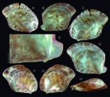
Electroma electra shell of types; (A) Holotype MZSP 166643, right valve, outer view (L 4.8 mm); (B) same, left valve, outer view; (C) same, right valve, inner view; (D) same, left valve, inner view; (E) same, left valve, detail of hinge, inner view, scale: = 0.5 mm; (F H) Paratype MZSP 164274#1 (L 4.0 mm), right, left and dorsal views.
The taxonomy of B. candida, previously a well-established species, has suffered a revulsion by Huber (2015). That author argued that the suppose type material of Arca domingensis Lamarck, 1819 represents the species that had so far been usually treated as B. cancellaria (Lamarck, 1819) in the literature. He argued that because Arca cancellaria Lamarck, 1819 does not have a type locality and bears doubtful type material, use of Arca domingensis is preferred, even though it disrupts prevailing usage (MolluscaBase, 2023). However, what Simone & Chichvarkhin (2004), for example, called B. dominguensis (now Acar domingensis) remains unnamed. Analyzing the taxonomy of both, B. cancellaria and A. domingensis, and the resolution of Huber (2015), it is possible to realize that the author violated the ICZN article 24.2.1 - the principle of the “First Reviser”. Thus, a redefinition of both species is necessary, what is provided below:
Barbatia cancellaria (Lamarck, 1819) revalidated under the concept and review by Simone & Chichvarkhin (2004: 356-360, figs. 1-7, 33-36, 43-51). (Fig. 44A, C)
Synonymy see Lamy (1907: 55) and Simone & Chichvarkhin (2004: 356).
Remarks: See below.
Genus Acar Gray, 1857
Acar domingensis (Lamarck, 1819) revalidated under the concept and review by Simone & Chichvarkhin (2004: 363-365, figs. 15-25, 60-65). (Fig. 44B)
Synonymy see Lamy (1907: 80-82) and Simone & Chichvarkhin (2004: 363).
Remarks: This item reunites the taxonomic discussion both, of B. cancellaria and of A. domingensis. Both Lamarck’s (1819) species were relatively forgotten along the 19th century (Lamy, 1907). Both species were not mentioned neither in the magistral survey by Reeve (1844). Both are, however, present in another important survey (Martini-Cheminitz) - Kobelt (1891), which can be considered the first reviser. In that paper, both species are illustrated by the first time. Both illustrations are reproduced here, Fig. 44A is of the Arca cancellaria Lamarck representative (Kobelt, 1891: 72, sp. 52, pl. 20, fig. 5), while Fig. 44B represents Arca domingensis Lamarck (Kobelt, 1891: 195, sp. 178, pl. 47 fig. 5). Both illustrations and descriptions by Kobelt (1891) - the first reviser, clearly show what were considered as those species along the time up to Huber (2015). Besides, other studies deepened the concept of both species, including anatomical features (e.g.,Simone & Chichvarkhin, 2004).
Thus, based on the ICZN article 24.2.1 - the principle of the “First Reviser”. The previous taxonomic concept of both, Barbatica cancellaria and Acar domingensis are revalidated, resolving an important gap of the Western Atlantic arcid taxonomy. One of the B. cancellaria syntype is illustrated here (Fig. 44C), showing the exact characteristics of what is known for the species. This resolution is also adopted in a recent paper (Passos et al., 2024: 23).
Genus Bathyarca Kobelt, 1891 Bathyarca arcadia new species (Figs. 45, 46A-E) https://zoobank.org/6C0FC89C-AAE7-49CD-AB2D-E0622D676960
Types: Holotype MZSP 166512, shell. Paratypes: MZSP 166514, 20 shells, MNHN IM 2000-39812, 3 shells from type locality. BRAZIL. Espírito Santo; off Itaúnas, 19°41′S 37°48′W, 790-940 m, MZSP 166516, 40 shells, MNHN IM 2000-39813, 20 shells (R.V. Marión-Dufresne col., MD55 sta. CB77, 27.v.1987).
Type locality: BRAZIL. Espírito Santo; off Conceição da Barra, 18°59′S 37°50′W, 637 m [R.V. Marión-Dufresne col., MD55 sta. CB76, 27.v.1987].
Diagnosis: SE Brazilian species with oblique outline, delicate outer sculpture. Hinge with very oblique teeth in both ends, middle teeth minute, narrow, deformed.
Description: Shell of ~7 mm, rather oblique; length similar to height; maximum inflation ~80% of length (Fig. 45C, M). Color white (Fig. 46A-F) to pale beige (Fig. 45A-F), with hairy beige periostracum close to edges (Figs. 45A, B, K, 58E). Right valve slightly smaller than left valve, exceeding in ventral edge (Fig. 45A, F, K). Prodissoconch elliptic, smooth, simple, of almost 1 mm (Fig. 45J: arrow). Umbones prominent, blunt, elevating ~6% shell height beyond hinge; separated from each other, located in anterior end of mid third of hinge. Lunule narrow, concave, ~10% of shell width, slightly broader anterior to umbones (Fig. 45C, M). Dorsal edge straight; anterior edge rounded, slightly smaller than posterior edge; ventral edge bluntly angular in its division with posterior edge. Sculpture delicate reticulated; radial cords predominating in right valve (Figs. 45A, K, 46E), ~45 radial cords from anterior to posterior hinge edges; commarginal cords predominating in left valve (Figs. 45B, L, 46D). Hinge with 3-4 middle teeth very minute, narrow, rather deformed, located lightly posterior to umbo; anterior to them from 5 (Fig. 46C) up to 8 (Fig. 45G-I) very oblique teeth, prosogire, average angulation ~45° in relation to antero-posterior shell axis, second and third teeth as largest ones; posterior to narrow teeth 6 (Fig. 46C) up to 8-9 (Figs. 46G-H, 59B) very oblique teeth, opisthogire, average angulation ~45° in relation to antero-posterior shell axis, penultimate tooth as largest. Inner surface white, opaque; muscle scars of difficult delimitation; scars of anterior and posterior adductor muscles rounded, located below both hinge ends.
Etymology: The specific epithet is in apposition and a make-up word, although has a meaning in Latin of adventuresome, an allusion to difficulty in finding this isolated population.
Distribution: Rio Grande do Norte (Francisco, 2015) to Rio de Janeiro coasts.
Habitat: 223-1,050 m depth.
Measurements (in mm): Holotype MZSP 166512 (Fig. 45A-J): 7.6 by 7.2. Paratype: MZSP 166514#1 (Fig. 45K-M): 7.4 by 7.5; MZSP 166516#1 (Fig. 46C-D): 5.0 by 4.9; MZSP 166516#2 (Fig. 46E-F): 5.0 by 4.6.
Additional material examined: BRAZIL. Espírito Santo (R.V. Marión-Dufresne col., MD55, 1987); off Itaúnas, 19°00.4′S 37°48.8, 950-1,050 m, MZSP 166515, 3 shells (sta. DC72, 27.v.), 18°59′S 37°48′W, 1,200 m, MZSP 166520, 2 shell (sta. CB78, 27.v), 18°58′S 37°48′W, 682 m, MZSP 166522, 12 shells (sta. SY74, 27.v); off Linhares, 19°36′S 38°53′W, 640 m, MZSP 166517, 25 shells, MNHN, 14 shells (sta. CB93, 30.v). Rio de Janeiro; off Arraial do Cabo, 23°46.7′S 42°10.0″W, 610 m, MZSP 166521, 45 shells, MNHN, 25 shells (sta. CB105, 02.vi).
Remarks:B. arcadia already was reported by Francisco (2015: 159: fig. 53), identified as B. glomerula Dall, 1881. However, examining the syntypes of that species (USNM 63166 - Fig. 46G-K) B. arcadia differs in having a more oblique outline (while B. glomerura is more rounded); outer sculpture more delicate, in right valve, for example, it has ~45 radial cords versus 35 radial cords of B. glomerula (Fig. 46H, K), thus, the radial cords of B. glomerula are wider; and mainly in the hinge, as B. arcadia has a harrower hinge, mainly at middle, and its teeth are much more inclined (the hinge of B. glomerula is broader, and its teeth are almost vertical positioned, besides, it lacks central narrow region with minute, rather deformed teeth), also, the narrow, minute teethed region is allocated posterior to umbo, while B. glomerula has a subumbonal area lacking teeth (Fig. 46G, I, J), area absent in B. arcadia. From Bathyarca inaequisculpta (E.A. Smith, 1885), B. arcadia differs in having a more oblique shape, a more pointed and lower umbones, fewer radial sculpture in right valve, and more dorso-ventrally elongated shell.
There is no other Atlantic congener species that can be confused with B. arcadia. Undoubtedly it is the sibling species of B. glomerula, which looks to be restricted to northern waters, from Florida to Antilles. Bathyarca arcadia is its southern counterpart, occurring in E SE Brazilian coast, with a wide gap from Antilles to Rio Grande do Norte.
Genus Bentharca Verrill & Bush, 1898 Bentharca celeris new species (Fig. 47) https://zoobank.org/742020AF-4379-4C29-B1AA-4F0B4BC9F78A
Types: Holotype MZSP 166556, shell. Paratypes: MZSP 104210, 2 shells, MNHN IM 2000-39814, 1 shell from type locality. BRAZIL. Espírito Santo; off Linhares, 20°43.7′S 31°56.7′W, 944-945 m, MZSP 104139, 1 valve, (R.V. Marión-Dufresne col., MD55 sta. DC49, 15.v.1987).
Type locality: BRAZIL. Espírito Santo; off Linhares, Vitória-Trindade submarine mountain chain, 20°20.9′S 36°19′W, 1,417-1,440 m [R.V. Marión-Dufresne col., MD55 sta. CP32, 15.v.1987].
Diagnosis: SE Brazilian species with very oblique, elongated outline. Sculpture delicate, uniform reticulate. Lunula very narrow and short. Hinge with very perpendicular anterior teeth; posterior teeth connected in outer edges.
Description: Shell of ~10 mm, very oblique - with main carina ~35° in relation to tinge line; length ~70% of height; maximum inflation ~40% of length (Fig. 47A, I). Color whitish (Fig. 47H J), yellowish (Fig. 47K-L) to pale beige (Fig. 47A-E); periostracum wanting. Both valves symmetric. Umbones slightly prominent, bluntly pointed, elevating ~5% shell height beyond hinge; narrowly separated from each other, located in posterior end of anterior third of hinge. Lunule narrow, concave, ~10% of shell width, ~37% of shell length (Fig. 47A, I). Dorsal edge straight; anterior edge bluntly pointed with dorsal edge, ~⅓ size of posterior edge; ventral edge ample on posterior half, abruptly ascendent in anterior half; posterior edge straight, with wide, blunt ventro-posterior angulation with ventral edge. Hinge (Fig. 47D, E, K) with anterior ~¼ possessing 5-6 small, uniform-sized, rather perpendicular, aligned teeth; succeeded by ~¼ edentelous, in region adjacent to umbones; posterior half with 6-7 elongated, very oblique, successive low teeth, all of them connected with each other in their outer end by longitudinal, lateral, low fold (Fig. 47F, G). Inner surface white, glossy; muscle scars of difficult delimitation; scars of anterior and posterior adductor muscles rounded, located below both hinge ends; posterior scar slightly larger than anterior scar.
Etymology: The specific epithet is derived from the Latin celer, meaning velocity, an allusion to the elongated shape of the shell.
Distribution: So far endemic from off Espírito Santo coast, near Vitória-Trindade submarine mountain chain.
Habitat: 637-1,440 m depth.
Measurements (in mm): Holotype MZSP 166556 (Fig. 47A-G): 10.3 by 7.1. Paratype: MZSP 104210#2 (Fig. 47H-J): 8.1 by 4.7; MZSP 104210#3 (Fig. 47K, L): 11.1 by 6.9; MZSP 104139 (Fig. 47M): 11.5 by 7.0.
Additional material examined: BRAZIL. Espírito Santo (R.V. Marión-Dufresne col., MD55, 27.v.1987); off Itaúnas, 18°58.9′S 37°49.6, 637 m, MZSP 166557, 1 valve (sta. CB76), 19°40.6′S 37°48.1′W, 790-940 m, MZSP 166558, 4 valves (sta. CB77).
Material of B. asperula: BRAZIL. Rio de Janeiro; Cabo Frio, off, 24°12′39″S 40°23′39″W, 3,049 m (R.V.W. Besnard col., sta. RBT 225, 07.vi.1971).
Remarks:Bentharca celeris is most closely related to B. asperula (Dall, 1881). B. asperula exhibits an extraordinarily wide geographic distribution, spanning from Japan across the entire Indo-Pacific to the Atlantic (MolluscaBase, 2024; Passos et al., 2024). It is associated with 8 nominal synonyms, two of which originate from the Atlantic: Arca culebrensis E.A. Smith, 1885 - found off Culebra Island, West Indies, at 390 fathoms, and Arca profundicola Verrill & Smith, 1885 - from station 2226, at 2021 fathoms, 37°N 71°54′W [an Atlantic Ocean point off Maryland]. The remaining synonyms originate from the Indo-Pacific or Japan. The type localities of B. asperula include “station 33, 1568 fathoms; station 19, 310 fathoms; Cape San Antonio, 1002 fathoms” (Dall, 1881: 121), all situated in the Caribbean and Gulf of Mexico regions.
While it may be difficult to believe that such a small bivalve, seemingly limited in its planktonic dispersion and absent from glacial areas, can maintain gene flow among distant oceans via glacial seas, challenging the synonymies of B. asperula lies beyond the scope of this paper. Nevertheless, the extraordinary wide distribution and numerous synonyms naturally create uncertainty regarding related species. However, B. celeris appears reasonably distinct from B. asperula, indicating a convincing case for specific separation rather than simple regional variation. Furthermore, shells of both species were collected sympatrically at MD55 station 77, and the differences between them were evident: MZSP samples 166558 (B. celeris) and 166559 (B. asperula).
The shell of B. celeris is more elongated and oblique, with the umbo slightly more anterior, while B. asperula exhibits a more trapezoid-rectangular outline, with the umbo more centrally positioned. Additionally, the sculpture of B. celeris consists exclusively of delicate reticulate patterns, with equal predominance of concentric and radial cords (Fig. 47B, C, H, J, L, M), whereas B. asperula’s shell features strong, uniformly distributed commarginal threads. The lunule of B. celeris is much narrower and shorter (Fig. 47A, I) compared to that of B. asperula. The hinge structures also differ: the anterior teeth of B. celeris are perpendicular (Fig. 47D-G, K), while those of B. asperula are highly oblique; furthermore, the posterior set of teeth in B. celeris are connected along their lateral edges (Fig. 47F, G), whereas those of B. asperula remain free from each other.
Bentharca asperula has been documented in Brazil (Prado & Barros, 1994; Passos & Birman, 2009; Francisco, 2015). However, in all cases, the illustrated specimens unmistakably exhibit the characteristics of B. asperula rather than those of B. celeris. Thus far, this suggests that B. celeris appears to be endemic to the deep waters off the coast of Espírito Santo.
Superfamily Limopsoidea Family Limopsidae Genus Limopsis Sasso, 1827 Limopsis paucidentata Dall, 1886 stratum novum, revalidated (Fig. 48B-D)
Limopsis aurita:Dall, 1886: 237; Maury, 1920: 42; Poirier, 1954: 148; Rios, 1985: 211 (pl. 76, fig. 1072), 1994: 233 (pl. 81, fig. 1148), 2009: 477 (fig. 1341); Gofas et al., 2001: 206; Passos & Magalhães, 2011: 4; Janssen & Krylova, 2014: 56; Janssen, 2015: 18, 21 (figs. 31, 32); MolluscaBase (2024); Passos et al., 2024: 28 (non Brocchi, 1814).
Limopsis aurita paucidentataDall, 1886: 237 (unfigured).
Type locality: Sta. 117, near Jamaica, 874 fathoms (1,598 m).
Material examined: BRAZIL. Rio de Janeiro; Campos Bay, off Campos dos Goytacazes, 21°48′S 40°30′W, ~100 m, MZSP 156267, 1 spm (R.V. Astromar, 22.i.2019).
Remarks:Limopsis paucidentata (Fig. 48B D) was previously regarded as a Recent Atlantic occurrence of the Italian (Mediterranean) fossil Limopsis aurita, originally described as Arca aurita by Brocchi in 1814 (pg. 485, pl. 11, fig. 9) (Fig. 48A). In addition to significant differences in age and geographic distribution, the original illustrations (Fig. 48A) depict a shell with distinct characteristics, including a more dorso-ventrally elongated shape, pronounced inflation, coarse concentric sculpturing, and a much more prominent umbo.
Even if there were errors in the initial designation of figures, and assuming that the correct figure is fig. 8, that particular shell also differs notably. It exhibits a more crenulated margin, a more symmetrical shape (in contrast to L. paucidentata’s asymmetrical outline), and a hinge with a greater number of teeth.
Given these conchological differences, as well as the geographical and temporal disparities, the most appropriate taxonomic approach is to maintain L. aurita as a fossil species. This involves revalidating its synonym, identified as a subspecies by Dall, and recognizing it as a distinct full species.
Genus Paracratis Huber, 2010 Paracratis borealis (Jeffreys, 1869) revalidated, new combination (Fig. 48F)
Limopsis borealisJeffreys, 1869: 174-175.
Limopsis minuta:Maury, 1920: 43; Poirier, 1954: 148; Rios, 1975: 195 (pl. 62, fig. 952), 1985: 211 (pl. 76 fig. 1075), 1994: 233 (pl. 81, fig. 1152), 2009: 478 (fig. 1345); Gofas et al., 2001: 206; Janssen & Krylova, 2014: 56; Janssen, 2015: 16, 25; Molluscabase, 2024; Passos et al., 2024: 28 (non Philippi, 1836).
Type localities: 50 miles N of Hebrides, 189 fms. E Nordland, 70-100 fms. Upper Norway, 450 fms.
Remarks:Paracratis borealis (Fig. 48F) has previously been identified as Limopsis minuta, described by Philippi as Pectunculus minutus in 1836 (pg. 63, pl. 5, fig. 3) (Fig. 48E). This species was initially described from the Plio-Pleistocene deposits in Sicily, Italy (Mediterranean). It has been reported as present in the Recent period from Canada to Mexico, with some disjunct occurrences in South Brazil (e.g., Rios, 2009).
Despite the significant temporal and regional differences, which might suggest a strong case for specific separation, Limopsis minuta, as described and illustrated by Philippi (1836), still exhibits conchological distinctions (Fig. 48E). It is notably larger, reaching around 15 mm, whereas P. borealis measures approximately 6 mm. Furthermore, Limopsis minuta features a more arched hinge and a taller umbo compared to those of P. borealis (Fig. 48F). The shell outline is also much less inclined (around 80°), while P. borealis has an angulation of approximately 65°.
Based on these temporal, geographic, and morphological differences, the most appropriate taxonomical procedure is to maintain Limopsis minuta as a Plio-Pleistocene Italian fossil within the genus Paracratis. Concurrently, its previous synonym, P. borealis, should be revalidated to represent the Recent Atlantic species.
Order Mytilida Superfamily Mytiloidea Family Mytilidae Genus Brachidontes Swainson, 1840 Brachidontes ynous new species (Figs. 49-50) https://zoobank.org/AE6AA3AF-E95F-4664-B185-10EF4FC6BC4B
Types: Holotype MZSP 165671. Paratypes: MZSP 105355, 40 spm from type locality. BRAZIL. Espírito Santo; Trindade Island (ProTrindade Project), Farrilhões, 20°31′22.4″S 28°19′52″W, MZSP 109572, 25 spm (02.vi.2012), Tartarugas, 20°31′01.3″S 29°17′56.9″W, MZSP 115081, 7 spm (Abbate col., 2013), Enseada da Cachoeira, 20°30′57.1″S 29°20′15.2″W, MZSP 108114, 3 spm (J. Mendonça col., 2012), Ponta do Túnel, 20°31′36.8″S 29°18′14.3″W, MZSP 109914, 3 spm (J. Mendonça col., 2012).
Type locality: BRAZIL. Espírito Santo; Trindade Island, Enseada do Lixo, 20°31′43.5′S 29°19′28.1″W [17.ii.2012].
Diagnosis: Endemic species from Trindade Oceanic Island, living subtidal, isolated. Shell smaller than 10 mm. Color mostly light. Sculptured by relatively uniform, delicate, numerous radial cords. Hinge with 4 teeth anterior to ligament, 12-15 taxodont-like teeth posterior to ligament.
Description: Shell up to 10 mm, outline rather trapezoid-mytiliform. Anterior region 2-3 times smaller than posterior region; ~1.5 times longer than heigh; ~2 (Fig. 49F) to ~1.3 (Fig. 50E, K) times longer than wide. Color beige to light brown (Figs. 49B, G, F, 50A, B, F), with some few reddish (Fig. 50L, M) and brownish specimens (Fig. 50D, E). Umbones weakly protruded, subterminal, anterior. Sculpture relatively uniform, narrow, numerous, radial cords; located close from each other (interspaces smooth, ~⅓ of cords width); from umbo up to posterior, ventral and anterior edges, forming weak crenulate borders (Figs. 49A, H, 50G, H); cords slightly wide in some specimens, ~40 per valve (Fig. 49B, G), up to very narrow, ~60 per valve (Fig. 50F); cords rarely dichotomic, except for those located in strip between umbo and corner of ventral-posterior edges (Figs. 49B, 50B, D, L). Anterior edge narrowly rounded, sometimes as beak anterior to umbo (Fig. 50F-H, J), sometimes as softer slope (Figs. 49A, B, G, H, 50A, B, D, L); dorsal edge straight; posterior edge amply rounded; ventral edge straight (Figs. 49A, B, G, H, 50A, B, D) to weakly convex (Fig. 50F, G, H, L). Inner surface glossy, translucent. Hinge with ligament occupying ~⅓ of its length, from umbo beak towards posterior (Fig. 49A, D, H, J, G, H); set of 4 teeth located anterior to ligament, weakly oblique, slightly uniform, except for more posterior tooth, being larger in right valve (Figs. 49C, 50J), and very oblique in left valve (Figs. 49I, 50I); set of 12-15 aligned, taxodont-like teeth posterior to ligament, being anterior teeth slightly smaller than posterior teeth (Figs. 49D, J, 50G, H). Scars of adductor muscles very weak; scar of anterior adductor muscle antero-posteriorly elongated, located in ventral region of anterior edge (Figs. 49A, H, J, 50G, H, I), with ~1/₃₀ of inner valve area; scar of posterior adductor muscle united to that of pair of posterior retractor muscles of foot, located from posterior region of umbonal cavity up to posterior region of hinge, both occupying ~1/₁₀ of inner valve area. Pallial line simple.
Mantle lobes (ml) translucent, except for middle and dorsal regions, fulfilled by whitish gonad (Fig. 49L: go). Mantle edges (mb) relatively thick, unpigmented; fusion between both lobes only between incurrent (ih) and excurrent (ex) canals (Fig. 49M: un); both canals lacking siphons or papillae, only simple edges (Fig. 49M). Gill (gi) very large, almost as large as pallial cavity; inner (id) and outer (od) demibranchs of equal length and width (Fig. 49K, L). Foot very small, peduncle-like (Fig. 49K: ft). Anterior adductor muscle from flattened to elliptic in section (Fig. 49K, L: aa), located in antero-ventral region. Posterior adductor muscle large, located as posterior end of visceral mass (Fig. 49K-M: ap). Pair of anterior retractor muscles of foot (Fig. 49K: ar) narrow, long, ~⅓ of visceral mass length, running from foot base up to umbonal cavity, flanking ventral surface of local visceral mass. Pair of posterior retractor muscle (Fig. 49K: rr) with insertion as large as anterior adductor muscle, just anterior to it. Strong cruciform-like pallial muscle (Fig. 49M: pm) in region between incurrent and excurrent muscle.
Etymology: The specific epithet is a Latinization of the native Guarani word yno’õ, meaning island, or insular, an allusion to the endemism of the species to the Trindade oceanic island.
Distribution: Endemic from Trindade Island.
Habitat: Under rocks, from intertidal up to ~16 m. Shell usually covered by incrustations, rare specimens with valves exposed.
Measurements (respectively L, H in mm): Holotype (Fig. 49A J): 8.4 by 5.5. Paratypes: MZSP 105355#1 (Fig. 50A-C): 8.3 by 5.5; MZSP 105355#2 (Fig. 50D-E): 6.3 by 4.4; MZSP 109572#1 (Fig. 50F-K): 7.6 by 6.2; MZSP 109572#2 (Fig. 50L-M): 7.6 by 5.6.
Additional material examined: BRAZIL. Espírito Santo; Trindade Island (ProTrindade Project), Andradas, 20°30′50″S 29°18′20″W, MZSP 101235, 3 spm (J. Mendonça col., 24.vii.2011); Farrilhões, 20°31′28.9″S 29°19′34.8″W, MZSP 104352, 2 spm (Abbate col., 16.ii.2012), MZSP 104445, 1 spm (Abbate col., 31.i.2012), MZSP 104419, 3 spm (Abbate col., 21.ii.2012), Enseada do Lixo, 20°31′43.5′S 29°19′28.1″W, MZSP 105356, 6 spm (17.ii.2012), MZSP 105376, 25 spm (21.ii.2012), MZSP 108238, 1 spm, Ilha da Racha, 20°30′26.5″S 29°20′48″W, MZSP 108108, 5 spm, Calheta, 20°30′26.1″S 29°18′44.2″W, MZSP 108122, 12 spm, 20°30′37.6″S, 29°18′28.1″W, MZSP 109617, 3 spm (12.vi.2012), 20°30′18.72″S 29°18′31.67″W, 16.7 m, MZSP 162509, 1 spm (24.v.2022), MZSP 121639, 7 spm (26.x.2014), Ponta do Nordeste, 20°29′46.4″S 29°20′35.4″W, MZSP 108136, 4 spm, Parcel das Tartarugas, 20°31′01.3″S 29°17′56.9″W, MZSP 108398, 13 spm (11.vii.2012), Enseada do Meio, 20°29′32.3″S 29°20′32.6″W, MZSP 108409, 9 spm, Ponta do Túnel, 20°31′36.9″S 29°18′14.3″W, MZSP 108533, 2 spm (19.vi.2012), Ponta Norte, 20°29′18.7″S 29°20′18.3″W, MZSP 162608, 5 spm, MZSP 162499, 6 spm (26.v.2022), Praia do Noroeste, 20°29′46.4″S 29°20′35.4″W, MZSP 162607, 7 spm, MZSP 162580, 8 spm (28.xi.2017), MZSP 162595, 2 spm (06.viii, 2018), 9 m, MZSP 162538, 15 spm (30.vii.2018), Orelhas, 20°29′40.2″S 29°20′32.9″W, MZSP 162597, 1 spm (07.viii.2018), MZSP 121604, 5 spm (01.xi.2014), MZSP 162592, 8 spm (06.xii.2017), 9 m, MZSP 162529, 1 spm (08.viii.2018); Martin Vaz Island, 20°31′36.9″S 29°18′14.3″W, MZSP 108429, 3 spm (30.vi.2012).
Remarks: Despite the Brachidontes that occur in Brazilian coast are far in being taxonomically well-known, the morphology, conchology and lifestyle of B. ynous are sufficiently different to be convincing that it is a new species. It appears to be endemic of Trindade oceanic island, a very remote place, ~2,000 km off the coast of Espírito Santo. It is the only species that does not occur in aggregates, i.e., it does not make colonies covering rocks like other local species do. On contrary, it is usually found isolated, on the hidden surface of the rocks. Also, it differently occurs much deeper, up to ~15 m, while the other congener species are intertidal. Three species of Brachidontes are reported to Brazilian coast (Rios, 2009), but mostly possibly they are all misidentifications (personal observation), two of Caribbean species [B. exustus (Linné, 1758) and B. solisianus (d’Orbigny, 1842) (presently in the genus Mytilaster Monterosato, 1884)], and one of an Argentinian species [B. rodriguezii (d’Orbigny, 1842)]. Sometimes, the Argentinian B. darwinianus (d’Orbigny, 1842) is also reported to Brazil (e.g., Rios, 1975). All these species have some synonyms, but only 2 have type locality in Brazil [Mytilus exiguus Dunker, 1875 - Santa Catarina; and M. janeirensis Dunker, 1866 - Rio de Janeiro], both considered synonym of M. solisianus. From these species, the only one that have the richness of teeth in the hinge is B. exustus (Simone et al., 2015: figs. 55-58), as well as the Caribbean B. modiolus (Linné, 1767), a very similar species. Brachidontes ynous differs from these two species in being much smaller (they easily reach 30 mm, while B. ynous rarely reach 10 mm), by more rounded outline (they are much more elongated), in lacking yellow pigment, in having the radial sculpture much more dense (while the sculpture of those species have wider interspaces), and by the anterior set of teeth being curved, instead of forming a straight platform with 1-2 more teeth, projected beyond the umbonal level. The other species lack these teeth or have them much less developed; additionally, they are dark-brown pigmented, with coarser sculpture.
Order Ostreida Superfamily Ostreoidea Family Ostreidae Subfamily Crassostreinae Genus Crassostrea Sacco, 1897 Crassostrea mangle Amaral & Simone, 2014, revalidated
Types: Holotype MZSP 89462. Paratypes: MZSP 166513, 29 specimens (dismembered from MZSP 89462), MZSP 100495, from type locality (collected in 08.i.2011).
Type locality: BRAZIL. Alagoas; Barra de Camaragibe, Camaragibe River Estuary, 09°18′52.82″S 35°25′31.69″W [Tavares et al., col., 21.x.2008].
Remarks:Crassostrea mangle was described replacing what had previously been identified as C. rhizophorae (Guilding, 1828) in Brazil. Following the study conducted by Amaral & Simone in 2014, the distribution of C. rhizophorae was constrained to Granada in the Antilles, Caribbean, which is its designated type locality. Even the occurrence of this species in other regions of the Caribbean and surrounding areas has been questioned. However, it is important to note that in the aforementioned study, the type specimens and the type locality of C. mangle were not explicitly established; they were only mentioned in a list. This oversight is rectified in the current work as above stated.
The validity of C. mangle was recently challenged, being synonymized with C. rhizophorae (Ferreira et al., 2023). However, that paper did not study samples from the type locality of C. rhizophorae, nor from the Caribbean at all. Instead, they based their synonymy on another paper (Lapègue et al., 2002), which studied specimens from Martinique and found the same haplotype in specimens from Paraná, Brazil. On the one hand, it is well known that the same haplotypes can occur in different species. On the other hand, it is very possible that C. rhizophorae does not occur in Martinique, and the specimens there may represent a different species. As mentioned above and by Amaral & Simone (2014), preliminary results indicate that several species of Crassostrea inhabit the Caribbean, with a certain degree of endemism. An example in the genus is C. praia (Ihering, 1907), which is endemic to a mangrove in Rio Grande, Rio Grande do Sul, Brazil.
Superfamily Pterioidea Family Vulsellidae Genus Electroma Stoliczka, 1871 Electroma electra Simone, Gomes & Molozzi, new species (Fig. 51) https://zoobank.org/15E763A7-2599-40C7-B7E3-6281303A64D0
Types: Holotype 166643. Paratype: MZSP 163738, 6 spm, MZSP 164274, 6 spm, from type locality.
Type locality: BRAZIL. Paraíba; Cabedelo, off, 07°02′41″S 34°47′42″W, ~14 m [C.R. Medeiros col., 2022].
Diagnosis: NE Brazilian species of ~5 mm; inclination of ~70°. Dorsal base relatively straight, slightly forming small projections.
Description: Shell of very fragile, translucent walls; about as tall as long; anterior, dorsal and anterior edges of both valves turned to right (Fig. 51H), friable. Basic color whitish to light beige, with irregular, coalescent, brown chevrons, of variable distribution and density (Fig. 51A, B, F, G). Outline slightly inclined ~70° in relation to longitudinal axis; anterior and ventral edges performing wide rounded arc, blunt beak with slightly concave posterior edge; dorsal edge almost straight, with small protruded umbo located between middle and anterior thirds; dorsal edge forming weak projections in both ends; left valve with subterminal byssal notch (Fig. 51B, E, D, G); with dorsal edge bearing 3 successive, small nodes (Fig. 51E). Umbo small, pointed, with rounded prodissoconch at tip (Fig. 51E). Hinge edentulous, with ligament in its entire extension (Fig. 51C, D, E); slightly thick in sub-umbonal area. Inner surface weakly glossy and iridescent, lacking visible nacre (Fig. 51C, D). Muscular scar of difficult visualization; scar of posterior adductor and posterior retractor muscle rounded, located in middle-posterior region.
Etymology: The specific epithet has as inspiration the Latin word electra, which originated the word electricity. This is an allusion to the shell color chevrons, looking like sparce electric rays.
Distribution: From Maranhão to Espírito Santo, Brazil.
Habitat: From 14 to 83 m depth.
Measurements (in mm): Holotype MZSP 166643 (Fig. 51A-E): 4.8 by 3.8. Paratype: MZSP 164274#1 (Fig. 51F-H): 4.0 by 4.0.
Additional material examined: BRAZIL. Maranhão; Carutapera, off Baía do Iririmirim, 00°44.973′S 45°52.285′W, 83 m, MZSP 164611, 5 spm (Piatam sta. 207, 19.xi.2008). Espírito Santo; Linhares, off, Abrolhos slope, 19°34.2′S 38°55′W, 340-360 m, MZSP 166719, 2 valves (MD55 sta. CB92.; 29.v.1987).
Remarks:Electroma electra is coauthored by Wilma Izabelly Ananias Gomes [Laboratório de Ecologia de Bentos - LEB, Universidade Estadual da Paraíba - UEPB, Avenida das Baraúnas, 351, Bodocongó, Campina Grande, PB, CEP: 58429-500, Brasil. E mail: wilmaizabelly@hotmail.com], and by Joseline Molozzi [Pós-Graduação em Ecologia e Conservação - PPGEC, Laboratório de Ecologia de Bentos - LEB, Universidade Estadual da Paraíba - UEPB, Avenida das Baraúnas, 351, Bodocongó, Campina Grande, PB, CEP: 58429-500, Brasil. E mail: jmolozzi@gmail.com], who collected and selected the first samples.
Its most similar species is the Mediterranean E. vexillum (Reeve, 1857), from which it differs slightly in having a more inclined outline, a more pointed umbo, and a straight dorsal edge. In contrast, E. vexillum has a rounded dorsal edge. Electroma vexillum has been reported as introduced in Colombia (Ardila, 2024), but whether that report pertains to E. electra is still under analysis. All remaining congeners are fossil species or from the Indo-Pacific region, and they typically have larger and more inclined shells compared to E. electra.
Superfamily Pinnoidea Family Pinnidae Genus Pinna Linné, 1758 Pinna trindadis new species (Fig. 52) https://zoobank.org/FECE27D9-B6BA-481A-A2CA-402FF0FF5BD2
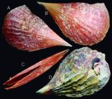
Pinna trindadis type shells: (A C) Holotype MZSP 108933 (L 301 mm), left, right and ventral views; (D) Paratype MZSP 105350, more usual dirty model (L 314 mm).
Types: Holotype MZSP 108933, shell. Paratype: MZSP 105350, 1 spm from type locality, 23 m (17.ii.2012).
Type locality: BRAZIL. Espírito Santo; Trindade Island, Enseada do Lixo, 20°31′33.9′S 29°19′33.6″W, 20 m [26.vi.2012].
Diagnosis: Brazilian from Trindade, with adult of ~300 mm; color brown. Umbonal angle 50-60°. Sculpture ~10 radial wide folds, relatively uniform, each one widening towards posterior.
Description: Shell easily over 300 mm. Basic color brown, being slightly lighter close to umbos. Antero-posteriorly long, ~1.8 times longer than heigh; flattened, ~6 times longer than wide. Middle blunt longitudinal carina developed, more pronounced in anterior half (Fig. 52A, B, D). Sculpture growth lines and concentric stairs, mainly in more posterior regions, where sometimes some isolated scale appearing (Fig. 52C); ~10 radial (longitudinal) folds, initially narrow, with interspaces equivalent to their width in anterior third; gradually becoming less nitid, as simple undulations in middle and posterior thirds, but present up to posterior edge, producing barely undulated posterior edge. Dorsal edge practically straight (Fig. 52A-B) or with weak posterior curve (Fig. 52D); ventral edge slightly concave; posterior edge amply rounded. Hinge edentulous, with ligament all along it, from anterior beak, up to short region posterior to posterior edge. Posterior region and posterior half of ventral region with opened gap (Fig. 52C).
Etymology: The specific epithet is derived from the local of occurrence - Trindade Island.
Distribution: Endemic from Trindade Island.
Habitat: Partially buried in sediment, 10-23 m. Shell mostly covered by incrustations (Fig. 52D), rarely found with clean shell (Fig. 52A-C).
Measurements (L, H in mm): Holotype (Fig. 52A C): 301 by 162. Paratype: MZSP 105350 (Fig. 52D): 314 by 190.
Additional material examined: BRAZIL. Espírito Santo; Trindade Island (ProTrindade Project), MZSP 131272, 1 spm (J. Mendonça col.), Calhetas, off Calhetas, 20°29′54″S 29°18′17″W, 35 m, MZSP 101104, 1 spm (23.vii.2011), praia, 20°30′28.3″S 29°18′38.8″W, 8 m, MZSP 104436, 1 spm, 1 shell (08.ii.2012), 20°30′36.1″S 29°18′44.2″W, MZSP 108253, 1 spm (03.vii.2012) 20°30′18.72″S 29°18′31.67″W, MZSP 118389, 1 spm (J. Braga col., 29.iv.2014), Enseada do Lixo, 20°31143.5!S 29°19′28.1″W, 15 m, MZSP 104434, 1 spm (20.ii.2012), Ilha Racha, 20°30′24.04″S 29°20′47.86″W, MZSP 104438, 1 spm, 1 shell (24.i.2012), 20°30′26.5″S 28°20′48″W, MZSP 109608, 1 spm, MZSP 109837, 1 spm (22.vi.2012), MZSP 122126, 1 spm (J. Braga col., 12.xi.2014), MZSP 131268, 2 spm, 22 m, MZSP 131270, 1 spm, 21 m (J. Mendonça col., 01.vii.2016), Andradas, 20°28′47.69″S 29°18′24.03″W, MZSP 105366, 4 spm (06.ii.2012), Orelhas, 20°29′32.5″S 20°20′32.6″W, MZSP 108934, 2 spm (22.vi.2012), 20°29′40.2″S 29°20′32.9″W, MZSP 115453, 2 spm (06.vii.2013), MZSP 118252, 1 spm, MZSP 118376, 1 spm (J. Braga col., 04.v.2014), MZSP 121994, 1 spm, MZSP 122173, 1 spm (J. Braga col., 24.x.2014), MZSP 131267, 1 shell (J. Mendonça col., 02.vii.2015), Farrilhões, 20°31′02.4″S 29°19′32.9″W, MZSP 110004, 1 spm (18.vi.2012).
Remarks: See below.
Pinna nembia new species (Fig. 53) https://zoobank.org/EE5017D1-5B54-4114-B247-C93BBF8FE602
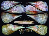
Pinna nembia type shells: (A D) Holotype MZSP 102181 (L 254 mm); (A) right valve, outer view; (B) same, inner view; (C) left valve, outer view; (D) same, inner view; (E G) paratype MZSP 165698#1 (L 237 mm), right, left and ventral views; (H I) paratype MZSP 105570 (L 185 mm), right and left views.
Types: Holotype MZSP 102181, specimen. Paratypes: MZSP 165698, 2 spm from type locality. BRAZIL. São Paulo; Ilhabela, Ilha dos Búzios, Costa do Aquário, 23°47.762′S 45°9.282′W, 10 m, MZSP 105570, 1 spm (Simone et al. col., 16.v.2012).
Type locality: BRAZIL. São Paulo; São Sebastião, Ilha dos Alcatrazes, Saco da Tartaruga, 24°06′24.95″S 45°42′17.19″S, 17 m (Simone et al. col., 05.x.2011).
Diagnosis: SE Brazilian species, with adult up to ~250 mm; color light beige-translucent. Umbonal angle ~40°. Sculpture ~10 radial wide folds, relatively uniform, each one entirely wide, usually rich in scales.
Description: Shell up to ~250 mm. Basic color light beige-translucent. Antero-posteriorly long, 2.4-2.1 times longer than heigh; flattened, ~6 times longer than wide. Middle blunt longitudinal weakly carina developed, more pronounced in anterior third (Fig. 53A, C, E, F, H, I). Sculpture growth lines, with weak concentric undulations in more posterior regions; sparse scales in posterior half (Fig. 53G); ~10 radial (longitudinal) folds, all of them appearing uniform undulations since anterior beak, increasing like that up to posterior margin, sometimes in straight manner (Fig. 53C, E), sometimes with weak undulations, mainly posterior (Fig. 53F, H, I). Dorsal edge practically straight (Fig. 53E, F) or with weak posterior curve (Fig. 53A-D, H-I); ventral edge straight (Fig. 53E-I) to sightly convex (Fig. 53A-D); posterior edge amply rounded (Fig. 53H-I) to weakly flattened (Fig. 53A-F). Hinge edentulous, with ligament all along it, from anterior beak, up to short region posterior to posterior edge. Posterior region and posterior quarter of ventral region with opened gap (Fig. 53G). Inner surface glossy, with pair of iridescent regions in anterior third, separated by narrow longitudinal opaque band (Fig. 53B, D); muscular scars not clear.
Etymology: The specific epithet is a Latinization of the native Guarani word ñemby, meaning “south,” in allusion to the southern occurrence of the species. It is the southernmost species of the genus in the Western Atlantic.
Distribution: Coasts of Rio de Janeiro to São Paulo.
Habitat: Mostly buried in sediment, 8-25 m.
Measurements (L, H in mm): Holotype (Fig. 53A-D): 254 by 105. Paratype: MZSP 165698#1 (Fig. 53E-G): 237 by 110; MZSP 105570 (Fig. 53H-I): 185 by 84.
Additional material examined: BRAZIL. Rio de Janeiro; Cabo Frio, Praia da Lagoa de Araruama, 22°53′10.12″S 42°00′12.66″W, MZSP 77163, 1 shell (Simone col., 15.i.1985); Angra dos Reis, Ilha Grande, 23°07′37″S 44°17′05″W, 10 m, MZSP 132104, 1 spm (Simone col., 2007), Praia dos Meros (Y. Bovi col.), 23°13′09.57″S 44°20′28.34″W, 8 m, MZSP 84227, 1 spm (ii.2007), MZSP 85981, 1 spm (vii.2007); São Paulo; Ubatuba, off Ubatuba, 23°31′S 44°54′W, MZSP 143087, 1 spm (SOL ISOTAN sta. 10.1, 10.i.1970); Ilhabela, Ilha dos Búzios, Costa das Etátuas, 23°47.408′S 45°00.455′W, MZSP 105782, 1 spm (Simone col., 16.v.2012), Parcel da Pedra Lisa, 23°47.450′S 45°08.718′W, MZSP 117182, 3 spm (Pastorino et al. col., 29.viii.2014); Santos, Laje de Santos, 24°18′S 46°11′W, 20 m, MZSP 151580, 1 spm (IOUSP col., i.2014); Itanhaém, Ilha da Queimada Pequena, 23°35′39″S 46°36′39″W, 12 m, MZSP 85627, 1 shell (Dornellas et al. col., 27.ii.2007); Peruíbe, Ilha da Queimada Grande, 24°28′57″S 46°40′41″W, 20 m, MZSP 35666, 2 spm (Souza & Simone col., 29.vi.2002).
Remarks: See below.
Pinna pereria new species (Fig. 54) https://zoobank.org/429DA8F7-89FD-4179-ADDD-BE96D7F2310D
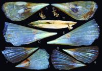
Pinna pereria type shells: (A C) holotype MZSP 165700 (L 105 mm), right, left, and ventral views; (D E) paratype MZSP 28508#1 (L 97 mm), right and left views; (F H) paratype MZSP 131559#1 (L 164 mm), (F) right valve outer view; (G) left valve inner view; (H) same, outer view; (I) specimen MZSP 141247#1 from Fernando de Noronha (L 106 mm).
Types: Holotype MZSP 165700, specimen. Paratypes: MZSP 28508, 4 spm from type locality. BRAZIL. Bahia; Salvador, Porto da Barra, 13°00′11.51″S 38°31′58.9″W, MZSP 131559, 2 shells (G.J.P. Queiroz col., i.1981; Colella collection).
Type locality: BRAZIL. Bahia; Salvador, Farol da Barra, 10°00′37.44″S 38°31′32.69″W, 5 m [Simone col., 22.ii.1997].
Diagnosis: NE Brazilian species, with adult up to ~150 mm; color whitish to light yellow, transparent. Umbonal angle ~45-50°. Sculpture 6-8 radial wide folds in dorsal ⅔ region only, relatively uniform or sinuous, each fold initially narrow; scales absent.
Description: Shell up to ~160 mm. Basic color whitish to light yellow; walls thin, rather flexible, transparent. Antero-posteriorly long, 2.3-2.5 times longer than heigh; flattened, ~5.6 times longer than wide. Middle blunt longitudinal weakly carina weakly developed, more pronounced in anterior third (Fig. 54A, B, D-H). Sculpture growth lines, with weak concentric undulations in more posterior regions; lacking scales; 6 (Fig. 54A, B, F-H) to 8 (Fig. 54D, E) radial (longitudinal) folds, all of them initially narrow, with interspaces equivalent to their width in anterior third; gradually becoming less nitid, as simple undulations in middle and posterior thirds, but present up to posterior edge, producing barely undulated posterior edge (Fig. 54E, G). Dorsal edge practically straight; ventral edge straight (Fig. 54E, D), slightly concave (Fig. 54A-B) to sightly convex (Fig. 54F-H); posterior edge ample. Hinge edentulous, with ligament all along it, from anterior beak, up to short region posterior to posterior edge (Fig. 54G). Posterior region and posterior half of ventral region with opened gap (Fig. 54C). Inner surface glossy, with pair of iridescent regions in anterior third, separated by narrow longitudinal opaque band (Fig. 54G); muscular scars not clear.
Etymology: The specific epithet is a Latinization of the native Guarani word pererĩ, meaning “fragile,” in allusion to the very fragile, almost friable shell.
Distribution: Coast of Rio Grande do Norte to Bahia, including Fernando de Noronha oceanic Island.
Habitat: Mostly buried in sediment among rocks, 4-25 m.
Measurements (L, H in mm): Holotype (Fig. 54A-C): 105 by 41. Paratype: MZSP 28508#1 (Fig. 54D-E): 97 by 37. Paratype: MZSP 131559#1 (Fig. 54F-H): 164 by 71; MZSP 141247#1 (Fig. 54I): 106 by 57.
Additional material examined: BRAZIL. Rio Grande do Norte; Areia Branca, Recife João da Cunha, 04°45′S 36°58′W, 9 m, MZSP 141617, 2 spm (Simone & Souza col., viii.2003). Pernambuco; MZSP 161823, 2 shells (Ex collection Vanin); Fernando de Noronha Archipelago, 03°52′S 32°25′W, MZSP 109129, 1 spm (Pronex), Laje Dois Irmãos, 03°49′47.28″S 32°25′01.89″W, 24 m (Simone et al. col., 26.ix.2018), MZSP 141247, 2 spm, MZSP 141298, 2 shells, Buraco das Cabras, 5 m, 03°52′39″S 32°28′39″W, MZSP 89955, 1 spm (Simone & Cunha col., 10.iii.2009), Ilha da Rata, Buraco do Inferno, 03°48′31.24″S 32°22′52.71″W, 10-15 m, MZSP 112964, 1 spm (Simone col., 05.v.2013), MZSP 31118, 1 shell (Simone & Souza col., 19.vii.1999), 03°48′28.11″S 32°22′56.9″W, 12 m, MZSP 141617, 1 spm (Simone et al. col., 27.ix.2018). Bahia; Salvador, Ribeira, 12°55′06.69″S 38°30′12.72″W, MZSP 81328, 1 shell (Simone col., 27.ii.1997), Farol da Barra, 13°00′37.44″S 38°31′32.69″W, MZSP 81329, 1 shell (Simone col., 22.ii.1997), 4 m, MZSP 63710, 1 shell (Simone col., 01.i.1985), Itapuã, 12°57′08″S 38°21′56″W, 3 m, MZSP 77151, 1 shell (Simone col., 21.i.1983); Alcobaça, off Alcobaça, 17°30′14″S 38°48′51″W, 10-20 m, MZSP 160543, 1 shell (W. Vailant col., 2022); Abrolhos, MSP 29040, 1 spm (R. Moura col., i.1997).
Remarks: The taxonomic discussion of the 3 species described herein is done once in these paragraphs. All of them have been reported as Brazilian occurrences of Pinna carnea Gmelin, 1971, a species in which they actually are similar. A clue for the misidentification stated with a study of the “true” P. carnea (Simone et al., 2015). In that study, specimens from Florida, a locality close to its type locality (Guantánamo Bay, Cuba) were described. Analyzing the Pinna samples collected in Brazilian coast, it was possible to easily separate them into 3 morpho-species and, additionally, realize that no one of them actually correspond to P. carnea. Each morpho-species occur in separated coastal region, being one of them the northeastern coast (Pinna pereria), the other in southeastern coast (P. nembia), and the third being endemic from Trindade (P. trindadis), the remote oceanic Island mentioned above.
As P. carnea has its anatomical features known (Simone et al., 2015) a preliminary anatomical investigation of the three Brazilian species was already performed. Interesting differences were found in all of them, but they will be explored in a future complementary paper. The present one is only devoted to conchological characters, as they are sufficiently convincing to base their taxonomy. As presently reported in the additional examined material, the argumentation is based on a large number of specimens, on a wide range of their occurrence, and, thus, very well supported. Related to the size, P. trindadis is the larger one, with most specimens being over 300 mm; P. nembia is the second larger, being the largest specimens about 250 mm, but must of them around 200 mm; P. pereria is the smallest one, rarely being over 150 mm, with the majority about 100 mm; P. carnea has some large specimens, easily reaching 300 mm, in south Caribbean region. Related to the shell color, P. trindadis has a strong brown to orange color, while P. nembia is light brown, and P. pereria is whitish or light yellow; P. carnea has a wider color variation, from brown, beige, up to reddish tones. The shell walls are thick in P. trindadis, not translucent; they are relatively thin, translucent in P. nembia; while the walls are very thin, flexible and transparent in P. pereria; P. carnea usually has a thickness comparable to that of P. nembia. The shell sculpture is relatively variable in pinnids, but a pattern is possible to be inferred to each species, in P. trindadis, the sculpture is of ~10 radial (longitudinal) folds, initially narrow, with interspaces equivalent to their width in anterior third, which gradually becomes less nitid, as simple undulations in middle and posterior thirds (Fig. 52); something similar occurs in P. pereria, but there are only 6-8 radial folds, and the ventral third region lacks folds, being smooth (Fig. 54D-I) or with non-radial sculpture (Fig. 54A-B); while P. nembia also has ~10 radial folds, but all of them appears as uniform undulations since anterior beak, increasing like that up to posterior margin; P. carnea has more radial folds, usually 10-12, also with distinction between anterior and posterior folds. The presence of scales in the posterior region is more developed in P. nembia, rare in P. trindadis, and never found in P. pereria; this is a very variable character in P. carnea. The umbonal angulation of all species are also different, P. trindadis is the ampler, with angle of ~50-60°, P. pereria has ~45-50°, P. nembia ~40°, and P. carnea is the sharper pointed, with angle ~25-30°.
Observing the abundant material of each species, it is possible to observe a relative uniformity of conchological features, which further supports the specific separation. A same uniformity is not found in the samples from Caribbean and Florida region, identified as P. carnea. It is a quite possible that, as much as occurred in Brazilian coast, under this epithet, a set of several similar-shelled species may exist. Pinna carnea has 4 nominal synonyms (MolluscaBase, 2023), 2 of them have no stated type localities and never where figured (P. degenera Link, 1807; P. pernula Röding, 1798), and 2 have type locality in the Caribbean Sea (P. flabellumLamarck, 1819 - West Indies; P. varicosa Lamarck, 1819 - Trinidad). Thus, no synonyms can be revalidated to shelter the Brazilian species.
There is no other valid Pinna species in the Western Atlantic. In the Eastern Atlantic, 2 species occur, the 3 Brazilian species differ from P. rudis Linné, 1758, mainly in not being so angulate (that species usually has umbonal angle ~25°), in not having reddish tones in shell color, and in lacking so developed scales on folds. They further differ from P. nobilis Linné, 1758 mainly in being much smaller (that species is huge, easily being over 500 mm), in not being so angulate (that species usually has umbonal angle ~25°), and differently sculptured (that species possesses striate sculpture, instead of folds).
Genus Servatrina Iredale, 1939 Servatrina amazonica new species (Fig. 55) https://zoobank.org/6E2EE385-EFB2-484B-86B3-395B1D1EDD4A
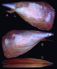
Servatrina amazonica Holotype MZSP 139952 (L 163 mm): (A) left view; (B) right view; (C) ventral view.
Types: Holotype MZSP 139952, specimen. Paratype: MZSP 139953, 2 specimens near type locality, 3.80012°N 50.39266°W, 42.3 m (o.t., sta. 2, 01.viii.2017, Wagner César R. Santos leg.).
Type locality: BRAZIL. Pará; off Rio Amazonas mouth, 3.94464°N 50.27423°W, 62.8 m [otter trawl, sta. 77, 25.v.2018, Wagner César R. Santos leg.].
Diagnosis: N Brazilian species, with adult size ~160 mm; color light brown. Umbonal angle ~30°. Sculpture 20 radial narrow folds in dorsal half, ~40 similar, but narrower folds in ventral half, relatively uniform, lacking scales; folds similar since anterior region.
Description: Shell ~160 mm. Basic color uniform light brown, being paler close umbo; walls relatively thick, not translucent. Umbonal angle ~30°. Antero-posteriorly long, ~1.7 times longer than heigh; flattened, ~5.4 times longer than wide (Fig. 55C). Middle longitudinal weakly wanting. Sculpture growth lines, with weak concentric undulations in more posterior regions; lacking scales in adult form, some present in young specimen; ~20 radial (longitudinal) folds in dorsal half (Fig. 55A, B), all of them narrow, uniform, interspaces 3-4 times wider than folds, in some regions narrow and wide folds intercalated; ventral half with ~40 folds similar to those folds of dorsal region, but narrower and located much closer from each other, interspaces 1-2 times folds width; border between dorsal and ventral set of folds gradual. Dorsal edge straight to weakly convex; ventral edge concave (Fig. 55A, B); posterior edge ample, with perpendicular to rounded superior region. Hinge edentulous, with ligament all along it, from anterior beak, up to short region posterior to posterior edge. Posterior region only with opened gap (Fig. 55C).
Etymology: The specific epithet is derived from the region of occurrence, off the Amazon river mouth; the term “amazonica” is the Portuguese feminine adjective relative to Amazonian origins.
Distribution: So far known off the mouth of Amazon River, between the Brazilian States of Amapá and Pará.
Habitat: Dredged by otter trawl, 42.3-62.8 m.
Measurements (L, H in mm): Holotype (Fig. 55A-C): 163 by 94.
Remarks:Servatrina amazonica has as closest species S. serrata (Sowerby I, 1825), a species that occurs from North Carolina to Suriname. Both species are the only ones that possess the delicate sculpture and the abundance of radial delicate folds. Servatrina amazonica differs in having slightly fewer folds, and, mainly, in haring simple, smooth folds, while those of S. serrata are mainly composed of successive micro-scales. Additionally, S. amazonica, as an umbonal angulation sharper (~30°), while that of S. serrata orbits ~40°. Also, S. amazonica appears to be smaller, being up to ~160 mm, while S. serrata easily is over 300 mm.
The other Western Atlantic species of Servatrina or Atrina Gray, 1842, such as A. rigida (Lightfoot, 1786) (occurrence from Caribbean to Venezuela) and S. seminuda (Lamarck, 1819) (occurrence from North Carolina to Chubut, Argentina) are very different, in having wider and much fewer radial folds, and a richness of scales (Schultz & Huber, 2013).
Subclass Autobranchia Infraclass Heteroconchia Superorder Archiheterodonta Order Carditida Superfamily Crassatelloidea Family Crassatellidae Genus Kalolophus DeVries, 2016 Kalolophus antillarum (Reeve, 1842)
Remarks:K. antillarum is designated with the type locality “ad insulam Margaritta, Antillarum,” along the Venezuelan Caribbean coast. Its distribution is relatively restricted, spanning from the Antilles to Venezuela, and typically found at depths ranging from 13 to 64 meters. Despite this, MolluscaBase (2023) lists numerous synonyms for K. antillarum, with type localities situated along the Pacific coasts of North, Central, and even South America. Some of these synonyms include fossil species, such as a Tertiary fossil from Brazil and another Cenozoic fossil from Peru. Upon thorough examination, it was observed that the shells of these synonyms exhibit distinctions from the K. antillarum types deposited in BMNH. Due to these differences, coupled with geographical and temporal disparities, these synonymies are not currently considered valid. Further research is required to establish the taxonomic status of these entities, which possibly are valid.
Superfamily Carditoidea Family Carditidae Genus Warrana Laseron, 1953 Warrana culmen Simone, Medeiros & Molozzi, new species (Fig. 56A-G) https://zoobank.org/B3A7914F-EFA4-4E19-B1DA-7C25C8FFA67F
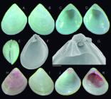
Warrana culmen shell of types and W. besnardi shell: (A) W. culmen holotype MZSP 166644 (H 1.3 mm), right valve, outer view; (B) same, left valve, outer view; (C) same, left valve, inner view; (D) same, right valve, inner view; (E) whole dorsal view; (F) paratype MZSP 163737#1 (H 1.3 mm), left valve in SEM, inner view; (G) same, detail of hinge, scale: = 100 µm; (H) W. besnardi, specimen MZSP 93035#1 (from Cananéia, São Paulo) (H 1.1 mm), right valve, outer view; (I) same, left valve, outer view; (J) left valve, inner view; (K) right valve, inner view.
Types: Holotype 166644. Paratype: MZSP 163737, 3 spm, from type locality.
Type locality: BRAZIL. Paraíba; Cabedelo, off, 07°02′41″S 34°47′42″W, ~14 m [C.R. Medeiros col., 2022].
Diagnosis: NE Brazilian species with umbonal angle of ~70°; height ~1.2× longer than wide. Umbonal transverse fold narrow. Hinge occupying ~21% of shell height. Crenulation in shell edges minute and numerous.
Description: Shell relatively thick; walls slightly translucent; outline oval, ~1.2× taller than long. Color white. Umbo central, blunt, with both sides straight, relatively symmetrical (anterior edge slightly shorter), with angle ~70°; ventral edge widely rounded (Fig. 56A-D, F). Prodissoconch smooth, bulging, occupying ~5% of shell height, ~14% of shell width, of 207 µm; flanked by narrow transverse umbonal fold (Fig. 56F, G). Sculpture uniform, concentric cords from transverse umbonal fold up to shell edge, interspaces deep, equivalent to half each cord width. Hinge thick, occupying ~21% of shell height, bearing only cardinal tooth, and wide anterior resilifer. Resilifer as simple, transverse groove, twice longer than wide, ligament circular in section, locater only in its dorsal, sub-umbonal region (Fig. 56F, G). Hinge teeth in posterior hinge half. Left valve with large anterior cardinal tooth, rather triangular, 3 times taller than long slightly bifid ventrally; posteriorly flanked by similar-sized socket (Fig. 56C, F, G). Right valve with tooth-socked counterpart (Fig. 56D). Inner surface glossy, with muscular scars of difficult visualization (Fig. 56C, D, F); scar of anterior and posterior adductor muscles relatively small, rounded, located in both sides of umbonal slopes; pallial line simple. Shell inner edge possessing minute crenulations in its entire ventral border (Fig. 56F).
Etymology: The specific epithet has the origin the Latin word culmen, meaning summit; an allusion to angulation of the shell apex (umbo), a species’ characteristics.
Distribution: From Maranhão to Alagoas, Brazil.
Habitat: From 14 to 35 m depth.
Measurements (in mm): Holotype MZSP 166644 (Fig. 56A-E): 1.0 by 1.3; MZSP 136737#1 (Fig. 56F, G): 1.0 by 1.3.
Additional material examined: BRAZIL. Maranhão; Apicum-Açu, off, 00°59.834′S 44°20.433′W, 35 m, MZSP 152577, 1 shell (PIATAM OCEANO sta. 307, 26.x.2008). Paraíba; Cabedelo, Praia Formosa, 06°59′05.4′S 34°49′38.2″W, MZSP 133438, 57 shells (Colella collection, A. Birman col., xi.1977). Alagoas; Japaratinga, off Praia de Japaratinga, 09°05′53.02″S 35°15′06.84″W, 4 m, MZSP 166758, 3 spm (F. Hartmann col.).
Remarks:Warrana culmen is coauthored by Carlinda Raílly Medeiros [Programa de Pós-Graduação em Engenharia Civil e Ambiental - PPGECA, Laboratório de Hidráulica II - LabHidII, Universidade Federal de Campina Grande - UFCG, Rua Aprígio Veloso, 882, Universitário, Campina Grande, PB, CEP: 58429-900, Brasil. E mail: carlindarailly@gmail.com] and by Joseline Molozzi [Pós-Graduação em Ecologia e Conservação - PPGEC, Laboratório de Ecologia de Bentos - LEB, Universidade Estadual da Paraíba - UEPB, Avenida das Baraúnas, 351, Bodocongó, Campina Grande, PB, CEP: 58429-500, Brasil. E mail: jmolozzi@gmail.com], who collected and selected the first samples.
Warrana culmen exhibits similarities with W. besnardi (Klappenbach, 1963), a species that has previously been confused with it. Warrana besnardi is confined to the southeastern Brazilian coast, whereas W. culmen is found along the northeastern Brazilian coast. Although W. culmen and W. besnardi share some resemblances, distinct differences exist among them, discernible in all examined samples (W. besnardi, represented by 7 lots in the MZSP collection, with numerous specimens each - Fig. 56H-K). Notable distinctions include the following: (1) The umbonal angulation of W. culmen is more acute, approximately 70°, whereas that of W. besnardi is broader, around 90°; (2) W. culmen possesses a slender outline with a height/length ratio of 1.2, while W. besnardi tends to be as wide as it is tall (~1.0 ratio) (Fig. 56H, I); (3) The hinge thickness of W. culmen is proportionally wider, occupying approximately 21% of the shell height, compared to W. besnardi, where this proportion is about 16%; (4) The crenulation along the ventral edge of W. culmen is more delicate and abundant (Fig. 56F) than that of W. besnardi (Fig. 56J, K); (5) Notably, the transverse umbonal fold of W. culmen is narrower and less conspicuous, whereas that of W. besnardi is more pronounced and obvious (Fig. 56H-K).
The classification of Brazilian species of Cuninae carditids was partly influenced by Middelfart (2002), who revealed a greater number of species than previously documented in Australia and the surrounding region, demonstrating species distinctions comparable to those observed presently.
Superorder Imparidentia Order Lucinida Superfamily Lucinoidea Family Lucinidae Genus Lucinoma Dall, 1901 Lucinoma apocalyptica new species (Fig. 57) https://zoobank.org/FCF81E9E-956D-43D3-9FE3-39799DA3A13B
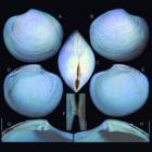
Lucinoma apocalyptica holotype MZSP 162656 shell (L 79.3 mm): (A) left valve, outer view; (B) right valve, outer view; (C) whole dorsal view; (D) left valve, inner view; (E) Right valve, inner view; (F) hinge of both valves almost articulating, ventral view; (G) hinge, left valve, right view; (H) hinge, right valve, left view. Scales: = 10 mm.
Type: Holotype MZSP 162656, shell.
Type locality: BRAZIL. Pará; off Salinópolis, 01°04′N 46°18′W, 250 m [Femorale-Ex collection Vanin; RV Oregon col., o.t., xi.1957].
Diagnosis: N Brazilian species of large size (~80 mm), thick walled. Sculpture simple, uniform, lacking undulations, carina or abrupt slope. Outline rounded. Umbo broad.
Description: Shell (Fig. 57), of ~80 mm; outline rounded; walls heavy, thick. Color white, opaque. Maximum inflation ~half length (Fig. 57C). Umbones central, slightly prosocline, anterior to it slightly concave edges, posterior to it slightly convex edges. Anterior, ventral and posterior edges performing soft rounded profile (Fig. 57A, B, D, E); posterior slope very weakly undulated, less acuminated than anterior region. Sculpture concentric, low scales, rather irregular, discontinuous; interspaces equivalent to 3-4 times each scales’ width; sculpture uniform from umbo up to margins (Fig. 57A, B). Pre-umbonal lunula present (Fig. 57C), sculptures only by very narrow, crowded, longitudinal lines. Ligament wide, mostly external, almost half of dorsal edge (Fig. 57C). Hinge (Fig. 57F-H) thick, ~8% of shell height; with pair of cardinal teeth in both valves, being anterior most tooth in right valve; posterior tooth in right valve and anterior tooth of left valve slightly bifid; weak anterior lateral tooth present, clearer in right valve (Fig. 57G, H); no detectable posterior lateral tooth. Ligament groove wide, from umbo up to almost posterior edge. Scar of anterior adductor muscle elongated, ~7 times longer than wide, situated ~18° in relation to vertical axis, ~13° in relation to adjacent pallial line. inner surface pure white, barely radially beaded (Fig. 57D, E). Scar of posterior adductor muscle elliptic, ~1.5 times longer than wide, with ~half of anterior muscle’s size. Scar of anterior pedal retractor muscle just superior and posterior to scar of anterior adductor muscle, rather rectangular, ~20 times smaller than adductor muscle. Pallial line complete, wide, anterior half nodulous; its anterior insertion in dorso-anterior edge of anterior adductor muscle edge; its posterior insertion in ventral end of posterior adductor muscle.
Etymology: The specific epithet is based on the Church Latin ‘apocalypsis,’ meaning “revelation, disclosure,” as well as from Greek ‘apokalyptein,’ meaning “uncover, disclose, reveal,” being an allusion to a large specimen being so far undiscovered in the north Brazilian deep waters.
Distribution: So far known from the type locality.
Habitat: 250 m depth.
Measurements (in mm): L 79.3 by H 71.4 by max. inflation 43.7.
Remarks: The taxonomy of the Lucinoma was very facilitated after the review by Taylor & Glover, 2016, who review the Western Atlantic species. The present described species belong to a set of 3 specimens collected by RV Oregon, acquired by Femorale, in which fortunately one specimen was donated to MZSP by Vanin’s family. The other specimens can be seen on the Femorale (2024) website. The large size, the only concentric sculpture, the thick tinge bearing a pair of cardinal teeth in each valve, and the anterior adductor muscle scar long, narrow, largely detached from pallial line, are all characters that induced the genetic attribution to L. apocalyptica.
The absence of well-spaced commarginal threads and absence of strong dorsal undulations easily differentiate L. apocalyptica from their Western Atlantic congeners L. filosa (Stimpson, 1851) and L. atlantis (McLean, 1936). The sculpture of L. apocalyptica is somewhat similar to that of L. blakeana (Bush, 1893), but it differs in being slightly more uniform. In the range of ~80 mm, L. apocalyptica is also much larger than its Western Atlantic congeners, that are in the range of 30-40 mm. Its umbo is blunt, while the umbo of those species is more pointed. Another interesting distinction is related to the hinge thickness, which is much broader in L. apocalyptica than those of the other species.
Lucinoma apocalyptica still differs from L. asapheusOliver, Rodrigues & Cunha, 2011, from NE Atlantic, in being larger (that species is ~25 mm long), more antero-posteriorly elongated (L. asapheus has a rounded outline), and by the hinge being thicker, with more robust cardinal teeth; on the other hand, the scar of the anterior adductor muscle has an equivalent length and format.
Order Cardiida Superfamily Tellinoidea Family Tellinidae Genus Lyratellina Olsson, 1961 Lyratellina juttingae Altena, 1965 (Fig. 58)
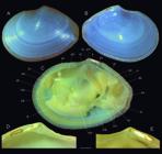
Lyratellina juttingae MZSP 166334, from off Pará: (A) left valve, outer view (L 35.3 mm); (B) same, inner view; (C) entire specimen, left view, left valve and left mantle lobe removed; (D) detail of hinge, left valve, inner view; (E) same, right valve. Scale: = 2 mm. Lettering: aa, anterior adductor muscle; ap, posterior adductor muscle; ar, anterior foot retractor muscle; cm, cruciform muscle; ex, excurrent siphon; ft, foot; go, gonad; hi, hinge; id, inner demibranch; in, incurrent siphon; li, ligament; mb, mantle border; od, outer demibranch; pc, pericardium; pl, posterior lateral tooth; pp, palps; pr, foot protractor muscle; rr, posterior foot retractor muscle; sc, siphonal chamber; sh, shell; sm, siphonal retractor muscle; um, umbo.
Lyratellina juttingaeAltena, 1965: 52 (fig. 1a e); MolluscaBase, 2023; Passos et al., 2024: 62.
Tellina (Merisca) juttingae:Rios, 2009: 546 (fig. 1520).
Type locality: N of the mouth of the Surinam River, 20 miles off the coast, 15 fms.
Remarks: This report is only an expansion of geographic distribution. The previous report is from Trinidad to Amapá, Brazil, but a sample of this species was collected off Pará, ~500 km southwards.
Interestingly, the very characteristic shell of L. juttindae (Fig. 58A-B, D-E), has a wide anterior region and walls relatively fragile, translucent walls. In the anatomy (Fig. 58C), it has a foot (ft) with a furrow along anterior edge, and relatively small and narrow siphons (ex, in). The retractor muscles of siphons (sm) are reunited in a pair of single central, longitudinal bundles. The palps (pp) are proportionally very large, comparable to the gill (id, od) size. As every tellinid, the protractor foot muscles (pr) have their origin partially dividing the anterior adductor muscle (aa).
Material examined: BRAZIL. Pará; off Rio Amazonas mouth, 3.94464°N 50.27423°W, 62.8 m, MZSP 166334, 2 spm (otter trawl, sta. 77, 25.v.2018, Wagner César R. Santos leg.).
Family Semelidae Genus Abra Lamarck, 1818 Abra americana (Verril & Bush, 1898) revalidated, new stratum (Fig. 59B-E)
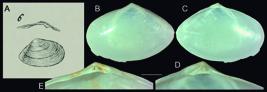
Studied Abra spp.: (A) replication of figure by Scacchi (1834: fig. 6) of his Italian fossil Tellina longicallus; (B E) shells details of A. americana; (B) MZSP 117015, off Rio de Janeiro, Brazil, left valve, outer view (L 15.4 mm); (C) same, inner view; (D) same, detail of hinge; (E) MZSP 116911, right valve, detail of hinge. Scale: = 2 mm.
Abra longicallis americanaVerrill & Bush, 1898: 778 (pl. 83, fig. 6-7); Warmke & Abbott, 1961: 201; Abbott, 19745: 515 (fig. 5805).
Abra longicallis:Morretes, 1949: 44; Rios, 1975: 249 (pl. 79, fig. 1194), 1985: 257 (pl. 91, fig. 1278); Díaz & Puyana, 1994: 14: 97 (fig. 271) (non Scacchi, 1835).
Abra longicallus:Rios, 2009: 556 (fig. 1551); MolluscaBase, 2023; Passos et al., 2024: 58 (non Scacchi, 1835).
Type locality: 6 stations between 39°49′N 68°28′30″W and 36°16′30″N 68°21′W, 924-2,620 fms.
Remarks: Until now, Abra americana has been identified as the Pliocene fossil Abra longicallus (Scacchi, 1835), originally classified within the genus Tellina Linné, 1758 (Fig. 59A), and described from Puglia, in the southern region of Italy. Initially proposed as a subspecies (technically a variety) of Abra longicallis (an unjustified emendation of longicallus) by Verrill & Bush in 1898, some authors adopted this classification, while most considered it a synonym (refer to synonymy above). In the original description, Verrill & Bush (1898: 778) emphasized that the variety then designated as americana differs from the European [fossil] form (longicallis) “in having the lateral tooth less remote, and the cartilage-pit or chondrophore longer, the antero-dorsal margin more convex, and the whole shell relatively broader.”
By examining the original image of the fossil (Fig. 59A) and comparing it to a typical shell of the living A. americana (Fig. 59B E), it becomes evident that, in addition to the features highlighted in the original description, the shells of the living population have a more pointed and centrally positioned umbo (in contrast to the fossil, which has a rounded umbo located more posteriorly). The sub-umbonal cardinal teeth are more developed, while the posterior cardinal teeth are shorter. Furthermore, the posterior edge is more rounded in the living species, whereas the fossil species exhibits a more angular shape. Additionally, the outer surface appears smoother in the living population, while the fossil species appears more concentrically sculptured.
Taking into account the temporal and geographical separation, along with the conchological differences explored above, the most appropriate taxonomic approach appears to be retaining A. longicallus solely for the Italian fossil species and designating the living Atlantic deepwater species as A. americana.
Order Venerida Family Veneridae Subfamily Petricolinae Choristodon typicum Jonas, 1844 revalidated
Choristodon typicumJonas, 1844: 185.
Type locality: insulam St. Thomas.
Remarks:Choristodon typicum, formerly recognized as Petricola typica and Rupellaria typica, has recently been synonymized with the Pacific species Choristodon robustus (Sowerby I, 1834) by, e.g.,Coan & Valentich-Scott (2012), MolluscaBase (2023), and Passos et al. (2024). Despite being considered sibling species, three factors suggest that this synonymy can be disregarded. Firstly, the southern region of South America does not host occurrences of C. typicum, making gene flow between both sides of the Americas impossible. In the Atlantic coast, for instance, the southernmost detected occurrence of C. typicum is in Santa Catarina, Brazil, significantly distant from the south curve to the Pacific region of the mainland. Secondly, Choristodon exhibits variable shell characteristics, making it challenging to identify secure conchological attributes justifying their separation. However, the Atlantic C. typicum generally displays a larger size and a coarser radial sculpture compared to the Pacific C. robustus, which tends to have a more delicate sculpture. Thirdly, the presence of C. typicum shells in pre-Columbian deposits, albeit rare, indicates that the species is not of anthropogenic introduction. Therefore, until a more robust database supports the improbable synonymy, Choristodon typicum will be maintained as a distinct taxon.
Superfamily Chamoidea Genus Pseudochama Odhner, 1917 Pseudochama radians (Lamarck, 1819) revalidated
Chama radiansLamarck, 1819: 96.
Chama cristellaLamarck, 1819: 96 (l’Océan de grandes Indes).
Type locality: “l’Océan de grandes Indes?”
Remarks: In the case of Pseudochama radians, the species has a notably problematic taxonomic history. Consequently, our approach to this species differs from the conventional method applied to other species. Instead of adhering strictly to the orthodox approach, we opt for a chronological explanation.
Currently, P. radians has been considered a synonym of P. cristella, both of which were described in the same paper, on the same page, and share the same type locality. It is worth noting that although P. radians precedes P. cristella (being species 12 and 13, respectively), this chronological order does not hold significance according to the International Code of Zoological Nomenclature (ICZN). The resolution of simultaneously published names is guided by the Principle of First Reviser (Art. 24.2).
Accordingly, Reeve (1847) was the first reviser, but he covered both species:
C. radians - pl. 4, fig. 19, local: ? (Lamarck described for “Ocean des grades Indes?”);
C. cristella - pl. 8, fig. 42, local “Batavia” (?) (Lamarck described it for “grades Indes”).
Reeve (1847) included both species, rendering the paper unsuitable for resolving the priority of names. Notably, the author modified the locality information for both species. In the case of C. radians, he retained only the original designation’s question mark. Conversely, for C. cristella, the location changed from the initially stated ‘undoubtful grandes Indes’ to ‘Batavia.’ Notably, Batavia appears to have been a region in the Netherlands, existing from 1795 to 1806.
The second reviser was Clessin, 1889, who also studied both species:
C. cristella (sp. 18, pg. 17, for Portorico);
C. radians (sp. 50, pg. 36, for Indischer Ocean, with the remark: “Reeve gibt keinen Fundort an.” - means Reeve does not provide a location, but Lamarck did… see above).
The examination of both species by Clessin (1889) also fails to resolve the priority of names. Nevertheless, the author did relocate C. cristella to the Caribbean (specifically Puerto Rico), associating it with C. radians.
Subsequent to these studies, C. cristella seemingly vanished from the literature on the Western Atlantic, with only occasional citations, such as those by Steyn & Lussi (1998) for South Africa and other papers in the Indo-Pacific region. In contrast, C. radians continued to be extensively mentioned, with approximately 40 papers citing the species. Notably, Huber (2010) resurrected an Atlantic C. cristella, designating it as the valid name and providing justification on page 678:
“Against page priority the type species Pseudochama, SD Gardner, 1926 P. cristella is here selected to represent this well known Caribbean species. The type locality of Pseudochama cristella (Lamarck, 1819) is corrected to the P. radians type locality Virgin Islands, St. Croix. There also the closest matching specimen has been found.”
Consulting Gardner (1926: 92), in the supposed “subsequent designation” of the type of Pseudochama there is:
“Type: Pseudochama cristella (Lamarck). (Recent from the Gulf of Siam to Java, the Molluccas, and Australia.)”
An apparent anomaly arises when considering Gardner’s concept of P. cristella, which pertains to the Indo-Pacific region. In contrast, Huber (2010) replaces the Western Atlantic P. radians with this very entity. However, there are additional complexities:
Upon consulting the original description of the genus Pseudochama by Odhner (1917: 30), the following details emerge:
“LYNGE (1908) gives a satisfactory criticism of CLESSIN’s opinion as to the Chama cristella of Reeve, which, according to the last-named author, is specifically distinct from the type of Lamarck, a view that cannot be maintained. The species is distributed from the Gulf of Siam to Java, the Moluccas, and Australia (Lynge, 1909). It is at once recognized by its orange colour and raised crista-shaped form, due to its attachment with the front half of the right valve, while its hind part is sharply bent from the substratum. I have referred this species to a new genus, Pseudochama, which comprises the so-called “inverse” Chamas, opposite to the normal or dextrally…” [my bold for emphasis].
These concluding paragraphs lead us to three key observations:
1) Pseudochama cristella does not emerge as a subsequent designation by Gardner in 1926. Odhner (1917) distinctly established it as the type species of the genus through original designation and monotypy.
2) There is a possibility that the Indo-Pacific ‘Chama cristella of Reeve’ mentioned by Odhner (1917: 30) does not correspond to Lamarck’s (1819) species from the Caribbean. It appears to be a misidentification, as Odhner seems to have had a different species in hand, distinct from the true C. cristella. Despite labeling it as the ‘C. cristella of Reeve,’ the author acknowledged and discussed the observed differences.
3) The alteration of the type locality of Chama cristella to ‘Virgin Islands, St. Croix’ by Huber (2010) seems inappropriate. This information lacks support in any published source, and the justification provided concerning P. radians type locality lacks clarity.
The priority dilemma finds resolution in the work of Pilsbry & McGinty (1938), the third reviser. In their comprehensive study, the authors addressed P. radians (pg. 77-78), establishing the species, along with some subspecies, for the Florida-West Indies region. Notably, P. cristella was seemingly overlooked, suggesting it may have been considered a non-regional species.
In summary, applying the Principle of First Reviser (ICZN Art. 24.2), and considering that the first two detected revisers (Reeve, 1847; Clessin, 1889) addressed both species without resolving priority, the ultimate determination came from the third reviser - Pilsbry & McGinty (1938), favoring P. radians.
Anything documented about C. or P. cristella between Clessin (1889) and Pilsbry & McGinty (1938) appears to be the result of misidentifications. These errors were rooted in the inclusion of Indo-Pacific populations, which represent distinct species. This is evident in Huber’s (2010) justification for considering P. cristella as valid (indirectly influenced by Odhner, 1917), wherein he referred to the ‘Chama cristella of Reeve,’ implying a species distinct from Lamarck’s description.
Order Adapedonta Superfamily Hiatelloidea Family Hiatellidae Genus Hiatella Bosc, 1801 Hiatella marisqueira new species (Figs. 60-61) https://zoobank.org/37E1F27B-9C6E-43A2-9698-020852D76AE9
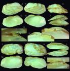
Hiatella marisqueira types. (A I) holotype MZSP 166300 (L 12.4 mm); (A) right valve, outer view; (B) left valve, inner view; (C) dorsal view; (D) left valve, inner view; (E) right valve, inner view; (F) ventral view, valves slightly opened to show hinge; (G) same, detail of hinge; (H) detail of hinge, right valve, inner view; (I) same for left valve; (J O) paratype MZSP 28813#1 (L 11.2 mm); (J) right valve, outer view; (K) left valve, inner view; (L) ventral view, valves slightly opened to show hinge; (M) left valve, inner view; (N) right valve, inner view; (O) hinge, ventral view. Scales: = 2 mm.
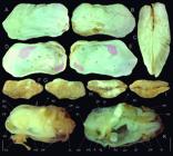
Hiatella marisqueira paratypes. (A E) MZSP 14160 (L 12.1 mm); (A) right valve, outer view; (B) left valve, inner view; (C) whole dorsal view; (D) left valve, inner view, muscles scars artificially colored; (E) same for right valve; (F I) MZSP 20120 (from Morretes collection) (L 14.6 mm), right, left, dorsal and ventral views; (J) Holotype soft parts, whole left view; (K) paratype MZSP 28813#1 soft parts, left view, left structures mostly removed, both siphons sectioned longitudinally. Scales: = 2 mm. Lettering: aa, anterior adductor muscle; ap, posterior adductor muscle; ar, anterior foot retractor muscle; by byssus; dg, digestive diverticula; ex, excurrent siphon; fg, foot byssal gap; ft, foot; gi, gill; go, gonad; ih, incurrent siphon; mb, mantle border; pe, periostracum; pp, palps; rr, posterior foot retractor muscle; st, stomach; un, fusion between both mantle boles.
Saxicava arctica:Morretes, 1949: 47 (non Linné, 1767).
Saxicava solida:Morretes, 1949: 47 (non Sowerby I, 1834).
Hiatella solida:Rios, 1970: 210; Narchi, 1973: 332-337 (figs. 1-7) (non Sowerby I, 1834).
Hiatella arctica:Rios, 1975: 253 (pl. 80, fig. 1212), 1985: 272 (pl. 95, fig. 1339), 1994: 294 (pl. 100, fig. 1432), 2009: 589 (fig. 1627) (non Linné, 1767).
Hiatella cf. solida: Passos et al., 2024: 46.
Types: Holotype MZSP 166300. Paratypes: MZSP 28813, 4 spm from type locality. BRAZIL. São Paulo; Ilhabela, Praia do Gato, 23°50′37.36″S 45°16′50.3″W (vii.1936). Paraná; Paranaguá, Ilha do Mel, Praia do Farol, 25°32′19.15″S 48°17′30.41″W, MZSP 20120, 16 shells (Morretes col., 15.iv.1933).
Type locality: BRAZIL. São Paulo; São Vicente, Ilha Porchat, 23°58′47.96″S 46°22′21.8″W, intertidal [O. Domaneschi col., vi.1993].
Diagnosis: SE S Brazilian species living usually associated to other intertidal invertebrates. Shell of rather rectangular outline, up to ~15 mm; lacking posterior carina. Muscle shell scars, both adductor muscles and foot retractor muscles proportionally large. Palps wide. Stomach lacking muscular belt. Intestine intensely coiled. Pair of siphons mostly separated from each other, with single line of terminal papillae.
Description: Shell variable, rather moldable, usually twice longer than wide, twice longer than height; outline slightly rectangular. Color white, with light beige periostracum, usually eroded in umbonal region (Figs. 60A, B, 61G, H), exceeding shell borders, mainly in posterior edges (Fig. 61J, K: pe). Umbones broad, occupying ~¼ of dorsal surface, slightly prominent; from terminal (Figs. 60J-N, 61A-E) to subterminal (Fig. 60A-E). Valves symmetric (Figs. 60L, 61C) to slightly asymmetric (Figs. 60C, 61H, I). Surface matte; sculpture lacking, except for growth lines and strong concentric undulations (Fig. 60A-B, J-K); in some few specimens with weak oblique carina present between ventral and posterior slope (Fig. 61A-B). Anterior and posterior regions rounded, similar-sized or with posterior region slightly larger; dorsal edge usually straight; ventral edge from convex (Fig. 60D-E) up to slightly concave (Fig. 61D-E). Inner surface glossy, pure white, weakly iridescent. Muscular scars shallow (Fig. 61D-E); scar of anterior adductor muscle slightly rectangular, located in ventro-anterior corner, occupying ~1/₁₅ of inner surface; scar of posterior adductor muscle rounded, slightly larger than anterior muscle, located between middle and posterior thirds of dorsal shell edge; scar of posterior foot retractor muscle located adjacent to posterior adductor, slightly anterior and dorsal to it, rather triangular, with ~75% of posterior adductor scar size; other scar, including pallial line, not detectable. Hinge only of cardinal teeth (Fig. 60F-I, L, O); left valve with small anterior, flattened cardinal teeth, corresponding to anterior socket in right valve; right valve with broad, slightly bifid posterior teeth, posteriorly interlocking with anterior teeth, and fitting in socket of left valve. Ligament occupying about double length as hinge, located posterior to hinge, protruding outside posterior to umbo (Figs. 60C, H, I, 61H).
Anatomical details in Narchi (1973, as H. solida). Complement (Fig. 61J-K): foot (ft) small, strongly attached to byssus fibers (by) in its ventral furrow. Mantle edge (mb) mostly fused between both lobes (un), only small gap present (fg) for foot and byssus access. Pair of siphons more than 50% separated from each other; incurrent siphon (ih) slightly broader and longer than excurrent siphon (ex); tip rounded, simple, with row of papillae in edge. Outer region of siphons protected by periostracum (pe). Anterior adductor muscle (aa) dislocated ventrally, close to ventral-anterior edge. Posterior adductor muscle (ap) slightly larger than anterior muscle, located between middle and posterior thirds of dorsal region. Pair of posterior foot retractor muscles (rr) strong, originated anterior to posterior adductor muscle, running almost vertically towards ventral, inserting in foot base, near byssus main insertion. Pair of anterior foot retractor muscles (ar) very narrow, originated in middle of anterior region, running directly towards posterior, inserting in same region of posterior retractor muscles.
Etymology: The specific epithet is derived from the Brazilian term marisqueira, meaning women that collect mussels. This is allusive to the habitat of some of the collected specimens, among the byssus of intertidal mussels.
Distribution: Rio de Janeiro to Rio Grande do Sul, Brazil.
Habitat: Intertidal, attached by byssus among mussels, usually Brachidontes spp., Perna perna (Linné, 1758), as well as to ascidian Polyandrocarpa zorritensis (Van Name, 1931) and among tubes of the polychaete Phragmatopoma caudata Krøyer in Mörch, 1863 (both Narchi, 1973).
Measurements (L, H in mm): Holotype MZSP 166300 (Fig. 60A I): 12.4 by 7.1. Paratype: MZSP 28813 (Fig. 60J-O): 11.2 by 7.0; MZSP 14160 (Fig. 61A-E): 12.1 by 6.5; MZSP 20120 (Fig. 61F-I): 14.6 by 5.1.
Additional material examined: 132 additional lots in MZSP collection from Rio de Janeiro up to Rio Grande do Sul.
Remarks:Hiatella marisqueira has been identified as both H. solida and H. arctica. Both species are part of a complex of ~60 taxa considered synonymous with H. arctica (MolluscaBase, 2023), occurring worldwide, from glacial up to tropical environments, from deep up to intertidal waters. Due to the wide plasticity of its shell, hard is to challenge this taxonomic insanity - a species exhibiting remarkable habitat tolerance, unknown dispersion patterns, and a wide range of forms and sizes. Focusing on what was referred to as H. arctica in SE Brazil up to N Argentina, a study started with samples collected in the latter region, in relative deep water (Simone & Penchaszadeh, 2008). In that study was concluded that the north Argentinian deep-water population could be secluded from the H. arctica complex, as it brings an own set of anatomical characters. A formerly synonym was revalidated - H. meridionalis d’Orbigny, 1846. The present paper refers to the neighbor population, occurring in SE Brazilian coast, up to Rio Grande do Sul. It occupies a distinct niche - intertidal, associated with some local invertebrates. As no one of the H. arctica complex taxa have type locality in SE Brazil, a new taxon was necessary to be introduced. The closest type locality is Santa Helena, of H. solida Sowerby I (1834: 88), collected at 33 m depth. Most remaining species are from Northern Atlantic region (H. arctica itself has “Oceano Norvegico” as type locality - Linné, 1767: 1113), or from Indo-Pacific.
Hiatella marisqueira differs from H. meridionalis (Simone & Penchaszadeh, 2008) in being slightly larger (12-15 mm) (while the other species rarely reaches 10 mm); in mostly lacking carina in shell posterior slope (almost always present in H. meridionalis), and by the muscle scars much larger, proportionally about double than those of H. meridionalis. Anatomically, the differences in muscle size, as referred above in the shell, are evident in both adductors and foot retractors; H. marisqueira still lack developed such quantity of papillae in the siphons, as that of H. meridionalis. The palps are also proportionally larger in H. marisqueira, as well as the stomach lacks a muscular belt, and the intestine is much more intensely coiled in H. marisqueira (Narchi, 1973) if compared to H. meridionalis.
An important feature of H. marisqueira, that looks exclusive if compared to all known Hiatella, including H. meridionalis (Simone & Penchaszadeh, 2008), and what is known about H. arctica (Hunter, 1949) is the conformation of the siphons. It has both siphons mostly separated from each other (Fig. 61J, K: ex, ih). The other species they are fused almost up to their tip, being a single large tube in an external view, and both incurrent and excurrent apertures are only distinguishable at tip of this tube.
Order Myida Superfamily Pholadoidea Family Pholadidae Genus Cyrtopleura Tryon, 1862 Cyrtopleura angelicalis Stakowian & Simone, new species (Fig. 62) https://zoobank.org/0648B886-E2EC-4C95-BD1B-CCDB0E6A0864
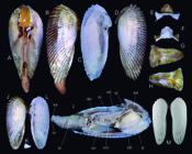
Cyrtopleura angelicalis types and C. costata shell: (A I) Holotype MZSP 143461 (L 105.2 mm); (A) whole dorsal view; (B) whole left view; (C) right valve, inner view; (D) same, outer view; (E) metaplax, outer view (W 22.1 mm); (F) same, inner view; (G) protoplax, outer view (L 30.0 mm), (H) same inner view; (I) whole right view, right valve and right mantle lobe removed; (J L) paratype MZSP 166518 (L 100.0 mm); (J) left valve, outer view; (K) its extracted umbonal appendix, inner view; (L) left valve, inner view; (M) C. costata typical Floridian specimen (from Goodland Bay, Collier County, courtesy E. Petuch; L 153 mm). Lettering: aa, anterior adductor muscle; ap, posterior adductor muscle; ex, excurrent siphon; ft, foot; id, inner demibranch; in, incurrent siphon; ki, kidney; od, outer demibranch; pp, palps; si, siphons; un, fused ventral edge between both mantle lobes (sectioned); vm, visceral mass.
Barnea (Scobinopholas) costata:Morretes, 1949: 49 (non Linné, 1758).
Cyrtopleura costata:Rios, 1970: 214, 1975: 254 (pl. 81, fig. 1214), 1985: 273 (pl. 95, fig. 1343), 1994: 295 (pl. 100, fig. 1437), 2009: 592 (fig. 1632); Passos & Magalhães, 2011: 6; Simone, 2019b: 10 (fig. 25); Stakowian & Simone, 2021: 1-7 (figs. 1-2); Passos et al., 2024: 52 (non Linné, 1758).
Types: Holotype MZSP 166519, spm. Paratypes: MZSP 143461, 13 spm, 166518, 1 spm (voucher of Stakowian & Simone, 2021) from type locality.
Type locality: BRAZIL. Paraná; São Miguel do Iguaçú, Baía de Paranaguá, Saco do Tambarutaca, near Port of São Miguel, 25°26′07.14″S 48°26′51.7″W [N. Stakowian col., 2018, estuarine mud].
Diagnosis: Shell height ~40% of length. Umbones between middle and anterior thirds. Sculptured by ~25 radial, tall threads, highly divergent anteriorly. Dorsal edge posterior to umbo wide. Infra-umbonal appendix broadly angulate (~40°).
Description: Shell of ~120 mm, ~2.5 times longer than tall; shell height ~40% of length; length ~2.1× maximum inflation (Fig. 62A). Color white, with thin, dark periostracum mainly preserved among threads. Anterior edge rounded; posterior edge bluntly pointed, almost half of anterior edge; ventral edge widely convex; all these edged undulating because of terminal prominence of radial sculpture, mainly in anterior edge. Umbones weakly prominent, located between anterior and middle thirds of dorsal edge; anterior to it rounded edge as flap of insertion of anterior adductor muscle, outer concavity; posterior to umbo similar edge flap, but ~twice longer and lower; dorsal flaps lacking sculpture. Sculpture ~25 radial threads (Fig. 62A, B, D, J), relatively uniformly spaced, interspaces ~3 times each thread width; each thread composed of successive, blunt nodes; interspaces smooth, only with growth lines; 4-5 anterior threads more spaced and more prominent, extending further shell edge. Inner surface opaque, white (Fig. 62C, L); anterior adductor muscle scar in umbonal region (also working as ligament and abductor muscle); posterior adductor muscle located in middle region between umbo and posterior end, close to dorsal edge, ~3 times longer than wide; pallial line wide, parallel to shell edge in anterior and middle region, in its region preceding posterior third suddenly turning dorsally, possessing siphonal retractor muscle scar, running as concave line up to posterior adductor muscle scar. Infra-umbonal appendix with base firmly attached, broadly angulate (~40°) (Fig. 62K, L). Metaplax (Fig. 62E, F) with flattened ventro-posterior flap; wide, laterally expanded anterior component, with wide lateral ends and 4 divergent longitudinal folds in middle. Mesoplax (Fig. 62G, H) entirely corneous, thin, flexible, attached almost directly to anterior adductor muscle (Fig. 62I: aa), outline slightly triangular, ~1.5 times longer than wide, blunt anterior end; inner scar triangular, occupying most of posterior third area.
Remaining characteristics, including anatomical description (Fig. 62I), see Stakowian & Simone, (2021: 1-7, figs. 1-2).
Etymology: The specific epithet has as inspiration the species’ common name “angel wings”, as its shell looks like, from the Latins angelicalis, meaning angelic.
Distribution: From Rio Grande do Norte to Santa Catarina. Brazil.
Habitat: Estuarine, intertidal level; muddy bottoms, buried in sediment up to ~1 m deep (Stakowian & Simone, 2021).
Measurements: Holotype MZSP 143461 (Fig. 62A I): 105.2 by 42.0. Paratype: MZSP 166518 (Fig. 62J L): 100.0 by 40.8.
Remarks:Cyrtopleura angelicalis, coauthored by Nicole Stakiwian, was previously identified as C. costata on the Brazilian coast. However, at the time of the publication of its anatomical description (Stakowian & Simone, 2021), several colleagues brought to our attention that the depicted shells did not entirely match those of the supposedly conspecific northern populations, specifically those from Florida and the Caribbean. One of the more emphatic colleagues was E. Petuch, who even sent a photo of a typical Floridian specimen (Fig. 62M), highlighting that the Floridian “true” C. costata is more elongated and slender, a feature that can also be observed in Caribbean populations. He also emphasized differences in rib count and the structure of the ribs, especially at the anterior end (Petuch, personal communication). Upon studying samples from the Caribbean and Florida regions, we found that these differences were consistent, and we identified additional distinctions, as discussed below.
Cyrtopleura angelicalis is thus formally introduced to the Brazilian population, coauthored by Nicole Stakowian, distinguished from C. costata by its broader valve (shell height approximately 40% of its length, compared to approximately 30% in C. costata). Additionally, C. angelicalis exhibits around 25 radial threads, while C. costata has roughly 35. Furthermore, the interspaces of the threads in C. angelicalis are wider, approximately three times the width of each thread, whereas in C. costata, the interspaces are narrow, equivalent to the width of the threads themselves. Moreover, the threads are more spaced out and prominent in the anterior region of C. angelicalis compared to those of C. costata. The infra-umbonal appendix of C. angelicalis also differs from that of C. costata (Fig. 62K), being slightly shorter and wider, with the base forming an angle of approximately 40°. In contrast, the appendix of C. costata is slightly narrower, with an angle of approximately 30°, and easier to detach, to the extent that it can be found isolated on beaches and was described as a peculiar capulid gastropod Capulus shreevei Conrad, 1869, with the type locality on Long Island, South Carolina (Conrad, 1869: pl. 13 fig. 3, 3a). Another synonym for C. costata is Leuconyx tyleriana H. & A. Adams, 1863, which lacks a type locality and has a scanty description. As no type material has been located and there is no indication that this species was based on Brazilian samples, the best course of action is to maintain it as a synonym of C. costata or possibly consider it a nomen dubium.
The distinctive characters mentioned above between C. angelicalis and C. costata obviously exhibit some degree of variation. The described distinctions are based on common patterns observed in both species. However, there is overlap of characters in some rare specimens, particularly in the anterior projected threads. The ideal scenario would be to investigate northern samples of C. costata with the same degree of detail as the Brazilian C. angelicalis ones (as conducted by Stakowian & Simone, 2021), along with DNA sequencing; undoubtedly, further distinctions would emerge.
Superorder Anomalodesmata Superfamily Verticordioidea Family Lyonsiellidae Genus Lyonsiella Sars, 1872 Lyonsiella angulosa new species (Fig. 63) https://zoobank.org/C869C843-38B7-4627-BC2D-F7874BCD3A0B
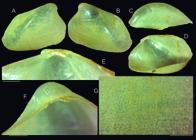
Lyonsiella angulosa holotype MZSP 166503 right valve (L 10.2 mm): (A) outer view; (B) inner view; (C) dorsal view; (D) ventral-slightly left view; (E) hinge region, ventral-slightly left view; (F) same, left view; (G) detail of outer surface of middle area of ventral region. Scales: = 1 mm.
Type: MZSP166503, shell (right valve and fragments of left valve).
Type locality: BRAZIL. Espírito Santo; off Itaúnas, continental slope, 18°59′S 37°50′W, 600-637 m [MD55 sta. CB76, Bouchet, Leal & Métivier col., 27.v.1987].
Diagnosis: S Atlantic species with prominent umbo as apex of high, blunt carina; outline rectangular. Micro-sculpture as radially aligned nodes.
Description: Shell of ~10 mm; wall fragile, slightly translucent; outline slightly rectangular, 1.4 times longer than tall, 2.8 times wider than long (each valve) (Fig. 63A-C). Umbo anterior, subterminal, very prominent; blunt, prosocline; from umbo initiating blunt carina, running obliquely towards ventral and posterior, separating posterior slope. Edge anterior to umbo slightly concave, edge posterior to umbo straight, bluntly angled ~150° with straight posterior edge; anterior edge rounded, slightly smaller than posterior; ventral edge vaguely convex. Sculptured by minute nodes radially aligned, ~8 lines per mm in middle region, apparently no concentric alignment (Fig. 63G); each node separated from each other by average space equivalent to 3 times its width. Hinge only in cardinal region, ~⅙ of shell length (Fig. 63D); with small, low, sub-umbonal tooth; and posterior thickness flanking ligament (Fig. 63E, F); ligament with narrow intervalvar component, and thick, oblique component (~⅛ of shell length) just posterior to cardinal tooth, flanking ventral edge of hinge thickness. Inns surface glossy, slightly iridescent (Fig. 63B, D); muscular scar of difficult individualization, both adductor scars on both ends of dorsal edge; pallial line entire.
Etymology: The specific epithet refers to the angularity of the shell outline, from the Latin angulus.
Distribution: So far known from the type locality.
Habitat: 600-637 m depth.
Measurements (in mm): 10.2 by 7.0.
Remarks:Lyonsiella angulosa differ from the species that occur in Western Atlantic, such as L. smidti Friele, 1886, in lacking circular outline in the valves. It differs from L. subquadrata (Jeffreys, 1882) in having a more pronounced carina, in lacking concentric undulations, and by delicate radial lines. It further differs from L. magnifica Dall, 1913, from Western Mexico coast, in having a more pronounced carina, and the umbo more prominent and more anteriorized. Comparing to the L. magnifica syntype (USNM 266802) it is possible to deduce that L. angulosa may be its Atlantic sibling species.
Class Scaphopoda Order Dentaliida Family Dentaliidae Genus Graptacme Pilsbry & Sharp, 1897 Graptacme obtura Fogarolli & Simone, new species (Fig. 64A F) https://zoobank.org/41CB788D-9EA1-4011-8DD2-3354945D0AC4
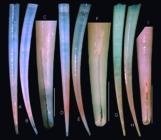
Shell of types of Graptacme obtura and G. semistriolata: (A) G. obtura holotype MZSP 166760 (L 35.6 mm), dorsal view; (B) same, right view; (C) same, detail of posterior region showing slit; (D F) G. obtura paratype MZSP 121837 (L 36.4 mm), same views as preceding specimen; (G I) G. semistriolata MZSP 47357 (L 26.4 mm), same views as first specimen. Scales: = 2 mm.
Types: Holotype MZSP 166760, spm. Paratypes: MZSP 121837, 6 spm from type locality.
Type locality: BRAZIL. Espírito Santo; Trindade oceanic island, Calheta beach, 20°30′37.6″S 29°18′28.1″W, 17 m [Protrindade Project, J.B. Mendonça col., 26.x.2014].
Diagnosis: Species from Trindade oceanic island with striae and slit in posterior region longer than 11% of shell length. Rate of increase 0,078 mm/mm of length. Strong bilabial posterior aperture.
Description: Size about 36 mm; walls thin, translucid, color white; well curved, section circular (Fig. 64A, B, D, E). widening smooth (tax of increase about 0.078 mm/mm of length). Sculpture absent, shell smooth, glossy; except for posterior 11-15% of shell length possessing longitudinal, delicate striae in entire circumference; striae gradually disappearing anteriorly (Fig. 64C, F). Anterior aperture rounded; peristome simple, with cutting edges. Posterior aperture with very narrow, elongated slit in ventral, sometimes in lateral surface (Fig. 64C, F), from 11 to 15% of shell length, sometimes longitudinal (Fig. 64F), sometimes slightly oblique (Fig. 64C).
Etymology: The specific epithet is derived from the Latin word obturus, meaning “plug”, an allusion to the shell format.
Distribution: Endemic from Trindade Island, Brazil.
Habitat: Sandy bottoms, about 10 m depth.
Measurements (in mm): Holotype MZSP 166760 (Fig. 64A-C): 35.6 by 2.9. Paratype: MZSP 121837 (Fig. 64D-F): 36.4 by 2.9; mean size of analyzed shells 36.3 mm. Apex slit mean 11,2% of total shell length. Anterior aperture width ~30 mm; posterior aperture width ~10 mm. Rate of increase of 0.078 mm/mm of length.
Additional material examined: BRAZIL. Espírito Santo; Trindade oceanic Island (J.B. Mendonça col.), Ponta da Calheta, 20°30′18.72″S 29°18′31.67″W, 17.8 m, MZSP 162545, 2 shells (10.viii.2018), 20°30′16.59″S 29°18′30.11″W, 14.6 m, MZSP 162575, 1 spm (30.i.2019), Ponta da Calheta, 20°30′18.72″S 29°18′31.67″W, 17.1 m, MZSP 162494, 1 spm (16.xi.2017), 17.2 m, MZSP 162593, 7 spm (04.xii.2017), Orelhas, 20°29′40.2″S 29°20′32.9″W, 14.5 m, MZSP 162559, 1 spm (23.vii.2018).
Remarks:Graptacme obtura was initially identified as G. semistriolata (Guilding, 1834), a species widely distributed from Florida to São Paulo (Caetano et al., 2006). However, upon closer examination, specimens from Trindade, a remote oceanic island approximately 2,000 km off the coast of Espírito Santo, were found to differ significantly from those on the mainland. Their shells exhibit a much longer slit on the posterior surface, extending up to 12-15% of the shell’s length, compared to G. semistriolata, which typically has a slit extension of around 5% (Fig. 64G, H). Additionally, the outer sculpture of the shells from Trindade is longer, reaching approximately 15%, while in G. semistriolata, the striation remains shorter, at about 5-6% only (Fig. 64I). Both species have remaining sell portions smooth, glossy. Graptacme obtura displays a more developed bilabial shape in the posterior aperture (Fig. 64C, F) compared to G. semistriolata (Fig. 64I). Furthermore, the shell outline of G. obtura is slender, with a rate of increase of 0.078 mm/mm of length, whereas G. semistriolata exhibits a proportionally wider shell, with a rate of increase of 0.102 mm/mm of length.
Among the other congeners found in the Western Atlantic (Caetano et al., 2006), G. obtura is readily distinguishable. It can be differentiated from G. calamus (Dall, 1889) by its narrower, pointed posterior end. Furthermore, G. obtura differs from G. perlonga (Dall, 1881) and G. eborea (Conrad, 1846) in having a much broader anterior region, whereas those species possess a very slender shell.
Order Gadilida Suborder Gadilimorpha Family Gadilidae Genus Polyschides Pilsbry & Sharp, 1898 Polyschides tetraschistus (Watson, 1879) (Fig. 65A-G)
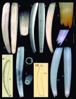
Polyschides tetraschistus shells of type specimens: (A) holotype of Siphodentalium tetraschistum BMNH 1887.2.9.66 (L 6.6 mm), left view; (B) same, dorsal view; (C) same, detail of posterior end; (D E) holotype of Polyschides xavante IBUFRJ 14190 (L 5.4 mm), left and dorsal views; (F) holotype of Polyschides noronhensis MZSP 88441, left view (L 8.6 mm); (G) paratype MZSP 46736, detail of posterior region, left view, scale: = 0.25 mm; (H) replication of Dall (1889: pl. 27, fig. 5) illustration of Cadulus quadridentatus (L 13.2 mm); (I) Polyschides quadridentatum holotype MCZ 7739 (L 9.5 mm), right view; (J) same, anterior view; (K) same, posterior view; (L) replication of Bush (1885: pl. 45, fig. 20) illustration of Cadulus incisus (L 8 mm); (M) ordinary specimen of P. quadridentatum, MZSP 119346 (L 6.1 mm) (from Itaparica, Bahia, Brazil) (courtesy Vitor A. Fogarolli), left view; (N) same, dorsal view; (O) same, detail of posterior end, right view, scale: = 1 mm.
Siphodentalium tetraschistumWatson, 1879: 521, 1886: 15-16 (pl. 2, fig. 8).
Cadulus tetraschistus:Pilsbry & Sharp 1898: 148 (pl. 23, fig. 1); Henderson, 1920: 3, 12, 96-97 (pl. 17, fig. 1); Morretes, 1949: 52; Lopes & Alvarenga, 1955: 181; Rios, 1970: 143.
Polyschides xavanteCaetano & Absalão, 2005: 1-8 (figs. 1-4) (new synonym).
Polyschides noronhensisSimone, 2009: 414-417 (figs. 16-22, 75-86) (new synonym).
Polyschides tetraschistus:Steiner & Kabat, 2001: 448.
Type localities:S. tetraschistum: Challenger sta. 113A: Anchorage off Fernando de Noronha, 7-25 fms [03°57′S 32°24′30″W, 13-46 m]. Polyschides xavante: Sancho Bay (3°51′S 32°26′W), Fernando de Noronha Island, Pernambuco, Brazil, 10-15 m. Polyschides noronhensis: Porto Beach, 03°50′06.67″S 32°24′10.00″W, 6 m.
Types:S. tetraschistum holotype BMNH 1887.2.9.66; P. xavante holotype IBUFRJ 14190. Paratype: MZSP 43518; P. noronhensis holotype MZSP 88441, MZSP 32011, 3 paratypes (all examined).
Remarks: A reanalysis of these gadilids, conducted in conjunction with the scaphopod specialist Carlos Henrique S. Caetano (Universidade do Estado do Rio de Janeiro), led me to the conclusion that the three species of Polyschides with a type locality from Fernando de Noronha actually constitute a single biological entity (Fig. 65A G). As discussed in the original description of the three species, they are differentiated by having a very slender shell, with relatively weak central obesity (Caetano & Absalão, 2005; Simone, 2009). Among the three names (see synonymic list above), priority is given to P. tetraschistus.
A challenge arises concerning the perception that the species is endemic to the Fernando de Noronha oceanic island. However, the former concept of “P. tetraschistus” describes a species with a more robust shell, distributed from North Carolina, USA, to Buenos Aires, Argentina. The former concept of P. tetraschistus has three nominal synonyms (MolluscaBase, 2023); the oldest one is Siphonodentalium quadridentatum Dall, 1881 (Fig. 65H), from Florida. This species can be revalidated to replace the previous concept, aiming to designate the species with a slightly more robust shell and a broader geographic range:
Polyschides quadridentatum (Dall, 1881) revalidated, new combination (Fig. 65H N)
Siphonodentalium quadridentatumDall, 1881: 36; Steiner & Kabat, 2004: 636, 655.
Cadulus quadridentatus:Dall, 1889: 428-429 (pl. 24, fig. 5); Henderson, 1920: 97-99; Morretes, 1949: 52; Davis, 1968: 135-138 (fig. 1); Abbott, 1974: 389 (fig. 4545); Penna-Neme, 1974: 115.
Cadulus tetraschistus quadridentatus:Pilsbry & Sharp, 1898: 149 (pl. 23, fig. 7; pl. 28, figs. 1-5).
Cadulus tetraschistus:Penna-Neme, 1974: 114; Rios, 1975: 182 (pl. 58 fig. 889), 1985: 202 (pl. 73, fig. 1034); Scarabino, 1980: 11-12 (fig. 1) (non Watson, 1879).
Polyschides tetraschistus: Días & Puyana, 1994: 257-258 (fig. 1048); Rios, 1994: 310 (pl. 107, fig. 1522), 2009: 453 (fig. 1281); Redfern, 2001: 191 (pl. 76 fig. 785); Absalão & Paula, 2004: 1-11 (fig. 1C); Steiner & Kabat, 2004: 557, 601, 606, 655, 656; Absalão et al., 2005: 175-178 (fig. 4); Caetano & Absalão, 2005: 1-8 (figs. 5-8); Caetano et al., 2006: 3, 32-33 (figs. 64, 65) (non Watson, 1879).
Type locality: West coast of Florida, 30 fms.
Remarks: In the original description, Dall (1881: 36) already highlighted the distinctions between his newly described species and the previously known P. tetraschistus, particularly in terms of size and degree of obesity. These differences, along with other subtle details, prompted him to distinguish the Fernando de Noronha sample from his Floridian one. Despite these disparities, Pilsbry & Sharp (1898) remained unconvinced and considered every relatively slender, 4 lobed posterior end species in the Western Atlantic as a single species (tetraschistus, quadridentatus, and incisus). This concept has been followed ever since, e.g., Penna-Neme (1974), Scarabino (1980).
However, subsequent collections revealed a consistent set of shell characteristics in the Fernando de Noronha species, setting it apart from specimens gathered in other regions. This evidence supports its classification as an isolated, endemic species. As much that it was described as new other two times (Caetano & Absalão, 2005: P. xavante; Simone, 2009: P. noronhensis). As discussed earlier, the species with a wide distribution and a more inflated shell requires renaming through the revalidation of the oldest synonym. A study on the separation among four species of Polyschides was conducted by Souza & Caetano (2020) based on morphometric analysis of the shell. They obtained an interesting result, summarized in their graphic of the fig. 3A, showing an overlap between their examined samples of P. xavante and P. noronhensis, and a separation of the sample of P. quadridentatum (what they called P. tetrachristus). That result is congruent with the taxonomic resolution proposed here.
ACKNOWLEDGMENTS:
To Serge Gofas, Universidad de Málaga, Spain, by permission of using the photos of Paracratis borealis available in MolluscaBase website https://molluscabase.org/aphia.php?p=image&tid=140255&pic=32164. For the photo of holotype of Lucapina itapema I thank the Technische Assistentin Senckenberg Forschungsinstitut, Authorized by Ronald Janssen, Sektion Malakologie Senckenberg Forschungsinstitut Sektion Malakologie. For the photo of Fissurella clenchi holotype and paratypes, and of the types of Fusinus brasiliensis, I am grateful to MCZ team, particularly Jennifer Trimble (Curatorial Assistant in IZ/Malacology), Melissa Merkel, Gonzalo Giribet and Adam J. Baldinger. For photos of live Pseudocyphoma intermedium I am grateful to Paulo Marcio S. Costa, Rio de Janeiro, and José Leal, BMSM. For photos of live Cyphoma gibbosum, I am grateful to Marcus and José Coltro, Femorale, and to Rick Stuart-Smith, Reef Life Survey https://reeflifesurvey.com. The gastropod material collected in the MD55 expedition in 1987, here studied, belongs to the MNHN, Paris, and a minor part was donated to MZSP by Philippe Bouchet; the collectors in that expedition were Bouchet, Leal and Métivier; the bivalve material belonged to Santa Úrsula University, Rio de Janeiro, and was donated to MZUSP, but part of it was also shared with NMHN. Photos of Dall types deposited in USNM were kindly sent by Ellen Strong, and are credited to the Department of Invertebrate Zoology, NMNH (USNM), Smithsonian Institution, particularly Yolanda Villacampa. The author gratefully acknowledge support by the Centro para Documentação da Biodiversidade, FFCLRP, University of São Paulo, Ribeirão Preto, Brazil for the tomography examinations. For Lara Guimarães. MZSP, for SEM examination of radulae. To William Lyons for the talking about Brazilian fasciolariid taxonomy. To Carlos E. Belz, Fabrizio M. Machado, Marcelo V. Fukuda and Airton Cruz for valuable comments on the manuscript in review process.
-
FUNDING INFORMATION:
Most of the samples of the new species of Electroma and Warrana, described jointed to Medeiros, Molozzi and Gomes, were obtained in the projects O PELD/RIPA (PELD/RIPA - #01300.003021/2021-06, FAPESQ/PELD #21/2020), FAPESQ (3149/2021) and CT HIDRO (CNPq/MCTI/FNDCT/CT Hidro #63/2022-409348/2022-8), with additional gratitude by logistic support. This project was part supported by projects CNPQ #301727/2022; Fapesp #2010/11400-1.
-
Published with the financial support of the “Programa de Apoio às Publicações Científicas Periódicas da Universidade de São Paulo”
REFERENCES
- Abbott, R.T. 1974. American Seashells, second edition. Van Nostrand Reinhold Company. New York, 663 pp., 240 pls.
- Absalão, R.S. & Gomes, R.S. 2001. The species usually reported in the subgenus Brochina (Caecum, Caecidae, Caenogastropoda) from Brazil and some relevant type specimens from western Atlantic. Bolletino Malacologico, 37: 9-22.
-
Absalão, R.S. & Paula, T. 2004. Shell morphometrics of three species of gadilid Scaphopoda (Mollusca) from the Southwestern Atlantic Ocean: comparing the discriminating power of primary and secondary descriptors. Zootaxa, 706: 1-12. https://doi.org/10.11646/zootaxa.706.1.1
» https://doi.org/10.11646/zootaxa.706.1.1 -
Absalão, R.S.; Silva, P.H.A. & Paula, T.S. 2005. Shell morphometrics in four species of Gadilidae (Mollusca, Scaphopoda) in southwestern Atlantic Ocean, Brazil. Revista Brasileira de Zoologia, 22: 175-179. https://doi.org/10.1590/S0101-81752005000100020
» https://doi.org/10.1590/S0101-81752005000100020 - Adams, C.B. 1850. Descriptions of supposed new species of marine shells, which inhabit Jamaica. Contributions to Conchology, 7: 109-123.
-
Adams, C.B. 1852. Catalogue of shells collected at Panama, with notes on synonymy, and habitat. Annals of the Lyceum of Natural History of New York, 5: 229-549. https://doi.org/10.1111/j.1749-6632.1852.tb00129.x
» https://doi.org/10.1111/j.1749-6632.1852.tb00129.x - Altena, C.O.v.R. 1965. A new Lyratellina from off the coast of Surinam. Basteria, 29(1-4): 52-24.
- Altena, C.O.v.R. 1975. The marine Mollusca of Suriname (Durch Guiana) Holocene and Recent. Part III. Gastropoda and Cephalopoda. Zoologische Verhandelingen, 139: 3-104, pls. 1-11.
-
Amaral, V.S. & Simone, L.R.L. 2014. Revision of genus Crassostrea (Bivalvia: Ostreidae) of Brazil. Journal of the Marine Biological Association of the United Kingdom, 94(4): 811-836. https://doi.org/10.1017/S0025315414000058
» https://doi.org/10.1017/S0025315414000058 -
Ardila, N. 2024. Especies marinas y costeras introducidas de Colombia. Available:. http://invasoresmarinos.invemar.org.co/ficha_electroma Access: 11/02/2024.
» http://invasoresmarinos.invemar.org.co/ficha_electroma -
Boss, K.J.; Rosewater, J. & Ruhoff, F.A. 1968. The zoological taxa of William Healey Dall. Bulletin of the United States National Museum, 287: 1-427. https://doi.org/10.5479/si.03629236.287
» https://doi.org/10.5479/si.03629236.287 - Bouchet, P. & Rocroi, J.P. 2005. Classification and nomenclator of gastropod families. Malacologia, 47(1-2): 1-397.
- Bratcher, T. & Cernohorsky, W.O. 1987. Living terebras of the world. A monograph of the recent Terebridae of the world. American Malacologists. Melbourne, 240 pp.
-
Brocchi, G.B. 1814. Conchiologia fossile subapennina con osservazioni geologiche sugli Apennini e sul suolo adiacente. Milano, 1: 56 + 240 pp., 2: 241-712, pl. 1-16. https://doi.org/10.5962/bhl.title.11569
» https://doi.org/10.5962/bhl.title.11569 - Bush, K.J. 1885. Additions to the shallow-water Mollusca of Cape Hatteras, N.C., dredged by the U.S. Fish Commission Steamer ‘Albatross,’ in 1883 and 1884. Transactions of the Connecticut Academy of Arts and Sciences, 6: 453-480, pl. 45.
-
Caetano, C.H.S. & Absalão, R.S. 2005. A new species of the genus Polyschides Pilsbry & Sharp, 1898 (Mollusca, Scaphopoda, Gadilidae) from Brazilian waters. Zootaxa, 871: 1-10. https://doi.org/10.11646/zootaxa.871.1.1
» https://doi.org/10.11646/zootaxa.871.1.1 -
Caetano, C.H.S.; Scarabino, V. & Absalão, R.S. 2006. Scaphopoda (Mollusca) from the Brazilian continental shelf and upper slope (13° to 21°S) with descriptions of two new species of the genus Cadulus Philippi, 1844. Zootaxa, 1267: 1-47. https://doi.org/10.11646/zootaxa.1267.1.1
» https://doi.org/10.11646/zootaxa.1267.1.1 - Calvo, I.S. 1987. Rádulas de gastrópodes marinhos brasileiros. Editora da FURG. Rio Grande, 202 pp.
- Clench, W.J. & Turner, R.D. 1950. The genera Sthenorytis, Cirsotrema, Acirsa, Opalia and Amaea in the Western Atlantic. Johnsonia, 2(29): 221-246.
- Clessin, S. 1889. Die Familie der Chamiden. Systematisches Conchylien-Cabinet, 8(5): 17-56, pls. 11-20.
- Coan, E.V. & Valentich-Scott, P. 2012. Bivalve seashells of tropical West America. Marine bivalve mollusks from Baja California to northern Peru. 2 vols., 1258 pp.
- Conrad, T.A. 1869. Notes on Recent Mollusca. American Journal of Conchology, 5: 104-108, pl. 13.
- Cossignani, T. & Allary, A. 2024. Voluta thevenini (Gastropoda: Volutidae: Voluta) nuova specie dal Brasile. Malacologia Mostra Mondiale, 122: 24-26.
-
Couto, D.R. & Simone, L.R.L. 2019. A morphology-based phylogenetic analysis of Fasciolariidae (Gastropoda: Buccinoidea). Zootaxa, 4684(1): 1-65. https://doi.org/10.11646/zootaxa.4684.1.1
» https://doi.org/10.11646/zootaxa.4684.1.1 -
Couto, D.R.; Bouchet, P.; Kantor, Y.I.; Simone, L.R.L. & Giribet, G. 2016. A multilocus molecular phylogeny of Fasciolariidae (Neogastropoda: Buccinoidea). Molecular Phylogenetics and Evolution, 99: 309-322. https://doi.org/10.1016/j.ympev.2016.03.025
» https://doi.org/10.1016/j.ympev.2016.03.025 - Crabos, O. 2024. Diversité des Volutidae, Mise en synonymie de Voluta thevenini Cossignani et Allary, 2024 avec Voluta ebraea Linné, 1758. Xenophora Taxonomy, 43: 37-38.
-
Cunha, T.J.; Lemer, S.; Bouchet, P.; Kano, Y. & Giribet, G. 2019. Putting keyhole limpets on the map: phylogeny and biogeography of the globally distributed marine family Fissurellidae (Vetigastropoda, Mollusca). Molecular Phylogenetics and Evolution, 135: 249-269. https://doi.org/10.1016/j.ympev.2019.02.008
» https://doi.org/10.1016/j.ympev.2019.02.008 - d’Orbigny, A. 1835-1846. Voyage dans l’Amerique meridionale exécuté pendant les années 1826…1833. Mollusques. P. Bertrand. Paris, vol. 5: pt. 3, 758 pp.
- Dall, W.H. 1881. Reports on the results of dredging, under the supervision of Alexander Agassiz, in the Gulf of Mexico, and in the Caribbean Sea, 1877-79, by the United States Coast Survey Steamer ‘Blake’. Bulletin of the Museum of Comparative Zoology, 9: 33-144.
- Dall, W.H. 1886. Reports on the results of dredging, under the supervision of Alexander Agassiz, in the Gulf of Mexico (1887-78), and in the Caribbean Sea (1879-80), by the U.S. Coast Survey Steamer ‘Blake’. Bulletin of the Museum of Comparative Zoology, 12(6): 171-318, pls. 1-9.
- Dall, W.H. 1889. Reports on the results of dredgings, under the supervision of Alexander Agassiz, in the Gulf of Mexico (1877-78) and in the Caribbean Sea (1879-80), by the U.S. Coast Survey Steamer ‘Blake’. Bulletin of the Museum of Comparative Zoology, 18: 1-492, pls. 10-40.
- Dall, W.H. 1892. Contributions to the Tertiary fauna of Florida, with especial reference to the Miocene silex-beds of Tampa and the Pliocene beds of the Caloosahatchie River. Part II. Streptodont and other gastropods, concluded. Transactions of the Wagner Free Institute of Science of Philadelphia, 3: 201-473, pls. 13-22.
- Dautzenberg, P. & Fischer, H. 1896. Dragages effectués par l’Hirondelle et par la Princesse-Alice, 1888-1895. Mémoires de la Société Zoologique de France, 9: 395-498, pls. 15-22.
- Dautzenberg, P. & Fischer, H. 1897. Dragages effectués par l’Hirondelle et par la Princesse-Alice, 1888-1896. Mémoires de la Société Zoologique de France, 10: 139-234, pls. 3-7.
- Davis, J.D., 1968. A note on the behavior of the scaphopod, Cadulus quadridentatus (Dall) 1881. Proceedings of the Malacological Society of London, 38: 135-138.
- Díaz, J.M. & Puyana, H.M. 1994. Moluscos del Caribe Colombiano. Colciencias. Fundación Natura. Invemar. Bogota, 292 pp., 74 pls.
- Dillwyn, L.W. 1817. A descriptive catalogue of recent shells. John and Arthur Arch. Cornhill, 1-2: xii, 1121 pp.
-
Dornellas, A.P.S. & Simone, L.R.L. 2011. Annotated list of type specimens of mollusks deposited in Museu de Zoologia da Universidade de São Paulo. Arquivos de Zoologia, 42(1): 1-81. https://doi.org/10.11606/issn.2176-7793.v42i1p1-81
» https://doi.org/10.11606/issn.2176-7793.v42i1p1-81 -
Dornellas, A.P.S. & Simone, L.R.L. 2013. Comparative morphology and redescription of three species of Calliostoma (Gastropoda, Trochoidea) from Brazilian coast. Malacologia, 5(1-2): 267-293. https://doi.org/10.4002/040.056.0215
» https://doi.org/10.4002/040.056.0215 - Duclos, P.L. 1846-1850. Colombella. In: Chenu, J.C. Illustrations conchyliologiques ou description et figures de toutes les coquilles connues vivantes et fossiles, classées suivant le système de Lamarck modifié d’après les progrès de la science et comprenant les genres nouveaux et les espèces récemment découvertes, 4: pls. 1-27.
- Espinosa, J.; Ortea, J.; Sánchez, R. & Gutiérrez, J. 2012. Moluscos marinos Reserva de la Biosfera de la Península de Guanahacabibes. Instituto de Oceanología. La Habana, 325 pp.
- Farfante, I.P. 1943. The genera Fissurella, Lucapina and Lucapinella in the Western Atlantic. Johnsonia, 1(10): 1-20.
- Farfante, I.P. 1947. The genra Zeidora, Nesta, Emarginula, Rimula and Puncturella in the Western Atlantic. Johnsonia, 2(24): 93-148.
-
Femorale, 2024. Lucinidae: Lucina sp. Available: http://www.femorale.com/shellphotos/detail.asp?species=Lucina%20sp.%2026324&url=/shellphotos/thumbpage.asp?family=lucinidae&cod=3043&prov=5 Access: 14/01/2024.
» http://www.femorale.com/shellphotos/detail.asp?species=Lucina%20sp.%2026324&url=/shellphotos/thumbpage.asp?family=lucinidae&cod=3043&prov=5 -
Ferreira, J.P.R.; Legat, A.P.; Lazoski, C.; Freire, T.B.; Gomes, C.H.A.M. & Melo, C.R.M. 2023. A historical and integrative taxonomic account of mangrove oyster species native to the Atlantic American coast: A re-evaluation of Brazilian Crassostrea species. Zoologischer Anzeiger, 305: 52-81. https://doi.org/10.1016/j.jcz.2023.06.002
» https://doi.org/10.1016/j.jcz.2023.06.002 - Francisco, J.A. 2015. Taxonomia das espécies da família Arcidae (Bivalvia: Pteriomorpha) no Brasil. PhD dissertation. Universidade Federal de Pernambuco. Recife, 224 pp.
-
Galindo, L.A.; Puillandre, N.; Utge, J.; Lozouet, P. & Bouchet, P. 2016. The phylogeny and systematics of the Nassariidae revisited (Gastropoda, Buccinoidea). Molecular Phylogenetics and Evolution, 99: 337-353. https://doi.org/10.1016/j.ympev.2016.03.019
» https://doi.org/10.1016/j.ympev.2016.03.019 -
Gardner, J. 1926. The molluscan fauna of the Alum Bluff group of Florida, Part II, Astartacea, Carditacea, Chamacea. United States Geological Survey Professional Paper, 142B: 81-95, pl. 16-17. https://doi.org/10.3133/pp142B
» https://doi.org/10.3133/pp142B - Gmelin, J.F. 1791. Systema naturae per regna tria naturae. Editio decima tertia. Lipsiae, 1(6): 3021-3910.
- Gofas, S.; Renard, J.L. & Bouchet, P. 2001. Mollusca. In: Costello, M.J.; Emblow, C. & White, R. [ed.]. European register of marine species: a check-list of the marine species in Europe and a bibliography of guides to their identification. Collection Patrimoines Naturels, 50: 180-213.
-
Gomes, R.S. & Absalão, R.S. 1996. Lista comentada e ilustrada dos Caecidae (Mollusca, Prosobranchia, Mesogastropoda) da operação Oceanográfica Geomar XII. Revista Brasileira de Zoologia, 13: 513-531. https://doi.org/10.1590/S0101-81751996000200019
» https://doi.org/10.1590/S0101-81751996000200019 -
Gould, A.A. 1846. New species of Rimula, Crepidula, Calyptraea, Hipponix, and Pileopsis, from the collection of the United States Exploring Expedition. Proceedings of the Boston Society of Natural History, 2: 159-162. https://doi.org/10.5962/bhl.part.3321
» https://doi.org/10.5962/bhl.part.3321 - Grabau, A.W. 1904. Phylogeny of Fusus and its allies. Smithsonian Miscellaneous Collections, 44(1417): 1-157, pls. 1-18.
- Gray, J.E. 1868. Notes on the specimens of Calyptraeidae in Mr. Cuming’s collection. Proceedings of the Zoological Society of London, 1867: 726-748.
-
Henderson, J.B. 1920. A monograph of the East American scaphopod mollusks. Bulletin of the Unites States National Museum, 111: 1-177. https://doi.org/10.5479/si.03629236.111.1
» https://doi.org/10.5479/si.03629236.111.1 - Huber, M. 2010. Compendium of bivalves. A full-color guide to 3,300 of the world’s marine bivalves. A status on Bivalvia after 250 years of research. ConchBooks. Hackenheim, 901 pp.
- Huber, M. 2015. Compendium of Bivalves 2. ConchBooks. Harxheim, 1-907.
-
Hunter, W.R. 1949. The structure and behavior of Hiatella gallicana (Lamarck) and H. actica (L.), with special reference to the boring habit. Proceedings of the Royal Society of Edinburg, 63: 271-289. https://doi.org/10.1017/S0080455X00011930
» https://doi.org/10.1017/S0080455X00011930 - Ihering, H.v. 1927. Die Fissureliden Brasiliens. Archiv für Molluskenkunde, 59: 95-111.
- Janssen, R. 2015. A review of the Oligocene Limopsidae of the North Sea Basin (Mollusca: Bivalvia). Geologica Saxonica, 61(1): 7-33.
-
Janssen, R. & Krylova, E.M. 2014. Deep-sea fauna of European seas: An annotated species check-list of benthic invertebrates living deeper than 2000 m in the seas bordering Europe. Bivalvia. Invertebrate Zoology, 11(1): 43-82. https://doi.org/10.15298/invertzool.11.1.06
» https://doi.org/10.15298/invertzool.11.1.06 - Jeffreys, J.G. 1862-1869. British Conchology. van Voorst. London, Vol. 1: pp. 341 [1862]. Vol. 2: pp. 479 [1864]; Vol. 3: pp. 394 [1865]. Vol. 4: pp. 487 [1867]. Vol. 5: pp. 259 [1869].
- Jonas, I.H. 1844. Vorläufig diagnosen neuer Conchylien. Zeitschrift für Malakozoologie, 1(12): 185-186.
- King, P.P. & Broderip, W.J. 1832. Description of the Cirrhipeda, Conchifera and Mollusca, in a collection formed by the officers of H.M.S. Adventure and Beagle employed between the years 1826 and 1830 in surveying the southern coasts of South America. Zoological Journal, 5: 332-349.
- Knudsen, J. 1970. The systematics and biology of abyssal and hadal Bivalvia. Galathea Report, 11: 1-241, 20 pls.
- Kobelt, W. 1891. Die Gattung Arca. Systematisches Conchylien-Cabinet, 8: 238 pp.
- Lamarck, J.B.P.M. 1819. Histoire Naturelle des Animaux sans Vertèbres, 4. Paris, 6(1) vi, 343 pp.
- Lamy, E. 1907, Revision des Arca vivants du Museum d’Histoire Naturelle de Paris. Journal de Conchyliogie, 55: 1-111.
-
Lapègue, S.; Boutet, I.; Leitão; A.; Heurtebise, S.; Garcia, P.; Thiriot Q., C. & Boudry, P. 2002. Trans-Atlantic Distribution of Mangrove Oyster Species Revealed by 16S mtDNA and Karyological Analyses. Biological Bulletin, 202: 232-342. https://doi.org/10.2307/1543473
» https://doi.org/10.2307/1543473 - Lea, H.C. 1843. Description of some new fossil shells from the Tertiary of Virginia. Proceedings of the American Philosophical Society, 3: 162-165.
- Leal, J.H. 1991. Marine prosobranch gastropods from oceanic islands off Brazil, species composition and biogeography. Universal Book Services, Dr. W. Backhuys. Oegstgeest, 419 pp.
- Lima, S.F.B.; Tenório, D.O. & Barros, J.C.N. 2007. New species of Brazilian deep-water Terebra (Caenogastropoda: Terebridae) with the first record of Terebra colombiensis Simone & Gracia, 2006 for the southwestern Atlantic. Miscellanea Malacologica, 2(4): 63-72.
- Linné, C. 1758. Systema Naturae, 10th ed. Laurentii Salvii: Holmiae. Stockholm, vol. 1: 824 pp.
- Linné, C. 1767. Systema Naturae, 12th ed. Laurentii Salvii. Holmiae, 1(2): 533-1327.
-
Longo, P.A.S.; Fernandes, M.C.; Leite, F.P.P. & Passos, F.D. 2014. Gastropoda (Mollusca) associated to Sargassum sp. beds in São Sebastião Channel - São Paulo, Brazil. Biota Neotropica, 14(4): e20140115. https://doi.org/10.1590/1676-06032014011514
» https://doi.org/10.1590/1676-06032014011514 -
Lopes, H.S. & Alvarenga, M. 1955. Contribuição ao conhecimento dos moluscos da Ilha Fernando de Noronha - Brasil. Boletim do Instituto Oceanográfico, 6(1-2): 157-190. https://doi.org/10.1590/S0373-55241955000100005
» https://doi.org/10.1590/S0373-55241955000100005 - Lorenz, F. & Fehse, D. 2009. The living Ovulidae. A manual of the families of allied cowries: Ovulidae, Pediculariidae and Eocypraeidae. Hackenheim: Conchbooks. 651 pp., 203 pls.
- Lynge, H, 1909. IV. Marine lamellibranchiata. The Danish Expedition to Siam 1899-1900. Mémoires de l’Academie Royale des Sciences et des Lettres de Danemark 5(3): 97-299.
- Lyons, W.G. & Snyder M.A. 2008. New genera and species of Peristerniinae (Gastropoda: Fasciolariidae) from the Caribbean region, with comments on the fasciolariid fauna of Bermuda. Veliger, 50(3): 225-240.
-
Lyons, W.G. & Snyder, M.A. 2019. Reassignments to the Genus Marmorofusus Snyder & Lyons, 2014 (Neogastropoda: Fasciolariidae: Fusininae) of species from the Red Sea, Indian Ocean, and southwestern Australia. Zootaxa, 4714(1): 1-64. https://doi.org/10.11646/zootaxa.4714.1.1
» https://doi.org/10.11646/zootaxa.4714.1.1 - Macsotay, O. & Campos V., R. 2001. Moluscos representativos de la Plataforma de Margarita - Venezuela. Editora Rivolta. Valencia, Venezuela, 280 pp., 32 pls.
-
Martini, F.H.M. 1769. Neues systematisches Conchylien-Cabinet/geordnet und beschrieben von Friedrich Heinrich Wilhelm Martini und unter dessen Aufsicht nach der Natur gezeichnet und mit lebendigen Farben erleuchtet. Nürnberg, 409 pp., pls. 1-31. https://doi.org/10.5962/bhl.title.119986
» https://doi.org/10.5962/bhl.title.119986 -
Maury, C.J. 1920. Recent molluscs of the Gulf of Mexico and Pleistocene and Pliocene species from the Gulf States. Bulletins of American Paleontology, 8(34): 35-115, pl. 1. https://doi.org/10.5962/bhl.title.10690
» https://doi.org/10.5962/bhl.title.10690 - Métivier, B. 1970. Mollusques prosobranches: Fissurellidae, Acmaeidae et Patellidae. Campagne de la Calypso aux large des côtes Atlantiques de l’Amérique du Sud (1961-1962). I (20). Annales de l’Institute Océanographique de Monaco, 47: 115-125.
-
Métivier, B. 1972. Sur quelques Fissurellidae (mollusques, gastéropodes) du nord, nord-est du Brésil. Bulletin du Muséum National d’Histoire Naturelle, series 3(38) Zoologie 32: 405-416. https://doi.org/10.5962/p.272574
» https://doi.org/10.5962/p.272574 -
Middelfart, P. 2002. Revision of the Australian Cuninae sensu lato (Bivalvia: Carditoidea: Condylocardiidae). Zootaxa, 112: 1-124. https://doi.org/10.11646/zootaxa.112.1.1
» https://doi.org/10.11646/zootaxa.112.1.1 -
MolluscaBase. 2023-2024. Mollusca. Available: https://molluscabase.org/aphia.php?p=taxdetails&id=51 Access: 17/08/2023.
» https://molluscabase.org/aphia.php?p=taxdetails&id=51 - Mörch, O.A.L. 1875. Synopsis familiæ Scalidarum Indiarum occidentalium. Videnskabelige Meddelelser fra den Naturhistoriske Forening i Kjöbenhavn, 1874(17): 250-268.
- Morretes, F.L. 1949. Ensaio de catálogo dos moluscos do Brasil. Arquivos do Museu Paranaense, 7: 1-216.
-
Narchi, W. 1973. On the functional morphology of Hiatella solida (Hiatellidae: Bivalvia). Marine Biology, 19: 332-337. https://doi.org/10.1007/BF00348903
» https://doi.org/10.1007/BF00348903 - Odé, H. 1996. A list of turbonillid taxa for the western Atlantic. Texas Conchologist, 32(2-3): 33-75.
- Odé, H. & Speers, A.B. 1972. Notes concerning Texas beach shells. Superfamily Pyramidellacea (continued). Texas Conchologist, 8: 74-79.
-
Odhner, N.H. 1917. Results of Dr. E. Mjöbergs Swedish scientific expeditions to Australia 1910-1912. Mollusca. Kungliga Svenska Vetenskapsakademiens Handlingar, 52(16): 1-115, 3 pls. https://doi.org/10.5962/bhl.title.12985
» https://doi.org/10.5962/bhl.title.12985 - Oliveira, M.P.; Rezende, G.J.R. & Castro, G.A. 1981 Catálogo dos moluscos da Universidade Federal de Juiz de Fora. Sinonímia de família, gênero e espécie. Universidade Federal de Juiz de Fora. Juiz de Fora, 520 pp.
-
Oliver, G.; Rodrigues, C.F. & Cunha, M.R. 2011. Chemosymbiotic bivalves from the mud volcanoes of the Gulf of Cadiz, NE Atlantic, with descriptions of new species of Solemyidae, Lucinidae and Vesicomyidae. ZooKeys, 113: 1-38. https://doi.org/10.3897/zookeys.113.1402
» https://doi.org/10.3897/zookeys.113.1402 -
Ourives, T.M.S.; Guerrazzi, M.C. & Simone, L.R.L. 2011. Gastropods from Camamu Bay, state of Bahia, Brazil. Check List, 7(3): 328-336. https://doi.org/10.15560/7.3.328
» https://doi.org/10.15560/7.3.328 -
Passos, F.D. & Birman, A. 2009. New records of Bentharca asperula (Dall, 1881) (Mollusca, Bivalvia, Arcidae) from Brazil. Biota Neotropica, 9(4): 1-3. https://doi.org/10.1590/S1676-06032009000400032
» https://doi.org/10.1590/S1676-06032009000400032 -
Passos, F.D. & Magalhães, F.T. 2011. A comparative study of the Bivalvia (Mollusca) from the continental shelves of Antarctica and Brazil. Biota Neotropica, 11(1): 143-155. https://doi.org/10.1590/S1676-06032011000100014
» https://doi.org/10.1590/S1676-06032011000100014 -
Passos, F.D.; Batistão, A.R. & Lima, L.L.C. 2024. Checklist of marine Bivalvia (Mollusca) from Brazil, with descriptive analyses of their bathymetric and geographical distribution. Zootaxa, 5488(1): 1-94. https://doi.org/10.11646/zootaxa.5488.1.1
» https://doi.org/10.11646/zootaxa.5488.1.1 - Pastorino, G. 2005. Recent Naticidae (Mollusca: Gastropoda) from the Patagonian coast. Veliger, 47(4): 225-258.
-
Pastorino, G. & Simone, L.R.L. 2021. Revision of the genus Buccinanops (Mollusca: Neogastropoda: Nassariidae), an endemic group of gastropods from the Southwestern Atlantic, including a new genus and accounts on the Buccinanopsinae classification. Journal of Zoological Systematics and Evolutionary Research, 59(6): 1-46. https://doi.org/10.1111/jzs.12479
» https://doi.org/10.1111/jzs.12479 -
Penna-Neme, L. 1974. Considerações sobre os Scaphopoda (Mollusca) da costa brasileira, com descrição de uma nova espécie. Papéis Avulsos de Zoologia, 28(6): 105-126. https://doi.org/10.11606/0031-1049.1974.28.p105-126
» https://doi.org/10.11606/0031-1049.1974.28.p105-126 - Petuch, E.J. 1979. New gastropods from the Abrolhos Archipelago and reef complex, Brazil. Proceedings of the Biological Society of Washington, 92(3): 510-526.
-
Petuch, E.J. & Berschauer, D.P. 2016. Six new species of gastropods (Fasciolariidae, Conidae, and Conilithidae) from Brazil. Festivus, 48(4): 257-266. https://doi.org/10.54173/F484257
» https://doi.org/10.54173/F484257 - Philippi, R.A., 1836. Enumeratio Molluscorum Siciliae. Simonis Schropii et Sociorum. Berolini, xiv, 267 pp., 12 pls.
- Philippi, R.A. 1850. Fusus. Abbildungen und Beschreibungen neuer oder wenig gekannter Conchylien, 3: 115-121, pl. 5.
- Pilsbry, H.A. & McGinty, T.L. 1938. Review of Florida Chamidae. Nautilus, 51(3): 73-79, pl. 7.
- Pilsbry, H.A. & Sharp, B. 1898. Scaphopoda. Manual of Conchology, 17: 1-280.
-
Pimenta, A.D. & Absalão, R.S. 2002. On the taxonomy of Turbonilla puncta (C.B. Adams, 1850) (Gastropoda, Pyramidellidae), with the description of a new species from Brazil and remarks on other Western Atlantic species. Zootaxa, 78: 1-16. https://doi.org/10.11646/zootaxa.78.1.1
» https://doi.org/10.11646/zootaxa.78.1.1 -
Poirier, H.P. 1954. An Up-to-date Systematic List of 3200 Seashells from Greenland to Texas. 434 pp. https://doi.org/10.5962/bhl.title.6530
» https://doi.org/10.5962/bhl.title.6530 - Prado, A.C.G. & Barros, J.C.N. 1994. Bentharca asperula (Dall, 1881) - Arcidae, Anadarinae. Nova ocorrência para a costa brasileira (Recife/Pernambuco). Conquiliologistas do Brasil, 9: 1-2.
- Radwin, G.E. 1977. The family Columbellidae in the Western Atlantic. Part IIa. The Pyreninae. Veliger, 20: 119-133, 4 pls.
- Récluz, C.A. 1850. Description de natices nouvelles. Journal de Conchyliologie, 1: 379-402, pls. 12-14.
- Redfern, C. 2001. Bahamian seashells. A thousand species from Abaco, Bahamas. Bahamianseashells.com, Inc. Boca Raton, Florida: 280 pp.
- Reeve, L. 1844. Monograph of the genus Arca. Conchologia Iconica, 2: pls. 1-17.
- Reeve, L. 1846-1847. Monograph of the genus Chama. Conchologia Iconica, 4: pls. 1-9.
- Reeve, L. 1858. Monograph of the genus Calyptraea. Conchologia Iconica, 11: pls. 1-8.
-
Reijnen, B.T. & Meij, S.E.T.v. 2017. Coat of many colours - DNA reveals polymorphism of mantle patterns and colouration in Caribbean Cyphoma Röding, 1798 (Gastropoda, Ovulidae). PeerJ.https://doi.org/10.7717/peerj.3018
» https://doi.org/10.7717/peerj.3018 - Rhind, P.M. & Allen, J.A. 1992. Studies on the deep-sea Protobranchia (Bivalvia): the family Nuculidae. Bulletin British Museum of Natural History (Zoology), 58: 61-93.
- Righi, G. 1963. Considerações sobre o estômago de Fissurella rosea Gmelin, 1791. Ciencia e Cultura, 15: 263-264.
- Rios, E.C. 1970. Coastal Brazilian seashells. Fundação Cidade do Rio Grande. Rio Grande, 255 pp. 60 pls.
- Rios, E.C. 1975. Brazilian marine mollusks iconography. Fundação Cidade do Rio Grande. Rio Grande, 331 pp., 91 pls.
- Rios, E.C. 1985. Seashells of Brazil. Fundação Cidade do Rio Grande. Rio Grande, 330 pp., 102 pls.
- Rios, E.C. 1994. Seashells of Brazil, 2nd edition. Fundação Cidade do Rio Grande. Rio Grande, 368 pp., 113 pls.
- Rios, E.C. 2009. Compendium of Brazilian seashells. Universidade Federal do Rio Grande. Rio Grande, 668 pp.
- Rolán, E. & Fernández G., R. 2014. Three new species of the genus Cheilea from the Atlantic Ocean (Gastropoda: Hipponicidae). Nautilus, 128(1): 1-8.
- Rosenberg, G.; Moretzsohn, F. & García, E.F. 2009. Gastropoda (Mollusca) of the Gulf of Mexico, pp. 579-699. In: Felder, D.L. & Camp, D.K. (Eds.). Gulf of Mexico-origins, waters, and biota. Biodiversity. Texas. A&M Press. College Station. Texas.
- Sanders, H.L. & Allen, J.A. 1977. Studies on deep-sea Protobranchia (Bivalvia). The family Tindariidae and the genus Pseudotindaria. Bulletin of the Museum of Comparative Zoology, 148(2): 23-59.
- Scacchi, A., 1834-1835. Notizie intorno alle conchiglie ed a’zoofiti fossili che si trovano nelle vicinanze di Gravina in Puglia. Annali Civili del Regno delle Due Sicilie, 6(12): 75-84.
- Scarabino, V. 1980. Cadulus tetraschistus (Watson), an earlier name for C. quadridentatus (Dall) and C. incisus Bush (Scaphopoda). Nautilus, 94(1): 11-13.
- Schultz, P.W. & Huber, M. 2013. Revision of the worldwide Recent Pinnidae and some remarks of fossil European Pinnidae. Acta Conchyliorum, 13: 1-164.
-
Schumacher, C.F. 1817. Essai d’un nouveau système des habitations des vers testacés. Schultz. Copenhague, 287 pp., 22 pls. https://doi.org/10.5962/bhl.title.35863
» https://doi.org/10.5962/bhl.title.35863 - Seguenza, G. 1863. Paleontologia malacologica delle rocce terziarie del distretto di Messina studiata nei suoi rapporto zoologici e geognostici. Annali dell’Accademia degli Aspiranti Naturalisti, 3(2): 2-77.
- Seguenza, G. 1875-1877. Studi paleontologici sulla fauna malacologica dei sedimenti plioceni depositatisi a grandi profondità. Bullettino della Società Malacologica Italiana, 1: 99-124 [1875]: 2: 17-49 [1876].
- Simone, L.R.L. 1996. Coronium, a new genus of Muricidae (Mollusca, Neogastropoda) from off the southeastern coast of Brazil, with description of two new species. Bulletin of Marine Science, 59(1): 45-52.
-
Simone, L.R.L. 1999. Comparative morphology and systematics of Brazilian Terebridae (Mollusca, Gastropoda, Conoidea), with descriptions of three new species. Zoosystema, 21: 199-248. http://www.moluscos.org/trabalhos/1999/Simone%201999%20Terebridae.pdf
» http://www.moluscos.org/trabalhos/1999/Simone%201999%20Terebridae.pdf -
Simone, L.R.L. 2000 [1998]. A phylogenetic study of the Terebridae (Mollusca, Caenogastropoda, Terebridae) based on species from the western Atlantic. Journal of Comparative Biology, 3(2): 137-150. http://www.moluscos.org/trabalhos/2000/Simone%202000%20Terebrinae%20phylogeny.pdf
» http://www.moluscos.org/trabalhos/2000/Simone%202000%20Terebrinae%20phylogeny.pdf -
Simone, L.R.L. 2002. Comparative morphological study and phylogeny of representatives of the superfamily Calyptraeoidea (including Hipponicoidea) (Mollusca, Caenogastropoda). Biota Neotropica, 2(2): 1-137. https://doi.org/10.1590/S1676-06032002000200013
» https://doi.org/10.1590/S1676-06032002000200013 -
Simone, L.R.L. 2004. Morphology and Phylogeny of the Cypraeoidea (Mollusca, Caenogastropoda). Papel Virtual. Rio de Janeiro, 185 pp. http://www.moluscos.org/trabalhos/2004/Simone%202004%20-%20Cypraeoidea.pdf
» http://www.moluscos.org/trabalhos/2004/Simone%202004%20-%20Cypraeoidea.pdf - Simone, L.R.L. 2005. Two new limpet-like gastropods from Canopus bank, N.E. Brazil (Caenogastropoda, Hipponicidae and Pediculariidae). Strombus, 12, suppl. 1: 5-11.
- Simone, L.R.L. 2006. Land and freshwater molluscs of Brazil. EGB. Fapesp. São Paulo, 390 pp.
- Simone, L.R.L. 2008. A new species of Fissurella from São Pedro e São Paulo Archipelago, Brazil (Vetigastropoda, Fissurellidae). Veliger, 50(4): 292-304.
-
Simone, L.R.L. 2009. Comparative morphology among representatives of main taxa of Scaphopoda and basal Protobranch bivalvia (Mollusca). Papéis Avulsos de Zoologia, 49(32): 405-457. https://doi.org/10.1590/S0031-10492009003200001
» https://doi.org/10.1590/S0031-10492009003200001 -
Simone, L.R.L. 2011. Phylogeny of the Caenogastropoda (Mollusca), based on comparative morphology. Arquivos de Zoologia, 42(4): 161-323. https://doi.org/10.11606/issn.2176-7793.v42i4p161-323
» https://doi.org/10.11606/issn.2176-7793.v42i4p161-323 -
Simone, L.R.L. 2019a. The caenogastropod brood pouches. Malacopedia, 2(5): 30-35. http://www.moluscos.org/trabalhos/Malacopedia/02-05Simone%202019%20Malacopedia-%20brood%20pouch.pdf
» http://www.moluscos.org/trabalhos/Malacopedia/02-05Simone%202019%20Malacopedia-%20brood%20pouch.pdf - Simone, L.R.L. 2019b. Modifications in adductor muscles in bivalves. Malacopedia, 2(1): 1-12.
-
Simone, L.R.L. 2021. A proposal to improve the resolution of the high phylogeny and taxonomy of the Neogastropoda. Malacopedia, 4(3): 31-37. http://www.moluscos.org/trabalhos/Malacopedia/04-03Simone%202021%20Malacopedia-%20Neogastropoda.pdf
» http://www.moluscos.org/trabalhos/Malacopedia/04-03Simone%202021%20Malacopedia-%20Neogastropoda.pdf -
Simone, L.R.L. 2023. The molluscan eyes. Malacopedia, 6(3): 16-24. http://www.moluscos.org/trabalhos/Malacopedia/06-03Simone%202023%20Malacopedia%20Eyes.pdf
» http://www.moluscos.org/trabalhos/Malacopedia/06-03Simone%202023%20Malacopedia%20Eyes.pdf -
Simone, L.R.L. 2024a. Molluscan higher classification: why is molecular technology not re-solving? Malacopedia, 7(4): 21-25. http://www.moluscos.org/trabalhos/Malacopedia/07-04Simone%202021%20Malacopedia-Molecular.pdf
» http://www.moluscos.org/trabalhos/Malacopedia/07-04Simone%202021%20Malacopedia-Molecular.pdf -
Simone, L.R.L. 2024b. The high classification of the Gastropoda, with emphasis on Caenogastropoda. Malacopedia, 7(5): 26-55. http://www.moluscos.org/trabalhos/Malacopedia
» http://www.moluscos.org/trabalhos/Malacopedia -
Simone, L.R.L. & Amaral, V.S. 2021. Phenotypic features of Dimya cf. japonica (Bivalvia, Dimyidae) from Niue Island (South Pacific) with accounts on its phylogeny and taxonomic relationships. Malacology, 64(1): 121-136. https://doi.org/10.4002/040.064.0107
» https://doi.org/10.4002/040.064.0107 - Simone, L.R.L. & Chichvarkhin, A. 2004. Comparative morphological study of four species of Barbatia occurring on the Southern Florida coast (Arcoidea, Arcidae). Malacologia, 46(2): 355-379.
-
Simone, L.R.L. & Cunha, C.M. 2012. Taxonomic study on the molluscs collected in Marion-Dufresne expedition (MD55) to SE Brazil: Xenophoridae, Cypraeoidea, mitriforms and Terebridae (Caenogastropoda). Zoosystema, 34(4): 745-781. https://doi.org/10.5252/z2012n4a6
» https://doi.org/10.5252/z2012n4a6 -
Simone, L.R.L. & Cunha, C.M. 2014. Taxonomical study on the mollusks collected in Marion-Dufresne (MD55) and other expeditions to SE Brazil: the Fissurellidae (Mollusca, Vetigastropoda). Zootaxa, 3835(4): 437-468. https://doi.org/10.11646/zootaxa.3835.4.2
» https://doi.org/10.11646/zootaxa.3835.4.2 -
Simone, L.R.L. & Gracia, C.A. 2006. Two new species of Terebra (Gastropoda, Conoidea) from Colombia. Papéis Avulsos de Zoologia, 46(11): 125-133. https://doi.org/10.1590/S0031-10492006001100001
» https://doi.org/10.1590/S0031-10492006001100001 - Simone, L.R.L. & Mezzalira, S. 1994. Fossil Molluscs of Brazil. Boletim do Instituto Geológico, 11: 1-202.
-
Simone, L.R.L. & Penchaszadeh, E. 2008. Redescription of Hiatella meridionalis d’Orbigny, 1846 (Mollusca, Bivalvia, Hiatellidae) from Argentina. Papéis Avulsos de Zoologia, 48(14): 119-127. https://doi.org/10.1590/S0031-10492008001400001
» https://doi.org/10.1590/S0031-10492008001400001 -
Simone, L.R.L.; Mikkelsen, P.M. & Bieler, R. 2015. Comparative anatomy of selected marine bivalves from the Florida Keys, with notes on Brazilian congeners (Mollusca: Bivalvia). Malacologia, 58(1-2): 1-127. https://doi.org/10.4002/040.058.0201
» https://doi.org/10.4002/040.058.0201 - Smith, E.A. 1885. Report on the Lamellibranchiata collected by HMS Challenger during the years 1873-76. Report on the Scientific Results of the Voyage of H.M.S. Challenger during the years 1873-76. Zoology, 13: 1-341, pls. 1-25.
- Snyder, M.A.; Vermeij, G.J. & Lyons, W.G. 2012. The genera and biogeography of Fasciolariinae (Gastropoda, Neogastropoda, Fasciolariidae). Basteria, 76(1-3): 31-70.
-
Souza, L.S. & Caetano, C.H.S. 2020. Morphometry of the shell in Scaphopoda (Mollusca): a tool for the discrimination of taxa. Journal of the Marine Biological Association of the United Kingdom, 1-12. https://doi.org/10.1017/S0025315420001216
» https://doi.org/10.1017/S0025315420001216 - Sowerby, G.B., I. 1828. On the recent species of the Genus Ovulum. Zoological Journal, 4(16): 145-162.
- Sowerby, G.B., I. 1834. Characters of new species of Mollusca and Conchifera, collected by M. Cuming. Proceedings of the Zoological Society of London, (1834): 87-89.
- Steiner, G. & Kabat, A.R. 2001. Catalogue of supraspecific taxa of Scaphopoda (Mollusca). Zoosystema, 23(3): 433-460.
- Steiner, G. & Kabat, A.R. 2004. Catalog of species-group names of Recent and fossil Scaphopoda (Mollusca). Zoosystema, 26(4): 549-726.
-
Stakowian, N. & Simone, L.R.L. 2021. Morphological description of Cyrtopleura costata (Bivalvia: Pholadidae) from southern Brazil. Papéis Avulsos de Zoologia, 61: e20216108. https://doi.org/10.11606/1807-0205/2021.61.08
» https://doi.org/10.11606/1807-0205/2021.61.08 - Steyn, D.G. & Lussi, M. 1998. Marine Shells of South Africa. An Illustrated Collector’s Guide to Beached Shells. Ekogilde Publishers. Hartebeespoort, South Africa, 264 pp.
-
Taylor, J.D. & Glover, E.A. 2016. Lucinid bivalves of Guadeloupe: diversity and systematics in the context of the tropical Western Atlantic (Mollusca: Bivalvia: Lucinidae). Zootaxa, 4196(3): 301-380. https://doi.org/10.11646/zootaxa.4196.3.1
» https://doi.org/10.11646/zootaxa.4196.3.1 - Terryn, Y. 2007. Terebridae: A Collectors Guide. Conchbooks & NaturalArt. 59 pp., pls.
-
Teso, V. & Pastorino, G. 2011. A revision of the genus Olivancillaria (Mollusca: Olividae) from the southwestern Atlantic. Zootaxa, 2889: 1-34. https://doi.org/10.11646/zootaxa.2889.1.1
» https://doi.org/10.11646/zootaxa.2889.1.1 - Thiele, J. & Jaeckel, S. 1931. Muscheln der Deutschen Tiefsee Expedition. Wissenschaftliche Ergebnisse der Deutschen Tiefsee-Expedition auf dem Dampfer “Valdivia” 1898-1899, 21: 161-268, pls. 6-10.
- Turner, R.D. 1959. The genera Amaea and Epitonium in the Western Atlantic. Johnsonia, 3(39): 344.
-
Vannozzi, A.; Pizzini, M. & Raines, B. 2015. Revision of South African Caecidae (Mollusca: Gastropoda). African Invertebrates, 56(1): 99-136. https://doi.org/10.5733/afin.056.0109
» https://doi.org/10.5733/afin.056.0109 - Vermeij, G.J. & Porter, J.W. 1971. Some characteristics of the dominant intertidal molluscs from rocky shores in Pernambuco, Brazil. Bulletin of Marine Science, 21: 440-454.
- Vermeij, G.J. & Snyder, M.A. 2003. Pleuroploca granulilabris, a new warm-temperate fasciolariid gastropod from Brazil. Journal of Conchology, 38(2): 119-123.
- Vermeij, G.J. & Snyder, M.A. 2018. Proposed genus-level classification of large species of Fusininae (Gastropoda, Fasciolariidae). Basteria, (4-6): 57-82.
-
Verrill, A.E. 1885. Third catalogue of Mollusca recently added to the fauna of the New England coast and the adjacent parts of the Atlantic, consisting mostly of deep sea species, with notes on others previously recorded. Transactions of the Connecticut Academy of Arts and Sciences, 6: 395-452. https://doi.org/10.5962/bhl.part.7414
» https://doi.org/10.5962/bhl.part.7414 -
Verrill, A.E. & Bush, K.J. 1898. Revision of deep-water Mollusca of the Atlantic coast of North America, with descriptions of new genera and species. Part 1, Bivalvia. Proceedings of the United States National Museum, 20(1139): 775-901, pl. 71-97. https://doi.org/10.5479/si.00963801.20-1139.775
» https://doi.org/10.5479/si.00963801.20-1139.775 - Warmke, G L. & Abbott, R.T. 1961. Caribbean seashells. Dover Publication, Inc. New York, 348 pp.
-
Watson, R.B. 1879. Mollusca of H.M.S. ‘Challenger’ Expedition. Part II. The Solenoconchia, comprising the genera Dentalium, Siphodentalium, and Cadulus. Zoological Journal of the Linnean Society, 14: 508-529. https://doi.org/10.1111/j.1096-3642.1879.tb02448.x
» https://doi.org/10.1111/j.1096-3642.1879.tb02448.x -
Watson, R.B. 1883. Mollusca of H.M.S. ‘Challenger’ Expedition. Part XVI. Journal of the Linnean Society (Zoology), 17: 26-40. https://doi.org/10.1111/j.1096-3642.1883.tb02040.x
» https://doi.org/10.1111/j.1096-3642.1883.tb02040.x - Watson, R.B. 1886. Report on the Scaphopoda and Gastropoda collected by H.M.S. Challenger during the years 1873-1876. Scientific Results of the Voyage of the H.M.S. Challenger, Zoology, 15(42): 756p., 50 pls.
-
Edited by:
Marcelo Veronesi Fukuda