A new species of Haminoea in Brazilian coast (Cephalaspidea, Haminoeidae) as part of H. elegans group
Abstract
Haminoea maray is introduced as a replacement for what was previously identified as H. elegans, now recognized as a complex comprising several species found on both sides of the Atlantic. This descriptive paper presents a comprehensive phenotypic study based on specimens collected from Ceará to São Paulo, Brazil, and had been previously recognized as a molecular variant. The newly described species inhabits estuarine environments with low salinity, exhibiting distinct conchological and anatomical features compared to other populations of H. elegans and related congeners. https://zoobank.org/936AAD86-91A6-459D-A6ED-9C3DC7F2E8FF.
Keywords
Gastropoda; Anatomy; Morphology; Taxonomy; Estuarine
INTRODUCTION
The cephalaspidean genus Haminoea Turton & Kingston [in Carrington], 1830 [Type species: Bulla hydatis Linnaeus, 1758, by monotypy] is found worldwide in temperate and tropical coasts. It has been relatively well-studied, with reviews based on both morphology and molecular approaches, providing additional insights into its history, biogeography, phylogeny, and supplementary bibliography (e.g.,Rudman, 1971; Malaquias & Cervera, 2006; Bharate et al., 2018; Oskars, 2019; Oskars & Malaquias, 2020; Turani et al., 2023).
On the Brazilian coast, three species have been reported - Haminoea antillarum (d’Orbigny, 1841), H. elegans (Gray, 1825), and H. petitii (d’Orbigny, 1841) (Marcus, 1977; Rios, 2009). The sole record for H. petitii was made by Marcus (1970), based on a single shell collected approximately 35 km off the coast, and it may possibly be a misidentification. Haminoea antillarum, frequently encountered in rocky shorelines, is notably distinguishable due to its diminutive size and the presence of vivid yellow and green dots distributed across its body (Valdés et al., 2006). In contrast, H. elegans is a larger species that has mainly been reported in estuarine environments (Matthews-Cascón et al., 2011).
Despite several interesting published studies, a cloud of uncertainties still surrounds the taxonomy of H. elegans. Its type locality is the “Mare Britannicum et Mediterraneum,” i.e., the NE Atlantic and Mediterranean (Gray, 1825: 408). However, it has been reported in the West Atlantic since Pilsbry (1895), and subsequently by multiple papers and sea slug guides (e.g.,Marcus, 1957, 1958; Thompson, 1977; Rios, 1994, 2009; Valdés et al., 2006; Redfern, 2013). Moreover, the amphiatlantic status of H. elegans was suggested after a morphological comparison between specimens from Africa and Florida (Martínez & Ortea, 1997). H. elegans from the Eastern Atlantic has been stated as a probable synonym of the Mediterranean species H. navicula (da Costa, 1778) due to its type locality and similar shell morphology (Malaquias & Cervera, 2006). Therefore, the W Atlantic H. elegans previously studied by Martínez & Ortea (1997) are probably a distinct species. Additionally, Redfern (2013) divided H. elegans from the West Atlantic into 5 different morphotypes, possibly species, based on shell morphology.
In a recent global molecular phylogeny of the genus Haminoea,Turani et al. (2023) corroborated with Malaquias & Cervera (2006), suggesting that H. elegans is not an amphiatlantic species. In the Western Atlantic, this name likely refers to four distinct species. However, Turani et al. (2023) have not confirmed if their candidate species matches those proposed by Redfern (2013). The data from Brazilian specimens included in that study have clustered in the phylogeny with specimens from Bermuda, Mexico, Cuba, Bahamas, and Venezuela, in what they called candidate species ‘H. elegans 2’ (Turani et al., 2023).
So far, data on Brazilian specimens are based on Marcus (1957), who studied samples from São Sebastião, São Paulo, but performed only a superficial anatomical approach on hard portions (shell, radula, gizzards, jaw) and an overview of a crawling specimen (Marcus, 1957: figs. 9-16). Later, Marcus (1958) analyzed 12 specimens in a more accurate analysis and included information on internal anatomy. Marcus (1957, 1958) mentioned the resemblance of his analyzed specimens to the original description of Bulla diaphana Gould, 1952 from Rio de Janeiro state (Brazil), synonymized with H. elegans by Pilsbry (1895).
Analyzing the data presented in papers based on Brazilian material presumed to be H. elegans, some differences are noted compared to papers based on other populations. Additionally, Brazilian specimens are collected in significantly lower salinity, reflecting the distinct environmental conditions of estuaries along the Brazilian coast. This suggests a potential environmental distinction within the Brazilian population.
Therefore, by associating the morphological and environmental differences between the Brazilian and other populations identified as H. elegans so far, with the molecular findings (Turani et al., 2023), it is not difficult to be convinced that the Brazilian population is a distinct species that needs taxonomic definition.
This paper is devoted to contributing to the taxonomy of what has been called “Haminoea elegans” on the Brazilian coast, particularly from Ceará to São Paulo, based on detailed morpho-anatomy and molecular data obtained elsewhere. Initially, introducing a new species name might not be necessary, as Bulla diaphana Gould, 1852, considered a synonym of H. elegans, has its type locality in the “harbour of Rio de Janeiro” (Gould, 1852: 222), undoubtedly referring to the Brazilian population. However, this species name is a homonym of Bulla diaphanaMontagu, 1803 (currently a synonym of Trivia arctica (Pulteney, 1799)), as well as B. diaphana Aradas & Maggiore, 1840 (currently a synonym of Weinhauffia turgidula Forbes, 1844, and already replaced by W. turgida Aartsen & Giannuzzi-Savelli, 1987: 281). A solution would be to attribute the concept “Bulla diaphana Gould, 1852” to the Brazilian population and introduce a new replacement name. However, this taxon is not well-defined by a good description; it has never been figured, and the type specimens have never been found (LR personal search in several pertinent museums). Therefore, it appears to be relatively problematic and seems to be a nomen dubium. A better decision would be to introduce a new species, including all formal attributes for that, considering Gould’s (1952) invalid homonym as a senior synonym.
MATERIAL AND METHODS
The studied samples were preserved in 70% ethanol, with no previous narcotization method. They belong to the collections of Museu de Zoologia da Universidade de São Paulo (MZSP), and Malacological Collection Professor Henry Ramos Matthews - Federal University of Ceará (CMPHRM). The list is in the formal description below. The anatomically studied specimens were simply extracted from their shells. The dissection was performed under usual techniques (e.g., Simone, 2011, 2018), under stereo-dissecting-microscope, with the specimens immersed under ethanol. Photos of all studied shells and all dissection steps were obtained by digital cameras (avulse and also connected to the microscope) and are stored in the MZSP Malacological Laboratory digital archives. The photos used in this paper were processed by image editor software (mainly Corel PhotoPaint), including multi-focus composition (mainly Axio-Vision). The drawings of anatomical features were obtained with the aid of a camera lucida. The anatomical features described here is a compilation of those present in the PhD dissertation of both senior authors (Lima, 2016; Galvão Filho, 2018).
Abbreviations in the figures: ab, abdominal ganglion; ag, albumen gland; am, tubular ampulla; an, anus; au, auricle; bc, bursa copulatrix; bm, buccal mass; br, subradular cartilages; ce, cerebral ganglion; cr, circular muscle of gizzard; cs, cephalic shield or propodium; dg, digestive gland; es, esophagus; ey, eye; fg, female gland; fp, female pore; ft, foot; ge, esophageal ganglion; gg, genital groove; gi, gill; go, gonad; gr, genital receptacle; or, bursa copulatrix; gz, gizzard; ho, Hancock’s organ; in, intestine; jw, jaw plate; ki, kidney; m4 m9, intrinsic and extrinsic odontophore muscles; mb, mantle border; mg, mucous gland; mj, jaw and peribuccal muscles; mja, transverse muscle crossing odontophore cartilages; mo, mouth; mp, penis muscle; mt, mantle; nr, nerve ring; oc, odontophore cartilages; od, odontophore; ot, oral tube; ov, spermoviduct; pc, pericardium; pd, pedal ganglion; pe, penis; pl, pleural ganglion; pp, penis papilla; pr, pt, prostate; py, pallial cavity; ra, radula; rs, radular sac; sb, subintestinal ganglion; sg, salivary gland; sp, supraintestinal ganglion; sr, seminal receptacle; st, stomach; vd, vas deferens; vt, ventricle.
RESULTS
Systematics
Haminoea maray new species (Figs. 1-11) https://zoobank.org/3392357B-6DE8-4382-95CA-7E79829BCE07
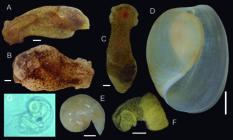
Haminoea maray, external and laying features. (A B) crawling living specimen, dorsal view; (C) same, ventral view; (D) shell, apertural view; (E) shell apical view; (F) egg laying; (G) intracapsular veliger larva (W 0.2 mm). Scales A C = 2 mm, D F = 5 mm.
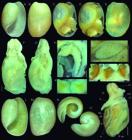
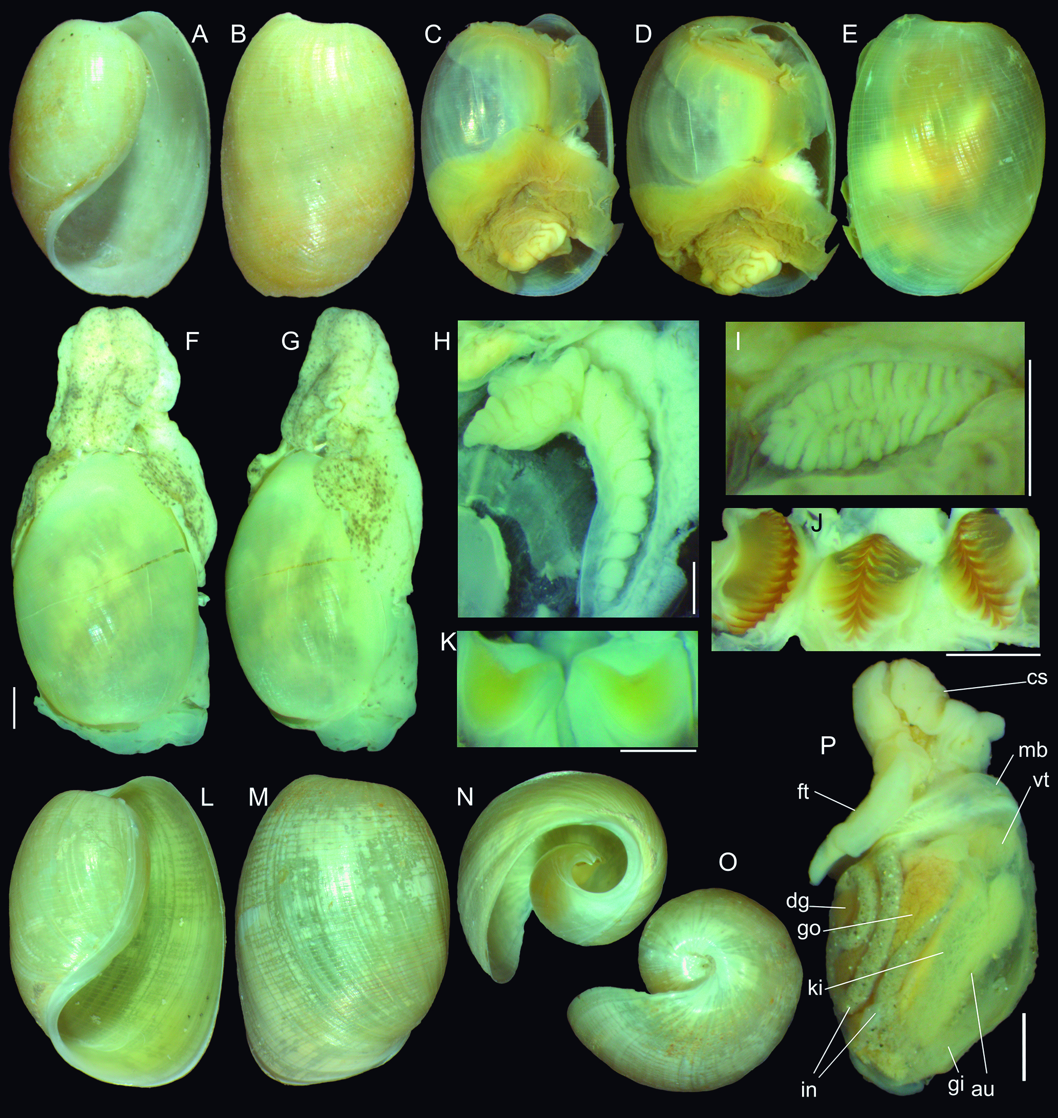
Haminoea maray, type photos. (A B) paratype MZSP 81748, from São Vicente, São Paulo (SP) (L 12.2 mm), shell in frontal and dorsal views; (C E) paratype MZSP 75350, from Ilhabela, SP, voucher of Marcus (1957) (L 16.8 mm) complete preserved specimen, frontal, frontal-slightly apical, and dorsal views; (F K) paratype MZSP 104911#1, from Guarujá, SP; (F) complete preserved specimen, dorsal view, scale = 2 mm; (G) same, right view; (H) detail of gill, ventral view, scale = 1 mm; (I) detail of Hancock’s organ, outer view, scale = 1 mm; (J) opened stomach showing gizzards in situ, scale = 1 mm; (K) pair of jaw plates in situ, ventral-inner view, scale = 0.5 mm; (L P) holotype MZSP 166112 from São Vicente, SP; (L) shell, frontal view (L 12.3 mm); (M) same, dorsal view; (N) same anterior view; (O) same, apical view; (P) specimen extracted from shell, left view. Scale = 2 mm.
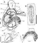
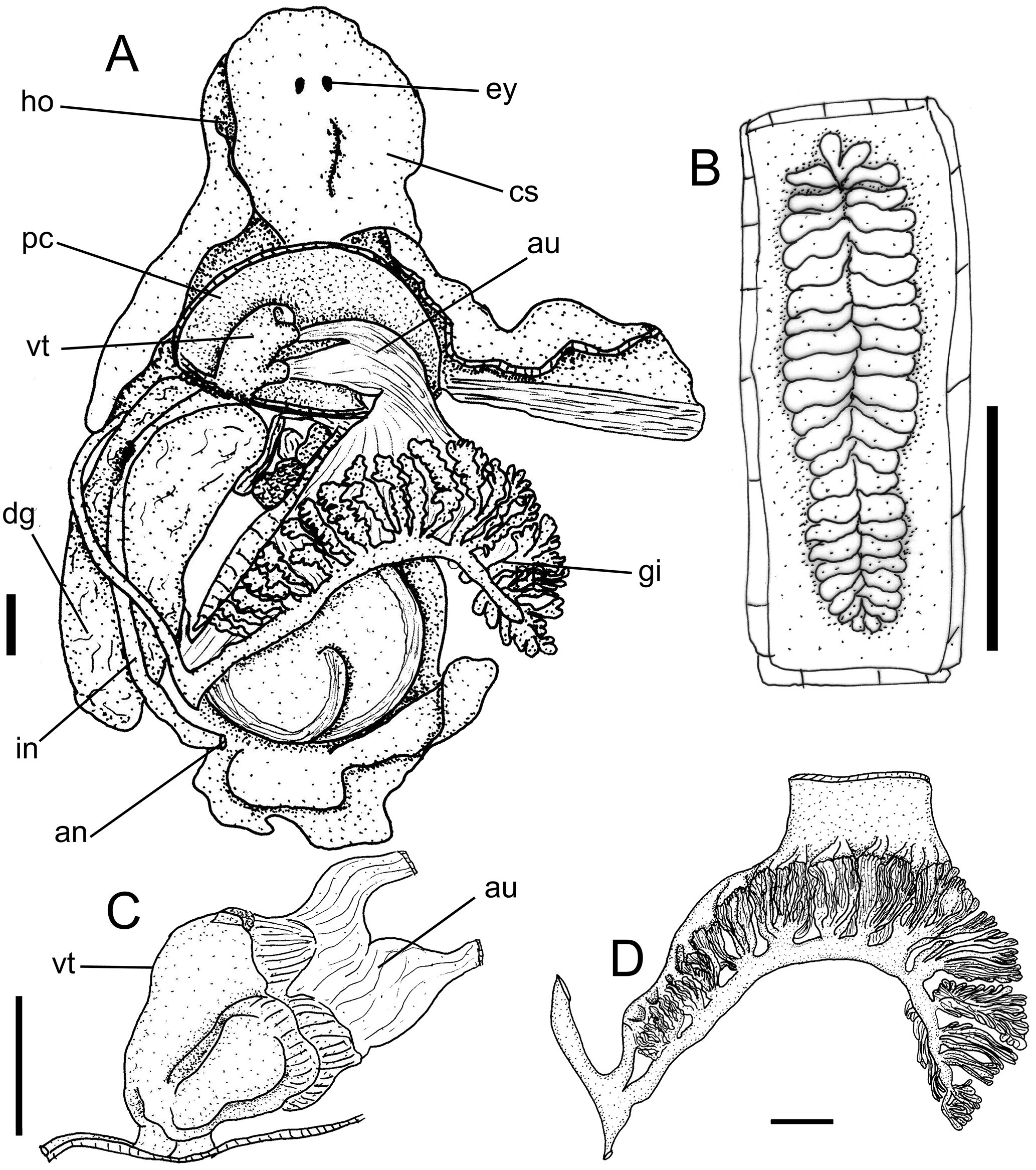
Haminoea maray, anatomical drawings: (A) whole dorsal view, shell and dorsal mantle removed, focusing pallial cavity structures; (B) Hancock’s organ; (C) heart, dorsal view; (D) gill, dorsal view, some adjacent structures also shown. Scales = 1 mm.
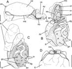
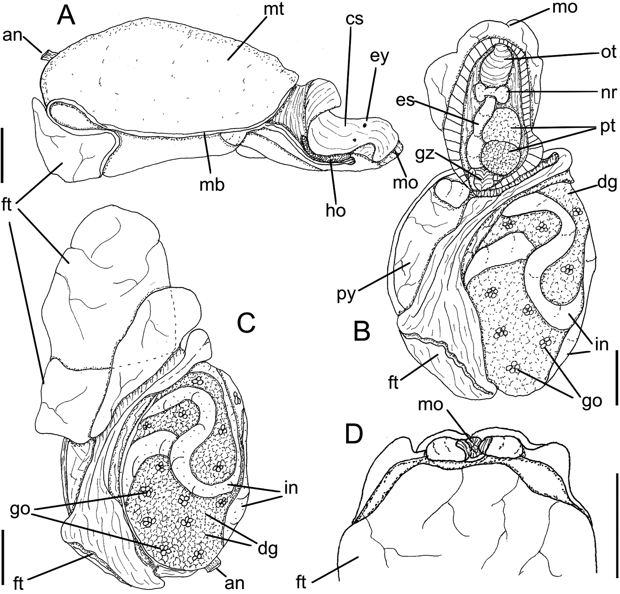
Haminoea maray, anatomical drawings. (A) right whole view, shell removed. (B) same, ventral view, anterior region sectioned longitudinally (C) same, whole ventral view. (D) detail of anterior region, mouth and flattened oral flaps. Scales = 1 mm.
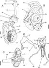
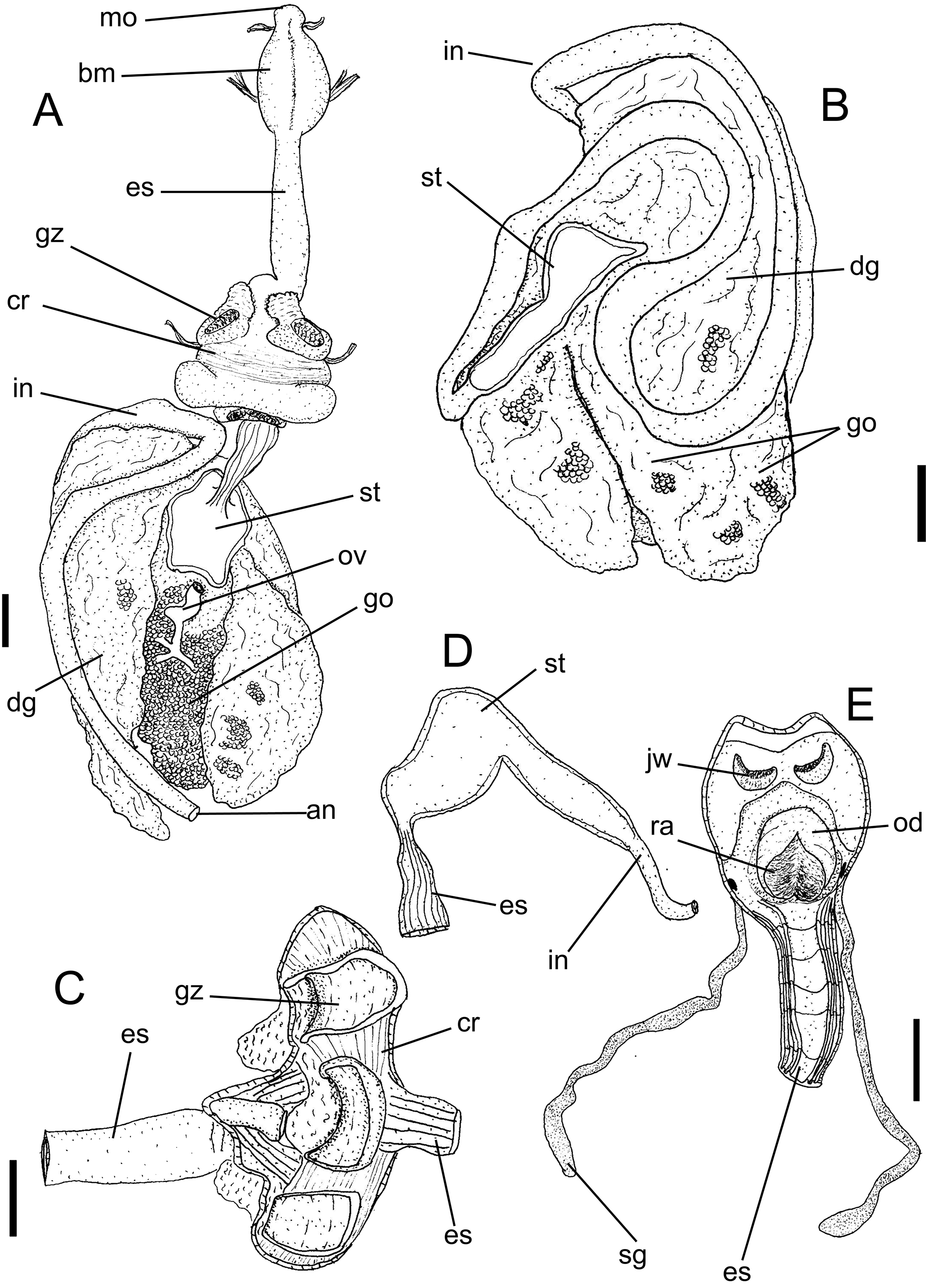
Haminoea maray, drawings of digestive system portions. (A) whole dorsal view; (B) digestive gland region, ventral view; (C) crop, opened longitudinally internal surface; (D) stomach opened longitudinally; (E) buccal mass opened longitudinally, internal view. Scales = 1 mm.
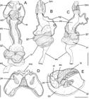
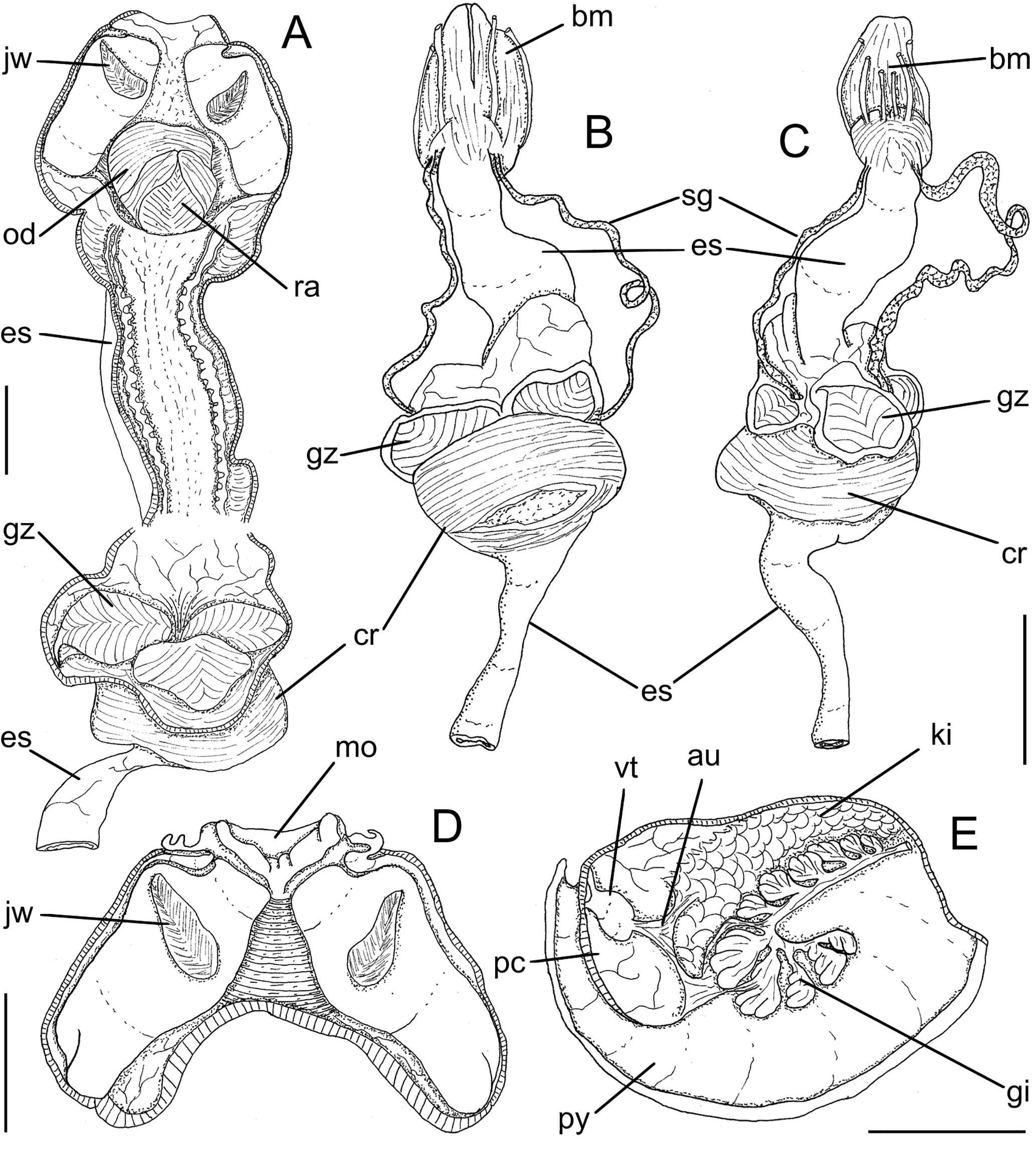
Haminoea maray, anatomical drawings. (A) foregut, longitudinally sectioned, ventral view, scale = 2 mm; (B C) same, whole dorsal and ventral views, scale = 2 mm; (D) oral tube with jaws, opened longitudinally, ventral view, scale = 1 mm; (E) pallial cavity and adjacent structures, pericardium ventral wall removed, ventral view. Scale = 5 mm.
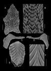
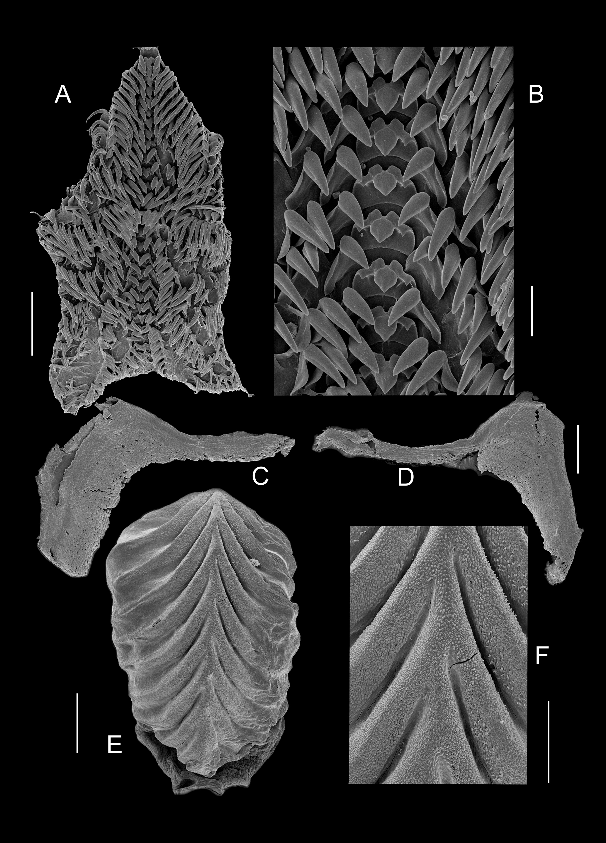
Haminoea maray, digestive hard portions in SEM: (A) radula, whole view, scale = 200 µm; (B) same, detail of central region, scale = 50 µm; (C D) Jaws, inner view, scales, 100 µm; (E) Gizzard plate, whole view, scale = 200 µm; (F) Same, detail of central region, scale = 100 µm.
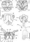
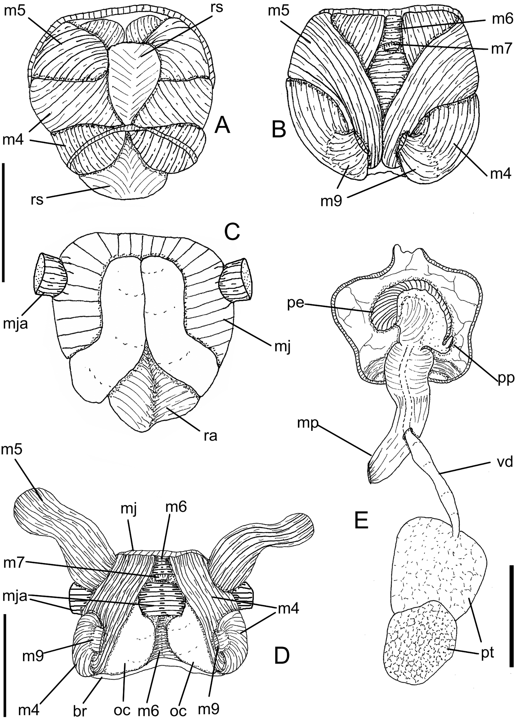
Haminoea maray, anatomical drawings. (A) Odontophore isolated, dorsal view, scale = 1 mm; (B) same, radula removed; (C) same, ventral view; (D) same, dorsal view, m4 and m5 deflected to expose cartilages, scale = 1 mm; (E) isolated male portion of genital structures, dorsal view, scale = 2 mm.
Bulla diaphanaGould, 1852: 222 (figs. 265a d [not found]) (non Montagu, 1803, nec Aradas & Maggiore, 1840).
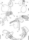
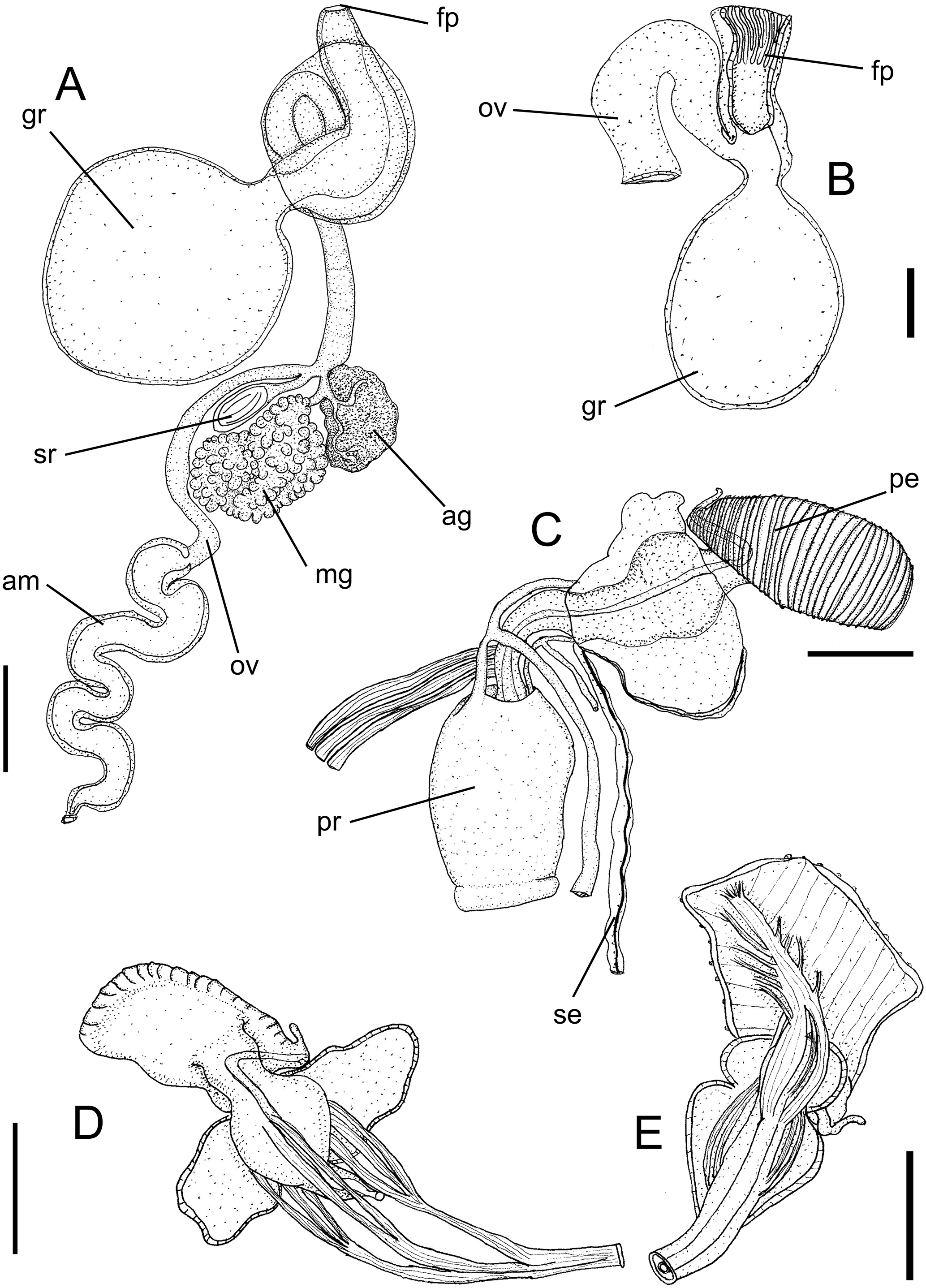
Haminoea maray, drawings of reproductive system. (A) proximal portion of reproductive system with female and hermaphrodite parts; (B) detail of female opening; (C) anterior (male) portion of reproductive system; (D E) penis, two views. Scales = 1 mm.
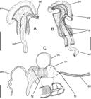
Haminoea maray, anatomical drawings. (A) penis isolated and opened longitudinally, dorsal view, penis muscle removed; (B) same, penis muscle still present; (C) female portion of genital structures, dorsal view, with some inner details of female grand. Scales = 1 mm.
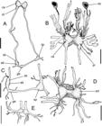
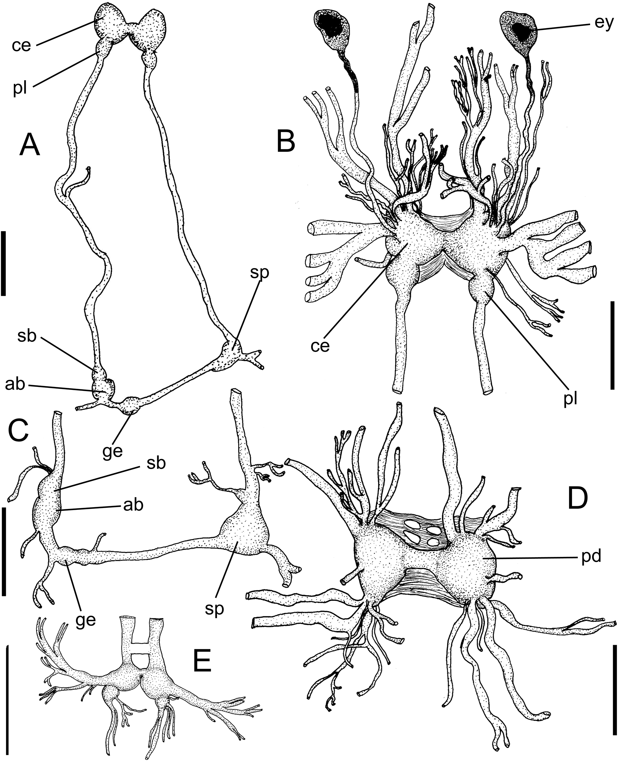
Haminoea maray, drawings of central nervous system. (A) Overview; (B) detail of cerebro-pleural ganglia, dorsal view; (C) detail of visceral ganglia; (D) detail of pedal ganglia; (E) detail of buccal ganglia. Scales = 1 mm.
Haminoea elegans: Marcus, Er., 1957: 395 (figs. 9-16); 1958: 35; Ev. Marcus & Er. Marcus, 1963: 6; 1967: 24, fig. 13; Rios, 1994: 198, fig. 931; 2009: 403, fig. 1091; Goodheart et al., 2016: 17, fig. 9A (non Gray, 1825).
Types: holotype MZSP 166112. Paratypes: BRAZIL. Ceará; Caucáia, Estuarine area in Ceara river, 03°42′12″S, 38°37′02″W (H Galvão Fo. col., 12.xii.2009), CMPHRM 3077, ~100 spm, MZSP 166110, 17 spm, MZSP 166111, 13 shells. São Paulo; Ilhabela (São Sebastião Island), 23°49′S, 45°22′W, MZSP 75350, 15 spm (voucher by Marcus, 1957, Marcus col., 10-20.iv.1954); Guarujá, Praia do Góes, 23°59′52″S, 46°18′55″W, MZSP 104911, 20 spm (CM Cunha col., 07.ii.2012); São Vicente, beach of Parque Bitaru, near Ponte Pênsil, 23°58′23.12″S, 46°23′29.12″W (Simone col.), MZSP 81748, ~100 shells (05.i.1978), MZSP 35597, ~100 spm (1985).
Type locality: BRAZIL. São Paulo; São Vicente, beach of Parque Bitaru, near Ponte Pênsil, 23°58′23.12″S, 46°23′29.12″W [Simone col., 05.i.1978].
Etymology: the specific epithet is a Latinization of the native Guarani word marã’ỹ, meaning translucent, in respect to the Gould’s (1852) idea of naming the Rio de Janeiro population as diaphana, which has a similar meaning.
Diagnosis: Brazilian coast species with shell very globose, spirally microscuptured. Radular rachidian with superior projections medially positioned and evident, and central cusp narrow and short; marginal teeth broad; marginal teeth narrow and long. Gizzard plates with chevrons tightly positioned, with pits less developed.
Description
External morphology: (Figs. 1A-E, 2F-G, P, 3A, 4) Cephalic shield (Figs. 3A, 2P, 4A: cs) trapezoidal, developed posteriorly into one tapered cephalic lobe or propodium, reaching most anterior part of shell, separated from foot by horizontal furrow (Figs. 3A, 7G, P). Hancock’s organ (Figs. 3A B, 4A: ho, 7I) with multiple, relatively symmetric lamellae. Parapodial lobes (Figs. 1A B, 2F-G) extended from foot sole to anterior dorsal side of shell. Gill plicate (Fig. 3A, 6E: gi), arched; multi-lobed, each lobe with several secondary, rather irregular filaments (Fig. 2H), gradually larger towards anterior; located internal to mantle cavity (Fig. 2P). Foot with posterior and anterior border rounded (Figs. 1A-C, 7C-D, F-G, P).
Shell: shape bulloid, globose, involute (mainly formed by body whorl); 1.3 (Fig. 1D) to 1.4 (Figs. 2A, C, L) times longer than wide. Outer surface with growth lines conspicuous forming grid pattern; spiral striae well-developed, thin, uniform, all-over surface (Figs. 1D, 2B, E, M). Periostracum thin, color orange-brown. Aperture ~50% of frontal view; narrow superior, expanding slightly superior to spire (~⅙ of length), wide interior (~40% of length); both superior and inferior regions rounded; spire concentrated in superior half. Outer lip with cutting edge. Inner lip amply convex in superior ⅔, slightly concave in inferior third (Figs. 1D, 2A, L). Callus weak, slightly more developed in inferior region. Anterior view with ample opened spiral aspect (Fig. 2N). Apical view not umbilicate (Figs. 1E, 2O). Umbilicus absent.
Circulatory and excretory systems: (Figs. 2P, 3A, 6E) Pericardial cavity dorsally positioned over anterior region of digestive gland, exposed in pallial cavity. Auricle (au) elongated thin-walled tissue anterior to gill and lateral to ventricle (vt), with two main connections. Kidney (ki) glandular-shaped positioned between gill and heart.
Digestive system: (Figs. 5-7) mouth in middle region of anterior furrow (Figs. 4A, B, D: mo). Buccal mass (Figs. 5A, 6B, C: bm) elongated, 3× longer than wide; no dorsal septate muscles. Oral sphincter bit elongated (Fig. 4B: ot). Odontophore muscles (Figs. 8A-D): mj, jaw and peribuccal muscles, originated from anterior edges of cartilages, directed towards anterior and dorsal, surrounding oral cavity; mja, unpaired string transverse muscle connecting both lateral sides of oral tube, passing through middle region of odontophore, transpassing, inclusive, both odontophore cartilages (Figs. 8C, D); m2, buccal retractors absent; m4, pair of main dorsal tensor muscles strong and broad, occupying ~⅖ of odontophore volume, twice as long as wide, covering ⅓ of cartilages, inserted on ventral portion of subradular membrane; m5, pair of dorsal auxiliary tensor muscles, occupying ~⅖ of odontophore volume, twice as long as wide, originating on most anterior region of odontophore cartilages, passing dorsally to m4, inserting on ventral side of subradular membrane, around radular sac; m6, unpaired horizontal muscle, with transverse fibers connecting median edge of both odontophore cartilages and anterior portion of m4, about same length and half as wide as m4, triangle-shaped (Figs. 8B, D); m7, unpaired muscle, originating on posterior region of m6 (Figs. 8B, D). Pair of odontophore cartilages (oc) elliptic, located more posteriorly, with ~half of odontophore length; m9, transverse muscle connecting both postero-ventral sides of both m4 (Figs. 8B, D). Radula formula 28 × 22.1.22 (Figs. 7A-B); rachidian tooth curved, with three cusps, smooth cutting edge; lateral teeth elongated, curved towards medially. Jaws (Figs. 4C-D, 5E, 6A, D: jw, 7K) shape rhomboidal, surface irregular; positioned on posterior portion of buccal mass. Anterior esophagus (es) thin and elongated, length bit longer than buccal mass length; inner surface with one pair of tiny folds. Salivary glands (Fig. 5E: sg) thin, elongated, aperture in latero-posterior region of oral cavity. Gizzard bulged, possessing three gizzard plates in anterior half (Figs. 2J, 5A, G, 6A-C: gz, 7E), posterior part very muscular and thick (Fig. 5A: cr). Gizzard plates (Figs. 7E-F, 4J) composed of many V shaped crests, with micro-irregular surface. Stomach thin walled, located inside digestive gland (Figs. 5A, D: st). Digestive gland (Figs. 4B-C, 5B: dg) dark beige, homogeneous, mixed with gonad, occupying most part of posterior region of visceral mass. Intestine narrow, walls thick, surrounding digestive gland (Figs. 4B-C, 5B: in); anus (Figs. 4C, 5A: an) postero-dorsal.
Genital system: (Figs. 9, 8E, 10) Gonad mixed with digestive gland (Figs. 4B, C, 5G: go). Hermaphrodite duct running anteriorly from central part of gonad up to right side of body; expanding anteriorly forming tubular ampulla (Fig. 9A: am). Ampulla length more than half of hermaphrodite duct. Seminal receptacle (sr) small, balloon-like, duct narrow, inserted laterally at end of hermaphrodite duct. Mucus gland (mg) and albumen gland (ag) located side by side, both connected to single duct shortly inserted in hermaphrodite duct slightly anteriorly to seminal receptacle’s insertion. Mucus gland (mg) not fused with oviduct. Genital receptacle (or bursa copulatrix) (gr, Fig. 10C: bc) as larger genital structure, ~4× mucus gland’s size, connected to oviduct in anterior position near to female opening (fp). Oviduct not glandular, elongated, anteriorly curved, opening on female bulb on right side of body. Male organs positioned right-anterior side of head-foot, inside right portion of cephalic shield and near to buccal mass. Prostate gland (Figs. 8E, 9C: pr) rounded, bilobed. Vas deferens narrow, penetrating through penis muscle (Fig. 8E: vd). Seminal groove running from male aperture to female bulb along right side of body. Penis (pe) curved, elongated, ~3 times longer than wide, elliptical, size bit smaller than prostate gland, with terminal slightly curved stylet. Vas deferens running along prostate up to tip of papilla (Figs. 10A-B).
Nervous system: (Fig. 11) Cerebral ganglia (ce) connected by short simple commissure, each ganglion with more than 10 innervations, with larger one directly branching and connecting to Hancock’s organ. Optic nerve ~4× longer than cerebral length. Pleural ganglia (pl) partially fused with cerebral ganglia and not innervated. Pedal ganglia (pd) highly innervated and connected to each other by one single and short pedal commissure. Buccal ganglia (Fig. 11E) small, connected to cerebral ganglia through elongated connective, united with single short buccal commissure. Visceral loop very elongated (Fig. 11A). Subintestinal and visceral ganglia slightly fused, size large as half of cerebral ganglion size. Supraintestinal ganglia size as larger as abdominal ganglia, connected to abdominal ganglia by elongated connective.
Distribution: Ceará to São Paulo.
Habitat: Estuarine species, with regions or relative low salinity; muddy bottoms, intertidal.
Measurements (L, W in mm): holotype: 12.3 by 8.6; paratypes: MZSP 81748 (Figs. 2A-B): 12.2 by 8.6; MZSP 75350 (Figs. 2C-E): 16.8 by 10.8; MZSP 35597#1 (Figs. 2L-O), 12.3 by 8.6.
Material examined: types.
DISCUSSION
Haminoea maray exhibits natural morpho-anatomical similarities to other populations previously identified as H. elegans. This resemblance led to misidentifications, a matter addressed and clarified in this paper. However, this study highlights the principal differences observed in the examined populations to establish a species-level distinction. The shell of H. maray itself is relatively more globose if compared to populations from Congo, São Tomé and Cuba, being slightly similar to those of Florida (Martínez & Ortea, 1997: figs. 1-6), despite in being still more inflated. Interestingly, all of them possess spiral microsculpture (Figs. 1D, E, 2B, E, L, M), which Pilsbry (1895: 356) called as “engraved spirals clearly visible without a lens”. This feature is shared with European H. navicula (Malaquias & Cervera, 2006). The radular rachidian tooth of H. maray has the pair of superior projections closer to median line and more evident than that of the Congo population, while its marginal teeth are broader, i.e., not so filiform than those of that population (Martínez & Ortea, 1997: figs. 7-12); the central cusp of the rachidian of H. maray is shorter and not so triangular as that of the population from Mexico-Florida-Caribbean (Martínez & Ortea, 1997: figs. 13 15), while the marginal teeth of H. maray are much narrower and longer (Figs. 7A-B). The gizzard plate of H. maray has the chevrons much close from each other, with tight interspaces (Figs. 2J, 7E-F), the populations from Congo and Florida have them more separated (Martínez & Ortea, 1997: figs. 19 22); besides, its pits, or elements, are less developed in H. maray.
Interestingly, all H. elegans-like populations have in the male genital apparatus a multifolded penis, including H. maray (Figs. 9C, 10A-B: pe). It differs from the penis of the São Tomé population (Martínez & Ortea, 1997: fig. 25) in being only half free, and in lacking an apparent longitudinal sperm groove; from the penis of the Congo population, H. maray differs in having a well-developed terminal papilla (pp) (Martínez & Ortea, 1997: figs. 26 27).
Related to other species, an interesting discussion on H. maray anatomical characters is possible based on available date in literature. The Hanckock’s organ of H. maray is longer and with much more filaments in both sides than that of H. cymbalum (Quoy & Gaimard, 1833) (Rudman, 1971, fig. 2D). The gill of H. maray has filaments more branched than that of H. zelandiae (Gray, 1843) (Rudman, 1971: fig. 3). The jaw plate of H. maray differs from that of H. navicula (Malaquias & Cervera, 2006: figs. 3E-F) in having medial slender projections (Figs. 7C-D). The radula H. maray is relatively similar to that of H. aptei Bharate, Oskars, Narayana, Ravinesh, Kumar & Malaquias, 2018 (Bharate et al., 2018: fig. 2E), differing in lacking medial sulcus in central cusp of rachidian, and by pair of distal elevations of the rachidian being more pointed (nor so rounded); from that of H. navicula (Malaquias & Cervera, 2006: fig. 4) it differs in lacking serrated edges of some cusps, and by rachidian having pair of secondary cusps and pair of dorsal elevations; this last two characteristics also distinguish it from H. orbignyana (Férussac, 1822) (Malaquias & Cervera, 2006: figs. 9A-E). The gizzard plate of H. maray is relatively similar to that of H. aptei (Bharate et al., 2018: figs. 2F-G), differing in having not so prominent chevrons (Fig. 7E), and by the pits being less developed (Fig. 7F); from H. navicula and from H. orbignyana (Malaquias & Cervera, 2006: figs. 5C-E, 8C-F) differs by the chevrons having not so rounded profile and by pits more amply distributed. The genital structures of H. maray differs from that of H. zelandiae (Rudman, 1971: fig. 4) in lacking exogenous sperm sac and secondary sperm sac, by smaller sized albumen gland, in having a single seminal receptacle (that species has two), in having a non-spiralized mucous gland (mg), and in having a larger, with shorter bursa with broader duct (gr). The prostate of H. maray differs from that of H. cymbalum in being simpler and cylindric, while that species it is spheric, with an accessory gland (Rudman, 1971: fig. 9B); it still differs from that of H. zelandiae in lacking transverse folds (Rudman, 1971: fig. 8). The penis of H. maray differs from those of H. crocata Pease, 1860, H. solitaria (Say, 1822) (Rudman, 1971: fig. 10), and H. orbignyana (Malaquias & Cervera, 2006: fig. 8A) in being strongly transversally folded and by the papilla in the tip; from that of H. aptei it differs in lacking fundus (Bharate et al., 2018: fig. 3); from that of H. navicula (Malaquias & Cervera, 2006: fig. 6) it differs in lacking apical penial spiny armature. The central nervous system of H. maray differs from that of H. zelandiae (Rudman, 1971: fig. 11) by the shortness of the cerebral commissure, as that species both ganglia are very separated from each other. It is recognized that, in a recent review (Oskars & Malakias, 2022), H. cymbalum, H. crocata and H. aptei were transferred to the related genus Haloa Pilsbry, 1921; and H. zelandiae to the genus Papawera Oskars & Malakias, 2019. Although H. cymbalum is also reported in the genus Lamprohaminoea Habe, 1952 (Oskars & Malakias, 2020).
Haminoea maray, distinct from other congeners (and allies) and from what has been identified as H. elegans in other regions, possesses its own unique identity. This distinctiveness is formally outlined in this descriptive paper, a classification supported by a preceding molecular study (Turani et al., 2023).
ACKNOWLEDGEMENTS:
The authors thank to Helena Matthews-Cascón, UFC, for the donation of the Ceará sample.
-
FUNDING INFORMATION:
This project did not use any external financial support.
-
Published with the financial support of the “Programa de Apoio às Publicações Científicas da Universidade de São Paulo”
REFERENCES
- Aartsen, J.J.v. & Giannuzzi-Savelli, R. 1987. On the dates of publication of Aradas & Maggiore’s “Catalogo Ragionato…” and its malacological implication. Bolletino Malacologico, 23(5-8): 269-273.
- Aradas, A. & Maggiore, G. 1840-1844. Catalogo ragionato delle conchiglie viventi e fossili di Sicilia. Atti dell’Accademia Gioenia di Scienze Naturali, 15(2): 187-217, 349-366.
-
Bharate, M.; Oskars, T.R.; Narayana, S.; Ravinesh, R.; Kumar, A.B. & Malaquias, M.A.E. 2018. Description of a new species of Haminoea (Gastropoda: Cephalaspidea) from India, with an account of the diversity of the genus in the Indo-West Pacific. Journal of Natural History, 52(37-38): 2437-245. https://doi.org/10.1080/00222933.2018.1533598
» https://doi.org/10.1080/00222933.2018.1533598 - Galvão Filho, H.C. 2018. Taxonomy and cladistic analysis of Plakobranchidae (Gastropoda: Sacoglossa). PhD dissertation. Museu de Zoologia, Universidade de São Paulo. São Paulo. 189p.
-
Goodheart, J.A.; Ellingson, R.A.; Vital, X.G.; Galvão Filho, H.C.; McCarthy, J.B.; Medrano, A.M.; Bhave, V.J.; García-Méndez, K.; Jimenéz, L.M.; López, G.; Hoover, C.A.; Awbrey, J.D.; Deus, J.M.; Gowacki, W.; Krug, P.J. & Valdés, A. 2016. Identification guide to the heterobranch sea slugs (Mollusca: Gastropoda) from Bocas del Toro, Panama. Marine Biodiversity Records, 9(56): 1-31. https://doi.org/10.1186/s41200-016-0048-z
» https://doi.org/10.1186/s41200-016-0048-z - Gould, A.A. 1852. Mollusca & shells of the United States Exploring Expedition. Gould & Lincoln. Boston. 510p.
- Gray, J.E. 1825. A list and description of some species of shells not taken notice of by Lamarck. Annals of Philosophy, New Series, 9: 134-140; 407-415.
- Lima, P.O.V. 2016. Análise filogenética de Cryptobranchia (Mollusca: Nudibranchia) com base em anatomia comparada. PhD dissertation. Instituto de Biociências, Universidade de São Paulo. São Paulo. 326p.
-
Malaquias, M.A.E. & Cervera, J.L. 2006. The genus Haminoea (Gastropoda: Cephalaspidea) in Portugal, with a review of the European species. Journal of Molluscan Studies, 72: 89-103. https://doi.org/10.1093/mollus/eyi052
» https://doi.org/10.1093/mollus/eyi052 -
Marcus, Er. 1957. On Opisthobranchia from Brazil II. Journal of the Linnean Society of London, Zoology, 43: 390-486. https://doi.org/10.1111/j.1096-3642.1957.tb01559.x
» https://doi.org/10.1111/j.1096-3642.1957.tb01559.x -
Marcus, Er. 1958. Notes on Opisthobranchia. Boletim do Instituto Oceanográfico, 7: 31-79, 8 pls. https://doi.org/10.1590/S0373-55241956000100002
» https://doi.org/10.1590/S0373-55241956000100002 - Marcus, Er. 1970. Opisthobranchs from northern Brazil. Bulletin of Marine Science, 20: 922-951.
- Marcus, Ev.B.R. 1977. An annotated checklist of the western Atlantic warm water opisthobranchs. Journal of Molluscan Studies Suppl. 4: 1-23.
- Marcus, Ev.B.R. & Marcus, Er. 1963. Opisthobranchs from the Lesser Antilles. Studies on the fauna of Curaçao and other Carribbean Islands, 19: 1-76.
- Marcus, Ev.B.R. & Marcus, Er. 1967. Opisthobranchs from the southwestern Caribbean Sea. Biological Investigations of the Deep Sea, 33. Bulletin of Marine Science, 17: 597-628.
- Martínez, E. & Ortea, J. 1997. Haminaea elegans (Gray, 1825) (Opisthobranchia: Cephalaspidea), a truly amphiatlantic species. Veliger, 40(4): 281-291.
- Matthews-Cascón, H.; Meirelles, C.A. & Martins, I.X. 2011. Retorno ao “lar” em moluscos pateliformes. In: Fernandez, M.A.; Santos, S.B; Pimenta, A. & Thiengo, S.C. Tópicos em Malacologia. Ecos do XIX Encontro Brasileiro de Malacologia. Rio de Janeiro, FAPERJ, Sociedade Brasileira de Malacologia. p. 146-151.
-
Montagu, G. 1803. Testacea Britannica or natural history of British shells, marine, land and freshwater, including the most minute: systematically arranged and embellished with figures. London, J. White. 606p. https://doi.org/10.5962/bhl.title.33927
» https://doi.org/10.5962/bhl.title.33927 - Oskars, T.R. 2019. The systematics and phylogeny of Haminoeidae (Mollusca, Cephalaspidea), with special focus on the taxonomy and phylogeny of the genus Haloa sensu lato. Bergen, University of Bergen. 96p.
-
Oskars, T.R. & Malaquias, M.A.E. 2020. Systematic revision of the Indo-West Pacific colourful bubble-snails of the genus Lamprohaminoea Habe, 1952 (Cephalaspidea: Haminoeidae). Invertebrate Systematics, 34(7): 727-756. https://doi.org/10.1071/IS20026
» https://doi.org/10.1071/IS20026 -
Oskars, T.R. & Malaquias, M.A.E. 2022. Systematic revision of the Indo-West Pacific bubble-snails of the genus Haloa (Pilsbry, 1921) (Cephalaspidea: Haminoeidae). Invertebrate Systematics, 36(5): 436-492. https://doi.org/10.1071/IS21011
» https://doi.org/10.1071/IS21011 - Pilsbry, H.A. 1895. Polyplacophora, Acanthochitonidae, Cryptoplacidae and appendix: Tectibranchiata. In: Manual of Conchology, structural and systematics, second series Pulmonata. Philadelphia, GWTJ. v. 15, p. 181-436.
- Redfern, C. 2013. Bahamian Seashells, 1161 Species from Abaco, Bahamas. Boca Raton, Florida, Bahamianseashells.com, Inc. 501p.
- Rios, E.C. 1994. Seashells of Brazil. 2.ed. Rio Grande, Fundação Universidade do Rio Grande. 368p., 113 pls.
- Rios, E.C. 2009. Compendium of Brazilian Seashells. Rio Grande, Ed. Evangraf. 668p.
- Rudman, W.B. 1971. On the opistobranch genus Haminoea Turton & Kingston. Pacific Science, 25(4): 245-559.
-
Simone, L.R.L. 2011. Phylogeny of the Caenogastropoda (Mollusca), based on comparative morphology. Arquivos de Zoologia, 42(4): 161-323. https://doi.org/10.11606/issn.2176-7793.v42i4p161-323
» https://doi.org/10.11606/issn.2176-7793.v42i4p161-323 - Simone, L.R.L. 2018. Lavajarus moroi, new cavernicolous Subulinidae from Ceará, Brazil (Gastropoda, Eupulmonata, Achatinidae). Spixiana, 42(2): 173-187.
- Thompson, T.E. 1977. Jamaican opisthobranch molluscs I. Journal of Molluscan Studies, 43: 93-140, pls. 1-3.
-
Turani, M.; Valdés, A. & Malaquias, M.A.E. 2023. Molecular phylogeny of the marine snail genus Haminoea (Gastropoda, Cephalaspidea): A framework to study marine diversity and speciation. Zoologica Scripta, 53: 52-77. https://doi.org/10.1111/zsc.12627
» https://doi.org/10.1111/zsc.12627 - Valdés, Á.; Hammann, J.; Behrens, D. & Dupont, A. 2006. Caribbean Seas Slugs. A Field Guide to the Opisthobranch Mollusks from the Tropical Northwestern Atlantic. Gig Harbor, WA, Sea Challengers. 289p.
Edited by
-
Edited by:
Marcelo Veronesi Fukuda